Publisher’s version / Version de l'éditeur:
Bioconjugation Protocols, pp. 505-532, 2011-07-01
READ THESE TERMS AND CONDITIONS CAREFULLY BEFORE USING THIS WEBSITE.
https://nrc-publications.canada.ca/eng/copyright
Vous avez des questions? Nous pouvons vous aider. Pour communiquer directement avec un auteur, consultez la
première page de la revue dans laquelle son article a été publié afin de trouver ses coordonnées. Si vous n’arrivez pas à les repérer, communiquez avec nous à [email protected].
Questions? Contact the NRC Publications Archive team at
[email protected]. If you wish to email the authors directly, please see the first page of the publication for their contact information.
NRC Publications Archive
Archives des publications du CNRC
For the publisher’s version, please access the DOI link below./ Pour consulter la version de l’éditeur, utilisez le lien DOI ci-dessous.
https://doi.org/10.1007/978-1-61779-151-2_32
Access and use of this website and the material on it are subject to the Terms and Conditions set forth at
Purification, functionalization, and bioconjugation of carbon nanotubes
Luong, John H. T.; Male, Keith B.; Mahmoud, Khaled A.; Sheu, Fwu-Shan
https://publications-cnrc.canada.ca/fra/droits
L’accès à ce site Web et l’utilisation de son contenu sont assujettis aux conditions présentées dans le site
LISEZ CES CONDITIONS ATTENTIVEMENT AVANT D’UTILISER CE SITE WEB.
NRC Publications Record / Notice d'Archives des publications de CNRC:
https://nrc-publications.canada.ca/eng/view/object/?id=81d64382-dff2-455f-b0b5-c89e77e17a6e https://publications-cnrc.canada.ca/fra/voir/objet/?id=81d64382-dff2-455f-b0b5-c89e77e17a6e505
Sonny S. Mark (ed.), Bioconjugation Protocols: Strategies and Methods, Methods in Molecular Biology, vol. 751, DOI 10.1007/978-1-61779-151-2_32, © Springer Science+Business Media, LLC 2011
Purification, Functionalization, and Bioconjugation
of Carbon Nanotubes
John H.T. Luong, Keith B. Male, Khaled A. Mahmoud,
and Fwu-Shan Sheu
Abstract
Bioconjugation of carbon nanotubes (CNTs) with biomolecules promises exciting applications such as biosensing, nanobiocomposite formulation, design of drug vector systems, and probing protein interac-tions. Pristine CNTs, however, are virtually water-insoluble and difficult to evenly disperse in a liquid matrix. Therefore, it is necessary to attach molecules or functional groups to their sidewalls to enable bioconjugation. Both noncovalent and covalent procedures can be used to conjugate CNTs with a target biomolecule for a specific bioapplication. This chapter presents a few selected protocols that can be performed at any wet chemistry laboratory to purify and biofunctionalize CNTs. The preparation of CNTs modified with metallic nanoparticles, especially gold, is also described since biomolecules can bind and self-organize on the surfaces of such metal-decorated CNTs.
Key words: Carbon nanotubes (CNTs), Gold nanoparticles, Purification, Noncovalent/covalent
biofunctionalization
Since their discovery (1), carbon nanotubes (CNTs) have attracted enormous attention owing to their unique structural, mechanical, and electronic properties (2–5). CNTs are also one of the few molecules that are known precisely at the atomic level with respect to the physical location of each atom. Arc-vaporization (6, 7) of two pure graphite rods (~1 mm apart) under helium, argon, or – even better – with methane (8) at low pressure (50–700 mbar) produces multiwalled carbon nanotubes (MWCNTs) with diameters of 2–100 nm. This mesoscale graphite system typically consists of two to ten incommensurate concentric cylinders of graphitic shells
1. Introduction
with a layer spacing of 0.3–0.4 nm. When the anode graphite rod contains a metal catalyst (Fe or Co), single-walled carbon nano-tubes (SWCNTs) are generated instead of MWCNTs (9). SWCNTs are truly a single large molecule comprised of a cylindri-cal graphite sheet of nanoscylindri-cale diameter capped by hemisphericylindri-cal ends with a typical diameter of 1 nm. Double-walled CNTs (DWCNTs) have also been synthesized and commercialized recently. The lengths of both SWCNTs and MWCNTs can range from hundreds of nanometers to upwards of 20 cm. CNTs thus display extremely high aspect ratios (length/diameter); and, indeed, the aspect ratio often exceeds 10,000 in most prepara-tions. Together with their pure hexagonal structure, CNTs are composed entirely of an sp2 bond structure, which is even stronger
than the sp3 bond structure in diamond. Consequently, CNTs are
virtually insoluble in most solvent systems. Nevertheless, surfac-tants such as sodium dodecyl sulfate are able to coat CNTs with negative sulfonate groups that are exposed to the surrounding environment. And Triton X-100, a nonionic detergent, also coats CNTs with their hydrophilic groups oriented toward the aqueous phase. Consequently, CNTs can be dispersed in an aqueous medium in the presence of ionic or nonionic surfactants.
The drawback of the arc-discharge method is the costly removal of non-nanotube carbon and metal catalyst materials. An alternative synthesis method, laser ablation, can be adapted for fullerene and SWCNT production by focusing a CO2 laser beam on a target with a high boiling temperature, e.g., carbon compos-ites doped with catalytic metals. The target is vaporized in a high-temperature argon atmosphere, and SWCNTs formed are conveyed via a gas stream to a collection chamber. The temperature, metal species, and the gas flow rate can all affect the resulting diameter of the nanotubes (10). Furthermore, large-scale pro-duction is a critical issue with this method, despite the high homogeneity of the SWCNTs produced. The chemical vapor deposition (CVD) synthesis method uses a hydrocarbon vapor, which is thermally decomposed at the surface of a metal catalyst to form the nanotubes. In this method, CNT growth is governed by the type of hydrocarbon gas and/or catalyst used and the growth temperature. Unlike the arc-discharge and laser ablation methods, the CVD approach allows for control over the location and the alignment of the synthesized CNTs (11).
Owing to the symmetry and unique electronic structure of graphene, the structure of a CNT strongly affects its electrical properties. SWCNTs are one-dimensional conductors, wherein all of the electrons are confined to move within a single atomic layer and all of the atoms in the SWCNT structure are surface atoms. To date, MWCNTs have received less attention than SWCNTs since their structures are more complex (each carbon
shell of a MWCNT can exhibit different electronic properties and chirality). Nevertheless, MWCNTs combine a very similar mor-phology and set of properties as compared to SWCNTs, while improving significantly their resistance to chemicals. This feature is especially important when the functionalization of CNTs is required for bioconjugation and bioapplications as discussed later. In the case of SWCNTs, covalent functionalization will break some C=C bonds, leaving “holes” in their structure that can affect both their mechanical and optoelectronic properties. In contrast, only the outermost wall of MWCNTs is modified during such functionalization reactions.
Remarkable advances have been made in the synthesis and functionalization of CNTs over the past decade, and this has helped to arouse considerable interest in potential applications of CNTs in the fields of biomedical materials, drug delivery, and tissue engineering. In addition, the quest for probing important analytes with very low detection limits and high specificity using biosensors incorporating nanoscale components has intensified, with several avenues being explored. In particular, CNTs have become an extremely popular theme in recent bioapplication research because of their nanoscale diameter, high electrocatalytic activity, and decreased vulnerability to surface fouling. CNTs can also be utilized as novel electrode materials; owing to their high surface-to-volume ratio, the variation of their electronic conductance to adsorbed surface species could potentially provide a sufficient level of sensitivity for single-molecule detection. Moreover, on the basis of their well-defined structure and high surface area, CNTs appear to be ideal materials for studying interactions with biomolecules, such as proteins, receptors, enzymes, etc. CNT-based sensors are also more stable than metal oxide sensors since they are not affected by mass transfer phenomena or by chemical changes to the surface carbon atoms. Thus, CNTs are promising materials for electrochemical sensors and show great potential for use in the next generation of biosensor devices. And finally, func-tionalized CNTs may also find useful applications in medicinal chemistry. The use of CNTs as drug delivery scaffolds and substrates for vaccines is feasible, and CNTs functionalized with bioactive moieties are well suited for targeted drug delivery appli-cations, since they exhibit a high propensity to traverse cell mem-branes with low cytotoxicity.
Like graphite, CNTs are relatively inert except at the nanotube caps, which are more reactive due to the presence of dangling bonds. To date, several strategies have been devised to solubilize, purify, and functionalize CNTs. For bioconjugation applications, the most successful approach is to functionalize sp2 carbons at the
sidewalls of nanotubes with organic pendant groups. Another important procedure is the noncovalent functionalization of
CNTs through supramolecular interactions (e.g., p–p stacking interactions), which allows the formation of stable suspensions of the nanostructures. This chapter presents some selected protocols to purify and bioconjugate CNTs that can be performed at any wet chemistry laboratory.
Both pristine and chemically modified CNTs can be obtained from various commercial sources with high purity (see Note 1). The user should request a copy of the “Product Specifications” from the supplier, which states the purity, dimensions, and the size-length distribution of the CNTs. If possible, transmission electron microscopy (TEM) images and the Raman signatures of the CNTs should also be obtained from the supplier.
1. Concentrated sulfuric acid: Although nearly 100% (w/w) sulfuric acid can be prepared, this loses SO3 at the boiling point to produce a solution that is only 98.3% (w/w) sulfuric acid. The ~98% (w/w) grade is more stable in storage, and is the usual form of what is typically described as “concentrated sulfuric acid” (approximately 18 M).
2. Concentrated nitric acid: Nitric acid is miscible with water, and distillation gives an azeotrope with a concentration of 68% (w/w) HNO3 and a boiling temperature of 120.5°C at 1 atm. Two solid hydrates are known; the monohydrate (HNO3·H2O) and the trihydrate (HNO3·3H2O). Nitric acid is isoelectronic with the bicarbonate ion. The concentrated nitric acid of commerce consists of the maximum boiling azeotrope of nitric acid and water. Technical grades are normally 68% (w/w) HNO3 (approximately 15 M), while reagent grades are specified at 70% (w/w) HNO3.
3. Concentrated hydrochloric acid: Commercial aqueous HCl is 35–38% (w/w) or approximately 11.5–12.4 M, respectively.
4. N-N-Dimethylformamide (DMF). 5. Dimethyl sulfoxide (DMSO). 6. Toluene.
7. Sodium dodecyl sulfate (SDS). 8. Ethanol. 9. Chloroform. 10. Methanol. 11. Anhydrous tetrahydrofuran (THF).
2. Materials
2.1. Carbon Nanotubes 2.2. General Chemicals and Solvents12. Acetone. 13. Acetic acid. 14. Sodium chloride. 15. Triton X-100. 16. Glycerol. 17. Ethylene glycol. 18. 2-Mercaptoethanol. 19. Calcium carbonate. 20. Sodium tetraborate. 21. 11-Aminoundecanoic acid. 22. Ammonium iron sulfate. 23. Potassium hydroxide. 24. Ammonium peroxodisulfate. 25. Sodium hydroxide.
26. Hydrogen peroxide. 27. Sodium borohydride.
28. Cystamine (2,2¢-diaminodiethyl disulfide). 29. Thionyl chloride.
1. 1-Ethyl-3-(3-dimethylaminopropyl)-carbodiimide (EDC). 2. 1-Pyrenebutanoic acid, succinimidyl ester (PASE).
3. Poly(diallyldimethylammonium chloride) (PDAC, 20% w/w in water, MW 100,000–200,000).
4. Poly(sodium 4-styrenesulfonate) (PSS, MW 70,000). 5. Polyethyleneimine (PEI, Mn = 423).
6. 1-(3-Aminopropyl)-3-methylimidazolium bromide (IL-NH2). 7. Nafion perfluorinated ion-exchange resin, 5% (w/w) aqueous
dispersion.
8. Glutaraldehyde (25% w/w in water).
9. Gold(III) chloride trihydrate (HAuCl4·3H2O). 10. Positively charged gold nanoparticles.
11. a-Cyclodextrin. 12. N-Hydroxysulfosuccinimide (Sulfo-NHS). 13. Pyrene aldehyde. 14. N-(3-(Trimethoxysilyl)propyl)ethylenediamine (AEAPTMS, (CH3O)3SiCH2CH2CH2NHCH2CH2NH2). 15. 3-Aminopropyltriethoxysilane (APTES). 16. Ethylenediamine.
2.3. Buffers and Other Specialty Chemicals for Coupling and Bioconjugation
17. Dithiobis(N-succinimidyl propionate) (DTSP).
18. 50 mM (N-morpholino)ethanesulfonic acid (MES) buffer solution, pH 5.7.
19. Phosphate-buffered saline (PBS) solutions, pH 6.8–7.5. 20. 88 mM sodium periodate solution in deionized water. Protect
from light.
21. Biomolecules for conjugation: Various oxidases/reductases useful for the construction of biosensing platforms are com-mercially available. Other biomacromolecules of interest such as proteins, receptors, and peptides can also be obtained from various commercial sources.
1. Zirconium oxide (100 nm particle size).
2. Track-etched polycarbonate membrane filter (0.4 mm pore size).
3. Filter paper (0.45 mm pore size).
4. Fritted glass support for filtration (47 mm diameter). 5. Filter membrane (100 nm pore size).
6. Nylon membrane (0.22 mm pore size). 7. Ultrafiltration membrane (50 kDa MWCO). 8. Polycarbonate membrane filter (0.22 mm pore size). 9. Dialysis membrane (10 kDa MWCO).
10. TEM grids. 11. Silicon wafers.
12. Indium tin oxide (ITO) wafers. 13. Quartz wafers.
14. Glassy carbon electrodes (3 mm in diameter). 15. Ultrasonic probe sonicator.
16. Ultrasonic water bath sonicator. 17. Centrifuge.
18. Centrifuge tubes.
19. Hotplates and stirring motors. 20. Laboratory oven for baking at 120°C 21. Vacuum oven for drying.
22. Reflux apparatus. 23. Ice bath.
24. UV–visible spectrophotometer. 25. Magnet (0.1 T).
2.4. Other Materials and Small Equipment
As-prepared CNTs contain carbonaceous impurities, such as amorphous carbon and graphite nanoparticles, and particles of the transition-metal catalysts (see Note 2). The use of high-temperature oxidation in air is effective in removing amorphous and graphitic contaminants from MWCNTs; for SWCNTs, however, metal catalysts must first be removed before this oxidation step since such metals are known to catalyze the low-temperature oxidation of CNTs (12, 13). In principle, the purification of SWCNTs is feasible by using a combination of the following: gas- or vapor-phase oxidation; wet-chemical oxidation/treatment; and centrif-ugation or filtration (including chromatography techniques) (13–24). These procedures, however, require several steps using special setups and instrumentation at elevated temperatures (800–1,200°C). If CNTs with high purity are not available, the following two protocols described below can be used to purify SWCNTs and MWCNTs.
SWCNTs are often produced using metal catalysts such as cobalt, nickel, iron, or their mixtures; therefore, the final products will typically contain such metallic nanoparticles (10–20 nm) embedded within a capsule of several graphene sheets. The magnetic proper-ties of SWCNTs are thus overwhelmed by the presence of these ferromagnetic materials; and since the magnetic particles strongly adhere to the bundles and are not easily dispersed, there is great importance in purifying as-prepared SWCNTs. The usual treat-ment of the SWCNT soot with strong acids cannot eliminate these metallic clusters without attacking the nanotubes them-selves. Furthermore, purification methods using high tempera-ture are also not desirable since they tend to collapse the nanotubes, thus changing their dimensions. On the other hand, SWCNTs can be purified mechanically using inorganic nanoparticles in an ultrasonic bath, which removes the particles from their graphene shells. Permanent magnetic poles are then used to trap the released particles (25).
1. Prepare a concentrated suspension of SWCNTs in either a SDS detergent (soap) solution or toluene.
2. Disperse the suspension in a solvent such as DMF (see Note 3) or 30% (w/w) nitric acid.
3. Add a powder of inorganic nanoparticles such as zirconium oxide (100 nm in size) or calcium carbonate to the suspension. 4. Sonicate the slurry for 24 h using a water bath sonicator. Trap
the magnetic particles released during the sonication procedure
3. Methods
3.1. Purification of CNTs
3.1.1. Purification of SWCNTs
onto the side of a container using a permanent magnet of 0.1 T. Retain the remaining SWCNT slurry.
5. Dissolve and remove the zirconium oxide or calcium carbonate from the slurry by additional acid treatment.
MWCNTs produced by the arc-discharge method typically have a wide distribution of lengths and diameters. They also tend to be heavily contaminated with nanoparticles (up to 50% by weight) consisting of nested, closed graphitic layers of a polyhedral shape. Purification of these nanotubes by oxidation in either the gas or liquid phase frequently leads to damage at the openings of the tube ends as well as at the sidewalls. Furthermore, solvents such as methanol, ethanol, or acetone are unable to provide a stable suspension, as aggregation (~100 mm in size) quickly occurs. However, this aggregation can be overcome by the use of surfac-tants such as SDS, leading to a simple protocol for the separation of the MWCNTs from such contaminated nanoparticles (26). 1. Prepare a suspension containing 2.5 g of SDS and 50 mg of
raw MWCNTs in 500 mL of distilled water. Sonicate the suspension for 15 min. (For this protocol, the SDS concen-tration was optimized to be twofold higher than the critical micelle concentration, CMC = 0.0082 M in pure water at 25°C).
2. Sediment and centrifuge the suspension at 5,000 × g for 10 min to remove large (>500 nm) graphitic particles. Recover the supernatant containing a colloidal suspension of the MWCNTs.
3. Filter the colloidal suspension (containing nanotubes) through a track-etched polycarbonate membrane (0.4-mm pore size) placed on a 47-mm diameter fritted glass support. During the filtration process, sonicate the suspension with an ultrasonic probe placed 5 mm above the filter (see Note 4). Collect the filtrate (containing primarily nanoparticle con-taminants) and recover the residue (containing primarily CNTs) from the filter by scraping.
4. Resuspend the recovered CNT residue in an SDS solution (as in Step 1) and repeat Steps 2 and 3 to further improve the purity of the MWCNT sample (see Note 5).
Alternatively, the MWCNTs can also be purified without using filtration steps as follows:
1. Prepare a suspension containing 15 g of SDS and 50 mg of raw MWCNTs in 500 mL of distilled water. Sonicate the suspension for 15 min. (For this protocol, the starting SDS concentration was optimized to be 12-fold higher than the CMC.)
3.1.2. Purification of MWCNTs
2. Sediment the suspension for a few hours and collect the aggregates (containing CNTs). The suspension contains smaller nanoparticle contaminants.
3. Resuspend the sediment in a solution containing 10 g of SDS in 500 mL of distilled water (SDS concentration = 8× CMC). Sediment the suspension for a few hours and collect the aggregates.
4. Resuspend the sediment in 7.5 g of SDS in 500 mL of dis-tilled water (SDS concentration = 6× CMC). Sediment the suspension for a few hours and collect the aggregates for further purification if necessary.
CNTs must first be derivatized either covalently or noncovalently followed by bioconjugation to achieve the best stability, accessibil-ity, and selectivity of the conjugated biomolecule of interest. The noncovalent functionalization of CNTs allows their sp2 bond
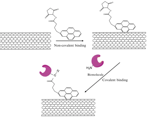
structure – and thus their important electronic characteristics – to be preserved. In this approach, the heterobifunctional molecule, PASE is irreversibly absorbed to SWCNTs, as the pyrenyl group interacts strongly with the hydrophobic surface of the SWCNTs due to p-stacking. The succinimidyl ester group located at the other end of the PASE molecule is highly reactive toward nucleophilic substitution with the free amines typically found on the surfaces of most proteins, resulting in the formation of an amide bond (Fig. 1). This concept has been successfully used to immobilize biomolecules to SWCNTs on gold grids (27) (see Subheading 3.2.1). Similarly, glucose oxidase can be immobilized via noncovalent functionalization of SWCNTs deposited on a silicon wafer to form a sensitive pH sensor (28). The protocol can also be used to attach SWCNTs to oxide substrates, such as quartz, SiO2 on Si, and ITO (29, 30) (Subheading 3.2.2). However, in this scheme the succinimidyl ester moiety of PASE is first used to link the molecule to an amino-modified substrate; this is then followed by the noncovalent attachment of SWCNTs to the available pyrenyl groups. And finally, as discussed in Subheading 3.2.3, noncovalent functionalization can also be used to attach magnetic nanoparti-cles to SWCNTs through a carboxylic derivative of pyrene rather than the succinimide derivative (31).
1. Incubate a TEM grid (27) or silicon wafer (28) with a sample of SWCNTs in a solution containing 6 mM PASE in DMF for 1–2 h at room temperature. (NB: Alternatively, the solution can be prepared at 1 mM in methanol (27).)
2. Wash the SWCNT-modified grid or wafer three times with pure DMF (or methanol) to wash away excess PASE reagent. 3. Add an aqueous protein solution to the functionalized
SWCNTs for 18 h at room temperature. For example, add 3.2. Noncovalent Sidewall Functionalization of SWCNTs for Protein Immobilization (see Notes 6 and 7) 3.2.1. Protein Immobilization to Pyrene-Functionalized SWCNTs Deposited on a TEM Grid or Silicon Wafer
ferritin at 5 mg/mL in a 7.5-mM NaCl solution (27) or 10 mg/mL glucose oxidase in filtered deionized water (28). 4. Rinse the TEM grid or wafer thoroughly with pure water for
6 h to remove excess protein.
5. Dry the grid or wafer and store at 4°C (see Note 8).
1. Clean the oxide substrate, such as quartz, SiO2 on Si, or ITO, with a 1:1 (v/v) mixture of concentrated HCl/methanol for 30 min, and then rinse with water.
2. Immerse the cleaned oxide substrate in concentrated sulfuric acid for 30 min to maximize the number of surface hydroxyl groups, and then rinse with water.
3. Boil the acid-cleaned oxide substrate in water for several minutes, and then blow dry with a stream of N2 gas.
4. React the surface hydroxyls of the clean oxide substrate with a 1% (v/v) solution of a heterobifunctional linker molecule
3.2.2. Noncovalent Immobilization of SWCNTs to Pyrene-Functionalized Oxide Substrates Non-covalent binding O O N O O O O N O O Covalent binding O N H H2N Biomolecule
Fig. 1. Noncovalent functionalization of single-walled carbon nanotubes (SWCNTs). The bifunctional molecule, 1-pyrene-butanoic acid, succinimidyl ester is irreversibly absorbed to SWCNTs since the pyrenyl group interacts strongly with the hydrophobic surface of the SWCNTs via p-stacking. The succinimidyl ester groups are highly reactive to nucleophilic substitution with the amines found on the surfaces of most proteins, resulting in the formation of an amide bond.
(i.e., an organosilane containing a free amino group on one end and a silane group at the opposite end, such as, e.g., AEAPTMS) in 1 mM acetic acid for 20 min at room tempera-ture, resulting in a surface covered with amino functional groups. Rinse three times with water, blow dry, and then bake the substrate for 3–4 min at 120°C.
5. Mix together 500 mL of 0.1 M sodium tetraborate (pH 8.5) with 400 mL of DMF containing 1 mg (2.9 mM) of PASE. Immerse the substrate containing free amino groups into this solution for 12 h, resulting in a pyrene-covered surface. 6. React the pyrene-functionalized surface for 2 h with oxidized
SWCNTs (prepared as described in Subheading 3.3) (see Note 9).
1. Prepare the carboxylic acid pyrene derivative (PyAH) by dis-persing 1 mmol of pyrene aldehyde and 1 mmol of 11-amin-oundecanoic acid in 30 mL of absolute ethanol. Stir the reaction mixture at room temperature for 24 h and separate the solution from unreacted pyrene aldehyde by filtration. Evaporate the ethanol slowly and precipitate the characteris-tic yellow Schiff base.
2. Disperse 1 mg of purified SWCNTs (refluxed in 2.5 M HNO3 for 12 h followed by washing with water and drying) in a solu-tion of 5 mg of carboxylic acid pyrene derivative (obtained from Step 1) in 50 mL of chloroform/ethanol (1:1, v/v). Stir overnight until the yellow color of the supernatant disappears. 3. Prepare capped magnetic iron oxide nanoparticles by adding
1.14 g of KOH in 20 mL of distilled water to 2 g of ammo-nium iron sulfate in 50 mL of water. Add 0.38 g of ammoammo-nium peroxodisulfate in 10 mL of water and 1.5 mL of oleic acid in 30 mL of toluene. Heat the mixture at 80°C for 30 min. Separate the organic phase and precipitate the capped iron oxide nanoparticles with ethanol.
4. Add 5 mg of capped magnetic iron oxide nanoparticles (obtained from Step 3) in 5 mL of chloroform to the PyAH-functionalized SWCNTs and stir for 2 days (see Note 10). 5. Separate the solution from the residue by filtration and wash
with acetone.
In order to covalently attach a biomolecule to CNTs, the introduc-tion of reactive funcintroduc-tional groups on the CNTs is a prerequisite. The carboxylic acid group is widely used since it is easily formed on CNTs via oxidizing treatments. The oxidatively introduced car-boxyl groups present useful sites for further modifications, as they enable the covalent coupling of molecules through the creation of amide and ester bonds. By this method, a wide range of functional
3.2.3. Immobilization of Capped Magnetic Nanoparticles to SWCNTs Functionalized with a Carboxylic Acid Pyrene Derivative (31) 3.3. Preparation of Oxidized Carbon Nanotubes for Covalent Functionalization with Biomolecules
moieties may be incorporated onto nanotube surfaces, for which purpose bifunctional molecules (e.g., diamines) are often utilized as linkers. A few illustrative examples of this approach include nanotubes equipped with dendrimers, nucleic acids, enzymes, metal complexes, or semiconductors and metal nanoparticles.
The oxidation of CNTs can be achieved by extensive ultra-sonic treatment in a mixture of concentrated nitric and sulfuric acid (Subheading 3.3.1) (32). Such treatment leads to the opening of the nanotube caps with holes formed in the tube sidewalls, followed by oxidative etching along the walls. The resulting CNTs that are obtained typically display lengths ranging from 100 to 300 nm, with both ends and the sidewalls decorated predomi-nantly with a high density of carboxyl groups. In an alternative oxidation protocol (Subheading 3.3.2), nanotube shortening can be minimized by refluxing the CNTs in concentrated nitric acid alone (33). Using this approach, the resulting CNTs have most of their pristine mechanical and electrical properties preserved; how-ever, oxidation only occurs at the opening of the tube caps and carboxylic acid functional groups are only formed at defect sites found along the sidewalls.
1. Suspend 10 mg of CNTs in 40 mL of a 3:1 (v/v) mixture of concentrated H2SO4/HNO3 in a 100-mL test tube and soni-cate in a water bath for 24 h at 35–40°C (see Note 11). 2. Dilute the resultant suspension with 200 mL of distilled
water, and collect the (larger sized) oxidized CNTs on a filter membrane (100-nm pore size).
3. Wash the collected CNTs extensively with a 10-mM NaOH solution.
4. Polish the cut tubes by suspending them in a 4:1 (v/v) mixture of concentrated H2SO4/30% aqueous H2O2 with stirring at 70°C for 30 min. This step will result in CNTs containing carboxylic acid groups rather than carboxylate groups. 5. After washing in water and filtering on a filter membrane
(100-nm pore size), resuspend the cut CNTs at a concentra-tion of 0.1 mg/mL in a 0.5% (w/w) Triton X-100 soluconcentra-tion. 1. Reflux 8.5 g of CNTs in 1.2 L of 2.6 M nitric acid for 45 h. 2. Cool the solution, and then transfer it to
polytetrafluoroeth-ylene centrifuge tubes and spin at 2,400 × g for 2 h.
3. Decant the supernatant acid and replace it with deionized water. Shake vigorously to resuspend the solids, followed by a second centrifugation–decanting cycle.
4. Resuspend the collected solids in 1.8 L of water with 20 mL of Triton X-100 surfactant, and adjust the mixture to pH 10 with sodium hydroxide.
3.3.1. Oxidation of Carbon Nanotubes in a Concentrated Sulfuric Acid/Nitric Acid Mixture
3.3.2. Oxidation of Carbon Nanotubes in Concentrated Nitric Acid
In order to deposit CNTs onto electrodes, their hydrophobic nature should be reduced by introducing hydrophilic groups onto the nanotube surface (34). Numerous methods have been reported in the literature for the functionalization and dispersion of CNTs (35). The important factors necessary for a good dispersion include having intact CNT structures without any major damage or modi-fications, and achieving a uniform homogeneous solution with long-term stability. The dispersion of CNTs is typically carried out by using physical methods (e.g., ultrasonication and milling) and chemical-based approaches (via covalent or noncovalent function-alization). Among the various creative methods that have been reported, we have selected a few representative protocols for the preparation of CNT/nanoparticle composites. The incorporation of nanoparticles into CNT-based materials can lead to an enhanced level of electrocatalytic activity for many electrochemical processes, potentially giving rise to several highly efficient applications such as photoelectrochemical cells and sensor devices. Gold or plati-num nanoparticles (NPs), e.g., can be attached to the walls of CNTs by various approaches, including physical evaporation, chemical reaction with functionalized CNTs, and layer-by-layer deposition methods (36–40). This section describes some key pro-tocols for the preparation of thiolated CNTs, and the self-assembly of gold and other metallic nanoparticles onto such thiol-modified CNTs. The gold nanoparticles can be easily prepared with con-trolled size and dispersion in the presence of a-cyclodextrin, as described in Subheading 3.4.5 (41–43).
1. Add potassium hydroxide (200 mg) and MWCNTs (10 mg) to a ruby mortar and grind them together for 1 h at room temperature.
2. Dissolve the reaction mixture in double-distilled, deionized water (10 mL) and precipitate several times in methanol to remove the potassium hydroxide.
3. Sonicate the MWCNTs in 10 mL of water for 6 h to obtain a uniform dispersion. The preceding steps make the MWCNTs hydrophilic in nature and help break down larger bundles of MWCNTs into smaller ones (44).
4. Prepare a suspension of the MWCNTs (1 mg; obtained from Step 3) in a 1% (w/w) HAuCl4 solution (1 mL) by sonication for 5 min to make the nanotubes disperse evenly throughout. 5. Dilute the MWCNT/HAuCl4 suspension to 100 mL with
double-distilled water and heat to boiling while stirring. 6. To obtain MWCNTs coated with gold nanoparticles, add
1.5 mL of 1% (w/w) sodium citrate to the boiling MWCNT/ HAuCl4 solution, and continue heating for 5–10 min until the color of the solution does not change (45).
3.4. Preparation of Metallic Nanoparticle/ CNT Composites 3.4.1. Preparation of MWCNT/AuNP Composites by Mechanical Processing and One-Step Citrate Reduction in Aqueous Solution (44, 45)
1. Heat pristine MWCNTs in air at 600°C for 2 h and then soak in hydrochloric acid for 24 h and centrifuge.
2. Rinse the resulting precipitate with deionized water and dry under air.
3. Chemically functionalize the MWCNTs by ultrasonication in a mixture of sulfuric acid/nitric acid (3:1, v/v) for 8 h. 4. Centrifuge and wash the acid-treated MWCNTs with
deion-ized water three times and oven dry. Redisperse the MWCNTs (containing carboxylic acid groups) in deionized water. 5. For layer-by-layer assembly, prepare a solution of PDAC (MW
100,000–200,000) at a concentration of 0.1 mg/mL in deionized water containing 0.05 M NaCl. Similarly, prepare a solution of 0.1 mg/mL PSS(MW 70,000) in deionized water containing 0.05 M NaCl.
6. Disperse the MWCNTs in the PDAC solution (0.1 mg/mL) for 3 h with sonication to obtain MWCNTs coated with a layer of PDAC.
7. Mix the PDAC-modified MWCNTs with the PSS solution (0.1 mg/mL) for 1 h with sonication, in order to obtain MWCNTs coated with a PDAC/PSS bilayer.
8. Centrifuge and wash the polymer-modified MWCNTs three times with deionized water.
9. Add a solution containing positively charged gold nanopar-ticles to the dispersion of polyelectrolyte-coated MWCNTs to obtain self-assembled MWCNT/AuNP complexes.
1. Disperse 10.5 mg of purified MWCNTs with 0.70 g of PSS in 70 mL of distilled water with continuous sonication for 15 min, and then hold the temperature at 50°C for 12 h under vigorous agitation.
2. Remove excess PSS by performing three centrifugation/ resuspension cycles, spinning at 12,000 × g for 30 min each time. Dry the product under vacuum at 60°C overnight to obtain the MWCNT/PSS product as a powder.
3. Dissolve 42 mg of IL-NH2 (72) in 11.6 mL ultrapure water, and then add 0.20 mL of an aqueous solution of MWCNTs/ PSS (2 mg/mL) dropwise into the mixture under stirring to form a well-dispersed solution.
4. To prepare the MWCNT/polymer/gold nanoparticle-ionic liquid (MWCNT/PSS/Au-IL) composites, add 0.20 mL of HAuCl4 aqueous solution (20 mM) dropwise over several minutes to the above mixture (MWCNTs/PSS).
5. After stirring for 10 h, filter the mixture through a nylon membrane filter (0.22 mm pore size). Wash the recovered
3.4.2. Preparation of MWCNT/AuNP Composites by Electrostatic Layer-by-Layer Assembly (39) 3.4.3. In Situ Synthesis of AuNPs on PSS Polymer-Wrapped MWCNTs Using Ionic Liquids (46)
MWCNT/PSS/Au-IL composite product thoroughly with water, and then dry overnight at 60°C under vacuum. 1. Disperse 0.4 mg of purified MWCNTs (containing carboxylic
acid groups) in 15 mL of distilled water under ultrasonic treatment.
2. Transfer 1.5 mL of the MWCNT dispersion into a vial, and then add 0.04–0.1 mg of cationic PEI polymer using a 1.0 M PEI aqueous stock solution. Mix the solution by a combina-tion of vigorous stirring and sonicacombina-tion.
3. Add 0.2 mL of HAuCl4 (24.3 mM) to the above mixture to give the desired molar ratio of PEI (monomer unit) to gold. Previous work has shown that the packing density of the attached gold NPs generally increases as the initial molar ratio of PEI:HAuCl4 decreases (i.e., from 400:1 to 9.8:1) (47). 4. Heat the resulting mixture at 60°C for 20 min.
5. After washing and centrifugation for two cycles, disperse the resulting MWCNT/AuNP composite product in 1 mL of water.
1. Disperse 1 mg of purified, acid-treated SWCNTs into 2 mL of deionized water and sonicate for 2 h to generate a black suspension of nanotubes.
2. Add HAuCl4 (1 mM, 0.8 mL) and a-cyclodextrin (a-CD) (2 mM, 1 mL) to the SWCNT suspension.
3. Add 20-mL aliquots (to a maximum volume of 100 mL) of freshly prepared, ice-cold 0.1 M sodium borohydride (NaBH4) with slow mixing using a magnetic stir bar.
4. Continue stirring the reaction mixture until a stable reddish color is observed.
5. Incubate the solution for 24 h at room temperature (~22–24°C) to let it stabilize.
6. Purify the AuNP/SWCNTs by washing and centrifugation (8,000 × g) three times to remove the unbound AuNPs. The supernatant should appear clear after the first washing cycle. Bioconjugated CNTs have numerous potential biomedical applications based on their unique properties. Some examples of such applications include biosensing, cell tracking and labeling, tissue engineering (as scaffolds), as well serving as vehicles for the intracellular transport of drugs, genes, and/or proteins. In particular, the efficient intracellular delivery of anticancer drugs, siRNA, proteins, and DNA combined with their corresponding effective therapeutic effects suggests that CNTs may provide a promising approach for transporting bioactive molecules in the
3.4.4. Deposition of AuNPs on PEI Polymer-Wrapped MWCNTs (47) 3.4.5. Preparation of SWCNT/AuNP Composites by Borohydride Reduction in the Presence of Cyclodextrin (41–43) 3.5. Bioconjugation of Carbon Nanotubes
next generation of biomedicines. CNTs may also serve as important components in novel bioengineered nanomaterials that can potentially impart several important features, such as greater mechanical strength per unit mass, good electrical and thermal conductivity, and the ability to guide cell growth and tissue regen-eration. CNTs can also be used as a platform for probing surface–protein or protein–protein interactions toward the devel-opment of highly specific biomolecule detectors. Indeed, it is anticipated that the next wave of advances in the nanobioelec-tronics field is the synthesis and fabrication of high-density, mul-tiplexed nanotube device microarrays for proteomic applications for simultaneously detecting large numbers of different target proteins in “real world” samples.
Although the noncovalent modification of CNTs with a pyrene moiety can be used to conjugate a biomolecule of inter-est as discussed earlier (see Subheading 3.2.1), the specific bio-conjugation of target biomolecules is most commonly achieved by covalent methods. Another interesting procedure is the use of APTES to solubilize CNTs to form a very stable CNT/ APTES complex. Biomolecules can then become covalently conjugated to the amino group of APTES via glutaraldehyde activation. This procedure, described below in Subheading 3.5.1, has been used to prepare enzyme electrodes with efficient direct electron transfer (48).
1. Dissolve 2.0 mg of MWCNTs in a mixture containing 100 mL of APTES, 100 mL Nafion perfluorinated ion-exchange resin and 800 mL of ethanol.
2. Sonicate the mixture for 20 min to obtain a suspension of uniformly dispersed MWCNTs.
3. Drop 3 mL of the MWCNT solution onto the surface of a solid hydrophobic substrate, e.g., a glassy carbon (GC) electrode (3 mm in diameter) and dry in air to form a uniform film con-taining a network of MWCNTs.
4. Drop 3 mL of a solution of enzyme (e.g., 20 mg/mL glucose oxidase in 50 mM phosphate buffer, pH 7.2) onto the MWCNT/APTES-modified GC electrode and dry in air. (Other enzymes can easily replace the glucose oxidase used here to form biosensors for other analytes.)
5. Drop 3 mL of a 2.5% glutaraldehyde solution (diluted tenfold from a 25% (w/w) stock solution in 50 mM phosphate buf-fer, pH 7.2) onto the electrode to crosslink the enzyme with APTES and dry.
6. Store the enzyme-modified electrode at 4°C in 50 mM phos-phate buffer, pH 7.2.
3.5.1. Solubilization and Bioconjugation of MWCNTs using APTES (48)
1. Prepare a 5-mL solution of 5 mM EDC and 10 mM sulfo-NHS in 50 mM MES buffer solution (pH 5.7) containing 0.5 M NaCl (see Notes 13 and 14).
2. Suspend carboxylated CNTs (1 mg/mL) in 5 mL of 50 mM MES buffer solution containing 0.5 M NaCl (pH 5.7). 3. Add the solutions from Steps 1 and 2 together (to give a total
volume of 10 mL) and let the resulting mixture react for 30 min at room temperature.
4. Remove the excess reactants from the EDC/sulfo-NHS-acti-vated CNTs by successive centrifugation and dilution with deionized water, followed by filtering and concentrating with an ultrafiltration membrane (50 kDa MWCO).
5. Dissolve the protein to be coupled at a concentration of 1–10 mg/mL in 500 mL of 0.1 M phosphate buffer, 0.5 M NaCl, pH 7.2.
6. Resuspend the activated CNTs from Step 4 in the buffered protein solution prepared in Step 5.
7. Allow the mixture to react under continuous agitation with a magnetic stirrer for at least 2 h at room temperature.
8. Add 2-mercaptoethanol to the reaction solution to obtain a final concentration of 20 mM to quench the coupling reac-tion. Alternatively, if the protein being activated is sensitive to this concentration level of 2-mercaptoethanol, quench the reaction by performing the following: wash the mixture by extensive centrifugation/resuspension using a suitable buffer (e.g., 10 mM sodium phosphate, 0.15 M NaCl, pH 7.4),
3.5.2. Covalent Coupling of Carboxylated CNTs with Biomolecules Using EDC/ NHS (see Note 12 and Fig. 2)
Fig. 2. Carbon nanotubes containing carboxylate groups (COOH-CNTs) can be coupled to amine-containing biomolecules using an aqueous two-step coupling process based on EDC/NHS (or sulfo-NHS) chemistry to form an amide bond.
followed by dialysis using an ultrafiltration membrane (50 kDa MWCO).
9. Resuspend the conjugated CNTs in 10 mM sodium phosphate, 0.15 M NaCl, pH 7.4, and store at −20°C.
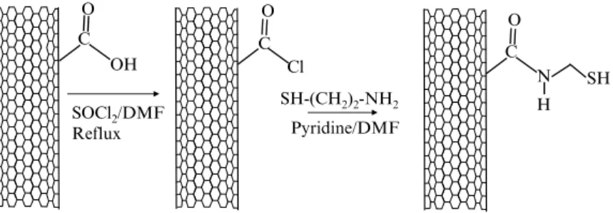
In this protocol, the carboxyl groups of CNTs are to be converted to amino groups by refluxing the CNTs in DMF in the presence of thionyl chloride. The resulting CNTs are modified with ethyl-enediamine to form an intermediate that can react with aldehyde groups, which can be introduced into the biomolecules of inter-est through various methods. For example, as described below, enzymes and proteins that are glycosylated (e.g., glucose oxidase and horseradish peroxidase) can be oxidized with sodium perio-date to produce aldehyde groups for reductive amination cou-pling to amino-modified CNTs (Fig. 3).
1. Place oxidized MWCNTs (containing carboxyl groups after treatment with sulfuric and nitric acids; see Subheading 3.3.1) in a vacuum oven and dry at 70°C overnight.
2. Suspend the dried CNTs (1 mg/mL) in anhydrous DMF using an ultrasonic water bath.
3. Add the CNT dispersion to thionyl chloride and reflux for 24 h to convert the carboxylic acids to acyl chlorides.
4. Wash the resulting nanotubes with anhydrous THF (five times) to remove excess SOCl2 using a polycarbonate membrane filter (0.22 mm pore size).
5. React the nanotubes with ethylenediamine for 3 days under continuous stirring to form CNTs with amino groups. 6. Wash the amino-modified CNTs with deionized water
exten-sively to remove excess ethylenediamine using a polycarbonate membrane filter (0.22 mm pore size).
7. Prepare a solution of a carbohydrate group-containing enzyme of interest in PBS buffer (e.g., glucose oxidase in PBS, pH 6.8).
3.5.3. Conversion of CNT Carboxyl Groups to Amino Groups for Covalent Bioconjugation (49) SOCl2/DMF Reflux SH-(CH2)2-NH2 C O O OH C Cl Pyridine/DMF C O N SH H
Fig. 3. Conversion of carboxylate group-containing carbon nanotubes (COOH-CNTs) to thiolated CNTs, which can be used for covalent coupling with gold nanoparticles or gold-labeled biomolecules.
8. Add 100 mL of 88 mM sodium periodate solution immediately to each milliliter of the enzyme solution to give a final perio-date concentration of 8 mM in the reaction mixture.
9. Incubate the reaction mixture from Step 8 for 1 h in an ice bath with gentle stirring in the dark.
10. Terminate the reaction by adding either glycerol (0.1 mL glycerol per mL of reaction solution) or ethylene glycol (250 mM) and incubating further for 30 min at room temperature.
11. Purify and concentrate the oxidized enzyme product by dialysis (10 kDa MWCO).
12. Disperse a predetermined amount of the amino-modified CNTs (from Step 6) in PBS buffer (pH 7.5) for 1 h in an ultrasonic water bath.
13. Slowly add the concentrated solution of oxidized glucose oxidase (from Step 11) to the suspension of amino-modified CNTs while gently stirring. Incubate the reaction mixture for 2 h to obtain CNTs covalently conjugated to glucose oxidase.
CNTs decorated with gold nanoparticles (prepared as described in Subheading 3.4) can be conjugated with biomolecules using a variety of different procedures. In the example protocol below, DTSP ( also known as Lomant’s reagent) adsorbs to the surface of the gold nanoparticles through the disulfide group, so that the terminal succinimidyl groups allow further covalent immobili-zation of amino-containing biomolecules such as enzymes, proteins, etc. (Fig. 4).
3.5.4. Covalent Attachment of Biomolecules to Gold Nanoparticle-Decorated CNTs
Fig. 4. Adsorption of dithiobis(N-succinimidyl propionate) (DTSP) to gold nanoparticles decorated on CNTs. The adsorbed DTSP can be used to couple the gold/CNT complexes to the free amino groups of biomolecules.
1. Incubate gold nanoparticle-decorated CNTs in 10 mM DTSP in DMSO for 30 min (see Note 15).
2. Wash the resulting nanotubes with DMSO three to five times to remove excess DTSP using a polycarbonate membrane filter (0.22 mm pore size).
3. Wash the functionalized tubes with deionized water three to five times.
4. Incubate the active ester-functionalized gold-decorated CNTs in a buffer solution containing the enzyme of interest (e.g., horseradish peroxidase (6 mg/mL) in 0.1 M phosphate buffer, pH 7) for 1.5–3 h.
5. Rinse the bioconjugated CNTs with buffer three to five times to remove excess enzyme.
The bioconjugation of CNTs is currently a subject of intense endeavor; however, the controlled functionalization of CNTs has not yet been fully established. Modification of the CNT surface with metal nanoparticles can potentially impart unique and/or enhanced properties, leading to the production of novel nanoma-terials for diversified applications in electrocatalysis, biosensors, biofuel cells, and implantable bioassays.
1. A useful online listing of CNT suppliers may be found at http://www.nanoten.com/ntyp.html. Some specific examples of suppliers include the following: (1) NanoCarbLab (NCL; http://www.nanocarblab.com) offers pristine as well as modi-fied SWCNTs with different purities ranging from 40 to 90% (all percentage purities stated herein are referred to as % w/w values): SWCNT-COOH (70–80% purity, 2–5% of carboxyl (–COOH) groups) and SWCNT-NH2 (70–80% purity, 2–5% of amino (–NH2) groups). Shortened SWCNT-COOH and SWCNT-NH2 (length 200–500 nm) with higher purity (90%) and with a higher content of –COOH or –NH2 groups (5–10%) are also available. The company also offers DWCNTs with 20–30% to 90% purity. As expected, CNTs with a high purity rating are very expensive compared to their counter-parts with a low purity rating. (2) NanoLab (http://www. nano-lab.com) is another commercial source of MWCNTs and SWCNTs. The MWCNTs are produced by CVD and purified to >95% as measured by thermal gravimetric analysis (TGA). Two different types of MWCNTs are offered: tubes with lengths of 1–5 mm and diameters of 15 ± 5 nm or 30 ± 10 nm; and longer tubes (5–20 mm) with the same
4. Notes
diameters. However, the products still contain 0.94% Fe and 0.14% S compared to 98.92% C. SWCNTs with different purities (40–95%) can also be obtained from NanoLab. SWCNTs with high purity (95%) and shorter lengths (1–5 mm) are prepared by CVD, whereas SWCNTs prepared by arc dis-charge are longer (over 10 mm) and contain 25% Ni or 40–50% Fe. Even after extensive purification, SWCNTs with over 90% purity still contain about 10% Fe. The company also offers DWCNTs (outer diameter = 4 ± 1 nm with purity over 95%) produced by the CVD method. COOH- and NH2 -functionalized CNTs are also available from this company. Pristine CNTs produced by CVD are subject to a reflux in sulfuric/nitric acid to generate a large concentration of car-boxyl groups on the CNT surface (2–7% –COOH by titra-tion). Amino-functionalized CNTs are produced as a derivative of COOH-functionalized nanotubes, whereby the –COOH group reacts with SOCl2 to form an acyl chloride, which is then reacted with dimethylamine to generate NH2 -modified CNTs. (3) Carbon Solutions, Inc. (http://www. carbonsolution.com) offers functionalized SWCNTs, such as CNT-PEG (polyethylene glycol), and also supplies AP (as-prepared)-SWCNTs with low purity (40–60% carbon and 30% metal content) as well as purified SWCNTs with high purity (>90% carbon and 2–4% metal content). (4) Nanocs (http://www.nanocs.com) offers both SWCNTs (diameters of 2–10 nm, lengths of 50 nm to microns) and MWCNTs (diameters of 10, 20, 40, 60, and 80–150 nm, with lengths of 1–100 mm) with different purities (40% to over 90%). The metal content in such products ranges from 2 to 30%. The company also offers modified CNTs such as CNT-COOH, CNT-NH2, CNT-SH, and CNT-PEG. Such functional groups can be attached either at the ends or the walls of the nanotubes.
2. Both the purity and solubility of prepared CNTs continue to be an issue for practical applications, i.e., new purification and characterization techniques are still needed. For example, one purification method developed at the NASA Glenn Research Center can purify gram-scale quantities of SWCNTs. This method uses a combination of high-temperature oxidations and repeated extractions with nitric and hydrochloric acid, and is a modification of a gas-phase procedure reported previ-ously by Smalley and coworkers (14). Nanotubes purified by this method reveal near complete removal of iron catalyst particles, from 22.7% (w/w) in the crude preparation to less than 0.02% (w/w) in the final product.
3. Fine, homogeneous dispersion/suspension of CNTs (1 mg/10 mL solvent) can be prepared using DMF, decalin,
and xylenes. Chloroform is not able to disperse CNTs, whereas chlorobenzene and 1,2-dichlorobenzene can be used to prepare homogeneous dispersions/solutions and homoge-neous solutions, respectively. Furthermore, oleum (20% free SO3) can disperse CNTs without sonication; however, such solvents are not compatible for bioconjugation.
4. Sonication of CNTs in 1% (w/v) aqueous SDS results in a solu-tion (50–100 mg CNTs/L) that is stable for about 1 month. However, SWCNTs are less soluble compared to MWCNTs, and only short SWCNTs (150 nm) are able to be dissolved. 5. At each step, the purified products should be examined by
standard analytical methods such as TEM, atomic force microscopy (AFM), TGA, Raman spectroscopy, visible-near infrared (NIR) spectroscopy, etc. Unfortunately, sample volumes are typically very small and there is no algorithm that can be used to easily convert the images from AFM or TEM into numerical data to quantify purity. When working with small quantities of starting materials, it is always desirable to obtain CNTs with the highest grade of purity from commer-cial sources (such as those listed as examples in Note 1 above).
6. CNTs are difficult to disperse and cannot be wetted by liquids with surface tensions higher than 100–200 mN/m. Thus, only ~0.1 mg of MWCNTs can be solubilized (with the aid of sonication) in 1 mL of DMF – one of the most effective solvents for CNT solubilization (50). Furthermore, CNTs tend to agglomerate as bundles in solvents, and if dispersed can reagglomerate quickly thereafter due to electrostatic attraction. Nevertheless, wrapping CNTs with a polymer can enhance the solubility of the nanotubes without affecting their original properties (51). CNTs can also be suspended and solubilized in Nafion (52), a perfluorosulfonated polymer, to facilitate the modification of electrode surfaces with an enzyme. Other procedures such as end- (52) and/or side-wall- (53) functionalization, sonication in the presence of a surfactant (54), and protonation by acids (55) are also effective to a certain extent. A concentrated H2SO4/HNO3 mixture can be used to generate carboxylic acid groups at the ends and sidewalls of CNTs via a refluxing/sonication process (56). The resulting oxidized CNTs become more soluble and can be stabilized in aqueous suspensions. Induced defects in the structure of oxidized CNTs can provide sites for the covalent coupling of biomolecules using the standard water-soluble coupling agent EDC (57, 58). However, such aggressive oxidative procedures often cut the CNTs into shorter fragments, thus partly reducing their high aspect ratio (59).
7. The functionalization of CNTs with biomolecules can be real-ized by both noncovalent- and covalent-based modification, and several biomolecules can also become adsorbed directly to CNTs noncovalently. In particular, antibodies (60), enzymes (61), and peptides (62) can bind nonspecifically to the exte-rior surface of CNTs, whereas oligonucleotides can bind to the surface as well as the open cavity space of CNTs. DNA– CNT interactions in water are suggested to result from DNA base-stacking at the CNT surface, with the hydrophilic sugar-phosphate backbone oriented outward toward the aqueous medium (63). And finally, ionic liquids and polymers – for instance, 4-chlorobenzenediazonium tetrafluoroborate (64) and PDAC (65–71), respectively – are also very efficient in functionalizing CNTs, resulting in charged nanotube struc-tures that may have potential utility in biomedical applications due to their strong interactions with charged cell membranes. Various ionic liquids are able to solubilize CNTs (72–76); the presence of the ionic liquid leads to the exfoliation of SWCNT ropes followed by the addition of functionalized aryl groups to the SWCNT sidewalls, resulting in functionalized SWCNTs that remain predominantly as separated strands.
8. In some cases, after immobilization, enzymes may be deacti-vated or their inherent activity is greatly reduced. One way that may help circumvent this issue is to conduct the immo-bilization of the target enzyme in the presence of its substrate. 9. Prior to their deposition onto the pyrene-modified substrate,
the oxidized SWCNTs are suspended in an aqueous solution and sonicated for 1 h. Following this, the suspension is cen-trifuged at 10,000 × g for 5 min, and then the recovered supernatant is filtered through filter paper (0.45 mm) (29). Alternatively, the SWCNTs used for deposition may be stabilized through the addition of highly charged ZrO2 nanoparticles (30).
10. Alternatively, capped cobalt or cobalt/platinum nanoparticles may also be used (31). Sonication in concentrated H2SO4 results in a (20 mg/L) CNT suspension that is only stable for a few hours. A very stable CNT suspension of up to 200 mg/L can be obtained by stirring the CNTs in oleum (20% w/w SO3 in H2SO4).
11. As discussed earlier, acid treatment of CNTs at elevated tem-perature facilitates their dispersion in an aqueous environ-ment. This treatment also creates a variety of carbonyl functional groups at defect sites in the outer graphene sheet. Further treatment with perchloric acid (HClO4) or potassium permanganate (KMnO4) will convert all of these groups into carboxylic acids. The carboxylates, in turn, can be used to link
CNTs to the amino groups of biomolecules or proteins using the well-known carbodiimide procedure (77). In this approach, the reaction between the –N=C=N– moiety of the carbodiimide with the carboxyl group of the CNT forms a highly reactive o-acylisourea derivative with an extremely short half-life (Fig. 2). This active species will react with the amino group of a biomolecule (i.e., enzyme, protein, etc.) to form a stable amide bond. Carbodiimides are “zero-length” crosslinking agents as no additional chemical structure is introduced between the CNT and the biomolecule. In some cases, the activity of the immobilized biomolecules may be affected due to steric hindrance induced by the covalent bind-ing process. This carbodiimide couplbind-ing procedure is often used to crosslink biomolecules to each other (such as in the synthesis of protein–protein, peptide–protein, nucleotide– protein complexes) as well as to immobilize biomolecules to a substrate. The water-soluble carbodiimide EDC and
N-hydroxysulfosuccinimide (sulfo-NHS) are widely used for carrying out this type of coupling reaction. In general, the advantage of adding sulfo-NHS to EDC reactions is that an increase in the stability of the active intermediate is achieved (Fig. 2). In this reaction scheme, nucleophilic attack of the free amino groups of the biomolecule by the activated car-boxyl groups on the CNTs results in the CNT–protein con-jugate. The protocol provided here is generalized and simplified from several different sources that describe in detail methods to couple a protein molecule to CNTs through EDC/sulfo-NHS chemistry (78–80).
12. Besides EDC, N-Cyclohexyl-N ′-2(4′-morpholium) ethyl carbodiimide-p-toluenesulfonate can also be used for the coupling reaction. EDC and N-hydroxysuccinimide (NHS) are widely used together since the NHS produces a more stable reactive intermediate, which has been shown to give a greater reaction yield.
13. Carbodiimide coupling reactions using EDC can also be per-formed in a solution of N-2-hydroxyethylpiperazine-N ¢- 2-ethanesulfonic acid (HEPES) at an optimal pH of 7.2–7.5.
14. Alternatively, the gold nanoparticle-decorated CNTs can be incubated with 20 mM cystamine (2,2¢-diaminodiethyldisul-fide) in deionized water for 2–3 h to introduce –S–(CH2)2–NH2 onto the gold surface. Biomolecules containing –COOH groups can then be coupled to the free amino groups on the gold-decorated CNTs by carbodiimide coupling, similar to the procedure described in Subheading 3.5.2.
15. 1-(3-Aminopropyl)-3-methylimidazolium bromide (ILNH2) may be prepared in the laboratory, as described in (46).
References
1. Iijima, S. (1991) Helical microtubules of gra-phitic carbon. Nature 354, 56–58.
2. Dresselhauss, M.S., Dresselhauss G., and Eklund, P.C. (1996) Science of fullerenes and carbon nanotubes. Academic Press, San Diego, CA, pp. 1–985.
3. Dekker, C. (1999) Carbon nanotubes as molecular quantum wires. Phys. World 52, 22–28.
4. Treacy, M.M.J., Ebbesen, T.W., and Gibson, J.M. (1996) Exceptionally high Young’s mod-ulus observed for individual carbon nanotube. Nature 381, 678–80.
5. Dai, H. (2000) Controlling nanotube growth. Phys World 13, 43–47.
6. Ebbesen, T.W., and Ajayan, P.M. (1992) Large-scale synthesis of carbon nanotubes. Nature 358, 220–222.
7. Ando, Y., and Iijima, S. (1993) Preparation of carbon nanotubes by arc-discharge evapora-tion. Jpn. J. Appl. Phys. 32, L107–L109.
8. Zhao, X., Wang, M., Ohkohchi, M., and Ando Y. (1996) Morphology of carbon nanotubes prepared by carbon arc. Jpn. J. Appl. Phys. 35, 4451–4456.
9. Iijima, S., and Ichihashi, T. (1993) Single-shell carbon nanotubes of 1-nm diameter. Nature
363, 603–605.
10. Sen, R., Ohtsuka, Y., Ishigaki, T., Kasuya, D., Suzuki, S., Kataura, H, et al. (2000) Time period for the growth of single-wall carbon nanotubes in the laser ablation process: evidence from gas dynamic studies and time resolved imaging. Chem. Phys. Lett. 332, 467–473.
11. Che, G., Lakshmi, B.B., Martin, C.R., Fisher, E.R., and Ruoff, R.S. (1998) Chemical vapor deposition based synthesis of carbon nano-tubes and nanofibers using a template method. Chem. Mater. 10, 260–267.
12. Ebbesen, T.W., Ajayan, P.M., Hiura H., and Tanigaki, K. (1994) Purification of nanotubes. Nature 367, 519–519.
13. Chiang, I.W., Brinson, B.E., Smalley, R.E., Margrave, J.L, Hauge, R.H. (2001) Purification and characterization of single-wall carbon nano-tubes. J. Phys .Chem. B, 105, 1157–1161.
14. Chiang, I.W. Brinson, B.E. Huang, A.Y., Willis, P.A., Brownikowski, M.J., Margrave, J.L., et al. (2001) Purification and character-ization of single-wall carbon nanotubes (SWNTs) obtained from the gas-phase decom-position of CO (HiPco process). J. Phys. Chem. B 105, 8297–8301.
15. Haddon, R.C. Sippel, J. Rinzler, A.G., and Papadimitrakopoulos, F. (2004) Purification
and separation of carbon nanotubes. MRS Bull. 29, 252–259.
16. Bandow, S., Rao, A.M., Williams, K.A., Thess, A., Smalley, R.E., and Eklund, P.C. (1997) Purification of single-wall carbon nanotubes by microfiltration. J. Phys. Chem. B 101, 8839–8842.
17. Dujardin, E., Ebbesen,T.W., Krishnan, A., and Treacy, M.M.J. (1998) Purification of single-shell nanotubes. Adv. Mater. 10, 611–613.
18. Dujardin, E. Dny, C.M, Panissod, P. Kintzinger, J.P., Yao, N., and Ebbesen, T.W. (2000) Interstitial metallic residues in purified single shell carbon nanotubes. Solid State Commun.
114, 543–546.
19. Rinzler, A.G., Liu, J., Nikolaev, P., Huffman, C.B., Rodrigues, F.J., Macias, P.J. et al. (1998) Large-scale purification of single-wall carbon nanotubes: process, product, and characteriza-tion. Appl. Phys. A 67, 29–37.
20. Dillon, A.C., Gennet, T., Jones, K.M., Alleman, J.L., Parilla, P.A., and Haben, M.J. (1999) A simple and complete purification of single-walled carbon nanotube materials. Adv.Mater.
11, 1354–1358.
21. Tohji, K., Goto, T., Takahashi, Shinoda, H. Y., Shimizu, N., Jaya-devan, B., et al. (1996) Purifying single-walled nanotubes. Nature
383, 679–679.
22. Tohji, K., Takahashi, H. Y., Shinoda, N., Shimizu, B., Jayadevan, I., Matsuoka, Y., et al. (1997) Purification procedure for single-walled nanotubes. J.Phys.Chem. B, 101, 1974–1978.
23. Martinez, M.T., Callejas, M.A., Benito, A.M., Maser, W.K., Cochet, M., Andres, J.M., et al. (2002) Microwave single walled carbon nanotubes purification. Chem. Commun. 1000–1001.
24. Sen, R., Rickard, S.M., Itkis, M.E., and Haddon, R.C. (2003) Controlled purification of single-walled carbon nanotube films by use of selective oxidation and near-IR spectros-copy. Chem. Mater. 15, 4273–4279.
25. Thien-Nga, L., Hernadi, K., Ljubovic, E., Garaj, S., and Forro, L. (2002) Mechanical purification of single-walled carbon nanotube bundles from catalytic particles. Nano Letts. 2, 1349–1352.
26. Bonard, J-M., Stora, T., Salvetat, J-P., Maier, F., Stockli, T., Duschl, C., et al. (1997) Purification and size-selection of carbon nanotubes. Adv. Mater. 9, 827–831.
27. Chen, R.J., Zhang, Y., Wang, D., and Dai, H. (2001) Noncovalent sidewall functionaliza-tion of single-walled carbon nanotubes for
protein immobilization. J. Am. Chem. Soc. 123, 3838–3839.
28. Besteman, K., Lee, J-O., Wiertz, F.G.M., Heering, H.A., and Dekker, C. (2003) Enzyme-coated carbon nanotubes as single-molecule biosensors. Nano Lett. 3,
727–730.
29. Zhu, J., Yudasaka, M., Zhang, M., Kasuya, D. and Iijima, S. (2003) A surface modification approach to the patterned assembly of single-walled carbon nanomaterials. Nano Letts. 3, 1239–1243.
30. Zhu, J., Yudasaka, M., Zhang, M., and Iijima, S. (2004) Dispersing carbon nanotubes in water: a noncovalent and nonorganic way. J. Phys. Chem. B 108, 11317–11320.
31. Georgakilas, V., Tzitzios, V., Gournis, D., and Petridis, D. (2005) Attachment of magnetic nanoparticles on carbon nanotubes and their soluble derivatives. Chem. Mater. 17, 1613–1617.
32. Liu, J., Rinzler, A. G., Dai, H., Hafner, J. H, Bradley, R. K., Boul, P. J., Lu, A., et al (1998) Fullerene pipes. Science 280, 5367, 1253–1256.
33. Chen, J., Hamon, M. A., Hu, H., Chen, Y., Rao, A. M., Eklund, P. C., and Haddon, R. C.. (1998) Solution properties of single-walled carbon nanotubes. Science 282, 5386, 95–98.
34. Yan, Y., Zhang, M., Gong, K., Su, L., Guo, Z., and Mao, L. (2005) Adsorption of methylene blue dye onto carbon nanotubes: A route to an electrochemically functional nanostruc-ture and its layer-by-layer assembled nano-composite. Chem. Mater. 17, 3457–3463.
35. Breuer, O. and Sundararaj, U. (2004) Big returns from small fibers: A review of polymer/ carbon nanotube composites. Poly. Comp. 25, 630–645.
36. Xue, B., Chen, P., Hong, Q., Lin, J. Y., and Tan, K. L. (2001) Growth of Pd, Pt, Ag and Au nanoparticles on carbon nanotubes. J. Mater. Chem. 11, 2378–2381.
37. Li, J., Moskovits, M., and Haslett, T. L. (1998) Nanoscale electroless metal deposition in aligned carbon nanotubes. Chem. Mater.10, 1963–1967.
38. Azamian, B. R., Coleman, K. S., Davis, J. J., Hanson, N., and Green, M. L. H. (2002) Directly observed covalent coupling of quan-tum dots to single-wall carbon nanotubes. Chem. Commun. 366–367.
39. Kim, B. and Sigmund, W. M. (2004) Functionalized multiwall carbon nanotube/ gold nanoparticle composites. Langmuir 20, 8239–8242.
40. Li. F., Wang, Z., Shan, C., Song, J., Han, D., and Niu, L. (2009) Preparation of gold nanoparticles/functionalized multiwalled carbon nanotube nanocomposites and its glucose biosensing application. Biosens. Bioelectron. 24, 1765–1770.
41. Liu, Y., Male, K.B., Bouvrette, P., and J.H.T. Luong. (2003) Control of the size and distri-bution of gold nanoparticles by unmodified cyclodextrins. Chem. Mat. 15, 4172–4180.
42. Mahmoud, K. A., Hrapovic, S. and Luong, J.H.T. (2008) Picomolar detection of protease using peptide/single walled carbon nanotube/ gold nanoparticle-modified electrode. ACS Nano 2, 1051–1057
43. Mahmoud, K.A. and Luong J.H.T. (2008) Impedance method for detecting HIV-1 protease and screening for its inhibitors using ferrocene-peptide conjugate/Au nanoparti-cle/single-walled carbon nanotube modified electrode. Anal. Chem. 80, 7056–7062.
44. Shie, J.-W., Yogeswaran, U, and Chen, S.-M. (2009) Haemoglobin immobilized on Nafion modified multi-walled carbon nanotubes for O2, H2O2 and CCl3COOH sensors. Talanta,
78, 896–902.
45. Zhang, R. and Wang, X. (2007) One step syn-thesis of multiwalled carbon nanotube/gold nanocomposites for enhancing electrochemical response. Chem. Mater. 19, 976–978.
46. Li, F., Wang, Z., Shan, C., Song, J., Han, D., and Niu, L. (2009) Preparation of gold nano-particles / functionalized multiwalled carbon nanotube nanocomposites and its glucose biosensing application. Biosens. Bioelectron. 24, 1765–1770.
47. Hu, X., Wang, T., Qu, X., and Dong, S. (2006) In situ synthesis and characterization of multi-walled carbon nanotube/Au nanoparticle com-posite materials. J. Phys. Chem. B 110, 853–857.
48. Luong, J.H.T., Hrapovic, S., Wang, D., Bensebaa, F., and Simard, B. (2004) Solubilization of multiwall carbon nanotubes by 3-aminopropyltriethoxysilane towards the fabrication of electrochemical biosensors with promoted electron transfer. Electroanal. 16, 132–139.
49. Li, J., Wang, Y.-B., Qiu, J-D., Sun, D.-C., and Xia, X.-H. (2005) Biocomposites of covalently linked glucose oxidase on carbon nanotubes for glucose biosensor. Anal. Bioanal. Chem.
383, 918–922.
50. Yamamoto, K., Shi G., Zhou, T., Xu, F., Xu, J., Kato, T., et al. (2003) Study of carbon nano-tubes-HRP modified electrode and its applica-tion for novel on-line biosensors. Analyst 128, 249–254.