https://doi.org/10.1007/s00792-019-01102-x ORIGINAL PAPER
Structural determinants increasing flexibility confer cold adaptation
in psychrophilic phosphoglycerate kinase
David Mandelman1 · Lionel Ballut1 · David A. Wolff1 · Georges Feller2 · Charles Gerday2 · Richard Haser1 ·
Nushin Aghajari1
Received: 3 December 2018 / Accepted: 21 May 2019 / Published online: 30 May 2019 © Springer Japan KK, part of Springer Nature 2019
Abstract
Crystal structures of phosphoglycerate kinase (PGK) from the psychrophile Pseudomonas sp. TACII 18 have been determined at high resolution by X-ray crystallography methods and compared with mesophilic, thermophilic and hyperthermophilic counterparts. PGK is a two-domain enzyme undergoing large domain movements to catalyze the production of ATP from 1,3-biphosphoglycerate and ADP. Whereas the conformational dynamics sustaining the catalytic mechanism of this hinge-bending enzyme now seems rather clear, the determinants which underlie high catalytic efficiency at low temperatures of this psychrophilic PGK were unknown. The comparison of the three-dimensional structures shows that multiple (global and local) specific adaptations have been brought about by this enzyme. Together, these reside in an overall increased flexibility of the cold-adapted PGK thereby allowing a better accessibility to the active site, but also a potentially more disordered transition state of the psychrophilic enzyme, due to the destabilization of some catalytic residues.
Keywords Phosphoglycerate kinase · Psychrophile · Enzyme · Hinge bending · Pseudomonas sp. · TACII 18 · Cold adaptation
Abbreviations
PGK Phosphoglycerate kinase 3-PG 3-phosphoglycerate 1,3-PG 1,3-bisphosphoglycerate AMP-PNP Adenylyl imidophosphate
Introduction
Extremophilic micro-organisms grow in a wide variety of extreme conditions as acidic and alkaline environments, high pressure, high salinity and temperatures ranging from − 25 °C (Mykytczuk et al. 2013) to 122 °C (Takai et al.
2008). Habitats of cold temperature have been colonized by a number of different organisms known as psychrophiles, which are able to grow at temperatures near the freezing point of cellular water (Low et al. 1973). Psychrophilic bacteria have been able to survive within these conditions by developing several molecular adaptations. At low tem-peratures, enzymes from these organisms display a higher specific activity and a lower thermostability than their meso-philic counterparts (Feller and Gerday 2003), and from a molecular point of view, this enzyme adaptation seems to be favored by a lower number of salt bridges, hydrogen bonds, hydrophobic clusters and proline residues in loop regions. A lower arginine/(arginine + lysine) ratio as well as an increased hydrophobicity at the surface of the protein have also been observed (Feller and Gerday 2003; Jaenicke 1990; Smalås et al. 2000; Siddiqui and Cavicchioli 2006; Feller
2013). Altogether, these factors should lead to an increased flexibility or dynamics of the enzyme, particularly around
Communicated by L. Huang.
Electronic supplementary material The online version of this article (https ://doi.org/10.1007/s0079 2-019-01102 -x) contains supplementary material, which is available to authorized users. David Mandelman, Lionel Ballut, and David A. Wolff contributed equally to this work.
* Nushin Aghajari [email protected] https://mmsb.cnrs.fr/
1 Biocrystallography and Structural Biology of Therapeutic
Targets, Molecular Microbiology and Structural
Biochemistry, UMR 5086, CNRS, University of Lyon 1, 7 passage du Vercors, 69367 Lyon Cedex 07, France
2 Laboratory of Biochemistry, Center for Protein Engineering
- InBioS, Institute of Chemistry B6a, University of Liège, Sart-Tilman, 4000 Liège, Belgium
the catalytic site, which may explain the high specific activ-ity at low temperature and the lower thermostabilactiv-ity (Sid-diqui and Cavicchioli 2006). A number of three-dimensional structures of cold-adapted enzymes determined by X-ray crystallography indicate how the flexibility of these enzymes is maintained in a cold environment and how their stability at higher temperature could be improved. Together, these characteristics have resulted in a growing interest of the bio-technology industries for psychrophilic enzymes (Gerday et al. 2000; Struvay and Feller 2012).
Phosphoglycerate kinases (EC 2.7.2.3, PGK) catalyze the phosphate transfer from 1,3-bisphosphoglycerate (1,3-BPG) to Mg-ADP resulting in the formation of 3-phosphoglycer-ate (3-PG) and Mg-ATP during glycolysis. The protein is active in its monomeric state except those from some hyper-thermophilic organisms as Pyrococcus woesei (Hess et al.
1995) and Sulfolobus solfataricus (Jones et al. 1995). The first determined three-dimensional structure of PGK from horse muscle (Banks et al. 1979) showed that the enzyme has two domains: an N-terminal domain and a C-terminal domain of equal size and separated by a helical linker. Each domain plays a distinct role in substrate recognition; the N-terminal domain binds 1,3-BPG using a basic patch (Harlos et al. 1992) and Mg-ATP is bound to the C-terminal domain (Davies et al. 1993). These enzymes operate via a hinge-bending motion of the two domains which brings the substrates at spatial proximity to facilitate phosphate transfer (Bernstein and Hol 1997; Marston et al. 2010; Inoue et al.
2010; Cliff et al. 2010).
Here, we report the crystal structures in both native and product bound forms of PGK from a psychrophilic bacteria
Pseudomonas sp. TACII 18 (henceforth PsyPGK) isolated
from Antarctic ice water. It has been shown that this enzyme displays a high catalytic rate constant kcat to compensate for the reduction of chemical reaction rates at low temperature, but also a high affinity (low Km) for the nucleotide, thereby improving the kcat/Km ratio which is the relevant efficiency parameter for intracellular enzymes (Bentahir et al. 2000). Microcalorimetric studies have revealed that the psychro-philic PGK is heat labile with, however, a small heat-stable domain including the nucleotide-binding site. It has been proposed that the main heat-labile domain acts as a desta-bilizing domain, providing the required flexibility around the active site for catalysis at low temperature, whereas the heat-stable domain provides a compact structure improving the nucleotide-binding affinity (Zecchinon et al. 2005).
In the light of the well-characterized catalytic mechanism of PGK, comparative studies with PGK three-dimensional structures from the mesophilic T. brucei (Bernstein et al.
1998), the thermostable B. stearothermophilus (Davies et al.
1993) and the hyperthermophilic T. maritima (Auerbach et al. 1997) may contribute to our understanding of molecu-lar mechanisms governing adaption to extreme temperatures.
Materials and methods
Crystallization
The recombinant protein expressed and purified as described previously (Bentahir et al. 2000) was crystallized using the hanging drop vapor diffusion method at room temperature under two different conditions as described earlier (Mandel-man et al. 2001). One in which only protein concentrated to 10 mg ml−1 was used, a second one in which protein at a concentration of 10 mg ml−1 was mixed with 2.1 mM AMP-PNP, 4.2 mM 3-PGA, 1 mM DTT and 10 mM Tris–HCl at pH 7 prior to crystallization. For both conditions, crystals grew in a mixture of 30% polyethylene glycol (PEG) 4000, 0.2 M MgCl2 and 0.1 M Tris–HCl at pH 8.4. Droplets of 2 µl of protein solution mixed with 2 µl of precipitant solution were equilibrated over a reservoir containing 500 µl of the precipitant solution. These drops were incubated at 290 K. Crystals were cryo-protected by stepwise soaking in a solu-tion containing 5, 10 and 15% ethylene glycol during 2 min at each concentration.
X‑ray data collection
For the complex, X-ray diffraction was performed in house on a Nonius FU 581 generator and CuKα X-rays were focused with Osmic mirrors, and for the crystal of native PGK, the data were collected at the European Synchro-tron Radiation Facility (ESRF) in Grenoble, France, on the beamline BM-30A/FIP. The crystals were flash-frozen in supercooled N2 gas produced by an Oxford Cryosystems Cryostream (600 series) and maintained at 100 K during data collection. Diffraction data were collected up to 2.1 (complex)- and 2.0 (native)-Å resolution, respectively, on a MAR345 image plate detector (MAR Research) using a crystal being oscillated 1° per frame around the φ-axis. Indexing and integration of the diffraction images, as well as scaling and merging of the reflections, were performed with DENZO and SCALEPACK (Otwinoswki and Minor
1997). For both conditions, crystals belong to the trigonal space group P32, with data collection statistics as described in Supplementary Table 1. One molecule is present in the asymmetric unit and the Matthews coefficient (VM) of 2.1 Å3Da−1 indicates a solvent content of 39% (Matthews
1968).
Determination and refinement of the structures
The crystal structure of PsyPGK determined from a crystal-lization solution containing the substrate was solved by the molecular replacement method using the program AMoRe
(Navaza 2001) and the structure of Thermotoga maritima phosphoglycerate kinase (Auerbach et al. 1997) as a search model. No solution was found using the entire molecule, but a search using domains N (residues 2–168) and C (residues 185–399) separately omitting the helical linker between the two domains gave a solution as described earlier (Mandel-man et al. 2001). Manual fitting and replacement of amino acid residues were carried out using COOT (Emsley et al.
2010) and alternated with maximum-likelihood refinement using the program “phenix.refine” (Afonine et al. 2012) to final R- and Rfree factors of 17.4 and 22.2%. Electron den-sity was partly lacking for some of the side chains in the three-dimensional structure including Lys 27, Lys 102, Lys 116, Lys 183, Lys 231, Lys 248, Lys 327, Asp 348, and Tyr 358. In all refinements, data in the range 46.0–2.1 were used. Data refinement statistics are given in Supplementary Table 1. The free R-factor (Brünger 1992) was calculated on the basis of 5% of the data being randomly selected. Analyses with the program PROCHECK (Laskowski et al.
1993) indicate that 92.3% of the non-glycine and non-proline residues were in the most favorable region of the Ramachan-dran plot (Ramakrishnan and RamachanRamachan-dran 1965) and the remaining in additional allowed regions. This crystal struc-ture was used for phasing the native data, for which data col-lection and refinement statistics as described in Supplemen-tary Table 1. Alternating manual fitting (COOT, (Emsley et al. 2010)) and maximum-likelihood refinement (“phenix. refine”, (Afonine et al. 2012)) were performed to final R- and Rfree factors of 17.9 and 22.9%. Electron density for the native structure was partly lacking for the side chains of resi-dues Gln12, Lys102, Lys116 Lys248, Lys 183 and Ser266.
Structure analysis
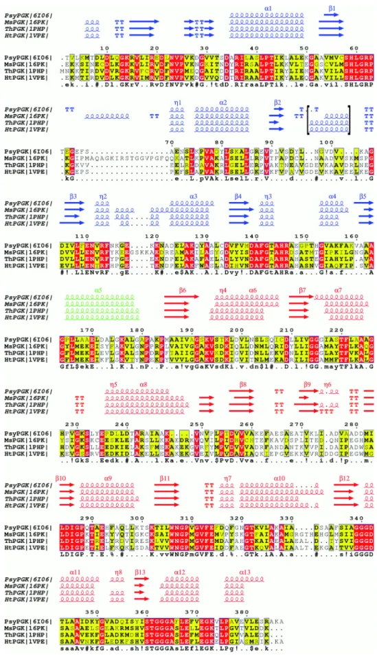
Protein sequences were aligned using CLUSTALW (Thomp-son et al. 1994) and rendered with the program ESPript (Robert and Gouet 2014). The secondary structure as indi-cated at the top of the alignment was calculated using the program DSSP (Kabsch and Sander 1983).
Structural superposition of Cα backbones was performed using the program “SUPERPOSE” (Krissinel and Henrick
2004; Winn et al. 2011) and the rmsd was calculated with NPS@ (Combet et al. 2000). Volumes and surfaces are cal-culated with GRASP (Nicholls 1993) and the number of hydrogen bonds was calculated using HBPLUS (McDonald and Thornton 1994). The hydrophobic surface area was cal-culated with the program BICEP (van Dijk et al., 2016), and the number of salt bridges was calculated using PIC (Tina et al. 2007). Three-dimensional structure representations were generated using PyMOL (DeLano 2002).
Atomic coordinates of PsyPGK have been deposited in the Protein Data Bank under accession numbers 6I06 (native) and 6HXE (complex).
Results
Overall structure description of psychrophilic PGK
The 387 amino acid residues phosphoglycerate kinase was crystallized in two open forms (one free and one with the product bound) under two distinct conditions and refined to 2.1-Å and 2.0-Å resolution, respectively. Data collection and refinement statistics are summarized in Supplemen-tary Table 1.
The RMSD between the two structures was calculated to be 0.5 Å The overall fold of PsyPGK, shown in Fig. 1, is very similar to the other known phosphoglycerate kinase structures. Two domains are separated by a helical linker (residues 165–179): the N-terminal domain (residues 2–164) which is made up of 4 α-helices and 8 β-strands forming a Rossmann fold and the C-terminal domain (resi-dues 180–387) containing 8 α-helices and 8 β-strands.
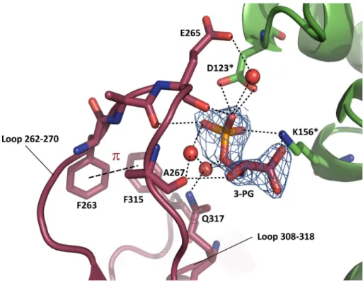
The largest differences between the three-dimensional structures compared herein are localized in the N-terminal domain. A two-turn helix between strand β5 and strand β6 in the thermophilic and hyperthermophilic enzymes is replaced by a one-turn helix in the mesophilic enzyme, and is com-pletely lost in the psychrophilic enzyme in which this stretch is unstructured (Fig. 2). This loss arises from a six-residue deletion, as indicated by the sequence alignment (Fig. 3). In the structure obtained by co-crystallization, a molecule of 3-PG (product of the catalyzed reaction) interacts with the main chain carbonyls of Ala264, Glu 265 and Ala267, as well as with the side chains of Asp123 and Lys156 from a symmetry-related molecule, in addition to three water mol-ecules (Fig. 4). 3-PG may contribute to the stabilization of loop 262–270 and loop 308–318 which interact via a hydro-phobic interaction between Phe263 and Phe315. This inter-action does not seem mandatory for the proper structuring of the loops since this is already seen in the Apo enzyme. However, it is interesting to notice that the two aromatic resi-dues are also part of a hydrophobic cluster involving Phe318.
Comparative studies with PGK counterparts
The psychrophilic enzyme was compared with counter-parts with which it displays high sequence identity (41.6% with the mesophile (Ms) T. brucei PGK, 48.1% with the thermophile (Th) B. stearothermophilus PGK and 46.1 with the hyperthermophile (Ht) T. maritima PGK). Three-dimensional structures of MsPGK, ThPGK and HtPGK superimposed on PsyPGK had rmsd values of 2.36/3.25 Å for MsPGK, 1.06/2.95 Å for ThPGK and 2.97/3.86 Å for HtPGK based on 113/387 (N-ter domain/entire protein) Cα atoms, respectively.
Fig. 1 Overall three-dimensional structure of the psychrophilic phosphoglycerate kinase (PDB-ID 6I06). N- and C-terminal domains are colored in blue and red, respectively. The helical linker is depicted in green. This figure was generated with PyMOL (DeLano 2002)
Fig. 2 Close-up on a region in the amino terminal part of the
enzymes in which a surface-exposed α-helix holding a number of charged residues and found in meso-, thermo- and hyperthermophilic
counterparts is unstructured in the psychrophilic enzyme. a ThPGK,
Fig. 3 Sequence alignments of PsyPGK (6I06), MsPGK (16PK), ThPGK (1PHP) and HtPGK (1VPE) performed using CLUSTALW (Thomp-son et al. 1994). Secondary structures of all the counter-parts, calculated with the DSSP algorithm (Kabsch and Sander
1983), are presented above the alignments. The N-terminal domain is colored in blue, the helical linker (α5) in green and the C-terminal domain in red. α-Helices are represented by α, β-strands by β and 310 helices by
η. Identities are shown on red background and similar residues on yellow background. For the psychrophilic enzyme, the miss-ing helix between strand β2 and strand β3 is indicated between brackets. Figure rendering was done using the program ESPript (Robert and Gouet 2014)
Amino acid composition
The overall amino acid composition of the four enzymes does not show significant differences apart from an increase in the number of alanine residues in PsyPGK (Table 1). This residue may play a crucial role in thermostability, due to its flexible nature, and is often found in loop regions of pro-teins. Furthermore, PsyPGK displays a lower Lys and Arg content, therefore, decreasing the potential to form stabiliz-ing ion pairs and H-bonds. By contrast, the proline content is unchanged, whereas this imino acid tends to rigidify the protein structure and is often found in β-turns and α-helices (Watanabe et al. 1997).
Hydrophobicity
Hydrophobic interactions play a major role in thermostabil-ity as they contribute to maintain the core of the protein. The percentages of hydrophobic residues (alanine, valine, isoleucine, leucine, phenylalanine, tryptophan, proline and methionine) in each enzyme are 51% in PsyPGK, 43% in MsPGK, 48% in ThPGK and 48% in HtPGK. The difference in hydrophobicity of the psychrophilic enzyme is mainly due to a higher percentage of alanine as noted above, but also a slight increase in the percentage of leucine and, at a lower degree, of valine. These residues tend to replace some polar residues at the surface of the protein, where they are ther-modynamically unfavorable, being exposed to the solvent, thereby increasing the overall flexibility of the outer shell. Similar observations have been made for other structures of
Fig. 4 Reaction product (glycerate 3-phosphate) binding contributes to stabilize loop 262–270 and loop 308–318. Stars indicate residues in a symmetry-related molecule. The 2Fo–Fc omit map is con-toured at 1.3 σ around glycerate 3-phosphate (3PG)
Table 1 Environmental temperatures and percentages of each amino
acid residue in the four phosphoglycerate kinases studied with charged residues highlighted in bold
Tenv PsyPGK MsPGK ThPGK HtPGK ~0 °C 37 °C 50–60 °C 85–90 °C Ala 15.5 9.1 12.4 9.5 Arg 2.3 3.6 5.1 3.0 Asn 2.6 3.0 3.3 2.3 Asp 7.0 4.5 7.9 6.0 Cys 0.8 1.4 0.3 0.0 Gln 1.8 2.0 0.5 1.5 Glu 5.4 5.7 8.4 9.3 Gly 8.8 10.2 8.9 9.5 His 1.3 1.8 2.0 1.5 Ile 4.7 6.1 5.3 6.5 Leu 11.1 9.5 9.6 9.0 Lys 7.5 9.5 8.1 10.8 Met 1.6 3.4 2.3 2.3 Phe 3.9 2.5 3.8 4.3 Pro 4.1 4.5 3.8 4.5 Ser 5.2 7.7 2.8 4.3 Thr 4.7 5.0 3.3 3.8 Trp 0.3 0.5 0.5 0.8 Tyr 1.6 2.7 2.0 0.8 Val 10.1 7.0 9.6 10.5
psychrophilic enzymes as citrate synthase from an Antarc-tic bacterial strain DS2-3R (Russell et al. 1998), α-amylase from Pseudoalteromonas haloplanctis (Aghajari et al. 1998), and metalloprotease from Pseudomonas sp. (Aghajari et al.
2003). Calculations of the hydrophobic surface area for the respective structure are 4300 Å2 for PsyPGK, 3059 Å2 for MsPGK, 3183 Å2 for ThPGK and 2859 Å2 for HtPGK which are in agreement with this tendency.
It has been reported that in the absence of substrates, the psychrophilic protein has two domains with different ther-mostabilities: one similar to the mesophilic PGK and one with lower stability (Bentahir et al. 2000). Parker et al. have shown, in studies on yeast PGK that the N-terminal domain unfolded 70 times faster than the C-terminal domain (Parker et al. 1996). When analyzing the structure of PsyPGK, we observed a cluster of polar amino acids in the core of the N-terminal domain including Arg19, Glu20, Cys57, Ser58, Tyr129, Ser150 and Thr151. This cluster is not found in mesophilic and thermophilic counterparts where Glu20 is replaced by a valine and Cys57 and Tyr129 by hydrophobic residues. The hydrophobic effect has been shown to play an important role in the stability of proteins (Dill 1990); hence, the substitutions observed may be responsible for the loss of hydrophobic interactions in the core of the protein and could explain the low stability of the N-terminal domain of PsyPGK.
The presence of these polar amino acids in the core of the protein and the increased number of valine, alanine and leu-cine at the surface of the protein suggest that the N-terminal domain may be the more thermolabile domain and could be responsible for the lower stability of the psychrophilic enzyme at higher temperatures.
Charge‑mediated interactions
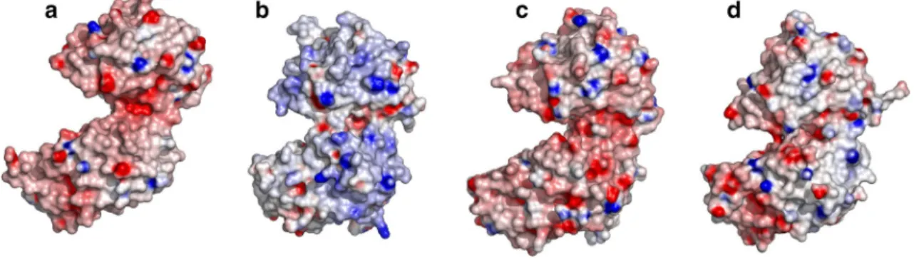
The number of charged residues increases from the psy-chrophilic PGK to the mesophilic one, and from the meso-philic to thermomeso-philic and hyperthermomeso-philic PGKs (24%
in PsyPGK, 25% in MsPGK, 31% in ThPGK and 31% in HtPGK, Table 1). Increasing charges are related with ther-mostability because these residues are able to make hydro-gen bonds and salt bridges as confirmed within these studies (Supplementary Table 2). Indeed, salt-bridge networks at the surface of thermophilic and hyperthermophilic proteins are frequently observed (Yip et al. 1995). For the PGKs stud-ied here, this tendency is confirmed (Fig. 5, Supplementary Fig. 1) with 0.02 salt bridges per residue in PsyPGK, 0.04 in MsPGK, 0.06 in ThPGK and 0.06 in HtPGK. It is also worth noting that the loss of one helix between strands β5 and β6 may contribute to the overall softness of the surface of the enzyme by decreasing the number of charged resi-dues turned towards the solvent (Fig. 2). Finally, the num-ber of salt bridges between the two domains increased with increased thermostability, with 0 inter-domain salt bridges in PsyPGK, 1 in MsPGK, 3 in ThPGK and 3 in HtPGK.
Hydrogen bonds
Despite the low energy of these interactions, they play a very important role in maintaining the three-dimensional struc-ture of the protein by their large number (Creighton 1991), and indeed the number of hydrogen bonds decreases drasti-cally in the psychrophilic enzyme compared to the other PGKs investigated herein (Supplementary Table 3).
Aromatic interactions
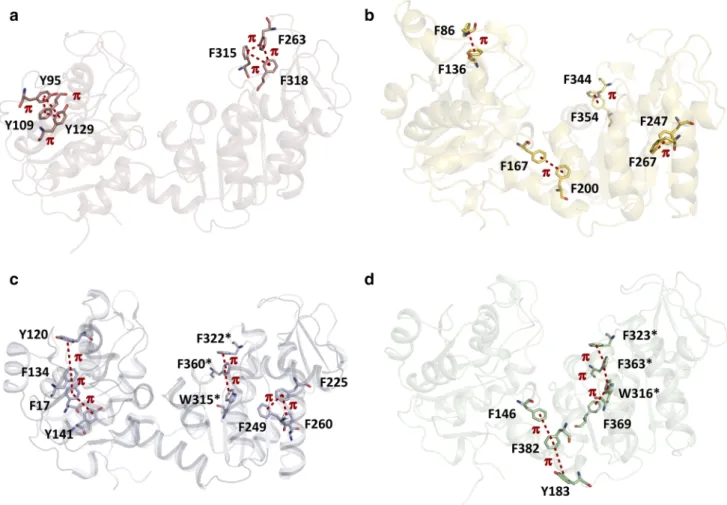
Aromatic interactions play an important role in protein stability (Burley and Petsko 1985). They are mediated by the Π electron cloud of aromatic rings being close in space (between 4.5 and 7 Å). Whereas the number of aromatic interactions is not strikingly different (9 in PsyPGK, 6 in MsPGK, 11 in ThPGK and 6 in HtPGK), these interactions are differently organized in the respec-tive structures (Fig. 6). Both domains in the psychrophilic enzyme are stabilized only locally by two clusters of three
Fig. 5 Surface representations of PsyPGK (a), MsPGK (b), ThPGK (c) and HtPGK (d) displayed at the same potential level. Color codes are red for negative charges and blue for positive charges. The figure was generated with GRASP (Nicholls 1993)
aromatic residues each. The thermophilic and hyperther-mophilic enzymes are stabilized by a series of aligned aromatic residues, spanning a larger distance, which highly stabilize the two respective domains (ThPGK) or the hinge region and the C-terminal domain (HtPGK). Finally, the mesophilic enzyme displays clusters being something in between compact and extended clusters, and stabilizing also the hinge region and the C-terminal domain.
Specific adaptations
The residues forming the active site are very well con-served among the four different enzymes suggesting that a more subtle mechanism is responsible for the increased catalytic efficiency of the psychrophilic enzyme observed at low temperature.
Increased flexibility around the hinge region
It is now well known how the enzyme performs the hinge-bending motions to get the two domains close together (Banks et al. 1979; Cliff et al. 2010; Auerbach et al. 1997). We hypothesized that this movement could be favored in the psychrophilic enzyme. It is clear that the overall amino acid composition and the reduced number of weak inter-actions tend to destabilize the enzyme but most probably, more specific modifications may play a role in the bending motion. To address this issue, we first focused our atten-tion on the three helices shown to form the inter-domain region and the surrounding regions (Marston et al. 2010; Inoue et al. 2010; Auerbach et al. 1997). These three helices (helices 5, 12 and 13 in PsyPGK) should be directly involved if an adaptive bending motion occurred in the psychrophilic enzyme. No differences could be found in the overall inter-domain organization. However, when observations were
Fig. 6 Formation of compact (a), extended (c, d) or semi-compact (b)
clusters of aromatic residues. The clusters are present in both N- and C-terminal domains for the psychrophilic and thermophilic enzymes, whereas the meso- and hyperthermophilic counterparts show
addi-tional stabilization at the hinge between the domains. (a) PsyPGK, (b) MsPGK, (c) ThPGK, (d) HtPGK. Conserved residues between ThPGK and HtPGK are indicated by a star
enlarged to the surrounding regions, we observed that some interactions between residues of helix 5 (residue 165–178) and helix 1 (residue 35–49) in the N-terminal domain, pre-sent in PGKs from meso-, thermo- and hyperthermophilic organisms (Fig. 7), were totally lacking in the psychrophilic enzyme. More concretely, in thermophilic and hyperther-mophilic PGKs, stabilization is mainly provided by an aromatic interaction between Phe169 and Tyr46 as well as a charge-mediated interaction between Glu172 and Arg7. In MsPGK, stabilization involves hydrogen bonds from Tyr191, Asn10 and Lys49, and a salt bridge between Lys49 and Glu53. Substitution of the aromatic Phe169 and charged Glu 172 (Ht- and ThPGKs)/Tyr191 and Glu194 (MsPGK) by a proline and an alanine, respectively, in PsyPGK (Pro165 and Ala168) which do not form similar interactions with the neighboring residues (Fig. 7), strongly suggests that the N-terminal domain should be under less constraint during the bending motion.
Destabilization of the 1,3‑biPG‑binding site
The reaction through which a phosphate monoester is trans-ferred to ADP has been extensively studied. The 3D structure determination of transition state analog (TSA) complexes
formed with trifluoromagnesate or tetrafluoroaluminate have largely contributed to the understanding of the mechanism (Cliff et al. 2010). In particular, TSA structures and muta-tional studies have shown that at least three basic residues are involved in the transfer process, namely Arg36, Lys193 and Lys197 in PsyPGK. Interestingly, Arg36 is positioned at the N-terminal of helix 1. In MsPGK, ThPGK and HtPGK, the N-terminal position of helix 1 is largely stabilized by a complex network of charge-mediated interactions, whereas in PsyPGK, only one interaction remains between Asp34 and Tyr80 (Supplementary Fig. 2). Furthermore, in the helix of the psychrophilic PGK, three polar or charged residues are substituted by the aliphatic residues Ala35, Leu38 and Leu46, Leu38 being in direct contact with the solvent. These reduced number of interactions and increased hydrophobic-ity support the idea that helix 1 is destabilized and more flexible, thus imparting a greater degree of movement to the catalytic residue, Arg36.
Another important feature related to the stabilization of the ligands bound to the enzyme is the presence of inter-domain bonds and in particular salt bridges observed in many organisms such as T. brucei, T. maritima or their human counterpart. Interestingly, one of the most conserved inter-domain charge-mediated interactions is lacking in the
Fig. 7 Different stabilizing interactions in the hinge region: a, b an
aromatic interaction between F169 and Y46 as well as a charge-medi-ated interaction between E172 and R7 are responsible for the (a) ther-mophilic and (b) hypertherther-mophilic enzyme stabilization. Hydrogen
bonds between Y191, N10 and K49, and a salt bridge between K49 and E53 are responsible for the (c) mesophilic enzyme stabilization. No stabilizing interactions are seen for the psychrophilic enzyme (d). This figure was generated with PyMOL (DeLano 2002)
psychrophilic enzyme. In the HtPGK crystal structure in complex with ligands (AMP-PNP and 3-PG), this interaction involves Arg62 and Asp200. These residues are conserved in the thermophilic and mesophilic counterparts, whereas Asp200 is substituted by Thr196 in PsyPGK (Supplementary Fig. 3). Despite this substitution, an interaction between the charged arginine and the hydroxyl group of the threonine would still be possible, but would probably be weaker than that of the salt bridge. Noticeably, Thr196 is positioned between Lys193 and Lys197 which form salt bridges with the transferred phosphate indicating that in addition to Arg36, the three basic residues involved in the transfer are less constrained.
Altogether, the weakly stabilized hinge domain as well as specific modifications surrounding the catalytic residues should strongly increase the flexibility, likely to enhance the catalytic rate.
Discussion
The comprehensive comparison of homologous proteins that display different optimal temperatures appears as a valid approach to elucidate the molecular mechanism of cold adaptation.
We have studied phosphoglycerate kinases for which crys-tal structures of psychrophilic, mesophilic, thermophilic and hyperthermophilic counterparts are available. All display a considerable difference in thermal stability despite identi-cal functions and very similar three-dimensional structures. The high specific activity of psychrophilic enzymes has been attributed to an increased flexibility at low tempera-tures (Feller and Gerday 2003). This property seems to ensure a better positioning of substrates in the active site and a rapid conformational change during the catalysis at low temperature.
Herein, we have shown that different factors may play a role in flexibility, for instance an increased number of hydro-phobic residues at the surface of the psychrophilic enzyme. These amino acids play a crucial role in stabilization of the three-dimensional structure when being in the core of the protein. At the surface, exposed to the solvent, they lead to destabilization of the structure and reduce thermostability (Privalov and Gill 1988).
Fewer salt bridges were found at the surface and between the two domains of PGK, and fewer aromatic interactions and a decreased number of hydrogen bonds were observed when its structure was compared to its meso- and thermo-philic counterparts. Similar trends have been observed in other enzymes of differing thermostabilities. In addition, the psychrophilic phosphoglycerate kinase lacks an α-helix compared to its counterparts. Indeed, secondary structure elements contribute to protein stability (Baldwin et al. 1996)
and play roles in local flexibility. Accordingly, the lack of such a helix should result in a more flexible protein (Jaenicke and Bohm 1998). Interestingly, it was reported that consecu-tive enzymes in a number of metabolic pathways can form complexes to transfer directly intermediary metabolites as it has been shown for GAPDH and PGK (Srivastava and Bernhard 1986; Srere 1987; Tomokuni et al. 2010). The for-mation of such weak and transient complexes is in agree-ment with functioning in cold environagree-ments (Sukenik et al.
2017), in which rapid responses are needed to cope with the low temperatures. An overall flexibility of PsyPGK, includ-ing a more flexible hinclud-inge domain coupled to destabilized conserved catalytic residues, is in favor of rapid modulation of the stability and the activity of these enzyme complexes.
Together, the overall increased flexibility of the cold-adapted PGK should allow a better accessibility to the active site compared to its counterparts and could contribute to explain the high specific activity at temperatures close to 0 °C. Differential scanning calorimetry studies have shown that in the presence of the substrate (Mg2+-ADP) and the product (3-PG) PsyPGK is stabilized compared to the free enzyme (Zecchinon et al., 2005). This indicates that the binding site is sufficiently stable in the presence of ligands despite an overall flexibility. Also, one cannot exclude the possibility of a more disordered transition state of the psy-chrophilic enzyme, due to the destabilization of the three catalytic residues Arg36, Lys193 and Lys197, and which hence could improve the reaction rate as well.
In the light of our results presented herein and other ear-lier studies on citrate synthase from an Antartic bacterial strain DS2-3R (Russell et al. 1998), α-amylase from
Pseu-doalteromonas haloplanctis (Aghajari et al. 1998), metal-loprotease from Pseudomonas sp. (Aghajari et al. 2003) and many others (Feller 2013), it appears evident that psy-chrophilic enzymes use various subtle strategies for cold adaptation.
Acknowledgements This work was supported by the Centre National de la Recherche Scientifique, by the European Union under the form of a TMR contract CT970131 (ColdNet), and by the Fonds National de la Recherche Scientifique, Belgium (Grants to GF and CG).
References
Afonine PV, Grosse-Kunstleve RW, Echols N, Headd JJ, Moriarty NW, Mustyakimov M, Terwilliger TC, Urzhumtsev A, Zwart PH, Adams PD (2012) Towards automated crystallographic structure refinement with phenix.refine. Acta Crystallogr D Biol Crystal-logr 68:352–367
Aghajari N, Feller G, Gerday C, Haser R (1998) Structures of the psy-chrophilic Alteromonas haloplanctis α-amylase give insights into cold adaptation at a molecular level. Structure 6:1503–1516 Aghajari N, Van Petegem F, Villeret V, Chessa JP, Gerday C, Haser
metalloprotease reveal new insights into catalysis by cold-adapted proteases. Proteins 50:636–647
Auerbach G, Huber R, Grattinger M, Zaiss K, Schurig H, Jaenicke R, Jacob U (1997) Closed structure of phosphoglycerate kinase from Thermotoga maritima reveals the catalytic mechanism and determinants of thermal stability. Structure 5:1475–1483 Baldwin E, Xu J, Hajiseyedjavadi O, Baase WA, Matthews BW
(1996) Thermodynamic and structural compensation in “size-switch” core repacking variants of bacteriophage T4 lysozyme. J Mol Biol 259:542–559
Banks RD, Blake CCF, Evans PR, Haser R, Rice DW, Hardy GW, Merrett M, Phillips AW (1979) Sequence, structure and activity of phosphoglycerate kinase: a possible hinge-bending enzyme. Nature 279:773–777
Bentahir M, Feller G, Aittaleb M, Lamotte-Brasseur J, Himri T, Chessa JP, Gerday C (2000) Structural, kinetic, and calorimet-ric characterization of the cold-active phosphoglycerate kinase from the antarctic Pseudomonas sp. TACII18. J Biol Chem 275:11147–11153
Bernstein BE, Hol WGJ (1997) Crystal structures of substrates and products bound to the phosphoglycerate kinase active site reveal the catalytic mechanism. Biochemistry 37:4429–4436
Bernstein BE, Williams DM, Bressi JC, Kuhn P, Gelb MH, Blackburn GM, Hol WGJ (1998) A bisubstrate analog induces unexpected conformational changes in phosphoglycerate kinase from
Trypa-nosoma brucei. J Mol Biol 279:1137–1148
Brünger AT (1992) Free R value: a novel statistical quantity for assess-ing the accuracy of crystal structures. Nature 355:472–475 Burley SK, Petsko GA (1985) Aromatic-aromatic interaction: a
mecha-nism of protein structure stabilization. Science 229:23–28 Cliff MJ, Bowler MW, Varga A, Marston JP, Szabo J, Hounslow AM,
Baxter NJ, Blackburn GM, Vas M, Waltho JP (2010) Transition state analogue structures of human phosphoglycerate kinase estab-lish the importance of charge balance in catalysis. J Am Chem Soc 132:6507–6516
Combet C, Blanchet C, Geourjon C, Deleage G (2000) NPS@: network protein sequence analysis. Trends Biochem Sci 25:147–150 Creighton TE (1991) Stability of folded conformations. Curr Opin
Struct Biol 1:5–16
Davies GJ, Gamblin SJ, Littlechild JA, Watson HC (1993) The struc-ture of a thermally stable 3-phosphoglycerate kinase and a com-parison with its mesophilic equivalent. Proteins 15:283–289 DeLano WL (2002) PyMOL. DeLano scientific, San Carlos, p 700 Dill KA (1990) The meaning of hydrophobicity. Science 250:297–298 Emsley P, Lohkamp B, Scott WG, Cowtan G (2010) Features and
development of Coot. Acta Crystallogr D Biol Crystallogr 66:486–501
Feller G (2013) Psychrophilic enzymes: from folding to function and biotechnology. Scientifica (Cairo), 512840
Feller G, Gerday C (2003) Psychrophilic enzymes: hot topics in cold adaptation. Nat Rev Microbiol 1:200–208
Gerday C, Aittaleb M, Bentahir M, Chessa JP, Claverie P, Collins T, D’Amico S, Dumont J, Garsoux G, Georlette D, Hoyoux A, Lonhienne T, Meuwis MA, Feller G (2000) Cold-adapted enzymes: from fundamentals to biotechnology. Trends Biotech-nol 18:103–107
Harlos K, Vas M, Blake CFF (1992) Crystal structure of the binary complex of pig muscle phosphoglycerate kinase and its substrate 3-phospho-D-glycerate. Proteins 12:133–144
Hess D, Kruger K, Knappik A, Palm P, Hensel R (1995) Dimeric 3-phosphoglycerate kinases from hyperthermophilic Archaea. Cloning, sequencing and expression of the 3-phosphoglycerate kinase gene of Pyrococcus woesei in Escherichia coli and char-acterization of the protein. Structural and functional comparison with the 3-phosphoglycerate kinase of Methanothermus fervidus. Eur J Biochem 233:227–237
Inoue R, Biehl R, Rosenkranz T, Fitter J, Monkenbush M, Radulescu A, Farago B, Richter D (2010) Large domain fluctuations on 50-ns timescale enable catalytic activity in phosphoglycerate kinase. Biophys J 99:2309–2317
Jaenicke R (1990) Protein structure and function at low temperatures. Philos Trans R Soc Lond B Biol Sci 326:535–551
Jaenicke R, Bohm G (1998) The stability of proteins in extreme envi-ronments. Curr Opin Struct Biol 8:738–748
Jones CE, Fleming TM, Cowan DA, Littlechild JA, Piper PW (1995) The phosphoglycerate kinase and glyceraldehyde-3-phosphate dehydrogenase genes from the thermophilic archaeon Sulfolobus
solfataricus overlap by 8-bp. Isolation, sequencing of the genes
and expression in Escherichia coli. Eur J Biochem 233:800–808 Kabsch W, Sander C (1983) Biopolymers 22:2577–2637
Krissinel E, Henrick K (2004) Secondary-structure matching (SSM), a new tool for fast protein structure alignment in three dimensions. Acta Crystallogr D Biol Crystallogr 60:2256–2268
Laskowski RA, MacArthur MW, Moss DS, Thornton JM (1993) PRO-CHECK: a program to check the stereochemical quality of protein structures. J Appl Cryst 26:283–291
Low PS, Bada JL, Somero GN (1973) Temperature adaptation of enzymes: roles of the free energy, the enthalpy, and the entropy of activation. Proc Natl Acad Sci U S A 70:430–432
Mandelman D, Bentahir M, Feller G, Gerday C, Haser R (2001) Crys-tallization and preliminary X-ray analysis of a bacterial psychro-philic enzyme, phosphoglycerate kinase. Acta Crystallogr D Biol Crystallogr 257:1666–1668
Marston JP, Cliff MJ, Reed MAC, Blackburn GM, Hounslow AM, Cra-ven CJ, Waltho JP (2010) Structural tightening and interdomain communication in the catalytic cycle of phosphoglycerate kinase. J Mol Biol 396:345–360
Matthews BW (1968) Solvent content of protein crystals. J Mol Biol 33:491–497
McDonald IK, Thornton JM (1994) Satisfying hydrogen bonding potential in proteins. J Mol Biol 238:777–793
Mykytczuk NC, Foote SJ, Omelon CR, Southam G, Greer CW, Whyte LG (2013) Bacterial growth at −15 degrees C; molecular insights from the permafrost bacterium Planococcus halocryophilus Or1. ISME J 7:1211–1226
Navaza J (2001) Implementation of molecular replacement in AMoRe. Acta Crystallogr D Biol Crystallogr 57:1367–1372
Nicholls AJ (1993) GRASP: graphical representation and analysis of surface properties. Biophys J 64:A116
Otwinoswki Z, Minor W (1997) Processing of X-ray diffraction data collected in oscillation mode. Methods Enzymol 276:307–326 Parker MJ, Spencer J, Jackson GS, Burston SG, Hosszu LLP, Craven
CJ, Waltho JP, Clarke AR (1996) Domain behaviour during the folding of a thermostable phosphoglycerate kinase. Biochemistry 35:15740–15752
Privalov L, Gill SJ (1988) Stability of protein structure and hydropho-bic interaction. Adv Protein Chem 39:191–234
Ramakrishnan C, Ramachandran GN (1965) Stereochemical crite-ria for polypeptide and protein chain conformations. Biophys J 5:909–933
Robert X, Gouet P (2014) Deciphering key features in protein structures with the new ENDscript server. Nucleic Acids Res 42:W320–W324
Russell RJ, Gerike U, Danson MJ, Hough DW, Taylor GL (1998) Structural adaptations of the cold-active citrate synthase from an Antarctic bacterium. Structure 6:351–361
Siddiqui KS, Cavicchioli R (2006) Cold-adapted enzymes. Annu Rev Biochem 75:403–433
Smalås AO, Leiros HK, Os V, Willassen NP (2000) Cold adapted enzymes. Biotechnol Annu Rev 6:1–57
Srere PA (1987) Complexes of sequential metabolic enzymes. Annu Rev Biochem 56:89–124
Srivastava DK, Bernhard SA (1986) Metabolite transfer via enzyme– enzyme complexes. Science 234:1081–1086
Struvay C, Feller G (2012) Optimization to low temperature activity in psychrophilic enzymes. Int J Mol Sci 13:11643–11665
Sukenik S, Ren P, Gruebele M (2017) Weak protein-protein interac-tions in live cells are quantified by cell-volume modulation. PNAS 114:6776–6781
Takai K, Nakamura K, Toki T, Tsunogai U, Miyazaki M, Miyazaki J, Hirayama H, Nakagawa S, Nunoura T, Horikoshi K (2008) Cell proliferation at 122 C and isotopically heavy CH4 production by a hyperthermophilic methanogen under high-pressure cultivation. Proc Natl Acad Sci U S A 105:10949–10954
Thompson JD, Higgins DG, Gibson TJ (1994) CLUSTAL W: improv-ing the sensitivity of progressive multiple sequence alignment through sequence weighting, position-specific gap penalties and weight matrix choice. Nucleic Acids Res 22:4673–4680 Tina KG, Bhadra R, Srinivasan N (2007) PIC: protein Interactions
Calculator. Nucleic Acid Res 35:W473–W476
Tomokuni Y, Goryo K, Katsura A, Torii S, Yasumoto K, Kemnitz K, Takada M, Fukumura H, Sogawa K (2010) Loose interaction between glyceraldehyde-3-phosphate dehydrogenase and phos-phoglycerate kinase revealed by fluorescence resonance energy transfer-fluorescence lifetime imaging microscopy in living cells. FEBS J 277:1310–1318
Van Dijk E, Varilly P, Knowles TPJ, Frenkel D, Abeln S (2016) Con-sistent treatment of hydrophobicity in protein lattice models accounts for cold denaturation. Phys Rev Lett 116:078101
Watanabe K, Hata Y, Kizaki H, Katsube Y, Suzuki Y (1997) The refined crystal structure of Bacillus cereus oligo-1,6-glucosidase at 2.0 Å resolution: structural characterization of proline substitu-tion sites for protein stabilizasubstitu-tion. J Mol Biol 269:142–153 Winn MD, Ballard CC, Cowtan KD, Dodson EJ, Emsley P, Evans PR,
Keegan RM, Krissinel EB, Leslie AGW, McCoy A, McNicholas SJ, Murshudov GN, Pannu NS, Potterton EA, Powell HR, Read RJ, Vagin A, Wilson KS (2011) Overview of the CCP4 suite and current developments. Acta Crystallogr D Biol Crystallogr 67:235–242
Yip KSP, Stillman TJ, Britton KL, Artymiuk PJ, Baker PJ, Sedelnik-ova SE, Engel PC, Pasquo A, Chiaraluce R, Consalvi V, Scan-durra R, Rice DW (1995) The structure of Pyrococcus furiosus glutamate dehydrogenase reveals a key role for ion-pair net-works in maintaining enzyme stability at extreme temperatures. Structure 3:1147–1158
Zecchinon L, Oriol A, Netzel U, Svennberg J, Gerardin-Otthiers N, Feller G (2005) Stability domains, substrate-induced conforma-tional changes, and hinge-bending motions in a psychrophilic phosphoglycerate kinase. A microcalorimetric study. J Biol Chem 280:41307–41314
Publisher’s Note Springer Nature remains neutral with regard to