TH `
ESE
TH `
ESE
En vue de l’obtention du
DOCTORAT DE L’UNIVERSIT´
E DE TOULOUSE
D´elivr´e par : l’Universit´e Toulouse 3 Paul Sabatier (UT3 Paul Sabatier)
Pr´esent´ee et soutenue le 18/11/2016 par :
Anne Combedazou
Caract´erisation des diff´erents mouvements collectifs au cours de la
migration des cellules de bordure chez la Drosophile
JURY
David CRIBBS Professeur d’Universit´e Membre du Jury
St´ephane NOSELLI Directeur de Recherche Membre du Jury Anne-Marie PRET Professeur d’Universit´e Membre du Jury
Pascal SILBERZAN Directeur de Recherche Membre du Jury
´
Ecole doctorale et sp´ecialit´e :
BSB : Biologie cellulaire
Unit´e de Recherche :
LBCMCP CNRS UMR 5088
Directeur(s) de Th`ese :
Damien RAMEL et Xiaobo WANG
Rapporteurs :
Remerciements
Je tiens à remercier tous les membres du jury: David Cribbs, Stéphane Noselli, Anne-Marie Pret et Pascal Silberzan, qui m’ont fait l’honneur d’évaluer ce travail.
Je tiens particulièrement à remercier mes directeurs de thèse.
Merci à toi, Damien, pour m’avoir encadrée et guidée dans des réflexions et expériences scientifiques tout au long des différentes étapes de ma thèse. Ta rigueur scientifique, ton expertise, et ta disponibilité m’ont énormément apporté. Je te remercie pour ta patience et ta gentillesse. Je retiendrai aussi ta joie de vivre communicative, et tes nombreux appartés, qui ont animé notre bureau.
Xiaobo, I want to thank you for having welcomed me in your team. Thank you for your supervision and your optimism who taught me a lot. Especially, you allowed me to learn numerous techniques of microscopy!
Je souhaite remercier toutes les personnes qui ont contribué à ces travaux : Céline et Thomas pour leur aide en microscopie, Guillaume pour les analyses, Nathalie pour les discussions scientifiques, et Valérie pour son aide technique. Je remercie également l’ensemble des membres de l’équipe et du LBCMCP pour tous les bons moments partagés. Je fais un petit clin d’œil à Amélie, Carole, Julie, et Marion pour les nombreuses pauses thé!
J’adresse mes remerciements à mes amis pour leur présence. Je remercie mes parents, Laure et Jonathan pour votre soutien et vos encouragements.
Pour finir, un grand merci à François pour ta patience et ton soutien permanent tout au long de cette aventure.
List of abbreviations ... 8
List of figures ... 12
INTRODUCTION ... 14
Chapter I: Cell migration... 16
I.1. Importance of cell migration ... 16
I.2. Modes of single cell migration ... 17
I.2.1. Amoeboid migration ... 19
I.2.2. Mesenchymal migration ... 20
I.3. Directed cell migration ... 23
I.4. Collective cell migration ... 24
I.4.1. Definition of collective cell migration ... 24
I.4.2. Morphological organisation of collective cell migrations ... 25
I.4.3. Models of collective cell movements in cancer ... 28
I.4.4. Models of collective cell movements during morphogenesis ... 30
I.4.4.1. Dorsal closure in Drosophila ... 30
I.4.4.2. Collective cell migration of the zebrafish posterior lateral line primordium ... 32
I.4.4.3. Collective cell migration during tracheal branching morphogenesis in Drosophila ... 34
I.4.4.4. Border cell migration in Drosophila ... 36
Chapter II: The cytoskeleton in cell migration ... 40
II.1. Actin Microfilaments ... 41
II.1.1. Organisation of actin network in protrusion (Lamellipodia) ... 43
II.1.2. Regulation of actin polymerisation ... 45
II.1.2.1. Regulation of actin nucleation at the barbed end mechanism of treadmilling ... 46
II.1.2.2. Regulation of actin polymerisation and depolymerisation ... 46
II.1.2.1.1. Profilin ... 46
II.1.2.1.2. Cofilin ... 47
II.1.2.1.3. Proteins containing the WH2 domain ... 48
II.1.2.1.4. Capping proteins ... 49
II.1.3. Regulation of branched nucleation ... 52
II.1.4. Organisation of actin network in protrusion (Filopodia) ... 55
II.2. Myosins ... 57
II.2.1. Non-Muscle Myosin II (NMII) ... 59
II.2.2. NMII activity on Actin filaments ... 62
II.2.3. Regulation of NMII ... 64
II.2.3.1. Proteins regulating NMII phosphorylation ... 65
II.2.3.1.1. Rho-associated Kinase (ROCK) ... 66
II.2.3.1.2. Other Kinases ... 66
II.2.3.1.3. Myosin light chain phosphatase (MLCP) ... 68
II.3. Small Rho GTPases ... 68
II.3.1. Regulation of small Rho GTPases ... 70
II.3.2. Rac1 GTPase ... 72
II.3.3. Cdc42 GTPase ... 73
Chapter III: Border cell migration ... 78
III.1. Oogenesis ... 78
III.2. Specification of border cell cluster ... 82
III.3. Timing of border cell migration ... 85
III.4. Guidance of border cell cluster ... 86
III.5. Adhesion of border cell cluster ... 89
III.6. Cohesion between border cells ... 91
III.7. Intercellular communication within border cell cluster ... 92
III.8. Border cell migration and actin cytoskeleton ... 93
III.8.1. Basis ... 94
III.8.2. Actin regulators ... 95
III.8.2.1. Actin nucleators ... 95
III.8.2.2. Monomers actin binding proteins ... 96
III.8.2.3. Actin filaments binding proteins ... 96
III.8.2.4. Severing proteins ... 98
III.8.2.5. Bundles proteins ... 99
III.8.2.6. Other regulators of actin dynamics ... 100
III.8.3. Myosins involved in border cell migration ... 102
III.9. Rho Small GTPases in Drosophila border cells ... 103
III.9.1. Rho Small GTPase Rac1 in Drosophila border cells ... 103
III.9.1.1. Regulation of Rac1 in Drosophila border cells ... 105
III.9.1.2. Regulation of actin by GTPase Rac signalling in Drosophila border cells ... 107
III.9.2. Rho small GTPase Cdc42 in Drosophila border cells ... 107
III.9.3. Rho small GTPase RhoA in Drosophila border cells ... 108
III.9.3.1. RhoA/ROCK/non-muscle myosinII pathway in Drosophila border cells ... 109
Chapter IV: Aim of this PhD thesis ... 112
RESULTS ... 114
Chapter I: Myosin II governs collective cell migration behaviour downstream of guidance receptor signalling ... 116
I.1. Summary ... 116
I.2. Results ... 117
ChapterII: The balance between Rac and Myosin II signalling pathway controls border cell migration behaviour. ... 138
II.1. Summary ... 138
II.2. Abstract ... 139
II.3. Introduction ... 140
II.4. Materials and methods ... 142
II.4.3. PA-Rac experiments ... 143
II.4.4. FRET analysis ... 143
II.4.5. Mathematical analysis of rotational and linear behaviour ... 143
II.4.5.1. Manual tracking ... 143
II.4.5.2. Measurement of rotation speed ... 144
II.4.5.3. Measurement of linearity index ... 144
II.4.6. Quantification of Myosin II intensity ... 144
II.4.7. Statistical analysis ... 144
II.5. Results ... 145
II.6. Discussion ... 149
Chapter III: Role of the Jun N-terminal kinase in the control of border cell behaviours . 152 III.1. Introduction ... 152
III.2. Results ... 153
III.3. Discussion ... 157
Chapter IV: Rotational movement quantification ... 160
IV.1. Introduction ... 160
IV.2. Results ... 161
IV.2.1. Two dimensional analysis method ... 161
IV.2.2. Three dimensional analysis of rotation (developed in collaboration with Guillaume Gay) 163 IV.2.2.1. Three dimensional tracking... 163
IV.2.2.2. Detection of the rotational movements ... 164
IV.2.2.3. Detection of the rotational movements: examples ... 166
IV.3. Discussion ... 168
IV.4. Methods (developed by Guillaume Gay) ... 169
IV.4.1. Nuclei segmentation method ... 169
IV.4.2. Rotation detection (described in Figure 37) ... 170
DISCUSSION ... 174
Chapter I: Introduction ... 176
Chapter II: Role of the microenvironment in the choice of the migration behaviour ... 178
II.1. Physical constraints might affect the transition between the two modes of migration 178 II.2. E-cadherin-mediated adhesionscould play function in the appearance of the rotation 180 Chapter III: Identification of the guidance receptors signalling pathways ... 182
III.1. EGFR and PVR signalling pathways in the control of border cell migration ... 182
III.2. Regulation of PVR and EGFR activities ... 183
III.3. Potential downstream targets of EGFR signalling ... 184
III.3.2. Small GTPase RhoA ... 185
III.4. Identification of an antagonism between Rac and ROCK/NMII during border cell movements ... 186 Chapter IV: Role of the cell-cell communication ... 188
Chapter V: Analysis and modelling 3D rotating border cell clusters (collaboration with Guillaume Gay) ... 190
Chapter VI: General conclusions ... 192
BIBLIOGRAPHY ... 196
List of abbreviations
AB: Abrupt
ADF: Actin Depolymerizing Factor ADP: Adenosine diphosphate Arp: Actin-RelatedProteins
ARPC: Actin related protein 2/3 complex AS: amnioserosa;
ATP: Adenosine triphosphate AVE: anterior visceral endoderm
C/EBP: CCAAT/enhancer-binding protein Ca: Calcium
CFP: Cyan fluorescent protein CK2: Casein kinase 2
CP: Capping Protein
CXCL12/SDF-1: chemokine (C-X-C motif) ligand 12 CXCR4: chemokine (C-X-C motif) receptor 4
CXCR7: chemokine (C-X-C motif) receptor 7
DE-cadherin: Drosophila melanogaster epithelial cadherin mDia: Diaphaneous
DIAP1: Drosophila inhibitor of apoptosis ECM: extra-cellular matrix
EcR: Ecdysone receptor
EGFR: Epidermal Growth Factor Receptor ELMO: Engulfment and cell motility protein F-actin: Filamentous actin
Fgf: Fibroblast growth factor FH: formin homology FP: Filopodia
FRET: Förster Resonance Energy Transfer G-actin: Globular actin
Gal4: Galactose 4
GAPs: GTPase activating proteins
GDI: Guanosine nucleotide dissociation inhibitors GEFs: Guanosine nucleotide exchange factors GFP: Green Fluorescent Protein
GMF: gliamaturation factor GDP: guanosine diphosphate GTP: Guanosine Triphosphate
GTPase: hydrolase Guanosine Triphosphate ILK: Integrin-like kinase
IQ: Isoleucine and Glutamine JNK: Jun N-terminal kinase LE: leading edge epidermis;
LIMK: Lin-11, Isl-1 and Mec-3kinases LM: Lamella Region
LOV: light, oxygen, or voltage LP: Lamellipodia
MELC: Myosin Essential Light Chains Mg: Magnesium
MHC: Myosin Heavy Chains MIM: Missing is metastasis
Mlc-c: Myosin light chain cytoplasmic MLCK: Myosin light chain kinase MLCP: Myosin light chain phosphatase
MRCK: myotonic dystrophy kinase-related CDC42-binding kinase MRLC: Myosin Regulatory Light Chains
MRTF: Myocardin-related transcriptional factor Mtl: Mig-2-like
MYPT1: myosin phosphatase targeting subunit 1 NLS:nuclear localisation signal
NMII: Non-Muscle Myosin II NPF: nucleation promoting factors PAK: p21-associated kinase
PA-Rac: photoactivatable analogues of Rac Pi: phosphate inorganique
PKC: protein kinase C PP1: protein phosphatase1 P-Tyr: Phospho-tyrosine
PVF1: platelet-derived growth factor and vascular endothelial growth factor-related factor 1
PVR: platelet-derived growth factor and vascular endothelial growth factor receptor RNAi: Ribonucleic acid interference
ROCK: (Drosophila Rok): Rho associated kinase RTK: Receptors tyrosine kinase
SF: Stress Fibres Slbo: slow border cells
Sqh: spaghetti squash (Drosophila MRLC) SRF: serum response factor
TAI: Taiman Upd: Unpaired USP: Ultraspiracle VE: ventral ectoderm
WASP: Wiskott–Aldrich syndrome protein WH2: WASP homology 2
Zip: zipper (Drosophila MHC)
List of figures
Figure 1: Different modes of cell migration. ... 18 Figure 2: Different morphologies between mesenchymal and amoeboid cells. ... 20 Figure 3: Mechanism of single cell migration. ... 22 Figure 4: Examples of collective cell migration. ... 27 Figure 5: In vivo models to study collective cancer cell migration. ... 29 Figure 6: Dorsal closure of Drosophila embryo. ... 31 Figure 7: Collective cell migration of the lateral line primordium in zebrafish. ... 33 Figure 8: Tracheal development in the Drosophila embryo. ... 35 Figure 9: Collective cell migration of Drosophila border cells in a stage 9 egg chamber. ... 37 Figure 10: Confocal images of the lamella region (LM) of fish fibroblasts containing lamellipodia (LP) and filopodia (FP). ... 40 Figure 11: Actin‐enriched protrusions at the leading edge of migrating cells. ... 42 Figure 12: Formation of actin filament. ... 44 Figure 13: Spire, an actin nucleator. ... 49 Figure 14: Regulation of actin filament polymerisation (treadmilling process). ... 51 Figure 15: Actin nucleation model at the leading edge of migrating cells. ... 54 Figure 16: Model of actin nucleation by formins. ... 57 Figure 17: Non‐muscle myosin II structure. ... 60 Figure 18: NMII conformation. ... 61 Figure 19: Actin and NMII interactions. ... 63 Figure 20: Regulation of small GTPases by cycles between an active GTP‐bound form to an inactive GDP‐ bound form. ... 71 Figure 21: Rho GTPases in cell migration. ... 75 Figure 22: Ovarioles of egg chambers at increasing stages of maturity. ... 78 Figure 23: Border cell migration. ... 81 Figure 24: Signalling pathways regulating specification and timing of border cell migration. ... 83 Figure 25: Effect of the loss of guidance receptors for border cell migration ... 87 Figure 26: E‐cadherin in border cell clusters. ... 90 Figure 27: Photoactivatable analogues of Rac (PA‐Rac). ... 92 Figure 28: Actin localisation in border cells. ... 94 Figure 29: Role of Rac for border cell migration. ... 104 Figure 30: NMII localisation and activity in border cells. ... 110 Figure 31: The switch from linear to rotational movement induces loss of active Rac polarity. ... 146 Figure 32: Polarisation of Rac activity is responsible for linear movement. ... 147 Figure 33: Rac activity negatively controls NMII activity. ... 149Figure 34: The inhibition of JNK blocks the rotation of border cell clusters. ... 154
Figure 35: The inhibition of JNK decreases the accumulation of NMII at the cell cortex. ... 155 Figure 36: inhibition of JNK promotes the stability of a polarised Rac activity. ... 156 Figure 37: Simple discrimination between linear and rotational behaviour. ... 162 Figure 38: Rotation analysis of wild‐type border cell cluster. ... 165 Figure 39: Test of the rotation analysis. ... 167 Figure 40: Detection of rotations in the 3D trajectory of a border cell relative to the cluster centre. ... 171 Figure 41: Hypothetical model of the regulation of collective movements during border cell migration. ... 193
Chapter I: Cell migration
Cell motility was first describedby the microbiologist Antonie Van Leeuwenhoek, when he observed the movements of bacteria under his microscope, more than 300 years ago. This mechanism is crucial for bacteria to reach nutrients, adapt to their environment, and escape from dangers. In multicellular organisms, cell movements, from one location to another one, are also required during many biological and pathological phenomena. This process, called cell migration, is essential during embryo development, immunity response, wound repair and tissue homeostasis (Ridley, Schwartz et al. 2003).
I.1.
Importance of cell migration
During embryo morphogenesis, the establishment of tissues and organs are highly dependent on the cell motility. For example, the gastrulation phenomenon involves cell to migrate together as a sheet and, this leads to the setting up of three germ layers of the embryo, which will constitute future organs (Montero and Heisenberg 2004). Later, the primordial germ cells migrate via the gut and divide to develop gonads, in which they will differentiate in mature gametes (Kunwar, Siekhaus et al. 2006). In adults cell migration is required for wound healing or skin renewal. In all inflammation cases, immune cells must defend the organism. Leukocytes might have to migrate from the blood circulation to inflammation sites through signals secreted by broken cells and pathogens. Thus, cell motility is critical for immune
surveillance (Friedl, Borgmann et al. 2001). Moreover, as this mechanism is involved in many biological processes and at all developmental stages, cell migration must be strictly regulated.
Therefore, deregulation of cell migration can generate serious diseases. During embryonic development, migration defects may lead to a number of different pathologies including mental and physical retardation or congenital heart diseases (Kurosaka and Kashina 2008). As cell migration is a critical process for immune surveillance, an increase in the motility activity causes autoimmune diseases such as multiple sclerosis, or chronic inflammation. In contrast, reduced cell migration leads to immunodeficiencies (Pals, Horst et al. 1989). Moreover, cell migration is also implicated in tumorigenesis. Tumour cells are able to metastitize and invade healthy tissues (Simpson, Selfors et al. 2008). Two types of cell migration have been described. Cells may migrate either individually or collectively (Friedl, Hegerfeldt et al. 2004). Details on the mechanism involving for these two modes of migration are in the following sections.
I.2.
Modes of single cell migration
Cell cultures studies allowed characterising mechanisms of single cell migration. To migrate, two types of cell movements have been developed by cells. Depending of their environment, cells will either use “swimming” or “crawling” to reach a destination. In a liquid environment, cells will swim. It is often the case of unicellular organisms like some protozoans or of some eukaryotic cells such as sperm cells. Thus to move these cells require specific structures such as cilia or flagella. On the other hand, most of animal cells travel by crawling on a substratum. Basically, to move cells have to polarise, and extend a membrane protrusion in the migration
direction. The next step is the formation of an adhesion between the extremity of the protrusion and the substratum. Through this adhesion, the cell tract on the substratum, retract the lagging edge, and finally previous adhesions with the substratum are removed (Ananthakrishnan and Ehrlicher 2007).
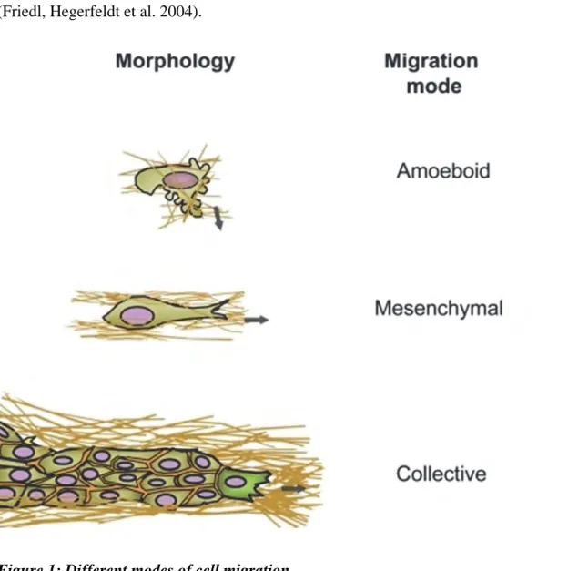
According to the innate cell morphology, different modes of single cell migration have been described: the mesenchymal and amoeboid cell motility. The nature of the substratum is also crucial for the choice in the mode of migration (Knight, Laukaitis et al. 2000, Friedl and Wolf 2003). A third mode, implicating numerous cells, is collective cell migration (Figure 1) (Friedl, Hegerfeldt et al. 2004).
Top and middle schemas represent amoeboid and mesenchymal single cell migrations, respectively. Bottom panel shows a group of cells migrating together. Adapted from (Friedl and Wolf 2010).
I.2.1. Amoeboid migration
Cells using amoeboid migration present a round shape (Figure 2 right). To maintain this morphology cells require strong cell contractility. During this type of migration, cells weakly interact and do not degrade the substratum, but squeeze between one another. Thus, cells migrate faster than mesenchymal cells undergoing migration. The environmental pressure allows cells to migrate, by pushing on its side the plasma membranes. However, when cells can interact with the matrix, the leading adhesion drives the translocation of the cell body from the rear to the front (Lammermann and Sixt 2009).
Amoeboid movements are permitted via the formation of protrusions, called blebs. Actin cytoskeleton, which controls the cell shape, is closely localised to the plasma membrane. Cells using this mode of migration present strong accumulation of the motor protein non-muscle myosin II to maintain the round cell shape and, to give them contractility properties. The formation of blebs is initiated by the separation between the cortical actin cytoskeleton from the plasma membrane. Once the bleb is formed, the actin cytoskeleton reforms at the membrane. The recruitment of the non-muscle myosin II causes cell contraction, leading to the cell translocation (Renkawitz, Schumann et al. 2009).
Figure 2: Different morphologies between mesenchymal and amoeboid cells.
Phase contrast images of mesenchymal K4 sarcoma cell line (left) and amoeboid A3 sarcoma cell line (right). From (Pankova, Rosel et al. 2010).
I.2.2. Mesenchymal migration
Many cells use the mesenchymal mode of motility, such as fibroblasts or various cancer cells. This type of migration can be divided in five steps: cell polarisation, formation of plasma membrane extensions, establishment of adhesions, translocation of the cell body, and retraction of the lagging edge (Figure 3). These mechanisms are controlled by many signalling pathways and by the environment (Lauffenburger and Horwitz 1996, Friedl and Wolf 2003).
A front/back polarity is established within the cell in the direction of migration. Indeed, in response to intra- or extracellular signals, the actin cytoskeleton becomes polarised, with a strong accumulation of actin at the leading edge (Mogilner and Oster 1996). Polymerisation
of actin filaments (called treadmilling) occurs close to the plasma membrane (Wang 1985). Thus, actin filaments push the plasma membrane and give rise to protrusions in the direction of the migration. In two-dimensional environment, these extensions are called pseudopodia, whereas, when cells migrate in three dimensions, the protrusions formed are filopodia and lamellipodia. While, filopodia are very thin and long extensions, lamellipodia are larger and flatter (Figure 3a) (Mattila and Lappalainen 2008, Friedl and Wolf 2009).
Once formed, the protrusion adheres to the substratum, generating an anchoring site allowing the cell to pull on the site. Adhesions link the cell via actin filaments to the substratum, mature and, become focal adhesion at the basis of the protrusion (Figure 3b). The nature of adhesion molecules will depend on the migrating cell type as well as the substratum. Many cells move on extra-cellular matrix (ECM) and adhere on it through numerous proteins including the integrin protein family. Cells may also migrate under other cells, and thus they will adhere on their substrate by cadherin molecules (e.g. in the case of epithelial cells, the adhesion is mediated by E-cadherin) (Even-Ram and Yamada 2005).
Once adhesions formed, cell body is translocated through the contraction of the actomyosin cytoskeleton (Figure 3c). The microtubule cytoskeleton also contributes to this process by promoting cytoskeletal changes and nuclear translocation.
To progress on their substratum, motile cells must retract their lagging edge. Stress fibres connect focal adhesions from the front and rear of cells, then contract and impair adhesions at the rear. Finally, the retraction occurs by the detachment of adhesions at the rear (Figure 3d).
This migration is slow due to the presence of strong adhesions and the necessity to degrade the matrix in order to create a path. The role of the actin cytoskeleton and its partners during cell motility is detailed in section II.
Figure 3: Mechanism of single cell migration.
a. Firstly, to move the cell extends a protrusion enriched in F-actin at the leading edge. Both
types of protrusions, lamellipodia and filopodia, can contribute to cell migration. Actin filaments grow in the direction of the migration and push the membrane to form protrusions.
b. Once formed, the membrane extension adheres to the substratum through focal adhesions. c. The contraction of the actin cytoskeleton allows the translocation of the cell body. The
contraction forces are generated by stress fibres linked to the focal adhesion. d. Finally, to move in a forward direction, motile cell retracts its lagging edge. Retraction fibres pull the back of the cell in the direction of migration. Adhesions at the rear of the cell are disassembled, and the lagging edge is retracted (Mattila and Lappalainen 2008).
I.3.
Directed cell migration
Motile cells move in a specific direction through the presence of positive or negative guidance signals (Lauffenburger and Horwitz 1996). The persistence in one precise direction of the migration may depend on extracellular chemicals (chemotaxis), or physical signals (durotaxis), as well as internal regulations. Thus, these mechanisms allow for controlling the efficiency of cell migration, and for promoting the formation of protrusions in the good direction, since in the absence of guidance cues a cell migrates randomly (Haeger, Wolf et al. 2015).
Chemical extracellular signals form a gradient determining the direction of the migration. Migratory cells may be able to sense these signals in their environment through specific receptors at their surface and thus move towards the highest concentration of these molecules. This process is called chemotaxis. As soon as a gradient, even weak, is detected, the cell can begin the migration process. In some cases, the nature of the environment can guide cells to their final destination. When cells move on the extra-cellular matrix, chemical substances can induce cell motility (Porcionatto 2006). Indeed, cells will move up to the highest fibronectin concentration. Moreover, the stiffness of the environment also contributes to escort cell motility. Most of the time cells will go from the most rigid to the softest substrate (Joaquin, Grigola et al. 2016).
Intracellular signalling pathways also contribute to the polarisation of the actin cytoskeleton in motile cells, and will be discussed in chapter III of the introduction.
I.4.
Collective cell migration
Much evidence has shown that cells must migrate in interconnected groups in several biological processes. During embryo morphogenesis, cell migration involves many cells moving together, such as the trachea network branching, salivary glands formation, amongst others. Processes of wound healing in adult also required a collective cell migration. More recently, the collective cell migration has been involved in tissue invasion by cancer cells (Friedl and Gilmour 2009).
I.4.1. Definition of collective cell migration
Cell migration is defined collective when several cells move in a cohesive and coordinated manner. During collective migration, cells keep their physical and functional contacts between each other. Moreover, to migrate collectively, cell polarisation must happen at the level of the entire cell group. So, the dynamics of the actin cytoskeleton need to be coordinated between all cells. Therefore, the group of cell may function as a single unit (Friedl 2004, Montell 2008).
Extracellular factors, such as growth factors, chemokines, or ECM components may guide groups of cells in one direction. Therefore, a motile group of cells is polarised, and cells at the front of the migration, called leader cells, drive the movement of the entire group. Leader cells have the ability to detect guidance cues and to form protrusions, which adhere to the substratum, and guide the other cells, called follower cells. Leader and follower cells have
different morphological shape, gene expression, and actin cytoskeleton dynamics (Mayor and Etienne-Manneville 2016).
For an efficient collective migration, cell junctions are retained within a group, while cells are migrating. Several adherens junction proteins can mediate the cohesion between cells from one group, including cadherins, integrins and immunoglobulin superfamily members. These proteins allow connecting the actin cytoskeleton or intermediate filaments of different cells. As many cells involved in collective migration are derived from epithelia, intercellular adhesions are often mediated by cadherins (Friedl and Gilmour 2009).
I.4.2. Morphological organisation of collective cell migrations
Different organisations of motile cell groups have been observed (Rorth 2009).
In two dimensions, cells may move as a sheet across a tissue surface. In this case, cells migrate as a monolayer, and maintain close contacts and are still connecting during their forward migration. In vivo, collective cell migration of epidermal sheets occurs across a tissue surface, and leads to epidermal single layered wound closure (Figure 4a) (Solnica-Krezel 2005).
The motility of strand-like groups has been observed in processes such as cancer cell invasion, or during the migration of the lateral line in zebrafish (Ghysen and Dambly-Chaudiere 2007). Clusters of cells may also migrate through tissues, after detaching from their origin, as that observed for Drosophila border cell migration (Figure 4b) (Montell 2003).
Collective migration may also occur in three dimensional tissue environments as strands or groups of cells. Collective cell movements, sprouting and branching, play a role for angiogenesis or gland formation (Figure 4c) (Adams and Alitalo 2007, Affolter and Caussinus 2008).
Collective movements also comprise non-cohesive groups of cells. For example, neural crest cells migrate collectively, but forming loose inter-cellular connections. This type of organisation appears elongated and polarised (Figure 4d) (LaBonne and Bronner-Fraser 1999).
Generally, in cohesive cell groups, membrane protrusions are mainly emitted by leader cells. On the contrary, in non-cohesive cohorts of cells, protrusions may be emitted by all cells. However, in all cases, protrusion extensions are favoured in the direction of migration (Vitorino and Meyer 2008).
Figure 4: Examples of collective cell migration.
Schemas of different collective migration models are represented in the first column. The regions where cells interact are depicted as a red border. Examples of the corresponding types of collective movement are shown in the second column. Extension of membrane protrusions is indicated by yellow arrowheads. In the third column, examples of these physiologic (green background) and pathologic (red background) models of collective cell migration are cited. AVE: anterior visceral endoderm. a. Collective epithelial 2D sheet
migration; example of intestinal epithelial cells migration (from (Hopkins, Pineda et al. 2007)). b. Schematic representation of groups of cells migrating together, illustrated by the migration of zebrafish lateral line (from (Haas and Gilmour 2006)). c. Representation of collective cells migrating like chains; example of fibroblast-leaded squamous cell carcinoma invasion (from (Gaggioli, Hooper et al. 2007)).d. Cells migrating collectively but forming loose contact between them; example of neural crest cells (from (Rupp and Kulesa 2007)).
I.4.3. Models of collective cell movements in cancer
In 2006, from histopathological analysis of human cancer lesions, the migration of cancer cells appeared collective (Christiansen and Rajasekaran 2006). Two years later, the first evidence of collective cancer cell migration was described in vivo (Alexander, Koehl et al. 2008). Collective cancer cells can invade into the stroma as strands, cords or clusters. In vitro studies revealed that many cancers may migrate collectively such as squamous cell carcinoma, colorectal carcinoma, melanoma, or even breast cancer (Figure 5 right) (Sahai 2005, Friedl, Locker et al. 2012).
Many labs study cancer cell migration, in vitro, for example by using invasion or scratch assays. However, progress in dynamic live imaging has now allowed for the study of cancer cell invasion in vivo, in small animals, in particular in mouse. Several approaches are used, such as the skin-flap, intravital microscopy, and optical window models. For the first model, a piece of skin is removed above the tissue of interest. Thus, some organs can be accessible, but this surgical operation is irreversible and may modify cellular physiology of the organism. To avoid this issue, optical transparent glass windows can be implemented by surgical
intervention (Figure 5 left). This method allows longer periods of observation. Many organs may be observed with this technic, such as abdominal organs, brain and dorsal skin. Recently, the evolution of endomicroscopic technics used in mice permit to study cancer progression, such as colon cancer, etc. Although small surgical intervention may be necessary, this method is less invasive than the first two techniques that impliment increased exposesure (Alexander, Koehl et al. 2008, Karreman, Hyenne et al. 2016, Karreman, Mercier et al. 2016).
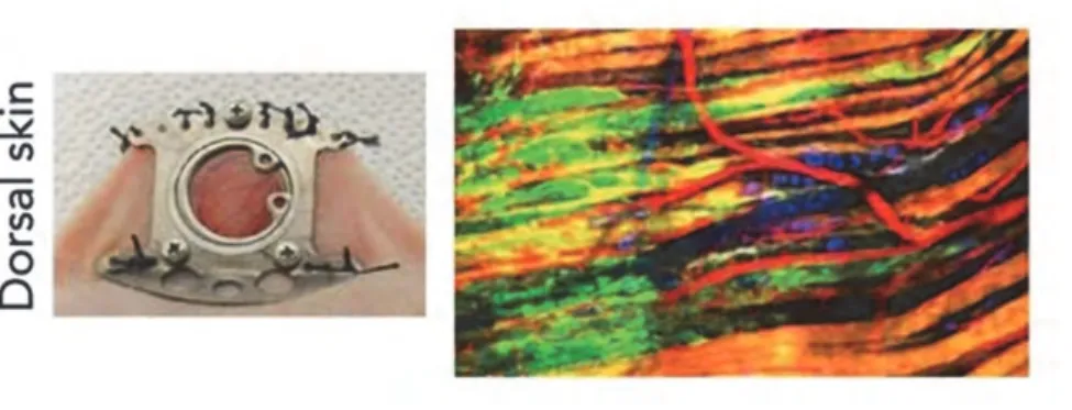
Figure 5: In vivo models to study collective cancer cell migration.
The left photography shows an optical window implanted in the dorsal skin of a nude mouse. At right, the image, from multi-photon microscope, shows the collective melanoma cells invasion. Melanoma cells in green are invading the skin tissue. In this image, blood vessels are coloured in red, collagen in grey, nerve fibres in blue, and muscle fibres in orange. Images from (Choi, Kwok et al. 2015) (left photography) and Bettina Weigelin & Peter Friedl (right image).
I.4.4. Models of collective cell movements during morphogenesis
Several organisms coupled with advanced microscopic technics have permitted the opportunity to track and study collective cell behaviours in vivo. Examples of collective movements during morphogenesis in zebrafishand Drosophila are detailed below.
I.4.4.1. Dorsal closure in Drosophila
During embryonic development in Drosophila, both lateral sides of the embryo stick together to form the dorsal closure (Young, Richman et al. 1993). This process, similar to wound healing, is a good model to study tissue repair. During dorsal closure, the entire epidermal epithelium migrates as a sheet over the squamous epithelial tissue that covers the dorsal side of Drosophila embryo and the vitellus, called amnioserosa (Figure 6) (Kiehart, Galbraith et al. 2000). Therefore, amnioserosa cells contribute to the migration of epithelial cells. Apical constriction of amnioserosa cells decreases the apical surface on which epidermal cells must migrate. Cell shape of amnioserosa and epithelial cells is modified due to reorganisation of the actin cytoskeleton. Leader cells in the epidermis acquire morphology of motile cells, and produce protrusions necessary to drive the migration. All cells within the sheet maintain cell-cell adhesions, as well as cell-cell polarity while migrating. Cells at the leading edges extend filopodia, which promote closure between the two segmented epithelial edges (Jacinto, Wood et al. 2000, Millard and Martin 2008).The amnioserosa starts to degenerate after dorsal closure (Hartenstein and Jan 1992).
During migration, leader cells keep epithelial properties (cell-cell adhesion, apico-basal polarity, and planar cell polarity). Several signals were identified during dorsal closure such as Jun N-terminal kinase (JNK), Wnt and Dpp signalling pathways, and cytoskeleton components and its regulators (Glise, Bourbon et al. 1995, Hou, Goldstein et al. 1997, Harden 2002). The canonical Wnt pathway, signalling through -catenin (Armadillo in Drosophila), controls the polarisation of epidermal cells. On the other hand, JNK signalling regulates the dynamics of actin cytoskeleton and thus contributes to the formation of membrane protrusion by leader cells. In filopodia, DE-cadherin (Drosophila melanogaster epithelial cadherin) and microtubules, regulated by the myosin XV, ensure adhesion between the two epithelial sheets at the end of the migration process (Jankovics and Brunner 2006, Liu, Woolner et al. 2008).
A-D. These confocal images show the successive epidermal hole closure of Drosophila
embryo expressing α-catenin-GFP. These images represent four steps of the closure: the initiation (A);the epithelial sweeping (B); the zippering (C); and the termination (D). LE: leading edge epidermis; AS: amnioserosa; VE: ventral ectoderm (from (Martin and Parkhurst 2004).
I.4.4.2. Collective cell migration of the zebrafish posterior lateral line primordium
The lateral line is a sensory system presents in fish and amphibians. In zebrafish, mechanosensory organs, called neuromasts, are located over the body. In each neuromast, a group of sensory hair cells can detect movements and vibrations in the water environment through the deformation of their cilia. The primordium of the zebrafish lateral line is composed of more than 100cells, which are migrating along the flank of the embryo, directionally from the anterior trunk to the tail (Figure 7A). The entire cohort migrates as a polarised and cohesive group of cells through different signals (Figure 7B) (Ghysen and Dambly-Chaudiere 2004). The cohort of cells exhibits a front-rear polarity, with leader cells extending lamellipodia and filopodia. Follower cells (also called trailing cells) assemble rosette-like mechanosensory organs of about twenty proneuromasts, which are deposited along the migration path, while the group of cells is migrating. Moreover, two receptors to the chemokine CXCL12/SDF-1, CXCR4 and CXCR7, are differentially activated and expressed between leading and trailing cells. This collective motility is guided by a chemokine gradient of CXCL12, which is expressed along the fish horizontal myoseptum.CXCL12 is recognised by the CXCR4 receptor (David, Sapede et al. 2002, Gilmour, Knaut et al. 2004). All cells
express CXCR4, but only leader cells activate it to polarise and guide the entire group of cells (Haas and Gilmour 2006). The formation of organs progenitors is controlled by the growth factor Fgf10 (Fibroblast growth factor), which is express locally by the surrounding tissue. Another chemokine receptor is express by cells at the trailing edge, the CXCR7. This last receptor could capture Sdf1 and block CXCR4 activity in the trailing edge (Valentin, Haas et al. 2007). Indeed, CXCR7 is even responsible for the formation of a self-generated gradient. By sequestering CXCL12 at the trailing edge, CXCR7 generates a gradient of chemoattractants within the primordium and guide its migration (Dona, Barry et al. 2013).
Figure 7: Collective cell migration of the lateral line primordium in zebrafish.
A.During migration, from behind the ear (right) to the tip of the tail (left), the primordium
(white arrow) deposits group of cells (asterisk) that will form the sensory organs called neuromasts. B.The migrating epithelial primordium is a cohesive polarised cluster of
A
B
cellscomposed of about one hundred cells and migrating directionally.ClaudinB promoter in lateral lime primordium cells drives the expression of GFP (Modified from Gilmour’s lab).
I.4.4.3. Collective cell migration during tracheal branching morphogenesis in Drosophila
Collective cell migration is required for epithelial tube branching for the morphogenesis of many organs, such as blood vessels, mammary glands, nephric ducts, or lungs. In Drosophila, the larval tracheal system is composed of hundreds of interconnected tubes. The role of this tracheal network is to supply the larva in oxygen (Affolter, Bellusci et al. 2003). Each tracheal tube is composed of an epithelial monolayer surrounding a central lumen. The tracheal network is formed from ten placodes of the ectodermal epithelium, presents on both sides of the body segments in the embryo. Each of these groups, containing about twenty tracheal precursor cells, constricts their apical surface that drives placode invagination, and undergoes two cell divisions, in order to form a tracheal sac of about eighty cells (Figure 8A). Therefore, primary branches grow out from these placodes. The directions of primary branches begin by the migration in defined directions of two cells from six fixed positions in each sac. Thus, at the tip, two tracheal sac cells (tip cells) lead the migration, produce membrane protrusions, and carry four to twenty placodal cells to form the primary branch (Ghabrial, Luschnig et al. 2003, Affolter and Caussinus 2008). This structure generates the primary branches by directed collective cell migration (Figure 8B-C). Finally, forces exerted by the migrating tip cells contribute to intercalation of stalk (follower) cells, necessary to the elongation of the branch. During tracheal development, only tip cells maintain a polarised shape, and a constricted
apical surface, which is in contact with trailing cells. Protrusions are extended from their basal surface, enriched in F-actin (Tanaka, Takasu et al. 2004).
The surrounding tissue secretes a ligand, Branchless, the homologue of an Fgf, which is recognised by the receptor Breathless. Tracheal cells express Breathless, and cells with the highest level of Breathless activity drive the migration toward the sources of Branchless, and remain attached to their tracheal neighbours (Sutherland, Samakovlis et al. 1996). Thus, the two tip cells are positioned at the terminal end of the branch, and display a more elongated shape combined with the highest protrusive activity than follower cells (Lebreton and Casanova 2014). Cells, with no or less Breathless activity, become trailing cells and form the branch stalk. Moreover Delta, produced in leader cells activate Notch signalling pathway in neighbouring cells to prevent these cells to take the lead position and become tip cells (Ghabrial and Krasnow 2006).
Slit and Robo guidance also contribute to tracheal branching. The two receptors interacting with these secreted ligands in the central nervous system are Robo and Robo2. Tip cells from different branches express differentially the two receptors Robo and Robo2. While activation of Robo2 signalling attracts tip cells to the source of Slit, the Robo signalling contributes to repulsive forces of motile tip cells. Thus, balance between promotion and inhibition of tracheal migration is required for a correct distribution of branches in the embryo (Englund, Steneberg et al. 2002).
The development of the embryonic tracheal system starts from 10 placodes of the ectodermal epithelium, presents on each side ofthe body segments in the embryo. A. After invagination of the tracheal cells and twice divisions, the tracheal sac consists of about eighty cells. B. Six primary branches are formed from these placodes. C,D. During development, these branches fuse and form the tracheal network, necessary for oxygenation of Drosophila larva. (Cabernard, Neumann et al. 2004).
I.4.4.4. Border cell migration in Drosophila
Drosophila ovary is composed of egg chambers at different stages of maturity. At stage 9, a cluster of four to eight motile border cells, surrounding two non-motile polar cells, migrates from the anterior pole until the oocyte. Egg chambers are surrounded by a monolayer of follicular epithelial cells. Border cell clusters delaminate from this epithelial layer, and migrate in between sixteen germ cells (Figure 9A and B). Fifteen of them are nurse cells, and one becomes the oocyte. Border cells carry two polar cells until the oocyte (Figure 9), where they will produce the eggshell structure, called micropyle, to allow entry of sperm (Rorth 2002). During their migration, border cells maintained DE-cadherin adhesions between them. The leader cells produce major membrane protrusions and drive the movement (Montell 2003).
Several signalling pathways control border cell migration. First, the polar cells secrete cytokine unpaired (Upd) that activates the JAK/STAT module in neighbouring cells, which then become the motile border cells. This signal is coordinated with the steroid hormone, ecdysone, to determine the correct migrating cells and the timing of the migration (Beccari, Teixeira et al. 2002). During migration, border cell clusters are guided by ligands, secreted at
highest concentration in the oocyte. Two receptors to these ligands are expressed at the surface of border cells, EGFR (epidermal growth factor receptor) and PVR (platelet-derived growth factor receptor (PDGFR) and vascular endothelial growth factor receptor (VEGFR) receptor related). These receptors contribute to the guiding of border cell clusters to the correct location, in particular by controlling protrusive activity (Duchek and Rorth 2001, Duchek, Somogyi et al. 2001, McDonald, Pinheiro et al. 2003, McDonald, Pinheiro et al. 2006).
Among other models, Drosophila border cell migration presents many advantages. For instance, Drosophila is a good genetically model, and, border cell clusters are composed only of six to ten cells, permitting their ease to track.
In early stage 9, border cells delaminate from the anterior follicular layer (A), migrate in between nurse cells (B), and reach the oocyte by stage 10 (C).Border cell clusters are designated by a white arrow. (Lucas, Khanal et al. 2013)
Among collective cell migration examples listed above, Drosophila border cells constitute a powerful model to study collective cell movements in vivo. Border cell cluster movements are easily imaged in their own environment. Indeed, the dissection of Drosophila ovaries permits keeping the entire egg chamber structure unaffected. Another advantage is the number of cells involved in this process. Indeed, it allows an easy tracking of border cells compared to models involving a hundred of cells like the primordium lateral line in zebrafish embryo.
Given the numerous advantages in studying drosophila, we employed this model in our study of collective migration.
Chapter II: The cytoskeleton in cell migration
In eukaryotes, the cytoskeleton is involved in numerous processes such as cell migration, contraction, vesicle trafficking, or cytokinesis. The cytoskeleton also has a role in the regulation of the cell shape, generating resistance to the cell, maintaining internal structures. Additionally, the cytoskeleton allows the interaction between cells within a tissue. Finally, during cell migration, it is responsible for the membrane deformation leading to the extension of membrane protrusions such as lamellipodia and filopodia (Figure 10) (Le Clainche and Carlier 2008).
Figure 10: Confocal images of the lamella region (LM) of fish fibroblasts containing lamellipodia (LP) and filopodia (FP).
Cells were transfected with mCherry-actin. Actin stress fibres (SF) are organised in parallel bundles. Scale bar, 10 μm. (Nemethova, Danielisova et al. 2008).
Three types of fibres, classified along their diameter, compose the cytoskeleton: actin microfilaments, intermediate filaments, and microtubules, each of them having their own roles in different cell processes. Many proteins bind these filaments to regulate them, thereby contributing to their dynamics. Several biological processes, including cell migration, can involve the three types of filaments (Ridley, Schwartz et al. 2003, Rodriguez, Schaefer et al. 2003, Raftopoulou and Hall 2004).
In this manuscript, I will focus on the role of actin cytoskeleton during cell migration. Indeed, its regulation is crucial for cell movements, including border cell migration in Drosophila.
II.1.
Actin Microfilaments
Actin microfilaments measures about five to nine nm of diameter, and are composed of actin monomers. The actin is a protein of 42 kDa extremely conserved, and present in muscle and non-muscle cells, representing10% of total proteins in eukaryotic cells (Pollard, Blanchoin et al. 2000). Actin polymerisation drives the morphological changes that allow cells to undergo dynamic processes, such as division, phagocytosis, and migration. During cell migration, the regulation of actin dynamics is crucial for the formation of membrane protrusions, and/or the establishment of cell-matrix adhesions. Depending on the cell type, different structures of membrane extensions, with long or short life-times will be extended, and, cell-matrix adhesions will be more or less strong. In a cell, protrusions are structures enriched in actin filaments, allowing steps of adhesion, translocation, and retraction; all of which permit cell movement (Figure 11A) (Cooper 1991). Several proteins control the actin dynamics, some of which participate in the regulation of actin filaments assembly, stability and organisation
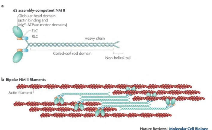
(Welch and Mullins 2002). During cell migration, actin structures are presents in two major types of protrusions: lamellipodia and filopodia (Figure 11A). Depending of its localisation within cell, actin filaments are differently organised. For cell migration or invasion, filaments can be branched and assembled, such as at the leading edge of lamellipodia, or organised in tight parallel bundles like in filopodia. These diverse actin filament organisations generate different protrusion structures. Indeed, filopodia are thin, about 0.1 to 0.3 µm, with a finger-like structure (Mitchison and Cramer 1996, Le Clainche and Carlier 2008). By comparison, lamellipodia are thicker sheet-like protrusions. Stress fibres in non-muscle cells represent another actin structure. These contractile filaments contain actin and non-muscle myosin II bundles, which anchor to focal adhesion sites and generate traction forces to the cell (Figures 10 and 11B). Anti-parallel organisation of actin filaments is observed in contractile stress fibres (Gardel, Schneider et al. 2010).
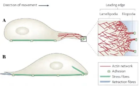
Figure 11: Actin-enriched protrusions at the leading edge of migrating cells.
A. During cell migration, the font of motile cells can be composed of lamellipodia and
filopodia. Actin filaments are the component of both protrusive structures. The elongation of A
F-actin occurs at the barbed ends extremities, oriented toward the plasma membrane. F-actin appears differently organised in these two types of protrusions. Actin filaments form a branched network in lamellipodia, while they are organised in tight parallel bundles in filopodia. B. Once cell adhered to the substratum, the nucleus and cell body move forward. The motor protein, myosin II interacts with stress fibres, which are linked to focal adhesions, in order to produce contraction forces allowing cell displacement. (modified from (Mattila and Lappalainen 2008)).
II.1.1. Organisation of actin network in protrusion (Lamellipodia)
The actin cytoskeleton is the key regulator of protrusions, adhesion formation, and retraction. Monomers of globular actin (G-actin) associate and polymerise to form filaments. The first step is nucleation. It consists of the assembly of oligomers of globular actin. As soon as they exceed the size of trimers, they adopt a helical structure, and become thermodynamically stable. The polymerisation can occur only if the concentration of monomeric actin is above the critical assembly concentration. Then, the elongation phase takes place, in order to form filamentous actin (F-actin). During this stage, monomers assemble together, forming a double helix of about 28 actin subunits. Microfilaments are polarised, with an extremity, barbed end (also called plus end), promoting the polymerisation, and another one, pointed end (also called minus end) which favours depolymerisation. Within the cell, the ratio between the globular and filamentary actin vary among species. This polarity of actin filaments allows the formation of protrusions (Pantaloni, Le Clainche et al. 2001).
The actin can interact with divalent cation calcium or magnesium (Ca2+ or Mg2+) as well as a nucleotide ATP (adenosine diphosphate) or ADP (adenosine diphosphate). Generally, monomeric actin binds Mg2+ and ATP. Then, actin monomers bound to ATP are added at the plus ends of filaments. Indeed, the critical concentration of free ATP-G-actin monomers is lower at the barbed end than at the pointed end, leading to a faster growth at the barbed end. Following, depolymerisation of actin filaments occurs at the extremity pointed end, where actin is mainly bound to ADP. The equilibrium of the filament dynamics is achieved when a monomer of actin disassembly from the pointed end and polymerisation at the barbed end is balanced and maintained by a critical concentration of monomers in the cytosol. This phenomenon is called treadmilling. The hydrolysis of ATP associated with polymerisation is at the origin of treadmilling and destabilises the filament (Figure 12) (Korn, Carlier et al. 1987, Reisler 1993).
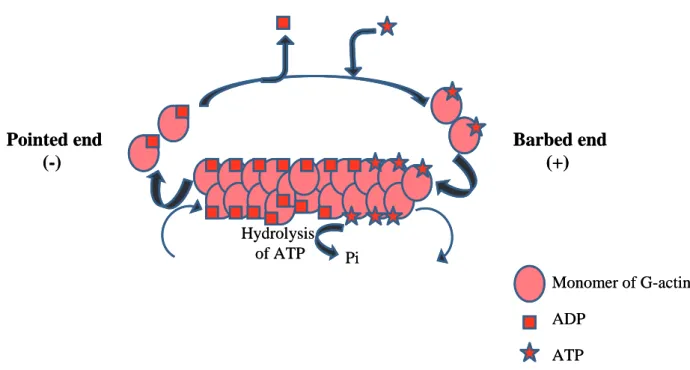
Figure 12: Formation of actin filament.
After the actin nucleus is formed, actin filaments elongate by the addition of G-actin monomers to both ends. However, the plus end (barbed end) filament extremity grows much
Monomer of G-actin ADP ATP Pi Hydrolysis of ATP Barbed end (+) Pointed end (-) Monomer of G-actin ADP ATP Pi Hydrolysis of ATP Barbed end (+) Pointed end (-)
faster. Actin filaments have a double helix structure. After filament assembly, G-actin, bound to ATP, is hydrolysed to ADP. When the dissociation of ADP-actin at the pointed end and the adding of ATP-actin at the barbed end are balanced, filaments reached the steady state.
The network of actin filaments is very dynamic. Therefore, if necessary, cells are able to adapt their actin cytoskeleton very rapidly and efficiently according to intra- or extra-cellular signals. The treadmilling process was well described in living fibroblasts (Wang 1985). Indeed, it has been revealed crucial for lamellipodia regulation and thus cell migration. The barbed end extremity composed of ATP-actin bound form and, demonstrating faster dynamics, is close to the cell membrane. The depolymerisation process is promoted at the pointed end, which means at the back of the lamellipodium, behind the lamella. Actin monomers released during depolymerisation can be used for polymerisation at the front. The actin is continuously dynamic, and oscillates between monomeric and filamentous forms. However, in order to maintain this network, polymerisation and depolymerisation processes are tightly regulated (Mogilner and Oster 1996, Pantaloni, Le Clainche et al. 2001).
II.1.2. Regulation of actin polymerisation
Several proteins regulate the balance between polymerisation and depolymerisation of actin, by interacting with actin monomers or actin filaments. Therefore, these proteins control the actin cytoskeleton dynamics according to the cell functions. Below is a review of some of the most important actin regulators involved during cell migration.
II.1.2.1. Regulation of actin nucleation at the barbed end
mechanism of treadmilling
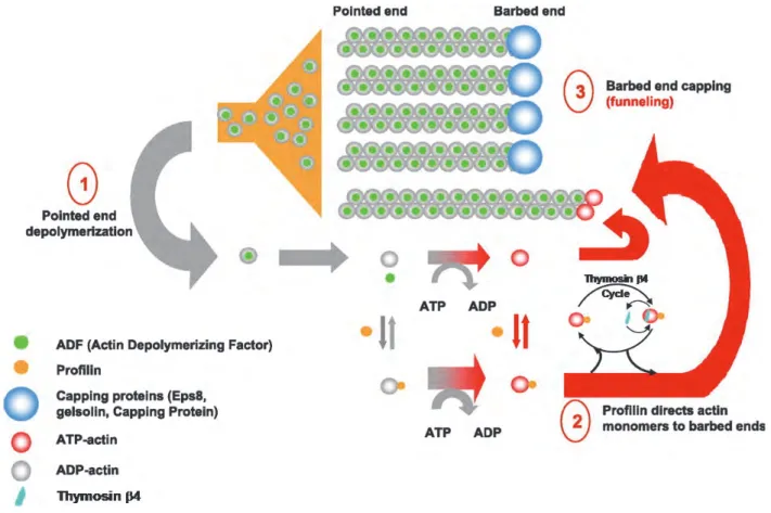
The treadmilling of actin monomers is too slow to be responsible for the fast locomotion of cells by itself. Several studies have been identified actin binding proteins that accelerate the treadmilling process. The following paragraphs detail the molecular mechanisms by which a set of proteins (Actin Depolymerizing Factor (ADF) also called cofilin, profilin, and capping proteins) cooperate to accelerate the treadmilling rate.
II.1.2.2. Regulation of actin polymerisation and depolymerisation
The polymerisation mainly and efficiently occurs at the barbed end extremity of actin filaments. Two groups of proteins contribute to this regulation: promotors of barbed end polymerisation and capping proteins.
II.1.2.1.1. Profilin
The profilin regulates the actin polymerisation by controlling the concentration of actin monomers. The profilin, one of the most abundant actin monomer binding proteins, is essential for actin cytoskeleton dynamics, by associating with G-actin to exchange ADP for ATP. The complex composed of profilin and ATP-G-actin (ratio 1:1) will associate at the barbed end of actin filaments (Figure 14-2). Combined to the role of different actin polymerisation nucleators and stimulators (such as cofilin, formins), the treadmilling process
is robustly improved. Profilin binds actin monomers and favours the exchange between ADP and ATP. Therefore, it allows recycling of ADP-G-actin in ATP-G-actin and increases polymerisation. Profilin-actin complexes are localised at the barbed end extremity of actin filaments. The profilin is negatively regulated by phosphatidylinositol (3,4)-bisphosphate (PtdIns(3,4)P2) (Yarmola and Bubb 2006).
II.1.2.1.2. Cofilin
The cofilin (also called ADF for Actin Depolymerizing Factor) is a factor of depolymerisation acting at the pointed end extremity of microfilaments. It binds preferentially actin filaments at ADP extremity and thereby promotes disassembly of actin filaments at the pointed end (Figure 14-1). The concentration of G-actin increases until polymerisation occurs at the barbed end extremity, in order to balance depolymerisation. Indeed, G-actin bound to ADP will be recycled and added in G-actin bound to ATP at the barbed end extremity. Thus, the cofilin activity leads to an increase of growing polarised filaments (Carlier, Laurent et al. 1997). Cycles of phosphorylation and dephosphorylation regulate cofilin activity. LIM (Lin-11, Isl-1 and Mec-3) kinases (LIMK) phosphorylate the cofilin and thereby inactive it, while the phosphatase slingshot activates cofilin by promoting its dephosphorylation (Arber, Barbayannis et al. 1998, Niwa, Nagata-Ohashi et al. 2002). Moreover, cofilin can act synergistically with profilin to improve elongation of actin filaments (Didry, Carlier et al. 1998).
II.1.2.1.3. Proteins containing the WH2 domain
The Wiskott–Aldrich syndrome protein (WASP) homology 2 (WH2) domain contributes to the recruitment of actin subunits on many actin nucleators, including the regulators of Arp2/3 complex, formins, ciboulot, and cordon bleu, which can promote barbed end assembly. It is also found in thymosin 4, which serve to sequester actin (Le Clainche and Carlier 2008). The thymosin 4 binds G-actin and thus plays a role in the availability of monomers for polymerisation. While the profilin preferentially interacts with the ADP-actin, the thymosin 4 has high affinity for ATP-actin. By interacting with actin monomers, the thymosin 4 sequesters them (Figure 14-2). Therefore, the reservoir pool of G-actin induced by the activity of thymosin 4 will then serve profilin role in promoting the growth of actin filaments (Pantaloni and Carlier 1993). Another protein, ciboulot, shares sequence homology with the thymosin 4, but promotes the polymerisation of actin at the barbed end by binding actin monomers, like the profilin (Boquet, Boujemaa et al. 2000, Pantaloni, Le Clainche et al. 2001).
Spire proteins also contain the actin-binding domain. However, spire is a part of the third class of the actin nucleators, after the Arp2/3 complex (see section II.1.3) and the formins family (see section II.1.4). Through the four WH2 domains, spire is able to bind four consecutive G-actin monomers, to form an unbranched actin filament (Quinlan, Heuser et al. 2005). Spire may also remain attached at the pointed end to block depolymerisation of actin filaments (Figure 13).
Figure 13: Spire, an actin nucleator.
The spontaneous nucleation of actin trimers allows the formation of actin filaments. The spire proteins are a class of actin nucleators. They contain four WH2 domains allowing the binding of four G-actin monomers. Thus, spire proteins lead to polymerisation of unbranched actin filament (Goley and Welch 2006).
II.1.2.1.4. Capping proteins
The density of the actin filament network as well as the concentration of G-actin is controlled by capping proteins. Capping proteins bind preferentially actin filaments at the barbed end extremity of microfilaments, in a calcium dependent manner, and block the assembly of actin monomers (Schafer, Jennings et al. 1996). Capping proteins terminate elongation, thereby limiting polymerisation of new filaments, and induce an increase of G-actin concentration. Thus, uncapped actin filaments will grow faster (funnelling process, Figure 14-3). However, some capping proteins also bind the pointed end extremity in order to block depolymerisation. Once fixed to this extremity, these capping proteins can form new nucleation sites where polymerisation will be initiated. According to the extremity capped, and, to the type of capping protein, the polymerisation can be favoured or inhibited.
The elongation of actin filaments occurs at barbed ends, in front of the protruding plasma membrane in protrusive structures such as lamellipodia, and filopodia (Rafelski and Theriot 2004). In cells, several capping proteins are involved in diverse processes and are regulated by different signalling pathways. The capping protein (CP) is a heterodimer of - and -subunits and is the homolog of striated muscle protein CapZ. The CP, known to control the extension of lamellipodia during cell migration, can interact with F-actin at the barbed end extremity to block the growth of microfilaments in non-muscle cells (Figure 14-3). The CP is the most ubiquitous and abundant capping protein. The Phosphatidylinositol 4,5-bisphosphate (PtdIns(4,5)P2)and the protein CARMIL (capping protein, Arp2/3 and myosin-I linker) inhibit actin filament capping by the CP (Schafer, Jennings et al. 1996). Among capping proteins, the gelsolin family, including at least gelsolin, CapG, severin, adseverin, villin, advillin, flightless I, and brevin control the actin organisation by severing filaments, capping filament ends and nucleating actin assembly. These proteins bind the barbed end extremity with very high affinity, and thus block the polymerisation at the barbed end of actin fibres. The PtdIns(4,5)P2 is one inhibitor of gelsolin activity and can promote F-actin growth against the membrane (Witke, Sharpe et al. 1995). Twinfilin, another ADP-G-actin sequestering protein that can also cap ADP-bound barbed ends, is composed of two ADF-homology domains. Twinfilin binds actin filaments at the barbed ends, by a similar mechanism as gelsolin, to promote actin filament severing and thus affect cell motility in vitro (Helfer, Nevalainen et al. 2006, Paavilainen, Hellman et al. 2007).This function of twinfilin was confirmed in vivo, in yeast. Its severing activity is inhibited by capping protein and PtdIns(4,5)P2. As such, this protein synchronises processes of filament severing and monomer sequestration at sites of rapid actin turnover (Moseley, Maiti et al. 2006). Twinfilin can also intact with CP, where it sequesters and maintain actin monomers in ADP-bound form (Palmgren, Vartiainen et al. 2002, Falck, Paavilainen et al. 2004). Another family, Eps8 proteins, are able to cap barbed end
extremities. In fibroblasts, Eps8 is localised in the cortex region and in protrusions (Scita, Tenca et al. 2001). This auto-inhibited capping protein is activated by its interaction with Abi1, controlling signalling pathway critical for cell motility (Disanza, Carlier et al. 2004, Hertzog, Milanesi et al. 2010).
However, tropomodulin binds the pointed end extremity and thus stabilises actin filaments. This protein blocks the release of G-actin from pointed end of the actin fibre. Thus, during cell migration, capping proteins limit actin polymerisation to the region activated by the Arp2/3 complex. Therefore, extremities of elongated filaments are re-capped to prevent depolymerisation and thus conserve the actin filament within the lamellipodium (Kostyukova, Rapp et al. 2005).
.
1. Cofilin (ADF) binds ADP-G-actin at the pointed end extremity of filaments and promotes
their depolymerisation. Cofilin activity leads to an increase of monomeric G-actin. 2. Profilin binds actin monomers and favours the exchange between ADP and ATP. Profilin enhances the recycling of ADP-G-actin in ATP-G-actin and increases the polymerisation at the barbed end extremity. Thymosin 4 binds and sequesters preferentially ATP-actin monomers and prevents their assembly into filaments 3. Most of capping proteins bind actin filaments at the barbed end and block the addition of G-actin at this extremity. At steady state, the depolymerisation of capped filaments leads to an increase of monomeric actin concentration.(modified from (Le Clainche and Carlier 2008)).
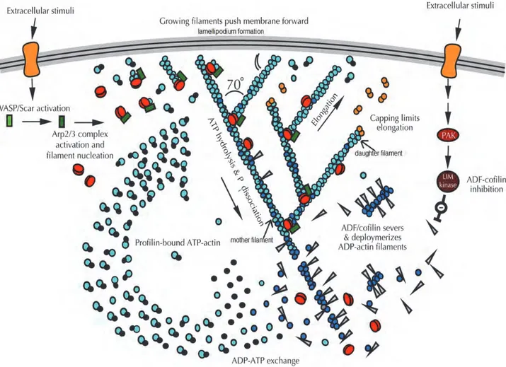
II.1.3. Regulation of branched nucleation
Migrating cells must maintain a constant treadmilling rate necessary for protrusive activity. In lamellipodia of motile cells, the complex of nucleation factors Ap2/3 ensures the actin filaments elongation. The Arp2/3 complex is composed of seven protein subunits including two actin-related proteins (Arp2 and Arp3) and five proteins ARPC1, ARPC2ARPC3, ARPC4 and ARPC5 (Higgs and Pollard 2001). Arp2 and Arp3 subunits, bound with actin filaments, generate new nucleation sites. The nucleation mediated by Arp2/3 is regulated by many proteins, the members of the Wiskott–Aldrich syndrome family of nucleation promoting factors (NPFs), such as WASP (Wiskott–Aldrich syndrome protein), N-WASP, Scar/WAVE, and WASH. These proteins were identified as Arp2/3 activators under the activity of the Rho small GTPase, Rac1 and Cdc42 (Nobes and Hall 1995). These proteins regulate actin dynamics in numerous cellular processes, such as cell migration, endocytosis, and phagocytosis (Stradal and Scita 2006, Takenawa and Suetsugu 2007). The contribution of