O R I G I N A L A R T I C L E
The ciliogenic transcription factor Rfx3 is required for
the formation of the thalamocortical tract by regulating
the patterning of prethalamus and ventral
telencephalon
Dario Magnani
1
, Laurette Morlé
2
, Kerstin Hasenpusch-Theil
1
, Marie Paschaki
2
,
Monique Jacoby
3
, Stéphane Schurmans
4
, Bénédicte Durand
2
and Thomas Theil
1,
*
1
Centre for Integrative Physiology, University of Edinburgh, Hugh Robson Building, Edinburgh EH8 9XD, UK,
2Centre de Génétique et de Physiologie Moléculaires et Cellulaires, CNRS UMR 5534, Université Claude Bernard
Lyon 1, Villeurbanne, Lyon F69622, France,
3Institute of Immunology, Centre de Recherche Public de la Santé/
Laboratoire National de Santé, Luxembourg, Luxembourg and
4Laboratory of Functional Genetics, GIGA-Signal
Transduction, GIGA B34, Université de Liège, Liège B-4000, Belgium
*To whom correspondence should be addressed at: Centre for Integrative Physiology, University of Edinburgh, Hugh Robson Building, Edinburgh EH8 9XD, UK. Tel: +44 131 650 3721; Fax: +44 131 650 6527; Email: thomas.theil@ed.ac.uk
Abstract
Primary cilia are complex subcellular structures that play key roles during embryogenesis by controlling the cellular response to several signaling pathways. Defects in the function and/or structure of primary cilia underlie a large number of human syndromes collectively referred to as ciliopathies. Often, ciliopathies are associated with mental retardation (MR) and malformation of the corpus callosum. However, the possibility of defects in other forebrain axon tracts, which could contribute to the cognitive disorders of these patients, has not been explored. Here, we investigate the formation of the corticothalamic/ thalamocortical tracts in mice mutant for Rfx3, which regulates the expression of many genes involved in ciliogenesis and cilia function. Using DiI axon tracing and immunohistochemistry experiments, we show that some Rfx3−/−corticothalamic axons abnormally migrate toward the pial surface of the ventral telencephalon (VT). Some thalamocortical axons (TCAs) also fail to leave the diencephalon or abnormally project toward the amygdala. Moreover, the Rfx3−/−VT displays heterotopias containing attractive guidance cues and expressing the guidance molecules Slit1 and Netrin1. Finally, the abnormal projection of TCAs toward the amygdala is also present in mice carrying a mutation in the Inpp5e gene, which is mutated in Joubert Syndrome and which controls cilia signaling and stability. The presence of identical thalamocortical malformations in two independent ciliary mutants indicates a novel role for primary cilia in the formation of the corticothalamic/thalamocortical tracts by establishing the correct cellular environment necessary for its development.
Received: September 9, 2014. Revised and Accepted: January 21, 2015
© The Author 2015. Published by Oxford University Press. All rights reserved. For Permissions, please email: journals.permissions@oup.com
Human Molecular Genetics, 2015, Vol. 24, No. 9 2578–2593 doi: 10.1093/hmg/ddv021
Advance Access Publication Date: 28 January 2015 Original Article
2578
at The University of Edinburgh on July 27, 2015
http://hmg.oxfordjournals.org/
Introduction
Primary cilia are complex subcellular organelles protruding from the cell surface, which mediate several sensory functions and are involved in regulating the cellular response to several signaling pathways and in particular to Sonic hedgehog (Shh) signaling (1). Moreover, primary cilia have important roles in human dis-eases, and defects in primary cilia formation and/or function underlie several human syndromes commonly referred to as ciliopathies (2,3). These disorders are associated with a large var-iety of manifestations that often include neurological features such as MR, but much less is known about the specific signaling pathways and the pathogenesis at the cellular and tissue level that ultimately result in neurological disease phenotypes (4,5).
A common feature of ciliopathies is malformations of the cor-pus callosum (CC) which as the major forebrain commissure pro-vides the interhemic exchange of information between the two cortical hemispheres. While the significance of callosal abnor-malities for the MR pathogenesis of ciliopathy patients remains unclear, thisfinding raises the possibility that other prominent axon tracts of the forebrain are affected in ciliopathies. To explore this, we here investigate the formation of the corticothalamic/ thalamocortical tracts in two ciliary mouse mutants. The thala-mocortical tract connects the dorsal thalamus with the cerebral cortex thereby conveying sensory information from the external environment to the cortex. The corticothalamic tract in turn sends processed sensory information back to the thalamus thereby providing the feedforward and feedback mechanisms es-sential in this processing unit (6). The formation of the thalamo-cortical tract requires a complex navigation of thalamothalamo-cortical axons (TCAs) through the prethalamus and ventral telenceph-alon (VT), and involves several guidepost cues telenceph-along the thala-mocortical path that control the navigation of thalamic axons. Two populations of pioneer neurons are located in the prethala-mus and the VT projecting their axons into the thalaprethala-mus and providing scaffolds for TCAs growing into the prethalamus and across the diencephalic/telencephalic boundary, respectively (7–10). In addition, cells from the lateral ganglionic eminence (LGE) migrate into the medial ganglionic eminence (MGE) to form a permissive corridor and guide TCAs through the other-wise non-permissive MGE (11). These corridor cells also mediate the sorting of TCAs according to their rostral/caudal origin in the thalamus by expressing the Slit1 and Nestin1 guidance factors (12). Interestingly, the development of the VT and of the pretha-lamus, which provide these guidepost cues, is under the control of Shh signaling, raising the possibility that the formation of the thalamocortical tract might be affected in primary cilia mutants. However, this has not been explored yet.
RFX transcription factors have been shown to play fundamen-tal roles in ciliogenesis by regulating the expression of genes involved in cilia assembly or function (13–15). Accordingly, Rfx3-deficient mouse mutants exhibit several hallmarks of cilio-pathies, in particular left–right asymmetry defects and hydro-cephalus (13,14), yet importantly they survive until birth, providing a rare opportunity to study the formation of forebrain axon connections in a cilia mouse mutant. Indeed, the Rfx3 mu-tation interferes with the formation of the CC (16). Here, we inves-tigate the formation of the corticothalamic/thalamocortical tracts in Rfx3-deficient mice. In these animals, some corticothala-mic axons (CTAs) abnormally migrate toward the amygdala. Moreover, only a small proportion of TCAs reach the dorsal telen-cephalon but many fail to migrate through the prethalamus, whereas others enter the VT but subsequently project ventrally toward the amygdala. These defects correlate with abnormal
patterning of the prethalamus. In addition, the rostroventral tel-encephalon forms neural heterotopias at its pial surface contain-ing a mixture of MGE- and LGE-derived cells expresscontain-ing the Slit1 and Nestin1 guidance molecules. Finally, the abnormal ventral deflection of TCAs toward the amygdala is also found in a second ciliary mouse mutant, which is deficient for Inpp5e, a Joubert syn-drome disease gene. Taken together, these analyses indicate a novel role for primary cilia in the development of thalamocortical tract.
Results
Rfx3−/−mutant embryos display defects in the development of thalamocortical connections
Recently, we showed that the ciliogenic transcription factor Rfx3 is required for CC development (16). Here, we investigate whether an Rfx3 null mutation also affects the formation of the thalamo-cortical/corticothalamic tracts. TCAs progress in a multistep fashion over their intermediate territories, the diencephalon and the VT (17). Thalamic neurons send their axons ventrally through the prethalamus toward the diencephalic–telencephalic boundary. After entering the telencephalon at E13.5, thalamic axons migrate through the VT via the internal capsule (ic) to reach the pallial–subpallial boundary (PSPB) and finally enter the cortical intermediate zone at E14.5. CTAs leave the cortex at around E14.5 and follow the same trajectory through the VT as TCAs but in the opposite direction. This trajectory is revealed in E18.5 control embryos by placement of DiI crystals into the cor-tex. This analysis also reveals the path of TCAs by retrograde labeling (Fig.1A–D). Rfx3−/−mutant brains show a similar labeling
pattern; however, we noted a number of backlabeled nuclei in the rostroventral telencephalon close to a region of the VT containing a heterotopia. This analysis also revealed an abnormal ventral projection toward the amygdala at intermediate and caudal levels (Fig.1E–H). To more specifically label TCAs, we placed DiI crystals into the thalamus. TCAs are highly fasciculated in the ic, are arranged in roughly parallel bundles in the striatum and reach the cortex (Fig.1I and J). In contrast, in Rfx3−/−mutant brains, thalamic axons appear disorganized in the striatum with some axons projecting toward the PSPB in more ventral re-gions (Fig.1M and N). At caudal levels, TCAs abnormally project into the VT toward the amygdala similar to ourfindings on the CTA trajectory.
Immunostaining for the panaxonal marker Neurofilament (NF) confirmed the pathfinding defects revealed by DiI labeling. In control embryos, this analysis showed parallel organized axons in the thalamus, prethalamus, ic, the striatum and in the intermediate zone of the cerebral cortex (Fig. 1K and L). In Rfx3−/−mutants, NF staining revealed several abnormalfiber tracts. Many axons are already disorganized in the thalamus, and the exit and entry zones of CTAs and TCAs into and out of the cortex appear broader (Fig.1O, arrowheads). Many NF+fibers ectopically project toward the amygdala forming a highly fasci-culated axon bundle at more caudal levels (Fig.1P, arrow).
To gain insights into the origin of these axonal defects, we performed NF immunofluorescence analyses and DiI labeling on E14.5 control and Rfx3−/−mutant brains. At this stage, NF labels axons in the developing thalamus, prethalamus and VT of control embryos (Supplementary Material, Fig. S1A and B). The ic contains highly fasciculated NF+axons, whereas axons are more
loosely organized in the striatum (Supplementary Material, Fig. S1C and D). In Rfx3−/−embryos, NF+axons are disorganized in
the VT projecting in several directions within the LGE and MGE,
at The University of Edinburgh on July 27, 2015
http://hmg.oxfordjournals.org/
Figure 1. Axon guidance defects in the E18.5 Rfx3−/−brains. Coronal sections through the brain of control (A–C and I–L) and Rfx3−/−(E–G and M–P) E18.5 embryos. (A–C and
E–G) DiI placements in the cortex (asterisks) reveal the anterior commissure (ac) (A and E) and the corticothalamic and thalamocortical tract through anterograde and
retrograde labeling, respectively. In Rfx3−/−mutants, both tracts are formed but note the backlabeled neurons close to a heterotopia at the ventral telencephalic
surface (inset and white asterisks in E) and the abnormal projections toward the amygdala at intermediate and caudal levels (arrowheads in F and G). (I, J, M and N)
Thalamic DiI labeling (asterisks) revealed the TCA trajectory in control embryos (I and J). In Rfx3−/−mutants, TCAs cross the PSPB in a broader entry zone (arrowheads
in M and N). Several axons leave the ic prematurely and form an ectopic axon bundle running ventrally toward the pial surface (arrows in M and N). (K, L, O and P)
Immunostainings for NF showed axonal pathfinding defects in the diencephalon (arrowheads in O) and an ectopic axon bundle running ventrally toward the
amygdala of Rfx3−/−embryos (arrows in O and P). (D and H) Schematic drawings summarizing the CTA and TCA pathfinding defects in Rfx3−/−mutants. Scale bar: 100 µm.
2580 | Human Molecular Genetics, 2015, Vol. 24, No. 9
at The University of Edinburgh on July 27, 2015
http://hmg.oxfordjournals.org/
and a thick axon bundle runs ectopically toward the pial surface (Supplementary Material, Fig. S1F and G, arrow), whereas caudal-ly no axons reached the PSPB (Supplementary Material, Fig. S1H and I). In addition, several NF+axons mis-project in the
prethala-mus resulting in the formation of abnormal bundles (Supple-mentary Material, Fig. S1I). Thesefindings were confirmed by DiI labeling experiments of TCAs. In contrast to control embryos, very few TCAs traverse the VT with no axons reaching the PSPB in Rfx3−/−mutants (Supplementary Material, Fig. S1K, L and Q), but a largefiber bundle abnormally projects ventrally toward the tele-ncephalic pia (Supplementary Material, Fig. S1R). To further dis-sect the path of thalamic axons, we performed calretinin (CR) immunofluorescence analysis. In control brains, CR selectively labels thefirst thalamic axons entering the VT and projecting through the ic (11) (Supplementary Material, Fig. S1O and P). In Rfx3−/−mutants, CR+axons were detected in the ic at rostral
le-vels (Supplementary Material, Fig. S1U) but consistent with the NF immunostaining and DiI labeling experiments, many CR+
axons migrated ventrally toward the pial surface after entering the VT at caudal levels (Supplementary Material, Fig. S1V). Also, some CR+axons form abnormal clusters in the thalamus. Finally,
placing DiI crystals into the cortex revealed CTAs in control em-bryos having penetrated into the VT and retrogradely labeled neurons in the thalamus (Supplementary Material, Fig. S1M and N). In Rfx3−/− mutants, backlabeled neurons were not observed, instead a large axon bundle projects from the cortex toward the pia of the VT (Supplementary Material, Fig. S1S and T). Collectively, these analyses indicate severe disruptions in the Rfx3−/− corticothalamic/thalamocortical tracts, with many Rfx3−/−thalamic axons being misrouted in the prethala-mus and VT.
Rfx3−/−mutants display abnormalities in the patterning of the prethalamus
From E9.5, Rfx3 is expressed throughout the forebrain (16) includ-ing the cortex and thalamus where corticothalamic and thala-mocortical neurons are born, respectively, and the prethalamus and VT through which CTAs and TCAs migrate. Therefore, the CTA/TCA pathfinding errors in Rfx3−/−mutants could result
from defects in all three tissues. To start to identify the tissue (s) primarily responsible for the TCA phenotype, wefirst investi-gated the development of the Rfx3−/−mutant thalamus. To this end, we examined the expression of several transcriptional regu-lators including Lhx2, Gbx2 and Ngn2 that play key roles in speci-fying thalamic nuclei and in determining their axonal projection patterns (18–20). We also analyzed neuronal differentiation mar-kers (Tuj1 and CR) and the expression of the axon guidance recep-tors Robo1/2 (21). Overall, these analyses showed no obvious defects in the development of the Rfx3−/− thalamus which could explain the TCA guidance mistakes (Supplementary Mater-ial, Figs S2 and S3). Furthermore, we have previously shown that cortical lamination is not affected in Rfx3−/−mutants (16). We therefore focused our further analyses on the development of the prethalamus and VT starting with the prethalamus.
Since development of the prethalamus is controlled by Shh signaling from the zona limitans intrathalamica (zli) (22–25) and since primary cilia are crucial for Shh signaling, wefirst in-vestigated Shh expression and that of its target genes Ptc1 and Gli1 in the Rfx3−/−diencephalon. This analysis did not reveal obvious defects in Shh zli expression (Fig.2A, D and G), whereas the expression domains of Ptc1 and Gli1 appear expanded in the prethalamus but no significant changes were found in the expres-sion levels of these genes as shown by qRT-PCR (Fig.2B, C, E, F,
H and I). Next, we investigated prethalamic patterning. In E14.5 control embryos, the prethalamic progenitor domain is charac-terized by Nkx2.2 expression (25) (Fig.3A and B). This expression domain is expanded in Rfx3−/−mutants (Fig.3C and D). Olig2, an-other marker for diencephalic progenitor cells, displays a more complex expression pattern. Immunofluorescence analysis showed a high-level Olig2 expression domain in prethalamic pro-genitors, and lower-level expression in thalamic propro-genitors, with a highventralto lowdorsalexpression gradient (Fig.3E, F, I
and J). In the Rfx3−/−mutant diencephalon, the dorsal thalamic expression of Olig2 and its expression gradient is maintained (Fig.3G, H, K and L). However, Olig2 expression is expanded in the prethalamus of Rfx3−/−mutants (Fig.3H and L). Finally, the expression domain of the Pax6 transcription factor normally ex-tends ventrally from the zli encompassing progenitors from the prethalamus and from the eminentia thalami, thereby delineat-ing the boundary between the thalamus and prethalamus (Fig.3E, F, I and J). In the Rfx3−/−mutants, Pax6 expression is maintained in prethalamic progenitors, but its expression in the diencephalic mantle is reduced (Fig.3G, H, K and L). Taken to-gether, these results suggest regionalization defects in the pre-thalamus of the Rfx3−/−mutants.
Abnormal MGE development and formation of heterotopias in theRfx3−/−VT
Since Rfx3−/−mutants show several corticothalamic/thalamocor-tical pathfinding defects in the VT, we next investigated its devel-opment. Recently, we reported a slight down-regulation of Shh signaling in the MGE of Rfx3−/−mutants using in situ hybridization (16), but qRT-PCR showed no effect on Shh and Gli1 expression, whereas Ptc1 expression was mildly up-regulated (P = 0.0384; n = 6; Supplementary Material, Fig. S4). Correct levels of Shh signal-ing are crucial for establishsignal-ing the PSPB separatsignal-ing the dorsal tel-encephalon and VT. However, expression analyses for Dlx2, Gsh2, Pax6 and Dbx1 did not reveal abnormalities at this boundary (Fig.4A and E, and Supplementary Material, Fig. S5). We next ana-lyzed the subdivision of the VT into the MGE and LGE. Dlx2 is ex-pressed throughout the proliferative zone of the VT, whereas Nkx2.1 expression is restricted to the MGE (Fig.4A and B). More-over, Nkx6.2 expression is confined to progenitors on either side of the interganglionic sulcus (Fig.4C). In Rfx3−/−embryos, these expression patterns are maintained except for a slight Nkx6.2 down-regulation (Fig.4E–G). We also noted an ectopic accumula-tion of Dlx2 and Nkx2.1 expressing cells outside the VT at its ven-tral surface (Fig. 4E–G) reminiscent of the heterotopia we observed at E18.5 (Fig.1E). These heterotopias were detected on both sides of the rostral telencephalon in all Rfx3 mutants but not in more caudal regions. Also, they varied in size with some extending toward the midline, whereas others were confined to the surface of the ventrolateral telencephalon. In addition, the heterotopias contain a mixture of progenitor cells and neurons. Expression of the MGE-specific genes Lhx6 and Lhx7, which regu-late Shh expression in the MGE and which are in turn Shh target genes (26), was detected in the heterotopias (Fig.4H and M) as well as that of Tuj1 characteristic of differentiating neurons (Fig.4N). Tuj1 immunofluroescence also showed a size reduction of the MGE mantle. Finally, we investigated whether disruption of the basal lamina, which normally surrounds the telencephalon, could account for heterotopia formation. Laminin immunofluor-escence showed that the basal lamina is disrupted in several po-sitions at the ventral pial surface of E12.5 and E14.5 Rfx3−/− mutants (Fig.4O and P). Taken together, these analyses show a size reduction of the MGE mantle and a disruption of the basal
at The University of Edinburgh on July 27, 2015
http://hmg.oxfordjournals.org/
lamina concomitant with the formation of neural heterotopias in Rfx3−/−embryos.
Cellular and molecular guidance cues are affected in the VT ofRfx3−/−embryos
Given these defects in the formation of the VT, we next analyzed whether ventral telencephalic guidance cues essential for TCA projection are also affected. Pioneer neurons located in the ros-tral MGE extend their axons into the thalamus and serve as scaf-folds to guide TCAs across the diencephalic–telencephalic boundary (7–9). Since no markers are available to selectively label these MGE pioneer neurons, we investigated their forma-tion by DiI placement into the E12.5 thalamus. In control em-bryos, these placements retrogradely labeled the pioneer axons and their cell bodies located in the rostral MGE (Fig.5A and B).
While this analysis also revealed pioneer neurons cell bodies and axons in the MGE mantle of Rfx3−/−mutants, their distribu-tion appeared less dense (Fig.5C and D). In addition, some DiI-la-beled cell bodies were located in the heterotopic tissue outside the MGE mantle.
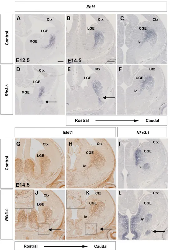
In addition to the MGE pioneer neurons, neurons derived from the LGE migrate into the MGE and form a corridor along which TCAs project through the otherwise non-permissive environ-ment of the MGE. In control E12.5 and E14.5 brains, these guide-post cells are marked by both Ebf1 and Islet1/2 expression from their origin in the LGE and during their migration into the MGE, where they form the ic (Fig.6A–C). In E12.5 and E14.5 Rfx3−/−
em-bryos, Ebf1-expressing cells originate normally in the LGE and mi-grate toward the MGE forming the corridor at caudal levels (Fig.6F). However, at rostral levels, Ebf1-expressing cells migrate toward the heterotopias thereby abnormally connecting the
Figure 2. Shh signaling in Rfx3−/−mutant diencephalon. Coronal sections through the diencephalon of E12.5 control (A–C) and Rfx3−/−mutant embryos (D–F) hybridized
with the indicated probes. (A and D) No major differences are observed in Shh expression in the Rfx3−/−zli compared with control embryos. Note the enlargement of the
third ventricle in mutant embryos (asterisks in D, E and F). (B and C) Ptc1 and Gli1 are expressed in the diencephalon of control brains, but not in the zli. (E and F) Expanded
expression of both Ptc1 and Gli1 is found in the prethalamus of Rfx3−/−mutants (arrowheads in E and F). (G–I) Quantification of Shh (G), Ptc1 (H) and Gli1 (I) expression in the
diencephalon using qRT-PCR revealed no significant changes in the expression levels of these markers (n = 6). Scale bar: 100 µm. 2582 | Human Molecular Genetics, 2015, Vol. 24, No. 9
at The University of Edinburgh on July 27, 2015
http://hmg.oxfordjournals.org/
surface of the MGE mantel with the LGE (Fig.6D and E). These ab-normalities were confirmed by immunostainings for Isl1/2. As in control embryos, Isl1/2+cells normally populate the corridor
(Fig.6G, H, J and K), but were also found ectopically in the hetero-topic tissue in Rfx3−/−embryos (Fig.6J and K). Finally, Nkx2.1 ex-pression labels the MGE ventricular zone and the globus pallidus of E14.5 control embryos, whereas the ic remains Nkx2.1 negative (Fig.6I). The Nkx2.1 expression pattern in the Rfx3−/−MGE mantle appears unaltered in Rfx3 mutants but as with the corridor mar-kers, Nkx2.1-expressing cells were also detected in the ectopic tis-sue (Fig.6L). Thesefindings suggest that the MGE corridor forms at caudal levels, but that corridor cells abnormally migrate toward the heterotopias at rostral levels.
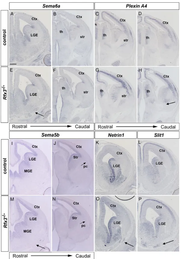
Next, we investigated whether the defects in forebrain pat-terning and in the formation of cellular guidance cues correlate with altered expression patterns of axon guidance molecules. Interestingly, Sema6a−/−mice show a very similar thalamocorti-cal pathfinding defect as Rfx3−/−mutants with TCAs being
de-flected toward the amygdala after entering the VT (27). Sema6a has a highly complex expression pattern in the developing fore-brain. Rostrally, Sema6a expression was mainly confined to the
ventricular zone of the VT, but weak expression was also found in the heterotopias of Rfx3−/−embryos (Fig.7A and E). Caudally, Sema6a transcripts were detected in thalamic neurons and in groups of cells along the path of TCAs in the prethalamus and dorsally and ventrally to the permissive corridor. This expression pattern is not affected by the Rfx3 mutation (Fig.7B and F). Plexi-nA4 encodes a semaphorin receptor, and groups of PlexiPlexi-nA4-ex- PlexinA4-ex-pressing cells delineate the corridor ventrally and dorsally and also reside in the caudal VT where TCAs normally not project (Fig.7C and D). In Rfx3−/−embryos, PlexinA4 expression largely persists in cells surrounding a smaller ic at rostral levels and in cells in the caudal VT where it is, however, adjacent to an ectopic axon bundle likely containing TCAs which mis-project toward the amygdala (Fig.7G and H). Sema5b encodes a chemorepellent for corticofugal axons, and is expressed in telencephalic progeni-tor cells and in neurons of the piriform cortex. This expression pattern is maintained in Rfx3−/−embryos with no Sema5b expres-sion in the heterotopias (Fig.7I, J, M and N). We also investigated expression of Netrin1 and Slit1 which interact in the rostral/caudal sorting of TCAs in the corridor (12). In addition, Slit1 regulates the migration of corridor cells in the MGE (28). Both genes are
Figure 3. Patterning defects in the prethalamus of Rfx3−/−mutant brains. (A–L) Coronal sections through the diencephalon of E14.5 control (A, B, E, F, I and J) and mutant
(C, D, G, H, K and L) brains were stained with the indicated antibodies. B, D, F, H, J and L are higher magnifications of A, C, E, G, I and K, respectively. (A and B) Nkx2.2
immunostaining labels the prethalamic progenitor domain. (C and D) Nkx2.2 expression was expanded in the diencephalon of Rfx3−/−mutants (arrowhead in D). (E, F,
I and J) In control brains, Olig2 displays a high expression domain in the prethalamic progenitor region and is also detected at lower levels in the progenitor domain of
the dorsal thalamus. (G, H, K and L) In the Rfx3−/−diencephalon, the dorsal thalamic expression of Olig2 and its gradient are maintained; however, the Olig2 prethalamic
expression is expanded (arrowhead in H and L). (E, F, I and J) Pax6 is highly expressed in the diencephalon of control brains at the boundary between prethalamus and
dorsal thalamus. (G, H, K and L) In the Rfx3−/−diencephalic ventricular zone, the Pax6 expression domain in the diencephalic mantle is reduced (arrows in H and L). Scale
bars: 100 µm.
at The University of Edinburgh on July 27, 2015
http://hmg.oxfordjournals.org/
expressed in ventral telencephalic progenitor cells, and Netrin1 transcripts were also found in the MGE mantle (Fig.7K and L). These expression patterns remain unaltered in Rfx3−/−embryos,
but both genes are ectopically expressed in cells located in the heterotopias (Fig.7O and P). Taken together, these expression analyses did not reveal changes in the overall Sema5b, Sema6a
Figure 4. Patterning defects in the VT of Rfx3−/−mutant embryos. In situ hybridizations (A–I and M) and immunostainings (J–L and N–P) on E12.5 (A–C, E–G, J, K, N and O) and
E14.5 (D, H, I, M, L and P) coronal sections with the indicated antibodies and probes, respectively. (A) Dlx2 is expressed in the VT, including LGE and MGE, and also defines the PSPB. (B) Nkx2.1 is specifically expressed in the MGE. (C) Nkx6.2 expression defines the sulcus between the LGE and MGE. (E and F) The expression patterns of Dlx2 and
Nkx2.1 are not changed in Rfx3−/−mutants, but Dlx2- and Nkx2.1-expressing cells ectopically accumulate outside the MGE mantle (arrows). (G) Nkx6.2 expression is reduced
in the Rfx3−/−VT. (D and I) Lhx6 and Lhx7 are strongly expressed within the MGE mantle. (H and M) In Rfx3−/−mutants, ectopic Lhx6 and Lhx7 expression is found in the
heterotopias (arrows in H and M). (J) Tuj1staining labels the mantle of MGE, LGE and the cortical preplate. (N) Rfx3−/−embryos show a thinner MGE mantle (asterisk) and an
ectopic accumulation of Tuj1+cells outside the MGE mantle (arrow). (K and L) In control brains, laminin labels the basal lamina surrounding the VT. (O and P) In Rfx3−/−
mutant brains, the basal lamina is abnormally disrupted (arrowheads). Scale bars: 100 µm. 2584 | Human Molecular Genetics, 2015, Vol. 24, No. 9
at The University of Edinburgh on July 27, 2015
http://hmg.oxfordjournals.org/
and PlexinA4 expression patterns, but show differences in the or-ganization of TCAs with respect to the expression domains of these guidance molecules and also indicate ectopic expression of axon guidance factors in the heterotopias.
TheInpp5e ciliary mouse mutant phenocopies the TCA pathfinding defects of Rfx3−/−mutants
The Rfx3 transcription factor has a prominent role in ciliogenesis by regulating the expression of many genes involved in cilia as-sembly and function, suggesting that the thalamocortical abnor-malities of Rfx3−/−embryos are likely caused by structural and/or functional ciliary defects. However, scanning electron micros-copy revealed that cilia are present in the diencephalon and telencephalon of E10.5 Rfx3−/−embryos without obvious mor-phological abnormalities (Supplementary Material, Fig. S6 and data not shown). To gain support for the involvement of cilia in the pathogenesis of these Rfx3−/−phenotypes, we characterized thalamocortical development in an additional ciliary mouse mu-tant that carries a mutation in the Inpp5e gene (29). Inpp5e en-codes inositol polyphosphate-5-phosphatase E that hydrolyzes the 5-phosphate of the second messengers PI(4,5)P2 and PI
(3,4,5)P3. The Inpp5e protein is localized at the axoneme of the primary cilium and has a role in cilia-mediated signaling and in regulating cilia stability (29,30). Moreover, its human homolog INPP5E is mutated in MORM and in Joubert syndrome (29,30). Inpp5e mouse mutants show defects in limb and kidney develop-ment typical for cilia dysfunction (29), but thalamocortical development has not been investigated yet. Similar to Rfx3−/− embryos, labeling of the thalamocortical tract in E18.5 Inpp5e−/− embryos by inserting DiI crystals into the thalamus revealed an abnormal broad trajectory of TCAs into the rostral cortex and a ventral deflection of TCAs toward the amygdala (Fig.8A, B, E and F). This phenotype was confirmed by NF immunohistochem-istry staining, which also showed the absence of heterotopias in the VT of Inpp5e−/−embryos (Fig.8C, D, G and H). Next, we inves-tigated the origin of this axonal phenotype. In the diencephalon of Inpp5e mutant embryos, Shh expression in the zli was more dif-fuse, and the Gli1 expression domain is broader but thalamic pat-terning and neuronal specification were not obviously affected (Supplementary Material, Fig. S7). Similarly, a slight expansion in Gli1 and Ptc1 expression in the MGE had no obvious effect on dorsal/ventral patterning of the telencephalon (Supplementary Material, Fig. S8). Cortical lamination and the expression of
Figure 5. Abnormal formation of MGE pioneer neurons in Rfx3−/−mutants. (A) DiI placements into the E12.5 thalamus of control embryos retrogradely labeled cell bodies
located in the MGE and their axons. (C) DiI placed in the E12.5 Rfx3−/−thalamus revealed pioneer neuron cell bodies and axons in the MGE mantle and in ectopic locations in
the heterotopias (arrows in C label the cell bodies and the asterisk the heterotopia). (B and D) Schematic representation of a series of coronal sections showing the location and axonal projections of MGE pioneer neurons. Note that these neurons reside in the rostral MGE and project their axons caudally toward the diencephalon. Scale bar: 200 µm.
at The University of Edinburgh on July 27, 2015
http://hmg.oxfordjournals.org/
Tbr1 and Ctip2 characteristic of corticofugal projection neurons also appeared normal in Inpp5e−/− embryos (Supplementary Material, Fig. S9). In contrast, the MGE corridor was broader and the groups of Sema6a- and PlexinA4-expressing cells delineating the corridor were not discernible in mutant embryos
(Supplementary Material, Figs S10 and S11), suggesting that de-fective ventral telencephalic development underlies the TCA path-finding defects. Moreover, the similar ventral deflection of TCAs in Rfx3−/−and Inpp5e−/−embryos strongly implicates cilia dysfunc-tion as the major cause of this phenotype in Rfx3−/−mutants.
Figure 6. Formation of the ventral telencephalic corridor in Rfx3−/−mutants. Coronal sections through the brain of control (A–C and G–I) and Rfx3−/−mutant embryos (D–F
and J–L) hybridized with the indicated probes or immunostained with the indicated antibodies. (A–F) Ebf1 expression marks the corridor cells in E12.5 (A and D) and E14.5
(B, C, E and F) control and Rfx3−/−mutant embryos. (D–F) Ebf1+cells are misdirected toward the heterotopias in Rfx3−/−mutant sections (arrows in D and E). (G, H, J and K)
Isl1/2 immunohistochemistry reveals the distribution of corridor cells in E14.5 control and mutant embryos. (J and K) In Rfx3−/−embryos, some Isl1/2+cells are ectopically
located in the heterotopias (insets and arrows). (I and L) Nkx2.1 expression labels the ventricular zone and mantle of the MGE, whereas the ic remains Nkx2.1 negative. (L) In
the Rfx3−/−MGE, some Nkx2.1 expressing cells are ectopically located in the heterotopias (arrow). Scale bar: 100 µm.
2586 | Human Molecular Genetics, 2015, Vol. 24, No. 9
at The University of Edinburgh on July 27, 2015
http://hmg.oxfordjournals.org/
Figure 7. The Rfx3−/−heterotopias express thalamocortical guidance molecules. Coronal sections through the brain of control (A–D, I–L) and Rfx3−/−mutants (E–H, M–P)
hybridized with the indicated probes. (A, B, E and F) At rostral levels, Sema6a shows expression in the ventral telencephalic ventricular zone and weak expression in the
heterotopia of Rfx3−/−embryos (arrow). Caudally, Sema6a is expressed along the path of TCAs in the prethalamus and VT, and its expression is not affected in Rfx3−/−
mutants. (C) PlexinA4 is expressed in the thalamus and delineates the corridor ventrally and dorsally (arrows). (D) PlexinA4-expressing cells also reside in the caudal
VT where TCAs normally not project. (G and H) In Rfx3−/−embryos, PlexinA4 expression is largely maintained in the VT and thalamus; however, in the caudal VT, it is
expressed adjacent to an ectopic axon bundle projecting toward the amygdala (arrow). (I, J, M and N) Sema5b expression in telencephalic progenitors and in neurons
of the piriform cortex ( pc) is maintained in Rfx3−/−embryos. (K, L, O and P) Netrin1 and Slit1 are expressed in ventral telencephalic progenitor cells, and Netrin1
transcripts were also found in the MGE mantle in both control and Rfx3−/−embryos, but both genes are ectopically expressed in the heterotopias (arrows in O and P).
Scale bar: 100 µm.
at The University of Edinburgh on July 27, 2015
http://hmg.oxfordjournals.org/
The Gli3 repressor does not rescue TCA pathfinding and heterotopia formation inRfx3−/−mutants
Finally, we addressed the cilia-regulated signaling pathway(s) which could underlie the TCA and heterotopia phenotype in Rfx3 mutants. During forebrain development, primary cilia are essential for the processing of the Gli3 protein thereby controlling the balance between Gli3 repressor (Gli3R) and activator forms (Gli3A). Recently, we showed that this balance is disturbed in the Rfx3−/−forebrain (16). Rfx3 mutants also phenocopy the tha-lamocortical abnormalities of the Gli3 hypomorphic mutant Gli3Pdn(10). In addition, the olfactory bulb phenotype of the Ftm
cilia mutant is rescued in Ftm−/−;Gli34699/+ embryos (31) in which the Gli34699allele exclusively produces a short Gli3 iso-form that resembles Gli3R (32) in a cilia-independent manner. Thesefindings raised the possibility that the TCA pathfinding de-fects in Rfx3−/−embryos are caused by disrupting the Gli3R/Gli3A ratio, and we therefore analyzed thalamocortical tract and het-erotopia formation in Rfx3−/−;Gli34699/+ mutants. However, Rfx3−/−;Gli34699/+mutants still showed an abnormal projection of TCAs toward the amygdala and formed heterotopias in the ventrorostral telencephalon (Supplementary Material, Fig. S12). Therefore, re-introduction of Gli3R is not sufficient to rescue these defects unlike the previously described rescue of the Ftm olfactory bulb phenotype (31). Thisfinding could be explained by a requirement for Gli activator and not Gli repressor function in VT patterning and indeed, Gli3 western blots showed that the levels of Gli3R are not altered in the VT of Rfx3−/−embryos (Sup-plementary Material, Fig. S13).
Discussion
Ciliopathies, syndromes caused by dysfunction of the primary cilium, are often associated with MR; however, the underlying
pathogenesis remains largely unknown. Here, we investigated the development of the forebrain in mice mutant for the cilio-genic transcription factor Rfx3. We show that Rfx3 mutants dis-play the formation of heterotopias in the VT and patterning defects in the prethalamus. Moreover, the development of guide-post neurons, which are provided by the VT and which guide TCAs from the thalamus into the cortex, is affected. In fact, many TCAs are unable to leave the thalamus or mis-project to-ward the amygdala after entry into the VT. As a similar ventral deflection of TCAs into the VT is also found in Inpp5e mutants, thesefindings provide a novel role for primary cilia in the devel-opment of the thalamocortical tract.
Rfx3 is required for patterning the VT and the diencephalon
Recent analyses have shown a prominent role of primary cilia in forebrain development, but only few cilia mouse mutants with forebrain phenotypes have been described yet. These analyses were also limited by severe patterning defects (33,34) or did not analyze diencephalic development (31). Therefore, roles of pri-mary cilia in the VT and in the diencephalon remain largely un-known or have not been analyzed at all. Rfx3 mutant mice provide a useful tool for studying such functions. In contrast to previously analyzed cilia mutants, these animals show a relative-ly mild alteration in the Gli3 activator/Gli3 repressor ratio in the forebrain (16), an important determinant of Shh controlled pat-terning processes.
Interestingly, Rfx3−/−embryos show multiple and complex ab-normalities in the VT. First, the size of the Rfx3−/−MGE mantle is reduced. Secondly, Rfx3−/−embryos form heterotopias in which the basal lamina is disrupted and telencephalic cells are located outside the mantle zone. These heterotopias are a complex mix-ture of different cell types including progenitor cells (Dlx2+and
Figure 8. TCA pathfinding defects in E18.5 Inpp5e−/−brains. Coronal sections through the brains of control (A–D) and Inpp5e−/−(E–H) embryos. (A, B, E and F) DiI placements
in the thalamus (asterisks) revealed the TCA trajectory in control embryos. (A and B) In Inpp5e−/−mutants, TCAs cross the PSPB in a broad area (arrows in E) and some TCAs
form an ectopic axon bundle running ventrally toward the pial surface (arrow in F). (C, D, G and H) NF immunostainings showed a broadened entry area of TCAs into the
cortex (arrows in G) and an ectopic axon bundle running ventrally toward the amygdala in Inpp5e−/−embryos (arrow in H). Scale bar: 100 µm.
2588 | Human Molecular Genetics, 2015, Vol. 24, No. 9
at The University of Edinburgh on July 27, 2015
http://hmg.oxfordjournals.org/
Nkx2.1+) and MGE- (Lhx6+and Lhx7+) and LGE-(Ebf1+and Isl1/2+)
derived neurons. The heterotopias also express Shh (16) and sev-eral genes encoding axon guidance molecules (Fig.7). The mo-lecular mechanisms underlying their formation remain unknown, but could relate to altered Shh signaling which could lead to an early overproduction of MGE neurons. While qRT-PCR analysis using the whole VT revealed a mild up-regulation for Ptc1 but not for Gli1 expression, in situ hybridization indicated reduced expression of the Shh target genes Ptc1, Gli1 and Nkx6.2 in the ventricular zone of the VT. This discrepantfinding could be attributed to the Shh expression in the heterotopias (16) and a concomitant up-regulation of Shh signaling in this ectopic tissue. Alternatively, the heterotopias could be formed by an over-mi-gration of ventral telencephalic neurons (35), which could also contribute to the size reduction of the MGE mantle region, with neurons settling outside the MGE mantle.
The Rfx3 mutation also affects the patterning of the prethala-mus as indicated by an expansion of Nkx2.2 and Olig2 expression and a concomitant reduction of Pax6 expression. These altera-tions in marker gene expression are consistent with known roles of Shh signaling. Prethalamic Nkx2.2 expression is reduced in the diencephalon of mouse embryos in which Shh signaling is inhibited (25), whereas Nkx2.2 and Olig2 expression is expanded after increasing Shh signaling in the diencephalon (25,36). In add-ition, tissue transplant studies in chick embryos show that ectop-ic expression of Shh negatively controls diencephalectop-ic Pax6 expression (37). Similarly, the expression of the Shh target genes Gli1 and Ptc1 is expanded in the prethalamus; however, our qRT-PCR analysis could not detect significant changes in the expression levels of these markers in the diencephalon. Due to the comparatively small size of the prethalamic progeni-tor domain within the diencephalon, it is possible that changes in Gli1 and Ptc1 expression in the prethalamus are too small to be detected within the whole diencephalic tissue. Alternatively, alterations in other signaling pathways could be responsible for the prethalamic defects. Wnt/β-catenin plays well-characterized roles in establishing the zli (38) and in specifying progenitor iden-tity in the thalamus (39), whereas Fgf signaling controls the de-velopment of GABAergic neurons in the prethalamus and rostral thalamus (40). However, roles for either pathway in pre-thalamic patterning have to be identified yet. In contrast, Rfx3−/ −embryos showed no obvious defect in thalamic development.
The specification of the thalamic nuclei appears to be delayed compared with the differentiation of prethalamic neurons and is concomitant with a down-regulation of Shh signaling, suggest-ing that molecules others than Shh play a more prominent role in specifying thalamic nuclei (41). Regardless of the exact mechan-ism, our data clearly indicate novel roles for primary cilia in the development of the VT and in the patterning of the prethalamus.
Corticothalamic/thalamocortical tract defects inRfx3−/−mutants
Our analyses also revealed pathfinding defects of thalamocorti-cal and CTAs in Rfx3−/−mutants occurring subsequently to pat-terning defects. Some thalamic axons reach the cortex whereas others are either not able to exit the dorsal thalamus, or after hav-ing entered the VT mis-project toward the amygdala. Since thal-amic patterning, production of post-mitotic neurons and initial growth of TCAs are not obviously affected, cell-autonomous de-fects in thalamic neurons are unlikely to underlie these pathfind-ing defects, but this requires further testpathfind-ing in transplantation experiments. In contrast, development of the prethalamus and
the VT is compromised in Rfx3−/−mutants. Both structures give rise to multiple neuronal cell types playing important roles in the guidance of thalamocortical/CTAs (17). Indeed, we show here an abnormal migration of corridor cells and MGE pioneer neurons into the ventral telencephalic heterotopias of Rfx3−/−mutants. Similarly, mis-patterning of the prethalamus might have affected the development of prethalamic pioneer neurons, which normally project their axons into the thalamus thereby providing a scaffold for TCAs. Given the importance of pi-oneer neurons in the development of forebrain connections (10,42–46), defects in the prethalamic pioneer neurons might be responsible for the clustering of TCAs in the diencephalon; however, we currently lack the molecular markers to test this hypothesis.
Moreover, our analysis providesfirst insights into the cellular and molecular mechanisms by which Rfx3 controls thalamocor-tical tract formation. The Rfx transcription factors regulate the transcription of genes important for ciliogenesis and function, suggesting that ciliary defects may underlie the abnormal TCA pathfinding in Rfx3−/−embryos. While cilia are present without
obvious morphological abnormalities in the Rfx3 mutant fore-brain, their function might be affected as in Ift88 hypomorphic mutants which have severe telencephalic patterning defects (34). Taken together with our finding that inactivating two genes with essential but different roles in cilia, namely Rfx3 or Inpp5e, which encode a transcription factor controlling cilia as-sembly or an enzyme regulating cilia signaling and stability, re-spectively, result in an highly similar TCA phenotype suggests that cilia dysfunction underlies this TCA pathfinding defect. The ectopic projection of TCAs toward the amygdala in Rfx3−/− and Inpp5e−/−embryos also phenocopies the TCA pathfinding er-rors in the Gli3 hypomorphic mutant Gli3Pdn/Pdn(10), suggesting
altered Gli3 processing as the key cilia controlled pathway re-sponsible for this phenotype. However, while the Gli3R/Gli3FL ratio is altered in the whole forebrain (16), Gli3 western blotting did not reveal significant changes in this ratio in the VT of Rfx3−/−embryos, similar to our previousfinding in Gli3Pdn/Pdn
mu-tants (10). Moreover, re-introducing a Gli3R allele in an Rfx3−/− mutant background does not ameliorate the TCA abnormalities, unlike the rescue of olfactory bulb formation in Ftm−/−embryos (31) or of the callosal defects in Rfx3−/−embryos (B. Durand, manuscript submitted). Thesefindings rule out the involvement of altered Gli3 to Gli3R processing. Alternatively, these data sug-gest that there could be different requirements for Gli3 repressor and activator functions in the dorsal telencephalon and VT. It will therefore be interesting to aim at rescuing the Rfx3 phenotype using a Gli3 mutant that predominantly forms Gli3A (47).
Finally, the formation of neural heteropias might also affect CTA pathfinding in the VT. The ventral TCA deflection and het-erotopia formation occur at different rostro/caudal levels. In con-trast, CTAs are deflected at rostral levels where heterotopias are present, and this deflection is absent in Inpp5e−/−and Gli3Pdn/Pdn
embryos which lack heterotopias. Moreover, the heterotopias contain Ebf1+and Isl1/2+neurons. These cells normally migrate from the LGE in the MGE to form a corridor permissive for TCAs and CTAs, but are abnormally scattered throughout the MGE mantle and even form a stream of cells connecting the LGE with the heterotopias. These also contain cells expressing Netrin1 and Slit1, which individually act as chemoattractant or repellent of TCAs, respectively, but their combined action is required for the rostral/caudal sorting of TCAs (12). Therefore, the heteroto-pias could provide alternative migration routes for CTAs toward the thalamus (27).
at The University of Edinburgh on July 27, 2015
http://hmg.oxfordjournals.org/
Conclusion
Our study highlights essential roles of Rfx3 for the correct devel-opment of the VT and the prethalamus and emphasizes how sub-tle defects in these tissues can lead to severe pathfinding defects in the thalamocortical tract. Thesefindings are highly relevant to human ciliopathies, which are often associated with MR. The thalamocortical tract as a major forebrain axon tract conveys most of the sensory information from the environment to the cerebral cortex and its disruption is predicted to interfere with the normal functioning of the cortex. Indeed, development of the thalamus and cortex is tightly linked and abnormalities in the thalamocortical pathways correlate with sensory and motor deficits in preterm born children (48,49). In addition, Bardet-Biedl syndrome patients have recently been reported with a re-duced thalamic size (50,51). Moreover, we show here that an iden-tical TCA pathfinding defect is present in mice mutant for the Inpp5e gene, whose human homolog is mutated in Joubert Syn-drome (29,30). It will therefore be interesting to analyze thalamo-cortical tract formation in ciliopathy patients which could lead to a new understanding of the pathogenesis of MR in these patients.
Materials and Methods
Mice
All animal research has been conducted according to relevant na-tional and internana-tional guidelines. Rfx3-deficient embryos were generated and genotyped as previously described (14). Gli34699 and Inpp5e null mutant mice used in this work have previously been described (29,32). For each marker and each stage, 3–6 mutant embryos were analyzed and compared with 3–6 controls. All reported phenotypes were fully penetrant.
In situ hybridization and immunohistochemistry
Antisense RNA probes for Dbx1 (52); Dlx2 (53); Ebf1 (11); Gbx2 (54); Gli1 (55); Ngn2 (56); Nkx2.1 (57); Lhx2 (EST2101448); Lhx6 and Lhx7 (58); Nkx6.2 (59); Ptc (60) and Shh (61) were labeled with digoxigen-in. In situ hybridization on 10 µm serial paraffin sections of mouse brains was performed as described (62).
Immunohistochemical analysis was performed as described previously (62) using antibodies against the following molecules: β-III-tubulin (Tuj1 antibody; 1 : 1000, Sigma); CR (1 : 1000, CHEMI-CON); Gsh2 (1 : 2500, a gift from K. Campbell, Cincinnati Children’s Hospital Medical Center, OH); Isl1/2 (1 : 100; DSHB); lam-inin (1 : 100, Sigma); NF (1 : 5; DSHB); Nkx2.2 (1 : 50, DSHB); Olig2 (1 : 1000, Millipore) and Pax6 (1 : 200; DSHB). Primary antibodies for im-munohistochemistry were detected with Alexa- or Cy2/3-conju-gated fluorescent secondary antibodies, and sections were incubated the nuclear counterstain TOPRO3 (0.2 µM) for 3 min after secondary antibody application. For non-fluorescent detec-tion, we used biotinylated goat anti-mouse/rabbit antibodies (Dako) followed by avidin-HRP and DAB detection (Vector Laboratories).
Carbocyanine dye placements and analysis
Brains were isolated andfixed overnight in 4% (w/v) paraformalde-hyde (PFA) at 4°C. For thalamic placements, caudal parts of the brains were removed with a coronal cut to expose the caudal sur-face of the dorsal thalamus. Depending on brain size, single crys-tals were placed at one to three positions along the dorsoventral extent of the dorsal thalamus. For cortical labelings, DiI was direct-ly placed on the cortical surface. For each axon tract and for each
stage, at least three control and three Rfx3−/−embryos were used. Dyes were allowed to diffuse at room temperature for 4–8 weeks in 4% (w/v) PFA in PBS. Brains were rinsed in PBS, embedded in 4% agarose and sectioned coronally on a vibratome at 100–120 µm. Sections were cleared in 9 : 1 glycerol : PBS solution containing the nuclear counterstain TOPRO3 (0.2 µM) overnight at 4°C.
Scanning electron microscopy
E10.5 embryos were dissected in PBS at 400 mOsm. Brains were removed, cut into small fragments andfixed overnight at 4°C in PBS (200 mOsm)/2% glutaraldehyde. Brain samples were then washed several times in PBS (400 mOsm) and postfixed for 15 min in PBS (400 mOsm)/1% OsO4(Euromedex, France). Fixed
brain samples were washed extensively with distilled water and dehydrated in a graded series of ethanol solutions andfinally in acetone. Brain samples were then prepared for scanning elec-tron microscopy by the critical point freeze–dry procedure (Bal-zers-Union, CPD020). Samples were surface-coated using a gold⁄ palladium spattering device (Hummer 2, Technics) under opti-mal conditions for 1 min 30 s, and observed with a scanning elec-tron microscope (S800, Phillips) at 15 keV. Observations were performed at the Centre for Microstructure Analysis of the Uni-versity of Lyon.
Quantitative RT-PCR
Total RNA was extracted from the dorsal telencephalon, the VT and the diencephalon of six wild-type and six Rfx3−/−E12.5 em-bryos using the Nucleospin RNA XS kit (Macherey Nagel). cDNAs were synthesized using 0.8 µg of total RNA, 200 ng of random primers (Promega) and 200 units of RevertAid H Minus M-MuLV Reverse Transcriptase (Fermentas) according to the manufacturer’s instructions in a final volume of 20 µl. Real-time PCR was performed as previously described (63) on 2 µl of cDNA diluted one-fifth using the SYBR Green fluorescent mix (Roche) in a LC480 LightCycler (Roche). Primer sequences are available upon request. According to melting point analysis, only one PCR product was amplified. RNA extracted from hetero-zygous samples was used to generate a standard quantification curve for each gene, allowing the calculation of relative amounts of transcripts in the samples. The expression of each gene was normalized using the housekeeping gene Tbp. Statistical analysis was performed with the non-parametric Mann–Whitney test using the GraphPad Prism software.
Western blotting
Protein was extracted from the VT of E13.5 wild-type and Rfx3−/− embryos as described previously (10). About 10 µg protein were subjected to gel electrophoresis on a 3–8% gradient Tris-acetate gel (Invitrogen), and protein was transferred to a nitrocellulose membrane, which was incubated with rabbit polyclonal anti-Gli3 antibody (1 : 500; Abcam). After incubating with a horserad-ish peroxidase-conjugated anti-rabbit IgG secondary antibody (1 : 2000; Dako), signal was detected using an ECL Plus detection kit (Amersham GE healthcare). Band intensity was determined using the ImageJ software. The levels of Gli3R, Gli3A and the Gli3R/Gli3A ratio were compared between wild-type and mutant tissue using the Mann–Whitney test.
Supplementary Material
Supplementary Material is available at HMG online. 2590 | Human Molecular Genetics, 2015, Vol. 24, No. 9
at The University of Edinburgh on July 27, 2015
http://hmg.oxfordjournals.org/
Acknowledgements
We thank Drs David J. Price, John Mason and Tom Pratt for critical comments on the manuscript and Isabel Martin Caballero and Kevin Adutwum-Ofosu for extremely helpful discussions. We are grateful to Trudi Gillespie for help with confocal imaging, Elo-die Vallin for animal husbandry, Carine Benadiba for performing scanning EM on embryos and Charline Maire for help with quan-titative RT-PCR.
Conflict of Interest statement. None declared.
Funding
This work was supported by grants from the Medical Research Council (T.T.). Work in the Durand Laboratory was supported by the Fondation pour la Recherche Medicale (FRM, Equipe DEQ20090515392 and DEQ2013029168) and the Agence Nationale de la Recherche (ANR).
References
1. Goetz, S.C. and Anderson, K.V. (2010) The primary cilium: a signalling centre during vertebrate development. Nat. Rev. Genet., 11, 331–344.
2. Badano, J.L., Mitsuma, N., Beales, P.L. and Katsanis, N. (2006) The ciliopathies: an emerging class of human genetic disor-ders. Annu. Rev. Genomics Hum. Genet., 7, 125–148.
3. Tobin, J.L. and Beales, P.L. (2009) The nonmotile ciliopathies. Genet. Med., 11, 386–402.
4. Guemez-Gamboa, A., Coufal, N.G. and Gleeson, J.G. (2014) Pri-mary cilia in the developing and mature brain. Neuron, 82, 511–521.
5. Valente, E.M., Rosti, R.O., Gibbs, E. and Gleeson, J.G. (2014) Pri-mary cilia in neurodevelopmental disorders. Nat. Rev. Neurol., 10, 27–36.
6. Grant, E., Hoerder-Suabedissen, A. and Molnar, Z. (2012) De-velopment of the corticothalamic projections. Front. Neurosci., 6, 53.
7. Metin, C. and Godement, P. (1996) The ganglionic eminence may be an intermediate target for corticofugal and thalamo-cortical axons. J. Neurosci., 16, 3219–3235.
8. Molnar, Z., Adams, R. and Blakemore, C. (1998) Mechanisms underlying the early establishment of thalamocortical con-nections in the rat. J. Neurosci., 18, 5723–5745.
9. Tuttle, R., Nakagawa, Y., Johnson, J.E. and O’Leary, D.D. (1999) Defects in thalamocortical axon pathfinding correlate with altered cell domains in Mash-1-deficient mice. Development, 126, 1903–1916.
10. Magnani, D., Hasenpusch-Theil, K., Jacobs, E.C., Campagnoni, A.T., Price, D.J. and Theil, T. (2010) The Gli3 hypomorphic mu-tation Pdn causes selective impairment in the growth, pat-terning, and axon guidance capability of the lateral ganglionic eminence. J. Neurosci., 30, 13883–13894.
11. Lopez-Bendito, G., Cautinat, A., Sanchez, J.A., Bielle, F., Flames, N., Garratt, A.N., Talmage, D.A., Role, L.W., Charnay, P., Marin, O. et al. (2006) Tangential neuronal migration con-trols axon guidance: a role for neuregulin-1 in thalamocorti-cal axon navigation. Cell, 125, 127–142.
12. Bielle, F., Marcos-Mondejar, P., Leyva-Diaz, E., Lokmane, L., Mire, E., Mailhes, C., Keita, M., Garcia, N., Tessier-Lavigne, M., Garel, S. et al. (2011) Emergent growth cone responses to combinations of Slit1 and Netrin 1 in thalamocortical axon topography. Curr. Biol., 21, 1748–1755.
13. Baas, D., Meiniel, A., Benadiba, C., Bonnafe, E., Meiniel, O., Reith, W. and Durand, B. (2006) A deficiency in RFX3 causes hydrocephalus associated with abnormal differentiation of ependymal cells. Eur. J. Neurosci., 24, 1020–1030.
14. Bonnafe, E., Touka, M., AitLounis, A., Baas, D., Barras, E., Ucla, C., Moreau, A., Flamant, F., Dubruille, R., Couble, P. et al. (2004) The transcription factor RFX3 directs nodal cilium develop-ment and left-right asymmetry specification. Mol. Cell. Biol., 24, 4417–4427.
15. Thomas, J., Morle, L., Soulavie, F., Laurencon, A., Sagnol, S. and Durand, B. (2010) Transcriptional control of genes in-volved in ciliogenesis: afirst step in making cilia. Biol. Cell., 102, 499–513.
16. Benadiba, C., Magnani, D., Niquille, M., Morle, L., Valloton, D., Nawabi, H., Ait-Lounis, A., Otsmane, B., Reith, W., Theil, T. et al. (2012) The ciliogenic transcription factor RFX3 regulates early midline distribution of guidepost neurons required for corpus callosum development. PLoS Genet., 8, e1002606. 17. Molnar, Z., Garel, S., Lopez-Bendito, G., Maness, P. and Price,
D.J. (2012) Mechanisms controlling the guidance of thalamo-cortical axons through the embryonic forebrain. Eur. J. Neuros-ci., 35, 1573–1585.
18. Chen, L., Guo, Q. and Li, J.Y. (2009) Transcription factor Gbx2 acts cell-nonautonomously to regulate the formation of lin-eage-restriction boundaries of the thalamus. Development, 136, 1317–1326.
19. Hevner, R.F., Miyashita-Lin, E. and Rubenstein, J.L. (2002) Cortical and thalamic axon pathfinding defects in Tbr1, Gbx2, and Pax6 mutant mice: evidence that cortical and thal-amic axons interact and guide each other. J. Comp. Neurol., 447, 8–17.
20. Miyashita-Lin, E.M., Hevner, R., Wassarman, K.M., Martinez, S. and Rubenstein, J.L. (1999) Early neocortical regionalization in the absence of thalamic innervation. Science, 285, 906–909. 21. Lopez-Bendito, G., Flames, N., Ma, L., Fouquet, C., Di Meglio, T., Chedotal, A., Tessier-Lavigne, M. and Marin, O. (2007) Robo1 and Robo2 cooperate to control the guidance of major axonal tracts in the mammalian forebrain. J. Neurosci., 27, 3395–3407.
22. Puelles, L. and Rubenstein, J.L. (1993) Expression patterns of homeobox and other putative regulatory genes in the embry-onic mouse forebrain suggest a neuromeric organization. Trends Neurosci., 16, 472–479.
23. Shimamura, K., Hartigan, D.J., Martinez, S., Puelles, L. and Ru-benstein, J.L. (1995) Longitudinal organization of the anterior neural plate and neural tube. Development, 121, 3923–3933. 24. Vue, T.Y., Aaker, J., Taniguchi, A., Kazemzadeh, C., Skidmore,
J.M., Martin, D.M., Martin, J.F., Treier, M. and Nakagawa, Y. (2007) Characterization of progenitor domains in the devel-oping mouse thalamus. J. Comp. Neurol., 505, 73–91.
25. Vue, T.Y., Bluske, K., Alishahi, A., Yang, L.L., Koyano-Nakaga-wa, N., Novitch, B. and NakagaKoyano-Nakaga-wa, Y. (2009) Sonic hedgehog signaling controls thalamic progenitor identity and nuclei specification in mice. J. Neurosci., 29, 4484–4497.
26. Flandin, P., Zhao, Y., Vogt, D., Jeong, J., Long, J., Potter, G., Westphal, H. and Rubenstein, J.L. (2011) Lhx6 and Lhx8 coor-dinately induce neuronal expression of Shh that controls the generation of interneuron progenitors. Neuron, 70, 939–950. 27. Little, G.E., Lopez-Bendito, G., Runker, A.E., Garcia, N., Pinon,
M.C., Chedotal, A., Molnar, Z. and Mitchell, K.J. (2009) Specifi-city and plastiSpecifi-city of thalamocortical connections in Sema6A mutant mice. PLoS Biol., 7, e98.
28. Bielle, F., Marcos-Mondejar, P., Keita, M., Mailhes, C., Verney, C., Nguyen Ba-Charvet, K., Tessier-Lavigne, M.,
at The University of Edinburgh on July 27, 2015
http://hmg.oxfordjournals.org/
Bendito, G. and Garel, S. (2011) Slit2 activity in the migration of guidepost neurons shapes thalamic projections during de-velopment and evolution. Neuron, 69, 1085–1098.
29. Jacoby, M., Cox, J.J., Gayral, S., Hampshire, D.J., Ayub, M., Blockmans, M., Pernot, E., Kisseleva, M.V., Compere, P., Schiff-mann, S.N. et al. (2009) INPP5E mutations cause primary cil-ium signaling defects, ciliary instability and ciliopathies in human and mouse. Nat. Genet., 41, 1027–1031.
30. Bielas, S.L., Silhavy, J.L., Brancati, F., Kisseleva, M.V., Al-Gaza-li, L., Sztriha, L., Bayoumi, R.A., Zaki, M.S., Abdel-Aleem, A., Rosti, R.O. et al. (2009) Mutations in INPP5E, encoding inositol polyphosphate-5-phosphatase E, link phosphatidyl inositol signaling to the ciliopathies. Nat. Genet., 41, 1032–1036. 31. Besse, L., Neti, M., Anselme, I., Gerhardt, C., Ruther, U., Laclef,
C. and Schneider-Maunoury, S. (2011) Primary cilia control telencephalic patterning and morphogenesis via Gli3 proteo-lytic processing. Development, 138, 2079–2088.
32. Böse, J., Grotewold, L. and Rüther, U. (2002) Pallister-Hall syn-drome phenotype in mice mutant for Gli3. Hum. Mol. Genet., 11, 1129–1135.
33. Stottmann, R.W., Tran, P.V., Turbe-Doan, A. and Beier, D.R. (2009) Ttc21b is required to restrict sonic hedgehog activity in the developing mouse forebrain. Dev. Biol., 335, 166–178. 34. Willaredt, M.A., Hasenpusch-Theil, K., Gardner, H.A.,
Kitano-vic, I., Hirschfeld-Warneken, V.C., Gojak, C.P., Gorgas, K., Bradford, C.L., Spatz, J., Wolfl, S. et al. (2008) A crucial role for primary cilia in cortical morphogenesis. J. Neurosci., 28, 12887–12900.
35. Cappello, S., Bohringer, C.R., Bergami, M., Conzelmann, K.K., Ghanem, A., Tomassy, G.S., Arlotta, P., Mainardi, M., Allegra, M., Caleo, M. et al. (2012) A radial glia-specific role of RhoA in double cortex formation. Neuron, 73, 911–924.
36. Kiecker, C. and Lumsden, A. (2004) Hedgehog signaling from the ZLI regulates diencephalic regional identity. Nat. Neuros-ci., 7, 1242–1249.
37. Vieira, C., Garda, A.L., Shimamura, K. and Martinez, S. (2005) Thalamic development induced by Shh in the chick embryo. Dev. Biol., 284, 351–363.
38. Martinez-Ferre, A., Navarro-Garberi, M., Bueno, C. and Marti-nez, S. (2013) Wnt signal specifies the intrathalamic limit and its organizer properties by regulating Shh induction in the alar plate. J. Neurosci., 33, 3967–3980.
39. Bluske, K.K., Vue, T.Y., Kawakami, Y., Taketo, M.M., Yoshika-wa, K., Johnson, J.E. and NakagaYoshika-wa, Y. (2012) beta-Catenin sig-naling specifies progenitor cell identity in parallel with Shh signaling in the developing mammalian thalamus. Develop-ment, 139, 2692–2702.
40. Kataoka, A. and Shimogori, T. (2008) Fgf8 controls regional identity in the developing thalamus. Development, 135, 2873–2881.
41. Hagemann, A.I. and Scholpp, S. (2012) The Tale of the Three Brothers—Shh, Wnt, and Fgf during development of the thal-amus. Front. Neurosci., 6, 76.
42. McConnell, S.K., Ghosh, A. and Shatz, C.J. (1989) Subplate neurons pioneer thefirst axon pathway from the cerebral cor-tex. Science, 245, 978–982.
43. De Carlos, J.A. and O’Leary, D.D. (1992) Growth and targeting of subplate axons and establishment of major cortical path-ways. J. Neurosci., 12, 1194–1211.
44. Super, H., Soriano, E. and Uylings, H.B. (1998) The functions of the preplate in development and evolution of the neocortex and hippocampus. Brain Res. Brain Res. Rev., 27, 40–64. 45. Lakhina, V., Falnikar, A., Bhatnagar, L. and Tole, S. (2007) Early
thalamocortical tract guidance and topographic sorting of
thalamic projections requires LIM-homeodomain gene Lhx2. Dev. Biol., 306, 703–713.
46. Piper, M., Plachez, C., Zalucki, O., Fothergill, T., Goudreau, G., Erzurumlu, R., Gu, C. and Richards, L.J. (2009) Neuropilin 1-Sema signaling regulates crossing of cingulate pioneering axons during development of the corpus callosum. Cereb. Cor-tex, 19 (Suppl 1), i11–i21.
47. Wang, C., Pan, Y. and Wang, B. (2007) A hypermorphic mouse Gli3 allele results in a polydactylous limb phenotype. Dev. Dyn., 236, 769–776.
48. Ball, G., Boardman, J.P., Rueckert, D., Aljabar, P., Arichi, T., Merchant, N., Gousias, I.S., Edwards, A.D. and Counsell, S.J. (2012) The effect of preterm birth on thalamic and cortical de-velopment. Cereb. Cortex, 22, 1016–1024.
49. Hoon, A.H. Jr, Stashinko, E.E., Nagae, L.M., Lin, D.D., Keller, J., Bastian, A., Campbell, M.L., Levey, E., Mori, S. and Johnston, M.V. (2009) Sensory and motor deficits in children with cere-bral palsy born preterm correlate with diffusion tensor im-aging abnormalities in thalamocortical pathways. Dev. Med. Child. Neurol., 51, 697–704.
50. Baker, K., Northam, G.B., Chong, W.K., Banks, T., Beales, P. and Baldeweg, T. (2011) Neocortical and hippocampal volume loss in a human ciliopathy: a quantitative MRI study in Bardet-Biedl syndrome. Am. J. Med. Genet. A, 155A, 1–8.
51. Keppler-Noreuil, K.M., Blumhorst, C., Sapp, J.C., Brinckman, D., Johnston, J., Nopoulos, P.C. and Biesecker, L.G. (2011) Brain tissue- and region-specific abnormalities on volumetric MRI scans in 21 patients with Bardet-Biedl syndrome (BBS). BMC Med. Genet., 12, 101.
52. Yun, K., Potter, S. and Rubenstein, J.L. (2001) Gsh2 and Pax6 play complementary roles in dorsoventral patterning of the mammalian telencephalon. Development, 128, 193–205. 53. Bulfone, A., Puelles, L., Porteus, M.H., Frohman, M.A., Martin,
G.R. and Rubenstein, J.L. (1993) Spatially restricted expression of Dlx-1, Dlx-2 (Tes-1), Gbx-2, and Wnt-3 in the embryonic day 12.5 mouse forebrain defines potential transverse and longitudinal segmental boundaries. J. Neurosci., 13, 3155–3172.
54. Wassarman, K.M., Lewandoski, M., Campbell, K., Joyner, A.L., Rubenstein, J.L., Martinez, S. and Martin, G.R. (1997) Specifica-tion of the anterior hindbrain and establishment of a normal mid/hindbrain organizer is dependent on Gbx2 gene func-tion. Development, 124, 2923–2934.
55. Hui, C.C., Slusarski, D., Platt, K.A., Holmgren, R. and Joyner, A. L. (1994) Expression of three mouse homologs of the Drosoph-ila segment polarity gene cubitus interruptus, Gli, Gli-2, and Gli-3, in ectoderm- and mesoderm-derived tissues suggests multiple roles during postimplantation development. Dev. Biol., 162, 402–413.
56. Gradwohl, G., Fode, C. and Guillemot, F. (1996) Restricted expression of a novel murine atonal-related bHLH protein in undifferentiated neural precursors. Dev. Biol., 180, 227–241.
57. Lazzaro, D., Price, M., de Felice, M. and Di Lauro, R. (1991) The transcription factor TTF-1 is expressed at the onset of thyroid and lung morphogenesis and in restricted regions of the foe-tal brain. Development, 113, 1093–1104.
58. Grigoriou, M., Tucker, A.S., Sharpe, P.T. and Pachnis, V. (1998) Expression and regulation of Lhx6 and Lhx7, a novel subfamily of LIM homeodomain encoding genes, suggests a role in mammalian head development. Development, 125, 2063–2074.
59. Qiu, M., Shimamura, K., Sussel, L., Chen, S. and Rubenstein, J. L. (1998) Control of anteroposterior and dorsoventral domains 2592 | Human Molecular Genetics, 2015, Vol. 24, No. 9
at The University of Edinburgh on July 27, 2015
http://hmg.oxfordjournals.org/
of Nkx-6.1 gene expression relative to other Nkx genes during vertebrate CNS development. Mech. Dev., 72, 77–88.
60. Goodrich, L.V., Johnson, R.L., Milenkovic, L., McMahon, J.A. and Scott, M.P. (1996) Conservation of the hedgehog/patched signaling pathway fromflies to mice: induction of a mouse patched gene by Hedgehog. Genes Dev., 10, 301–312. 61. Echelard, Y., Epstein, D.J., St-Jacques, B., Shen, L., Mohler, J.,
McMahon, J.A. and McMahon, A.P. (1993) Sonic hedgehog, a
member of a family of putative signaling molecules, is impli-cated in the regulation of CNS polarity. Cell, 75, 1417–1430. 62. Theil, T. (2005) Gli3 is required for the specification and
differ-entiation of preplate neurons. Dev. Biol., 286, 559–571. 63. Paschaki, M., Schneider, C., Rhinn, M., Thibault-Carpentier,
C., Dembele, D., Niederreither, K. and Dolle, P. (2013) Tran-scriptomic analysis of murine embryos lacking endogenous retinoic acid signaling. PLoS ONE, 8, e62274.
at The University of Edinburgh on July 27, 2015
http://hmg.oxfordjournals.org/