Université de Montréal
The role of transcription factor Nrf2 in osteoarthritis
By
Jamilah Abusarah
Biomedical Sciences program Faculty of medicine
This thesis was submitted to the Faculty of Graduate and Postdoctoral Studies in partial fulfillment of the requirements
of Master Degree (M. Sc.) in Biomedical Sciences Option: General
July, 2014
Université de Montréal
Faculty of Graduate and Postdoctoral Studies
Thesis heading:
The role of transcription factor Nrf2 in osteoarthritis
Presented by: Jamilah Abusarah
Has been evaluated by following jury members:
Muhammad Zafarullah President of jury Mohamed Benderdour Director of research Hassan Fahmi Co-Director of research Jean-François Gauchat Member of jury
Résumé
Introduction: L'arthrose est caractérisée par une destruction progressive du cartilage, une
inflammation synoviale, et un remodelage de l’os sous-chondral avec une production excessive des médiateurs inflammatoires et cataboliques. Nous avons démontré que le niveau du 4-hydroxynonénal (4-HNE), un produit de la peroxydation lipidique, est augmenté dans le cartilage humain arthrosique sans qu’on sache le mécanisme exacte impliqué dans l’augmentation de cette molécule. Des données de la littérature indiquent que l’accumulation du HNE est contrôlée par l’action de la glutathione S-transférase A4-4 (GSTA4-4), une enzyme impliquée dans la détoxification du HNE. Au niveau transcriptionel, l’expression de cette enzyme est régulée par la transactivation du facteur de transcription Nrf2. Objectif: L’objectif de cette étude vise à démontrer que l’augmentation du HNE dans le cartilage arthrosique est attribuée, en partie, à l’altération de l’expression de la GSTA4-4 et de Nrf2.
Méthode: Le niveau d’expression de la GSTA4-4 et de Nrf2 a été mesurée par Western blot et
par PCR en temps réel dans le cartilage humain arthrosique et dans le cartilage provenant des souris atteintes d’arthrose. Pour démontrer le rôle du Nrf2 dans l’arthrose, les chondrocytes humains arthrosiques ont été traités par l’interleukine 1beta (IL-1) ou par le H2O2 en
présence ou en absence des activateurs du Nrf2 tels que le Protandim®, AI, et du 6-Gingérol. Par ailleurs, les chondrocytes ont été transfectés par un vecteur d’expression de Nrf2 puis traités par l’IL-. En utilisant le modèle d’arthrose chez la souris, les animaux ont été traités par voie orale de 10 mg/kg/jour de Protandim® pendant 8 semaines. Résultats: Nous avons observé une diminution significative de l’expression de la GSTA4-4 et de Nrf2 dans le
cartilage humain et murin arthrosique. L'activation de Nrf2 bloque la stimulation de la métalloprotéinase-13 (MMP-13), la prostaglandine E2 (PGE2) et de l'oxyde nitrique (NO) par
l’IL-1. En outre, nous avons montré que l'activation Nrf2 protège les cellules contre la mort cellulaire induite par H2O2. Fait intéressant, l'administration orale de Protandim® réduit la
production du HNE par l'intermédiaire de l’activation de la GSTA4. Nous avons démontré que le niveau d’expression de la GSTA4-4 et de Nrf2 diminue dans le cartilage provenant des patients et des souris atteints d’arthrose. De plus, la surexpression de ce facteur nucléaire Nrf2 empêche la production du HNE et la MMP-13 et l’inactivation de la GSTA4-4. Dans notre modèle expérimental d’arthrose induite par déstabilisation du ménisque médial chez la souris, nous avons trouvé que l'administration orale de Protandim® à 10 mg / kg / jour réduit les lésions du cartilage. Conclusion: Cette étude est de la première pour démontrer le rôle physiopathologique du Nrf2 in vitro et in vivo. Nos résultats démontrent que l’activation du Nrf2 est essentielle afin de maintenir l’expression de la GSTA4-4 et de réduire le niveau du HNE. Le fait que les activateurs du Nrf2 abolissent la production de la HNE et aussi un certain nombre de facteurs connus pour être impliqués dans la pathogenèse de l’arthrose les rend des agents cliniquement utiles pour la prévention de la maladie.
Mots-clés : Arthrose, cartilage, hydroxynonénal, glutathione S-transferase, Nrf2, catabolisme,
Summary
Background: Osteoarthritis (OA) is characterized by progressive cartilage destruction,
synovial inflammation, and subchondral bone remodelling with increased inflammatory and catabolic responses. Elevated levels of oxidative stress lead to the accumulation of reactive oxygen species and subsequently the production of lipid-peroxidation products (LPO). The toxic aldehyde 4-hydroxynonenal (HNE) is a LPO product that was found, at pathological concentrations, strongly related to the release of different catabolic and inflammatory mediators in OA. Transcription factor nuclear factor erythroid 2-related factor 2 (Nrf2) acts as a key modulator for the expression of multiple cellular stress-response genes such as glutathione S-transferase A4-4 (GSTA4-4), an important HNE detoxifying enzyme. Protandim®, a commercial product composed of a mixture of natural antioxidant products is reported to activate and increase the levels of Nrf2 in different tissues. Objective: In this study we are evaluating the biological effects of Nrf2 activators such as, Protandim®, A-I, and 6-Gingerol. Results: The activation of Nrf2 can lead to indirect blockage of inflammatory and catabolic responses as well as oxidative stress. Using human OA chondrocytes, we demonstrated that Nrf2 activation by Protandim®, A-I, and 6-Gingerol abolished interleukin-1beta (IL-1)-induced metalloproteinase-13 (MMP-13), prostaglandin E2 (PGE2), and nitric
oxide (NO). Furthermore, we showed that Nrf2 activation protects cells against H2O2-induced
cell death. Interestingly, the oral administration of Protandim® reduces HNE production via GSTA4-4 activation. We found that Nrf2 protein and mRNA levels decrease in OA cartilage from human and mice and match GSTA4-4 changes. Moreover, the overexpression of this
nuclear factor abrogates IL-1-induced GSTA4-4 inhibition as well as HNE and MMP-13 production. In our experimental mouse model of OA induced by surgical destabilization of the medial meniscus (DMM), we found that oral administration of Protandim® at 10 mg/kg/day reduces cartilage damage. Conclusion: This is the first in vitro and in vivo study to demonstrate the pathophysiological role of HNE in OA. In addition, this study indicates that Nrf2 activators abolish HNE production and number of factors known to be involved in OA pathogenesis. These findings render such activators clinically-valuable agents in the prevention of OA.
Keywords: Osteoarthritis, cartilage, hydroxynonenal, glutathione S-transferase, Nrf2,
TABLE OF CONTENTS
Résumé ... i
Summary ...iii
TABLE OF CONTENTS ... v
LIST OF FIGURES ... viii
LIST OF TABLES ... x
LIST OF ABBREVIATIONS ... xi
ACKNOWLEDGMENTS ... xiv
CHAPTER I: INTRODUCTION ... 1
1. THE DIARTHRODAL JOINT ... 2
1.1 Articular Cartilage ... 2
1.2 Bone ... 7
1.3 Synovial Membrane ... 8
1.4 Synovial Fluid ... 8
2. OSTEOARTHRITIS (OA) ... 9
2.1 Definition and classification ... 9
2.2 Epidemiology of OA ... 10
3. PATHOPHYSIOLOGY OF OA ... 12
3.1 Pro-inflammatory mediators ... 13
3.2 Catabolic molecules ... 19
4.1. Oxidative stress and antioxidant molecules ... 25
4.2. Reactive oxygen species (ROS) ... 26
4.3 Lipids within the cell ... 27
4.4. Lipid peroxidation (LPO) ... 28
4.5. HNE ... 30
5- TRANSCRIPTION FACTOR Nrf2 ... 37
5.1 Definition and function. ... 37
5.2. Transcription regulation of HNE-detoxifying GSTA4-4 by Nrf2 ... 42
6. OBJECTIVES AND HYPOTHESIS... 44
CHAPTER II: MATERIALS AND METHODS ... 45
2.1 Specimen Selection ... 46
2.2 Sample Preparation and Isolation of Chondrocytes ... 46
2.3 Cell Culture ... 47
2.4 Protein Detection by Western Blotting ... 47
2.5 MMP-13 Assay ... 48
2.6 PGE2 Assay ... 48
2.7 NO Assay ... 48
2.8 Viability test ... 49
2.9 Cartilage homogenization ... 49
2.10 RNA extraction and reverse transcription-polymerase chain reaction ... 49
2.11 Surgically induced OA mouse model. ... 51
2.12 Cartilage histology ... 52
2.13 Plasmids and transient Nrf2 transfection ... 52
2.14 Cellular level of HNE-protein adducts ... 53
CHAPTER III: RESULTS ... 55
CHAPTER IV: DISCUSSION ... 71
CONCLUSION ... 84
LIST OF FIGURES
Figure 1 : The structure of a healthy typical Synovial joint. ... 3 Figure 2: Schematic diagram of the cellular organization in the zones of articular
cartilage (A) and of the collagen fiber architecture (B) (from Buckwalter et.al [3]). ... 4 Figure 3: Normal joint vs. OA joint shows changes such as loss of cartilage integrity and
erosion, loss of joint space and the formation of osteophytes. ... 7 Figure 4: Possible scheme for HNE role as modulator of cellular signaling pathways in its
free and protein bound forms. ... 37 Figure 5: General scheme depicts the regulation of gene expression through Keap1-Nrf2-ARE signaling pathway [142]. ... 39 Figure 6: Some reported Nrf2 activators in natural dietary supplements. ... 42 Figure 7: A scheme represents the proposed role of Nrf2 activation in regulating HNE
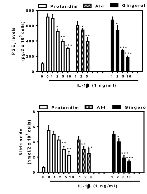
level. (Adapted from Abusarah et al. 2012) [157] ... 43 Figure 8: Effect of Nrf2 activators on cell viability and Nrf2 expression. ... 57 Figure 9: The activation of Nrf2 protects the cell against H2O2-induced cell death. ... 59 Figure 10: Isolated human OA chondrocytes were pre-treated with increasing concentrations
of three different Nrf2 activators; Protandim®, AI-1 or 6-Gingerol (0-10 µg, 0-5 µM and 0-10 µM respectively) for one hour. The cells were then treated with 1 ng/ml of IL-1β for 24 hours. Cell culture medium was collected and assayed against A) PGE2, B) MMP-13
and C) NO. Values represent the means ± SEM of 4 separate experiments performed in duplicate. *P < 0.05, **P < 0.01, ***P < 0.001 compared to IL-1β-treated cells. ... 61
Figure 11: Protandim®-activating Nrf2 reduces cartilage degradation in DMM mouse model of OA. ... 63 Figure 12: (A) Expression of Nrf2, GSTA4-4 was decreased in human OA cartilage (n = 3-4).
Cartilage extracts from healthy subject and OA patients are subjected to Western blot using monoclonal anti-human Nrf2 and GSTA4-4. (B). Nrf2 and GSTA4-4 mRNA levels were determined by real-time PCR in cartilage from sham and DMM WT mice 8-weeks post-surgery. Statistics: Student's unpaired t-test (n=3-4): **P < 0.01 (vs. sham). ... 65
Figure 13: The activation of Nrf2 by Protandim® abolished IL-1β -induced GSTA4-4 down-regulation (A), HNE production (B), and MMP-13 release (C). ... 67 Figure 14: (A) IL-1β inhibits Nrf2 expression in human OA chondrocytes. Cells were treated
with 1 and 2 ng/mL IL-1β for 24 hours. (B-D) Overexpression of Nrf2 (A, lower panel) prevented the IL-1β-induced GSTA4-4 down-regulation (B), IL-1β-induced HNE generation (C) and MMP-13 (D) release. Isolated chondrocytes were transiently transfected with empty or Nrf2 expression vector (1 ng/106 cells) and treated than after
for 24 hours with or without 1 ng/mL IL-1β. Cellular extracts were subjected to the determination of Nrf2 by Western blot (A lower panel), GSTA4-4 mRNA expression by real-time PCR (B), and HNE level by ELISA (C). MMP-13 level was measured in the culture media by ELISA (D). Statistics: Student's unpaired t-test (n=3): *P < 0.05, **P < 0.01, ***P < 0.001 (vs. empty vector; #P<0.05, &P<0.01 (vs. empty vector + IL-1β). .. 69
LIST OF TABLES
LIST OF ABBREVIATIONS
4-hydroxy-2-hexenal 4-HHE
4-Hydroxynonenal HNE
Activator protein 1 AP-1
Advanced LPO-end products ALEs
Alkoxyl radical LO
Anterior cruciate ligament transection ACLT
Antioxidant response element ARE
Arachidonic acid AA
Carnosine CAR
Chemokine (C-X-C motif) ligand 1 CXCL-1
c-Jun N-terminal kinase JNK
Collagen Col
Cyclooxygenase COX
Cyclooxygenase-2 COX-2
Destabilization of the medial meniscus DMM
Extracellular matrix ECM
Fibroblast-like synoviocytes FLS
Glutathione GSH
Glutathione peroxidase GPx
Glutathione S-transferase GST
Heme oxygenase-1 HO-1
Hydrogen peroxide H2O2
Hydroxyl radicals OH
.
IL-1 receptor antagonist IL-1Ra
Inducible nitric oxide synthase iNOS
Intercellular Adhesion Molecule 1 ICAM-1
Interferon-gamma INF-ɣ
Interleukin-1beta IL-1β
Kelch ECH associating protein 1 Keap1
Lipid hydroperoxide LOOH
Lipid peroxidation LPO
Lipid peroxyl radical LOO
.
Malondialdehyde MDA
Matrix metalloproteinase-13 MMP-13
Matrix metalloproteinases MMPs
microsomal-PGES m-PGES
Mitogen-activated protein kinase MAPK
Myeloperoxidase MPO
N-acetyl-cysteine NAC
NG-monomethyl-t-arginine NMMA
Nitric oxide NO Non-steroidal anti-inflammatory drugs NSAIDs
Nrf2 Activator II AI-1
Nuclear factor erythroid 2-related factor 2 Nrf2
Nuclear factor-kappa B NF-κB
OA Research Society International OARSI
Osteoarthritis OA
Peroxisome proliferator-activated receptor gamma PPARɣ
Peroxynitrite ONOO−
Polyunsaturated fatty acids PUFAs
Post-translational modification PTM
Prostaglandin PG
Prostaglandin E2 PGE2
Prostaglandin-E synthase PGES
Protein kinase C PKC
Reactive nitrogen species RNS
Reactive oxygen species ROS
Rheumatoid arthritis RA
small Maf sMaf
Superoxide dismutase SOD
Tissue inhibitors of metalloproteinases TIMPs
ACKNOWLEDGMENTS
In the name of Allah, the most beneficent, the most merciful,
I would like to sincerely thank my supervisor Dr. Mohamed Benderdour not only for his guidance in the research work but also for his continuous support, understanding and patience. For encouraging me to express my opinions and pose any questions or concerns even on the go, your mentorship and dedication inspire me to become a better researcher.
I’m also very thankful for my co-advisor Dr. Hasan Fahmi for his time and support and for Dr. Julio Frenandes and his lab staff. Special thanks for Qin Shi for providing insightful technical support and professional advice.
Words fail me to express my deepest feelings of gratitude to my parents, Mr. Majdi and Mrs. Wafa Abusara and my siblings for their ongoing unconditional support.
I would like to extend to a heartfelt thank you for my parents-in-law, Mr. Ahmad Alatawneh and Mrs Amina Almalty for their love and sincere prayers.
To my husband, friend and companion Natheer, I can’t thank you enough for your unlimited patience and encouragement. Your support is the reason I’m here today. You and our baby Amina are my source of strength, happiness and inspiration in this life.
Last but foremost I’ll be always thankful to Allah for blessing me with all the great loving people in my life and for granting me the strength to achieve my goals.
I would like to acknowledge the support of the Natural Sciences & Engineering Research Council of Canada (NSERC) for financing this project.
1. THE DIARTHRODAL JOINT
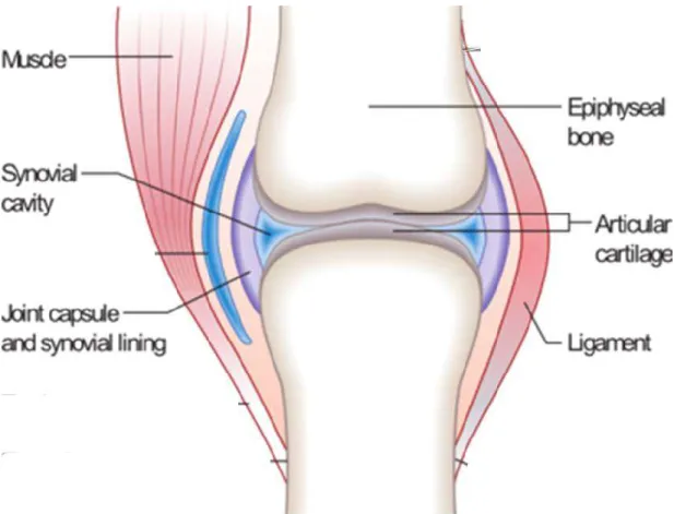
The diarthroidal or synovial joint is the most abundant movable type of joint in the body of a mammal. Its encapsulated system consists of bone, articular cartilage, synovial membrane and synovial fluid [1]. Fig.1 shows a simplified synovial joint.
1.1 Articular Cartilage
Cartilage is a non-innervated anaerobic connective tissue found in many areas around the human body. There are three main types of cartilage classified according to the relative amounts of main components; elastic cartilage, hyaline cartilage and fibrocartilage. Cartilage constitutes mainly of sole type of cells named chondrocytes distributed within a mesh of an extracellular matrix (ECM). It can be divided into four zones according to the morphological changes in chondrocytes and matrix from the articular surface to the subchondral bone [1].
Fig.2 illustrates the structure and different zones within hyaline cartilage. A synovial joint
contains hyaline cartilage which forms a protective layer covering the two bone ends of the joint to reduce contact stresses there and facilitate bone movement without pain. Moreover, articular cartilage protects bone surface from impact stresses, and minimizes friction and wear in the joint [1, 2]. Therefore, loss or destruction of cartilage can be associated with extreme pain especially during movement, which is often the case in advanced OA patients.
Figure 2: Schematic diagram of the cellular organization in the zones of articular cartilage (A) and of the collagen fiber architecture (B) (from Buckwalter et.al [3]).
The composition, structure and functions of chondrocytes vary depending on their zone or location within the cartilage [4, 5]. In the superficial zone chondrocytes are in contact with the synovial fluid, cells are closely spaced and aligned with the cartilage surface. This zone, serves mainly to support the tensile stresses generated when compressive loads are applied to the cartilage. The transitional intermediate zone holds lower density of randomly dispersed chondrocytes and high concentration of proteoglycan and collagen. In the deep radial zone chondrocytes forms rows of cells parallel to the collagen fibers. The calcified zone is the innermost zone that connects the cartilage to the subchondral bone. This zone consists of lower count of chondrocytes uniquely capable of synthesizing type V collagen (Col V) which was presented as an important factor in the structural integrity to the less elastic subchondral bone [6].
1.1.1 Chondrocytes
Chondrocytes, the sole cellular component, form only 1-5% of cartilage volume, yet the highly specialized cells are responsible for producing other macromolecular components of the ECM. Depending on their location within the matrix the highly metabolically active chondrocytes can produce multiple collagen types including type II Collagen (Col II), Col VI, Col IX, Col X, Col XI, and proteoglycan among other mediators [2]. Chondrocytes are responsible for maintaining ECM integrity by controlling both the degradation and synthesis of matrix macromolecules. Though the exact process in still under study, it is believed to be under careful balance between multiple catabolic and anabolic mediators produced by the cells. Interleukin-1 beta (IL-1β) presents an example as it triggers a cascade of signals that can induce the expression of catabolic enzyme matrix metalloprotease-13 (MMP-13), up-regulate the expression of pro-inflammatory enzymes like cyclooxygenase-2 (COX-2) and molecules like prostaglandin E2 (PGE2), and affect the transcription of genes needed for the synthesis of
proteoglycans [7]. On the other hand, cytokines, such as insulin-dependent growth factor-I, oppose these catabolic activities by stimulating matrix synthesis and cell proliferation. Chondrocytes can also produce tissue inhibitors of metalloproteinases (TIMPs) that antagonize the catabolic effect of MMPs. Furthermore, the interaction between chondrocytes and matrix in an interdependent pattern is essential for tissue maintenance and has huge effect on the metabolic activity of chondrocytes. [2, 7].
1.1.2 Extracellular Matrix (ECM)
Articular cartilage derives its form and mechanical properties from its matrix [2].Water, the main component contributing up to 80% of the matrix, collagen and proteoglycans interact to gives tensile strength to the cartilage. The matrix also functions as an environment to protect chondrocytes from mechanical stress, store cytokines and growth factors needed to maintain cell function and activity, act as a media for nutrient diffusion to chondrocytes and as signal transductor for the cells [8]. Col II is the principal component of ECM accounts for 90-95% of collagen. Deformations in the matrix due to the of loss of balance between catabolic and anabolic factors in the cartilage can further alter the response of chondrocytes, and lead to overall abnormal cartilage structure and function such as the case in advanced OA [2] . Fig.3 shows main differences between normal and OA joint.
Figure 3: Normal joint vs. OA joint shows changes such as loss of cartilage integrity and erosion, loss of joint space and the formation of osteophytes.
1.2 Bone
The calcified zone of the cartilage is bordered by the subchondral bone plate, a very thin cortical bone structure to which the articular cartilage is anchored. Arterial terminal branches in the subchondral bone plate it can supply approximately 50% of the glucose, oxygen, and water required by articular cartilage [9, 10]. Bone cells include osteoblasts and osteoclasts secret factors that can affect the activity and differentiation of chondrocytes
under investigation it is well reported that, changes affecting metabolic activity of bone cells, especially in the subchondral bone, may alter local signaling factors and trigger changes affecting near chondrocytes and articular cartilage in general [11].
1.3 Synovial Membrane
The Synovial membrane is bi-layer membrane of connective tissue that lines the cavities of joints. There are two types of cells forming the innermost or internal layer of the membrane; fibroblast-like synoviocytes (FLS) and macrophages. While FLS are responsible for producing hyaluronan long chain sugar polymers [12], macrophages act to clear unwanted substances and debris [13]. This membrane seals off the synovial fluid from the surrounding tissue and plays essential role in supporting joint lubrication by secreting synovial fluid and other important macromolecules like hyaluronan. Moreover, the membrane acts to regulate the amount and content in it by controlling the passage or filtration of nutrients into the synovial capsule and the clearance of waste products from it [1, 11] .
1.4 Synovial Fluid
The synovial fluid is a viscose plasma dialysate secreted by the joint tissues especially synovial membrane characterized by high content of hyaluronic acid. The synovial fluid holds three main advantages; the viscous fluid lubricates the articulating surfaces, provides a media for carrying nutrients to chondrocytes, and clearing waste products from the cartilage while acting as a shock absorber [1]. Whereas cell counts of synovial fluid are normally low, healthy
function of the mononuclear cells of synovial fluid is the removal of debris that normally appear in joints. However, the fact that vascular and synovial membrane permeability are altered by inflammation, accounts for changes in protein and cellular content in diseased synovial fluid [14]. Moreover, a direct correlation between high levels of leukocytes and synovial fluid pH was described in samples from patients with various forms of acute and chronic arthritis [15]. In addition, the level of different enzymes present in the synovial fluid can vary in the presence of articular disease. These enzymes could be either produced locally by the synovial membrane or released by synovial fluid macrophages or entering from plasma. Such changes can have strong implication on disease progression and symptoms development [1, 16].
2. OSTEOARTHRITIS (OA)
2.1 Definition and classification
OA is one of the most prominent chronic degenerative disease of the joints; it is the most common joint disorder in the world and the second most common diagnosis among ageing population [17]. Disease diagnosis and stage determination can be achieved through analyzing clinical and idiographic outcomes. Clinical examination evaluates symptom such as pain, local inflammation and change or limitation in joint function, while radiographic imaging can aid in viewing articular changes like deformations and joint space narrowing. The most commonly affected joints are the distal interphalangeal joints of the hands and weight baring joints such as the hips and the knees [18].
OA is a multifactorial disease that is mainly characterised pathologically by progressive loss of articular cartilage in synovial joints. Moreover, disease development and progression also involves synovial inflammation, bone remodeling and increased inflammatory and catabolic responses. Therefore, OA is being considered a disease that affects the joint as an organ including cartilage, bone, synovium, muscles and ligaments [19]. The structural integrity of the matrix of human articular cartilage is maintained by a dynamic equilibrium between synthesis and degradation. In OA disruption of the differentiation and function of chondrocytes influences the composition and structure of the cartilage matrix. This disruption causes catabolic factors to overcome the anabolic capacity of chondrocytes leading to overall loss of normal ECM composition and cartilage properties.
2.2 Epidemiology of OA
Advanced OA is considered a major socio-economical problem not only in Canada but in the Western countries and the world. The disease significantly reduces the productivity, activity and quality of life of patients as well as exerting high stress on the health system. Moreover, an anticipated increase in OA prevalence within the next two decades is expected to make it the fourth leading cause of discordance, this increase is mainly attributed to ageing population, sedentary lifestyle and obesity epidemic [20]. According to a report published by the Arthritis Alliance of Canada in 2011 titled “The impact of arthritis in Canada: today and the next 30 years”, the prevalence of OA is expected to further increase in Canada to reach 71% by 2040 among seniors above the age of 70 years old.
OA can be generally classified into two forms either as primary or secondary OA. While the primary form of the disease is the most common type, it is considered as an idiopathic phenomenon that is usually related to aging process, typically occurring in older individuals. Though primary OA has no specific apparent trigger or initiating factor, multiple risk factors can play a role in disease progression. However, secondary OA refers to damage affecting the synovial joint as a result of predisposing condition like trauma or congenital deformity and can happen earlier in life [21]. Risk factors; OA can be considered as the product of an interaction between different factors; these factors can be categorized as systemic risk factors affecting the biological activity of the body or local risk factors that exert an influence on specific joint or joints. Systemic risk factors; age is considered as the strongest factor for OA development and progression where changes in biological functions are associated with cumulative exposure to different risk factors [21]. Women have higher prevalence not only for developing OA but for having more severe form of the disease especially during and after menopause [22, 23]. Therefore, both gender and hormonal factors are considered to be important risk factors. Race and ethnicity can also play a role where studies on different location revealed variation in OA prevalence and joints affected. For example, African Americans reported more joint and hip symptoms than whites in general [24]. Further study is needed to comprehend the origin of this variation. Moreover, factors such as genetics and the presence of congenital or developmental conditions also have an influence on disease progression [25]. Local risk factors; obesity is highly targeted as a potent risk factor for developing OA particularly in weight-bearing joints especially the knee. However, weight loss and exercise were found to reduce pain and risk of developing symptomatic OA. Knee surgery or injury are considered among the strongest risk factors
especially for early OA of the knee, also having an occupation or playing certain sports that exerts strong impact or strain on certain joints can increase the prevalence for developing OA [21, 26].
3. PATHOPHYSIOLOGY OF OA
OA is described as a disease affecting the whole joint. However, articular cartilage is considered a major target of tissue injury that is most related to symptoms development and a hallmark of disease progression regardless of triggering factors. Alterations in ECM and cartilage are associated with changes in chondrocyte and disruption of homeostasis leading to loss of the balance between anabolic and catabolic pathways necessary for maintaining the properties of cartilage tissue. Studies on OA indicate that cytokines and growth factors are implicated in the disease. Degradation of cartilage ECM and the suppression of its biosynthesis by chondrocyte are regulated by the release of several cytokines in the joint. According to the triggered response these factors can be classified as proinflammatory, which are generally also catabolic (e.g. IL-1, IL-6, IL-8, and tumor necrosis factor-alpha (TNF)), or anti-inflammatory (e.g. IL-4, IL-10, IL-13). Other factors like IL-1 receptor antagonist (IL-1Ra) and interferon-gamma (IFN-ɣ) are also classified as inhibitory cytokines, since they may block the actions of catabolic cytokines. Though many of these factors are necessary at low levels for normal homeostasis, in OA disruption in their level can indicate an important role in the disease. Despite the absence of clear or specific initiating factor, the progression of the disease is mostly regulated by pro-inflammatory cytokines mainly IL-1β and TNFα [27, 28]
3.1 Pro-inflammatory mediators
In OA, upregulation of cytokines production such as IL-1β and TNFα in synovial membrane leads to increase in their level in cartilage triggering the activation of chondrocytes. Upon activation by IL-1β, cells show a pronounced upregulation in gene expression of MMPs, inducible nitric oxide synthase (iNOS) and COX-2, as well as in the synthesis of inflammatory mediators such as PGE2 and nitric oxide (NO). Current research on OA suggests a central role
of IL-1β in joint destruction and other pathological features of the disease. While IL-1β is largely involved in the induction of catabolic effect on cartilage in OA, TNFα is presented more as an inducer of the inflammatory effect [29]. Further investigating its effect indicated cytokine ability to induce deleterious changes in chondrocyte function and in the integrity of ECM. In addition, understanding cytokine-induced signal transduction pathways involved the OA and/or normal cartilage homeostasis can provide new potential targets for intervention in OA [30, 31]. In fact, several clinical trials have tried to modulate the effect of major cytokines in order to control their role in OA pathogenesis. This approach had several successes in rheumatoid arthritis (RA). For example, the administration of TNF antagonists have been effective in treating RA, yet TNF antagonist therapy has less dramatic effects in patients with OA. Clinical trials on the use of several inhibitors of biological targets propose certain factors as part of the disappointing outcome such as side effects of medication, time of intervention and pharmacokinetics of the used agents [32].
3.1.1 Pro-inflammatory cytokine IL-1β
IL-1β protein is primarily produced as a precursor (pro-IL-1β) that needs to be processed by caspase-1 to generate its mature and active cytokine. Upon binding its type 1 receptor (IL-1RI), IL-1β triggers several pathways leading to activation of several signal transduction kinases as well as inducing nuclear translocation of number of transcription factors. These effects can be inhibited by naturally present IL-1Ra and type2 IL-1 decoy receptor. Several studies have presented strong indications of the significant role played by IL- 1ß in OA [30]. In normal articular tissue the cytokine is produced in limited amount. However, its level markedly increased in chondrocytes as well as synovial cells from patients with OA [33]. The reported increase in IL-1β production and also in cellular response to the cytokine might be explained by several observations; while the level of IL-1 antagonists in OA synovium is downregulated, there is a concomitant upregulation of IL-1β-converting enzyme and IL-1RI in human chondrocytes and synovial fibroblasts. Therefore, these observations may provide a molecular explanation for the enhanced catabolic effects of IL-1β in OA joint tissues.
The destructive effects of IL-1β in OA include both elevation of cartilage catabolism and suppression of cartilage anabolism. IL-1β can trigger multiple signal transduction pathways that upregulates gene expression and subsequently the levels of major extracellular proteolytic enzymes involved in cartilage degradation such as MMPs [34]. In addition, the cytokine has the capacity to induce several proinflammatory mediators involved in the increase of local hematopoietic cells during OA. An increase in articular hematopoietic cells
can cause increases in oxidative burst activity and decreases in O2 concentration, and
generation of additional inflammatory and proteolytic enzymes that can lead to progression of OA [30]. Among proinflammatory mediators induced by IL-1β the upregulation in gene transcription of iNOS and COX-2 that causes a subsequent increase in levels of NO and PGE2
respectively hold significant importance in the pathology of OA. The detrimental effects on articular tissue associated with significantly high levels of these proinflammatory mediators have been closely investigated in the literature [30, 31]. Moreover, the cytokine can also induce downregulation in biosynthesis of Col II, major articular joint protein as well as through stimulating chondrocyte apoptosis [35]. As a result, IL-1β can decrease ECM synthesis by affecting the anabolic activities of chondrocytes and/or the cell densities of articular cartilage. Consequently, the outcomes of triggering of IL-1β pathway in OA can be related to deleterious effects that include both elevation of cartilage catabolism and suppression of cartilage anabolism [30].
Several strategies aiming to inhibit or modify the activity of IL-1 have been investigated as possible treatments in OA such as the administration of 1Ra and soluble IL-1 receptors. The results of these investigations revealed potential agents that may be used in the treatment of OA. However, further research is needed to establish sufficient data about its safety and effectiveness. The role of different IL-1 inhibitors in OA has been discussed in an evidence based review [36].
3.1.2 COX-2 and PGE2
Cyclooxygenase (COX) also named prostaglandin H synthase is the main enzyme in the metabolism arachidonic acid (AA) and subsequent production of prostaglandins (PG). Membrane-bound phospholipids are converted into AA through phospholipase A2 activity. COX enzymes convert AA first to PGG2 and then to an unstable metabolite PGH2 that will be
rapidly converted into three types of prostanoids: prostaglandins, thromboxanes, prostacyclins in addition to leukotrienes [37]. Currently, three isoform of COX enzyme have been identified each encoded by a different gene. The inducible nature of the isoforme COX-2 expression by several mediators such as pro-inflammatory mediators like IL-1β and TNFα and lipid peroxidation (LPO) product 4-hydroxynonenal (HNE) made it a target for closer investigation [38, 39]. COX-2, but not other isoforms, synthesis was significantly increased in different diseases known to be of inflammatory nature is well reported in disease-related pattern. The increase in both mRNA and protein levels of COX-2 in arthritic joint tissues of animal models of joint inflammation indicate an important role played by the enzyme in arthritic articular tissues [40]. PGE2, the most abundant PG produced in the articular tissue, is involved in many
physiological events, such as cell growth, immune regulation, and inflammation. Three prostaglandin-E synthase (PGES) isozymes, each encoded by a separate gene, are responsible for PGH2 isomerization to produce PGE2. Further studying the activity of microsomal-PGES
(mPGES) isoforme showed inducible nature in the presence of inflammatory stimuli providing an interesting target in the development of inflammatory arthropathies. Both COX-2 and mPGES are key enzymes for PGE2 biosynthesis especially under inflammatory conditions
The study of OA cartilage specimens indicated not only the expression of COX-2 but also showed spontaneous increase in PGE2 levels that are at least 50-fold higher than normal
cartilage [41]. Therefore, the noticed increase in local PGE2 production, suggests an
attribution through COX-2 activity. However, recent evidence was provided that PGE2 in
human synovial fibroblasts can also play a role in controlling COX-2 expression through a positive feedback mechanism [42]. In OA, PGE2 is well reported to induce a catabolic effect
where it can inhibit collagen expression, increase the production of MMPs, and modulate bone resorption by stimulating osteoclasts activity [43, 44]. PGE2 has also apoptotic effect on
chondrocytes by enhancing NO-induced apoptosis. In addition, other researchers suggested a direct apoptotic role for PGE2 perhaps through cAMP activity [45].
The expression of COX genes is highly regulated. The inducible nature of COX-2 gene expression is regulated through its gene promoter which contains several inducible enhancer elements [46]. Several stimulators can interact with these elements to play a role in COX-2 gene expression through different pathways. Interestingly, 15d-PGJ2, the endpoint metabolite of PGD2 is found to prevent IL-1β induced COX-2 expression and PGE2 production [47]. Yet,
it also induced COX-2 expression but not PGE2 production in the absence of IL-1β. This
observation suggests the presence of another signaling pathway involved in PGE2 production
with IL-1β stimulation on which 15d-PGJ2 inhibitory effect takes place [39].
The use of COX-2 specific inhibitor caused marked suppression in COX-2 expression, PGE2 production, along with swelling, and cellular infiltration in the joints [48]. It was also
shown that pre-treating rat models with neutralizing anti-PGE2 monoclonal antibodies
the most widely prescribed drugs. They work by inhibiting COX enzymes in a non-specific way. Majority of the reported undesirable effects of NSAIDS are attributed to COX-1 isoform inhibition. Therefore, developing agents to specifically block COX-2 while simultaneously keeping COX-1 activity unaffected is considered a rational approach to obtain efficacy without deleterious side effects. COX-2 specific agents, generally called coxibs, are the most common clinically prescribed drugs as analgesics and for the management of several conditions including OA. Coxibs proved to be effective with significant reduction in known side effects usually associated with conventional NSAIDs usage [50]. However, recently concerns are rising regarding the permanence of some gastrointestinal and renal side effects, also regarding potential cardiovascular adverse effects. Up to date results released are considered inconclusive triggering the need for developing new agents or targets that help better control the expression and activity of COX-2 and PGE2 [51].
Amongst COX-2 and mPGES-1 gene expression inducers HNE is also reported to act as inflammatory mediator that enhances protein and mRNA levels by affecting gene promoter activity in dose-time dependant pattern [45, 52]. This induction is also associated with increases in PGE2 production. The expression of COX genes is highly regulated. The
inducible nature of COX-2 gene expression is regulated through its gene promoter which contains several inducible enhancer elements. Several stimulators can interact with these elements to play a role in COX-2 gene expression through different pathways. For example, the cytokine IL-1β induces gene expression via mitogen-activated protein kinase (MAPK) and protein kinase C (PKC) pathways while HNE appears to modulate different pathways that affect COX-2 and mPGES-1 expression such as p38MAPK signaling pathway [45, 52].
Interestingly, 15d-PGJ2, a potent activator of peroxisome proliferator-activated receptor gamma (PPARɣ), is found to prevent IL-1β induced COX-2 expression and PGE2
production. Yet, this endpoint metabolite of PGD2 could induce COX-2 expression but not
PGE2 production in the absence of IL-1β suggesting the presence of another signaling pathway
involved in PGE2 production with IL-1β stimulation on which 15d-PGJ2 inhibitory effect
takes place [39, 47].
3.2 Catabolic molecules
The maintenance of healthy cartilage requires a system of ongoing balance between building and breaking mechanisms. However, when a shift in the endogenous system causes increases in the catabolic activity it leads to increase in cartilage ECM degradation. In OA, the destruction of cartilage components is considered a central character of disease development especially the degradation of Col II by collagenases activity of MMPs such as MMP-13 [53]. Moreover, other molecules can also contribute to the catabolic process like the free radical NO which is considered among the main molecules that mediate catabolic changes [54] .
3.2.1. MMPs
MMPs are a group of endopeptidase that share similar structure. Since their discovery the enzymes were studied for their function within different tissue not only for connective tissue remodeling, but also in wound healing, angiogenesis, and metastasis as well [55]. Among the group four enzymes MMP-1, -8, -13 and -14 have collagenolytic activity and are capable of breaking the highly stable fibrillar collagen. Samples from synovium and
cartilage from OA and RA patients showed an increase in the activities of several MMPs. However, an upregulation in cartilage expression and activity of collagenase-3 (MMP-13) in OA cartilage present the enzyme as the primary collagenase in OA [53, 56]. In arthritis endogenous inhibitors of MMPs such as TIMPS are produced; however their activities are not sufficient to compensate for the increase in catabolic activity of MMPs [57].
In an effort to reduce/manage the catabolic activity of MMPs, a number of MMP inhibitors have been developed and tested in various animal models of arthritis. An inhibitor of the collagenases (MMP-1, -8, and -13) prevents cartilage degradation in animal models of both RA and OA [58]. Other MMP inhibitors were tested in arthritis clinical trials, but serious side effects mainly due to lack of specificity have largely hindered their further development [59]. MMP-13 is known to be activated by a number of mediators, such IL-1β. In OA, the increase in MMP-13 expression and activity might therefore be attributed, in part, to the increase in IL-1β release in the synovial fluid [60, 61].
Moreover, locally produced inorganic oxidants were found capable of degrading cartilage mainly by inducing the oxidation of ECM components and/or posttranslational
modification of MMPs [62, 63]. Moreover, reactive oxygen species (ROS) might shift the
balance of proteolytic potential by decreasing the production and/or the activity of TIMPs
[64]. In addition, HNE can also induce MMP-13 expression at protein and mRNA levels. [65-68]. Interestingly, an earlier study revealed that the pathway needed for activating the transcription and protein synthesis of MMP-13 by HNE is different from the pathways triggered by proinflammatory mediators like IL-1β and TNF-α. Whereas IL-1 induction of MMP-13 gene expression requires the activity of p38 and, c-Jun N-terminal kinase (JNK) with the translocation of NF-κB translocation [66], TNF-α triggered MMP-13 gene
expression involves MAPK, activator protein 1 (AP-1) and NF-κB [65]. Nevertheless, HNE induction of MMP-13 transcription requires p38 MAPK activity with possible role of AP-1 and c-Jun but not NF-κB [67].
3.2.2 Inducible nitric oxide synthase (iNOS) and NO in OA
NOS is the enzyme responsible for the production the highly reactive molecule NO through l-arginine metabolism. The enzyme is present within different tissues in three isoforms; neuronal (nNOS) and endothelial (eNOS) and inducible (iNOS). The constitutive nNOS and eNOS produced low level of NO play an important role in intercellular signaling and the homeostasis in neurons and endothelial cells [69, 70] . However, in the presence of certain stimuli like inflammation the release of certain cytokines and mediators such as IL-1β and TNFα can trigger the transcription and activity of iNOS; as a result large quantities of NO are produced [71]. In fact, the inducible production of NO is significant not only for inflammatory, and immunological host defense responses, where NO can exert cytotoxic effects against microbes, but for tumor cells and tissue repair as well [72]
Earlier studies indicated the presence of higher levels of NO in human synovial fluids from RA and OA patients when compared with normal samples [73]. The observation suggests an important role for the free radical as a mediator of inflammation in both diseases. In OA, sustained high levels of inflammatory cytokines especially IL-1β leads to prolonged activation of iNOS usually in the presence of highly oxidative environment. The release of high amount of NO in the presence of superoxide anion leads to the formation of the highly reactive peroxynitrite which in turn induces proapototic and proinflammatory responses in
additional predominant catabolic effect where it inhibits the synthesis of ECM components; Col II [78] and proteoglycan [79], as well as activating MMPs [80]. These observations indicate essential role of induced NO release and its derivatives as mediators in OA pathogenesis through different mechanisms [81].
The production of NO by chondrocytes and synoviocytes in OA is regulated by the level of NOS transcription and translation. Therefore, several studies tested the effect of different inhibitors of NOS in OA and RA. Such studies helped also elucidate the role of iNOS and NO as mediators in disease pathogenesis. Studies on animal models of OA using different inhibitors such as NG-monomethyl-t-arginine (NMMA) and specific iNOS inhibitor N-iminoethyl-L-lysine (L-NIL) showed interesting and promising findings in RA and OA respectively. The data showed that the inhibition of iNOS results in marked suppression in tissue destruction associated with chronic inflammation [82, 83]. In a study on an animal model of RA, the administration of NMMA greatly reduced leukocytes effusion, joint erosion and other signs of inflammation in the area. Interestingly, the beneficial effect was not limited to earlier stages of the arthritis, but it was also present at the chronic phase of the disease [84]. Similar results were also obtained by selectively inhibiting iNOS in experimental OA model using L-NIL. The inhibition of iNOS was associated with reduced production of NO in tissue which also resulted in a significant decrease in the production of catabolic factors such as MMPs and peroxynitrite [83]. In a more recent study in OA the use of L-NIL could also prevent NO-induced lipid peroxidation and subsequent production of highly reactive toxic compounds like HNE; a key mediator in OA [85]. Moreover, L-NIL significantly reduced the cytotoxic, proinflammatory and catabolic effects induced at different pathological concentrations of HNE. L-NIL also blocked the inhibitory effect of IL-1β on glutathione
S-transferase (GST) and the stimulation of ROS in a dose dependent pattern [85]. The reported beneficial effects of NOS inhibitors indicated a potential promising protective effect on chondrocytes in OA.
4. THE OXIDATIVE STRESS IN OA
Closer study of the chronic degenerative nature of OA led to significant shift in the search for therapeutic solutions for the disease. In fact, most available pharmacological agents targeting the formation of inflammatory mediators have not provided a real solution for OA. Therefore, there is an essential need for other secondary therapies designed to prevent the progression of this chronic disease. Current attempts based on recent data aim to develop effective chondroprotective disease-modifying agents to control different molecular pathways involved in the initiation and progression of OA. Oxidative stress within articular tissue has been pointed out as an aspect with a significant role in the pathogenesis of OA, an observation supporting the predicted therapeutic potential of targeting pathways involved in joint metabolism controlled or affected by oxidative stress [86, 87].
Though physical exercise is important for maintaining normal cartilage function, excessive repetitive mechanical load and sheer stress can trigger apoptotic and inflammatory responses in chondrocytes. The increase in ROS production within the articular tissue due to excessive mechanical load and stimulated pro-inflammatory mediators can lead to diffusion of free radicals into cartilage. Interestingly, studies also show a concomitant decrease in the protective antioxidant response within the joint leaving chondrocytes highly prone to cell
death through caspase activation mediated by the oxidative radicals [88]. Moreover, research on the effects of ageing show that cartilage homeostasis is highly affected by ageing process where chondrocytes are less responsive to mechanical anabolic stimuli. This observation can be correlated with our knowledge about OA to explain much higher incidence of the disease among older patients. Another condition affecting cartilage termed cellular senescence may lead to an irreversible arrest of chondrocytes growth causing detrimental outcomes on cartilage homeostasis relevant to cartilage ageing. A study by Yudoh et al. [89] investigated the role of oxidative stress and reactive radicals in cartilage senescence and in the development of OA. The study demonstrates that high level of oxidative stress affects chondrocytes in the presence of profound decrease in antioxidative capacity of articular tissue. Consequently, the oxidative stress induces a mechanism triggering the acceleration of chondrocyte senescence by causing abnormal erosion of cellular telomere end. Therefore, oxidative stress is considered closely involved in cartilage senescence and the development of OA. In addition, both mechanical and chemical stressors may alter cellular adaptation to hypoxia during disease progression, causing oxidative damage and changes in the microenvironment ultimately negatively affecting the synthesis of chondrocytes [89]. The same study reported an important reduction in the antioxidative potential in degenerating regions in comparison with the intact regions from the same OA articular cartilage sample. Obtained results suggested that oxidative damage can induce catabolic changes to cartilage matrix in articular cartilage. Furthermore, the treatment of cultured cartilage with an antioxidative agent showed a protective effect against the observed oxidative stress-induced chondrocyte dysfunction and cartilage damage. Collectively these findings support the theory focusing on oxidative stress as an inducer of catabolic changes in cartilage matrix [89].
4.1. Oxidative stress and antioxidant molecules
Reactive molecules are normally present within a biological system. Different reactive species can be produced as byproducts of normal activities such as aerobic metabolism, during pathological conditions or result from external resources such as pollutants and cigarette smoke. At certain concentration reactive radicals do play a role in regulatory functions, cell signaling and in immunological response [90]. However, when present in large volumes, reactive molecules can interact with and damage major cellular components like proteins, lipids and DNA [90]. The induced damage can be attributed to the production of peroxides and free radicals like ROS and reactive nitrogen species (RNS). To avoid excessive cellular damage the body contains number of defense and protective mechanisms including continuous repair machinery and the antioxidant system. Disturbances in the balance between produced oxidative molecules and available reducing agents due to uncontrolled production of free radicals with or without a decline in reduction capacity of the cells can cause tissue damage in several pathophysiological conditions [91]. Under such conditions the cell is considered under oxidative stress. The effect of this stress on the cell and the body overall depends on the extent of damage caused by the reactive species and on cell’s ability to constantly repair any damage. At different levels oxidative stress can cause several changes in cellular response. The induced effect may range from inducing altered cellular activity by affecting cell signaling pathways; either directly as chemical messengers or indirectly via modifying cellular components involved in the pathway such as lipids, to induce apoptosis. Moreover, under severe levels of oxidative stress the accumulating damage can prevent the
The antioxidant system contains different enzymes which protect against oxidative stress. Moreover, the role of antioxidants in controlling the production of inflammatory cytokines, LPO and the activation of transcription factors which trigger other inflammatory components is of major importance in several pathological conditions. The enzymes glutathione peroxidase (GPx), heme oxygenase-1 (HO-1) and superoxide dismutase (SOD) are among the best studied cellular antioxidants. In OA, studies indicated the importance of GSX, HO-1, SOD2 and SOD3 in the oxidative defense in cartilage [86, 94]. In addition, there is a well reported change in the expression of these genes in OA chondrocytes and in the presence of inflammatory cytokines such as IL-1β [86, 90].
Antioxidant molecules such as ascorbic acid, flavonoids, N-acetyl-cysteine (NAC) and glutathione (GSH) have been shown to play an important role in different inflammatory diseases such as atherosclerosis, arthritis and OA [95, 96]. In particular GSH, the substrate for GST and GPx, have shown a promising protective role in OA [97].
4.2. Reactive oxygen species (ROS)
Aerobic cellular activities such as cellular respiration can produce oxygen containing reactive molecules named ROS. The highly reactive molecules and the less reactive of these species are important for redox signaling pathways [98]. The immune system also benefits from the destructive activity of ROS in killing pathogens [99]. However, due to their high reactivity, ROS are widely linked to the induction or aggravation of different pathological conditions such as atherosclerosis, cancer, neurodegenerative disease and OA [90]. The production of ROS is not solely influenced by internal mechanisms but external sources like
antioxidants within the biological system is considered primordial to maintain an optimal level of these molecules at all times [100].
In OA, several ROS have been closely studied as main factors in the pathology of the disease. While articular cartilage is an avascular tissue that depends on nutrient supply needed for basic cellular functions from the synovial fluid, chondrocytes are adapted to work within low oxygen pressure to anaerobic environment [101]. In pathological conditions associated with OA, fluctuations in partial oxygen pressure, mechanical stress and inflammatory mediators such as IL-1β can put chondrocytes under the influence of oxidative stress. Subsequently, significant increase in the production of ROS such as NO and O2- takes
place. Different ROS molecules can further interact to generate reactive radicals, including peroxynitrite (ONOO−) and hydrogen peroxide (H2O2). Moreover, recent studies reported
increase in the production of myeloperoxidase (MPO) in OA as well [102]. In the presence of H2O2and Fe+2, chondrocytes produce hydroxyl radicals (OH
.
). The highly reactive radical caninitiate further series of reactions with unsaturated fatty acids within the lipids of cell’s membrane. Chain reactions can then result in the formation of other lipid radicals with much longer half life (RO
.
,ROO.
) such as LPO end product HNE [103]. The role of oxidative stress along with produced ROS and LPO end products in OA is a subject for deep investigation and research.4.3 Lipids within the cell
hormones are essential for the structure and function of living cells. Earlier opinions focused on lipids as a source of storage and provision of energy through their oxidation however, a much complicated and diverse role in the regulation of biological processes have been described later on [104]. For example, lipids can act as chemical messengers in cellular signaling; they are involved in hormone regulation, membrane lipid layer formation, and cholesterol synthesis (cholesterol is also the precursor of bile acids, vitamin D and steroidal hormones). The hydrophobic properties of lipids make them excellent candidates for signaling as they can diffuse through different membranes to carry signals by binding to either membrane-bound or intracellular receptors [105]. Lipids can act as potent chemical messengers for paracrine, autocrine, and endocrine signaling forming a complex signaling network. In fact, imbalances in this network can be associated with different pathological conditions [106]. Interestingly, the essential fatty acid linoleic acid has an important role as precursor for the formation of prostaglandins which was first described by Samuelsson et al in 1964 [107, 108]. Prostaglandins in turn play a significant role in inflammation and other diseases including OA [39, 107].
4.4. Lipid peroxidation (LPO)
The lipids within the cell can undergo non-enzymatic oxidative degradation by ROS, especially when the cell is under oxidative stress. Polyunsaturated fatty acids (PUFAs) within the cellular membrane are main target for different ROS due to the presence of multiple double bonds within their structures. The breakdown of PUFAs within cellular membrane can lead to further oxidative reactions and the production of potentially toxic LPO-end products.
reaction can result in significant tissue damage starting from the oxidation of only a few lipid molecules [109].
The production of advanced LPO-end products (ALEs), such as aldehydes, within the cell is of special interest because of their unique biological properties and potential role in different pathological conditions. ALEs are more stable and have longer half life than ROS, making their ability to form protein adducts causing protein dysfunction and affecting cellular response of major importance. Moreover, the rates of ALEs production and protein adduct formation increase along with reduction in antioxidant capacity, such as during ageing, causing the accumulation of ALEs and modified proteins [110]. Depending on their target protein, different ALEs can be associated with different pathological conditions, such as atherosclerosis, asthma and OA [67, 111-113]. Whether studied LPO-products are the cause of a certain disease or they are merely produced as a result of the pathological condition, is still under investigation. However, the role of LPO-products in disease progression is predominant [109]. The process of LPO can affect the cell on different levels; disturb the structure of the cellular membrane, causing changes in its fluidity and permeability, and affect metabolic processes and ion transportation through the membrane [114].
During LPO three main steps take place; initiation, propagation, and termination. The initiation phase generally results in the abstraction of a hydrogen atom. In the case of PUFA the initial reaction with radicals produces lipid radical (L•), which then reacts with oxygen molecules to form a lipid peroxyl radical (LOO•). In the next step, the LOO• can become lipid hydroperoxide (LOOH) by abstracting hydrogen atom from another fatty acid molecule which in turn becomes a second lipid radical [115]. In the presence of reduced metals, such as Fe+2, a reductive cleavage of LOOH can occur producing lipid alkoxyl radical
(LO•). Both alkoxyl and peroxyl radicals can abstract additional hydrogen atoms from other fatty acid molecules thereby triggering lipid peroxidation chain reaction [116]. Additionally, in the presence of reducing agents like ascorbate, LOOH can break to produce reactive aldehydes, such as malondialdehyde (MDA), HNE, and 4-hydroxy-2-hexenal (4-HHE) [117]. LPO products such as HNE can then interact with proteins, peptides, phospholipids, and nucleic acids within the cell. As a result, HNE can act as second toxic messenger affecting cellular pathways and causing cytotoxic, mutagenic and genotoxic changes [118, 119].
4.5. HNE
4.5.1. Synthesis and characteristics of HNE
The α,β-unsaturated hydroxyalkenal is the primary LPO product in cells from ω−6 PUFA such as linoleic acid and arachidonic acid [118]. HNE is normally found within different biological tissues at basal concentration where it acts as second messenger [120]. However, the increase in its level especially during oxidative stress is being linked to different pathological conditions. The electrophilic nature of HNE due to its α,β-unsaturated carbonyl makes it highly reactive to cellular nucleophiles such as proteins especially via 1, 2- and 1, 4-Michael addition. These properties make HNE at level beyond its basal concentration to possible toxicity to the cell [118, 121]
4.5.2. Metabolism of HNE
The role of HNE in the pathology of several diseases made intracellular HNE degrading pathways an important part of the antioxidative defense system. By preventing the
accumulation of aldehydic LPO-products, these pathways can protect proteins from modification by the reactive compounds. HNE is metabolized mainly through intracellular metabolism by different mechanisms including Michael additions, oxidation and reduction [122]. Several enzymes such as alcohol dehydrogenases, aldehyde dehydrogenases, and GST in addition to cofactors like NAD+ and NADH are needed for these reactions to take place [123]. The cellular capacity for HNE enzymatic metabolism depends on the level and availability of the metabolizing enzyme and/or cofactor. For example, though GST is present in almost all cell types the availability of this pathway is controlled by the level of GSH within the cell. Therefore, when a rapid or prolonged increase in HNE production leads to a fast decrease in intracellular level of GSH, the role of this antioxidant in HNE detoxification is greatly compromised [121].
4.5.3. HNE trapping molecules (Carnosine, N-acetyl-cysteine, Glutathione)
The high bioactivity of HNE can be attributed to mainly to the electrophilic nature of its α,β-unsaturation. This character makes cellular nucleophiles such as GSH amino acids cysteine, lysine and histidine within side chains of proteins main target for the aldehyde. The interaction between HNE and a nucleophile results in the formation of adducts especially Michael adducts formation, a stable and strong bond [121]. The same reaction is also a method for HNE detoxification where forming similar adducts with reducing molecules like GSH hinders free HNE molecules preventing their interaction with other cellular components. However, benefiting of this pathway as reliable detoxification method depends on the presence of elevated levels of the reducing agent GSH. Exogenous neucleophilic compounds such as Carnosine (CAR) and NAC can interact with and trap free HNE molecules by forming
stable covalent adducts. Therefore, such compounds referred to as HNE-trapping molecules are being studied for possible protective properties to prevent deleterious effect of free and protein-bound HNE molecules on cellular components [124]. Additionally, these compounds can be used to investigate the effect of free HNE molecules and HNE modified proteins on the cell. The natural antioxidant GSH (Glu–Cys–Gly) is essential for maintaining antioxidants in an active state, and exerts high detoxification reactivity against HNE. CAR, a natural antioxidant composed of β-alanyl-L-histidine dipeptide has been found to inhibit HNE-induced protein modification in cartilage and other tissues by directly binding to free HNE molecule or displacing bound HNE off protein surface. Therefore, CAR is being presented as a quencher of highly cytotoxic aldehydes [125]. Another amino acid derivative NAC has highly protective scavenging properties against HNE and MDA. Studies reported that the pretreatment with NAC protects chondrocytes against HNE-induced apoptosis in chondrocytes and several cell lines [97].
4.5.4. HNE and GSTA4-4
GSTs are important enzymes for cellular metabolism and detoxification. GST facilitates the conjugation between GSH molecule and an electrophilic center like HNE. In fact GST is more than 600 times more active than the non-enzymatic Michael addition reaction. Under oxidative stress or in case of pathological condition, high LPO rates accelerate HNE formation. The increase in GST activity to neutralize the produced HNE can lead to a concomitant fast drop in intracellular GSH concentration [122]. GSTs could also aid in reducing hydroperoxide formation during LPO process leading to less HNE generation [126].
within articular tissue due to its high catalytic efficiency with the toxic aldehyde [127]. On one hand, studies on OA showed that the loss of GSTA4-4 enhanced the cytotoxic effect of HNE on chondrocytes. On the other hand, enzyme’s overexpression succeeded in protecting the cells from HNE-induced cell death [97].
4.5.3. HNE in OA
Research on OA showed increased production of LPO-products especially MDA and HNE in articular tissue. Moreover, synovial fluid samples from OA patients revealed significant increase in the levels of HNE adducts in OA samples in comparison with control subjects There is also evidence that the influence of HNE follows dose dependant pattern, where at lower concentration (≤10 µM) HNE acted as an important mediator of catabolic and inflammatory processes in OA. At higher concentration ≥20 µM, HNE had pronounced cytotoxic effect on chondrocytes [97, 125] . This important influence of HNE on the cell is caused mainly by its interaction with cellular proteins especially through the formation of adducts. HNE-modified proteins may show various changes in their function and/or activity [125, 128]. HNE is capable of affecting a wide array of biological activities, such as signal transduction, gene expression, and modulation of cell proliferation [129] mainly by modulating the expression of different genes [130]. Such HNE-induced effects including apoptosis are well reported in several cell types of different origins including chondrocytes [131]. However, HNE cytotoxicity was preventable in the presence of antioxidant agent NAC or by the overexpression of HNE-detoxifying enzyme GSTA4-4 [97].
![Figure 2: Schematic diagram of the cellular organization in the zones of articular cartilage (A) and of the collagen fiber architecture (B) (from Buckwalter et.al [3])](https://thumb-eu.123doks.com/thumbv2/123doknet/2067764.6407/20.918.149.763.146.479/schematic-cellular-organization-articular-cartilage-collagen-architecture-buckwalter.webp)

![Figure 5: General scheme depicts the regulation of gene expression through Keap1-Nrf2- Keap1-Nrf2-ARE signaling pathway [142]](https://thumb-eu.123doks.com/thumbv2/123doknet/2067764.6407/55.918.121.811.201.740/figure-general-scheme-depicts-regulation-expression-signaling-pathway.webp)