Chronic low back pain
KCE reports vol. 48 C
Federaal Kenniscentrum voor de gezondheidszorg Centre fédéral d’expertise des soins de santé
Belgian Health Care Knowledge Centre 2006
The Belgian Health Care Knowledge Centre
Introduction : The Belgian Health Care Knowledge Centre (KCE) is an organization of public interest, created on the 24th of December 2002 under the
supervision of the Minister of Public Health and Social Affairs.
KCE is in charge of conducting studies that support the political decision making on health care and health insurance.
Administrative Council
Actual Members : Gillet Pierre (President), Cuypers Dirk (Deputy President), Avontroodt Yolande, De Cock Jo (Deputy President), De Meyere Frank, De Ridder Henri, Gillet Jean-Bernard, Godin Jean-Noël, Goyens Floris, Kesteloot Katrien, Maes Jef, Mertens Pascal, Mertens Raf, Moens Marc, Perl François, Smiets Pierre, Van Massenhove Frank, Vandermeeren Philippe, Verertbruggen Patrick, Vermeyen Karel. Substitute Members : Annemans Lieven, Boonen Carine, Collin Benoît, Cuypers Rita, Dercq
Jean-Paul, Désir Daniel, Lemye Roland, Palsterman Paul, Ponce Annick, Pirlot Viviane, Praet Jean-Claude, Remacle Anne, Schoonjans Chris, Schrooten Renaat, Vanderstappen Anne.
Government commissioner : Roger Yves
Management
Chief Executive Officer : Dirk Ramaekers Deputy Managing Director : Jean-Pierre Closon
Information
Federaal Kenniscentrum voor de gezondheidszorg - Centre fédéral d’expertise des soins de santé. Wetstraat 62 B-1040 Brussels Belgium Tel: +32 [0]2 287 33 88 Fax: +32 [0]2 287 33 85 Email : [email protected] Web : http://www.kce.fgov.be
Chronic low back pain
KCE reports vol 48C
NIELENS H.,VAN ZUNDERT J.,MAIRIAUX P.,GAILLY J.,VAN DEN HECKE N.,MAZINA D.,CAMBERLIN C.,
BARTHOLOMEEUSEN S.,DE GAUQUIER K.,PAULUS D.,RAMAEKERS D
Federaal Kenniscentrum voor de gezondheidszorg Centre fédéral d’expertise des soins de santé
Belgian Health Care Knowledge Centre 2006
Title : Chronic low back pain
Authors : Nielens H (Physical Medicine and rehabilitation service, UCL), Van Zundert J (Hospital Oost-Limburg), Mairiaux P (Department of occupational medicine and health education, ULG), Gailly J (Scientific Society of general practitioners, SSMG), Van Den Hecke N, Mazina D (Department of occupational medicine and health education, ULG), Camberlin C (KCE), Bartholomeeusen S (Academic Centre for general practice, KULeuven), De Gauquier K (Socialist Mutuality), Paulus D (KCE), Ramaekers D (KCE).
External experts : JP. Belgrado (ULB), L. Braeckman (UGent), AM. Depoorter (VUB), P. Donceel (KULeuven), C. Fauconnier (ULB), A. Gierasimowicz-Fontana (CHU Brugmann), D. Hennart (Hôpital Erasme, ULB), Y. Henrotin (ULG, Belgian Back Pain Society), J. Legrand (SPF Santé Publique), D. Lison (UCL), G. Moens (IDEWE), T. Parlevliet (UZ Gent), M. Redivo (INAMI), E. Simons (CEBAM), E. Van De Kelft (AZ Maria Middelares), M. Van Sprundel (Universiteit Antwerpen)
Validators : R. Lysens (KU Leuven), B. Timmermans (Scientific Society of general practice,SSMG), M. van Tulder (Vrije Universiteit, Amsterdam)
Acknowledgements These experts took part to the draft of the first part of the scientific report: X. Banse (Department of Orthopaedic and Traumatology, Cliniques universitaires Saint-Luc, UCL), J. De Bie (Department of Psychiatry, Ziekenhuis Oost-Limburg), C. Demoulin (Department of Motor Sciences (ISEPK), ULG), J. Grisart, (Department of Physical Medicine and Rehabilitation and Multidisciplinary Pain Centre, UCL), F. Lecouvet, (Department of Radiology, Cliniques universitaires Saint-Luc, UCL),B. le Polain (Department of Anesthesiology and Multidisciplinary Pain Centre, Cliniques universitaires Saint-Luc, UCL), D. Peuskens (Department of Neurosurgery, Ziekenhuis Oost-Limburg,Genk), L. Plaghki (Department of Physical Medicine and Rehabilitation and Multidisciplinary Pain Centre, Cliniques universitaires Saint-Luc, UCL), P. Vanelderen (Department of Anesthesiology, Critical Care and Multidisciplinary Pain Centre, Ziekenhuis Oost-Limburg, Genk), M. Vanderthommen (Department of Motor Sciences (ISEPK), ULG), J. Vandevenne (Department of Radiology, Ziekenhuis Oost- Limburg,, Genk), M. Vanhalewyn (Société Scientifique de Médecine Générale) Conflict of interest : none declared
Disclaimer: The external experts and validators collaborated on the scientific report but are not responsible for the policy recommendations. These recommendations are under full responsibility of the Belgian Health Care Knowledge Centre (KCE).
Layout: Dimitri Bogaerts, Nadia Bonnouh Brussels, February 2007
Study nr 2005-04
Domain : Good Clinical Practice (GCP)
MeSH : Low Back Pain ; Sciatica ; Review Literature ; Occupational Medicine ; Health Care Costs NLM classification : WE 755
Language : english
This document is available on the website of the Belgian Health Care Knowledge Centre. How to cite this report ?
Nielens H, Van Zundert J, Mairiaux P, Gailly J, Van Den Hecke N, Mazina D, et al. Chronic low back pain. Good Clinical practice (GCP). Brussels: Belgian Health Care Knowledge Centre (KCE); 2006. KCE reports 48 C (D/2006/10.273/71).
Foreword
Like the proverbial bad penny that keeps turning up, chronic low back pain is a real curse that seems to keep coming back the more you try to get rid of it. But in addition to the pain and discomfort caused to individuals, the social cost of this disorder in terms of medical treatments and absenteeism is also a problem that clearly needs to be addressed.
It was therefore inevitable that the KCE would one day be invited to tackle this problem in the hope that it would find, if not radical solutions, at least a number of clear and effective strategies.
It must be said that in this field few tests and treatments have made any difference, so there is no place for simplistic solutions. This is a caveat that should be borne in mind to avoid the temptation of simple throwing money at the problem.
However, the situation is not hopeless. After all, given the wealth of scientific data out there a number of specific diagnostic and therapeutic approaches can be recommended without any hesitation, and these approaches must be the cornerstone of any care program offered to patients suffering from chronic backache. In addition, time is of the essence: patients must be offered such treatments at the earliest opportunity.
Until now there is one vital link that has been missing in many recommended strategies: the prevention and care of chronic low back pain in the workplace. Not surprisingly, given the frequent association between backache and occupation, this point has been mentioned within the framework of occupational medicine and medical insurance. Against this background, therefore, the KCE decided to look more closely at what is for us a new discipline, and this yielded a number of promising avenues of investigation, as is often the case with a multidisciplinary approach.
We would like to thank the research teams who took part in this project for their exemplary cooperation. The researchers had very different scientific backgrounds, but it was this factor that produced wide-ranging results based on a synergy of views. Indeed, the results of this transversal approach have confirmed the raison d'être of a federal centre as the driving force behind individual and collective efforts to resolve complex problems such as chronic low back pain.
Jean-Pierre CLOSON Dirk RAMAEKERS
Executive summary
The purpose of this project is to analyse the problem of "common" chronic low back pain, which is defined as lumbar pain lasting more than three months, with or without sciatalgia (radiation towards the thigh or the leg following nerve compression) and without suspicion of a severe underlying pathology. The problem is examined from three angles. The first part analyses the available evidence on the diagnosis and treatment of chronic backache. The second part analyses the databases available in Belgium to assess the extent of this pathology and the related costs. The third part examines the consequences of low back pain on the working population, based on the data available in the field of occupational medicine. Furthermore, it analyses the data provided by the literature on the best treatment for this problem within the framework of occupational medicine.
Diagnosis and treatment of chronic backache: what
does the evidence say?
Methodology
Given the vast scope of this subject, the literature review focused primarily on systematic literature reviews (in particular, searches in Medline, in Embase and in the Cochrane Database of Systematic Reviews) and on guidelines. However, we also consulted other sources (including the databases of "Health Technology Assessment"). Additional research identified a number of randomised clinical trials that were printed after these publications. The systematic reviews and guidelines were assessed on the basis of the lists proposed by AGREE and by the Cochrane Collaboration. The conclusions were assigned a "level of evidence" based on the GRADE classification system.
Results
The literature search confirmed that there is a wealth of publications on low back pain. For certain procedures, the available studies concern a mixed population of patients (acute, sub-acute and/or chronic) or must be extrapolated based on data relating to acute low back pain. Other data relate specifically to "common" chronic low back pain, based on a diagnosis following the exclusion of "red flags" (warning signals to be taken into consideration within the framework of the anamnesis or the clinical examination to rule out the suspicion of a serious underlying etiology).
Many of the elements of the clinical diagnostic approach are based on traditions or the opinions of experts. In particular, in common chronic low back pain there is not sufficient evidence to recommend specific additional examinations (imaging, biology, electromyography, intervention techniques and assessment of physical condition). This lack of evidence concerning the validity of the diagnostic tests is partly due to the absence of a gold standard for the diagnosis of chronic low back pain.
Reassuring information for the patient supported by quality evidence and provided during the clinical examination is one essential element of the therapeutic care of low back pain. In the case of chronic low back pain, there are several noninvasive conservative treatments that can be recommended: exercise programs, behavioural-type interventions (although it is impossible to give a precise definition of their content), short-term programs involving patient education and multidisciplinary programs based on the biopsychosocial model. A multidisciplinary approach that includes several interventions (such as education, exercise programs, a behavioural approach, relaxation and visit to the workplace) is more effective than one-off interventions or conventional care. In contrast, there is quality evidence to suggest that traction and "EMG biofeedback" should not be used for the treatment of chronic low back pain.
There are a few quality clinical trials that give evidence of the efficacy of drug treatments (except for tramadol and codeine). In particular, there is a lack of trials for paracetamol and anti-inflammatory drugs.
The same conclusions can be drawn for non-surgical invasive treatments (injection techniques) and for surgery: few studies demonstrate their added value and no publications specifically analyse the side effects. Nonetheless, these techniques are often used. In addition, they generate high costs and can lead to serious complications and disabilities. More specifically, there is evidence to suggest that arthrodesis should not be recommended, whereas over 7,000 interventions of this type were performed in Belgium in 2004.
Summary of the available evidence on the diagnosis of “common” chronic low back pain
History taking Quality of evidence
"Red flags" (cf. definition in the text) Very low
"Yellow flags" * (outside the context of occupational medicine) Moderate
Waddell non organic signs Moderate against
Functional state and disability assessment tool Very low
Pain evaluation tools use Very low
Clinical examination
Orthopaedic examination Very low
Neurological examination Very low
Lasègue No evidence
Spinal palpation tests and pre-manipulative tests accuracy Moderate against
Biology Very low
Imaging
Conventional X-ray Moderate against
Magnetic resonance imaging Moderate against
CT scan Very low
Discography Moderate against
Electromyography
Conventional ENMG Very low
Surface EMG Very low
Invasive diagnostic techniques
Facet joint blocks Moderate, but conflicting
Selective nerve root blocks Very low
Physical capacity and fitness evaluation
Cardiorespiratory endurance Very low
Trunk muscle strength evaluation Very low
* Psychosocial risk factors associated with a risk of chronicity or a longer period of disability
Summary of the evidence on the treatment of “common” chronic low back pain
Noninvasive treatments Quality of evidence Drugs Quality of evidence
Patient information during examination High Paracetamol No evidence
Bed rest
No evidence (“high against” in
acute/subacute low back pain) Anti-inflammatory drugs
Low
Lumbar supports Very low Acetylsalicylic acid No evidence
Massage Low Codeine/tramadol Moderate
Heat and cold therapy No evidence Strong opioids Very low
Electrotherapy, thermotherapy Low Benzodiazepines Low
Ultrasound, laser therapy Low Myorelaxants Very low
TENS Low Antidepressants Moderate but conflicting
Balneotherapy Moderate Gabapentine Low
Hydrotherapy Low Phytotherapy Low
Tractions High against Topical NSAIDS No evidence
EMG biofeedback High against Invasive treatments Quality of evidence
Back schools (except occupational setting) Low Conventional epidural injections without sciatica No evidence
Brief educational intervention Moderate Conventional epidural injections with sciatica Very low
Psychotherapeutic cognitivo-behavioral
interventions Moderate Transforaminal epidural injections if sciatica Low
Physical reconditioning and exercises High
Other injections (facets, trigger points,
sacro-iliac, etc.) Very low
Multidisciplinary – intensive (education, exercises, relaxation, behavioural
interventions, etc.) High
Other invasive treatments Quality of evidence
Acupuncture Moderate, but conflicting
Intradiscal techniques Very low
Radiofrequency facet denervation Low
Radiofrequency lesioning dorsal root ganglion Very low
Radiofrequency neurotomy of sacro-iliac joint No evidence
Neuroreflexotherapy Low
Percutaneous electrical nerve stimulation Low
Adhesiolysis Very low
Spinal Cord Stimulation Low (failed back surgery syndrome)
Surgery Quality of evidence
Discectomy in case of disc prolapse without sciatica No evidence
Discectomy in case of discoradicular conflict with sciatica Low
Extent of the problem of chronic low back pain in
Belgium
Data sources
For first-line care, we used the INTEGO database to analyse the frequency of consultations and to assess the health care consumption. The data are collected by a sample of general practitioners in Flanders. The analysis of the hospital data was based on the 2004 Minimal Clinical Data (RCM - MKG). This analysis was supplemented by data supplied by the National Health Insurance Institution (INAMI/RIZIV) that included all the diagnostic and therapeutic procedures that can be performed in the context of the care of lumbar pain. The database of the Socialist Mutuality allowed us to make an approximate calculation of the cost of the consumption of care in 2004 by a population of patients suffering from chronic low back pain.
Low back pain is frequently encountered in general practice
In general practice, over one quarter of patients between 18 and 75 years of age have consulted their general practitioner about a problem of low back pain in the last ten years. The incidence remains stable. In 2004, 5% of patients registered with a general practitioner (the "practice population") consulted their doctor about low back pain. Compared with other patients, these low back pain patients are more prone to comorbidity, receive three times more prescriptions for anti-inflammatory drugs and have clinical biology tests more often.
Chronic low back pain: who foots the bill?
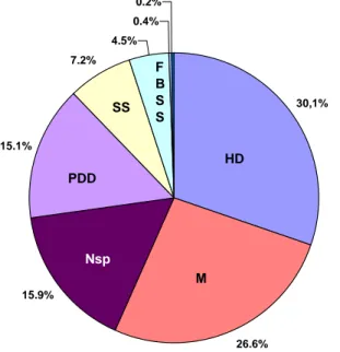
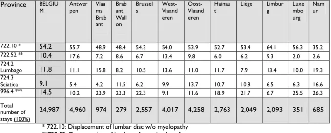
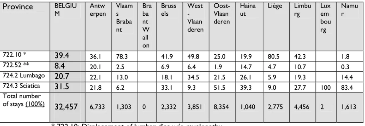
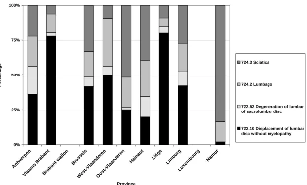
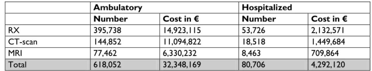
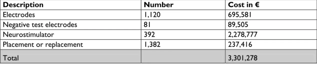
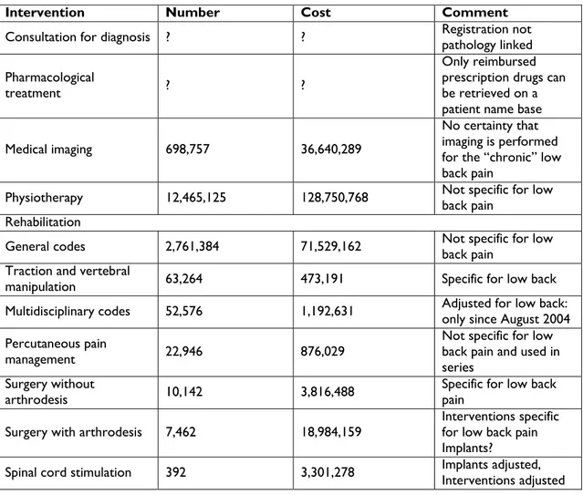
Around 40,000 classic hospital stays and 46,000 one-day hospital admissions have been recorded for low back pain problems. The most common diagnosis is "displacement of lumbar disc without radiculopathy" (a diagnosis for which discectomy is carried out in two thirds of cases). The interpretation of the hospital data is limited by coding errors (ICD-9-CM). Considerable regional disparities were recorded, with a higher proportion of admissions and surgical interventions in the north of the country and in Brussels. The INAMI/RIZIV data allow us to make an approximation of the costs connected with the treatment of low back pain: imaging (€ 36 640 000) physiotherapy (€ 128 750 000 for all disorders), rehabilitation (€ 73 200 000 for rehabilitation relating to all disorders, tractions and multidisciplinary treatment), percutaneous treatment of pain (€ 876 000), spinal cord stimulation (€ 3 301 278) and surgery with arthrodesis (€ 18 984 000) or without arthrodesis (€ 3 816 000). The limits inherent to these estimates are, on the one hand, the absence of specificity of the nomenclature codes for lumbar pain (especially chronic pain) and, on the other hand, the lack of many other sources of information on costs (such as consultations, hospitalization and other items of expenditure).
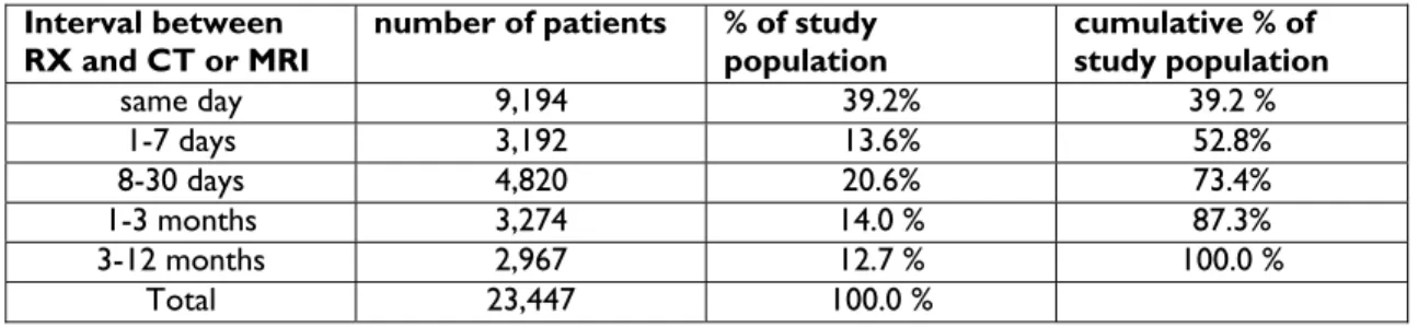
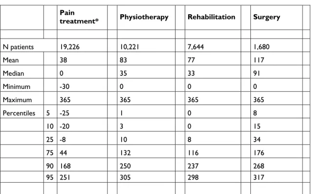
According to the longitudinal data of the Socialist Mutuality, the approximate annual medical cost connected with the care per patient suffering from chronic low back pain and for whom medical imaging codes have been invoiced is € 922. This estimate is also limited by several factors: the method used to select patients suffering from chronic lumbar pain, the absence of data relating to consultations, the lack of accuracy in terms of the anatomical region to which certain procedures are related and the unknown time interval between the diagnosis and a possible intervention.
This study concluded that the total direct medical cost of chronic low back pain in Belgium varies from 81 to 167 million euros. According to the literature, the medical costs paid by the health insurance sector account for only 10% to 30% of the overall
indirect costs for the patient and for society. The total amount could therefore be prudently estimated at between 270 million and 1.6 billion euros.
Grave consequences for social security
While the indirect costs cannot be accurately estimated, an analysis of the occupational medicine databases shows that the effects of chronic low back pain on society and on industry are harmful indeed. The results are based on the data of the Intermedicale (a service specialising in prevention and protection in the workplace) and of the Fund for Accidents at the workplace (FAT – FAO).
In occupational medicine, 11.9% of sick leave lasting 28 days or more is caused by a problem of low back pain. This type of disability is more prevalent among male employees with the status of manual workers who have recently joined the company. The sectors most frequently affected are cleaning, construction and food. As a result, one in every 20 patients is assessed as being permanently unable to return to work. In 15% of cases, the patient can go back to work provided the work is adapted, a fact that highlights the crucial role of the occupational physician when it comes to caring for low back pain.
The database of the FAT-FAO reveals that in Belgium every year twelve thousand occupational accidents lead to back pain, i.e. 6,63% of the total annual number of accidents recorded. The consequences are staggering: of the workers presenting an acute episode of low back pain connected with occupational accidents 72% were absent from work, and of this total figure 8,2% were absent for three months or more. A total of 62,4% and 95% of workers are temporarily or permanently disabled respectively. The sectors most affected are the timber industry, the construction industry and the metalworking industry. The construction and health/social sectors have the highest figures for permanent disability. Furthermore, the data reveal the geographic disparities, as the number of permanent partial disabilities is higher in Wallonia than in Flanders. Overexertion is the most frequently declared cause of accidents, while falling is the most frequent cause of injuries leading to permanent disability.
The best care within the framework of occupational
medicine: the role of occupational physicians and of
advisory physicians from the mutualities (médecin
conseil - adviserend geneesheer)
This project highlights the crucial role of occupational physicians and of advisory physicians when it comes to reducing the consequences of back pain, not only for the patient but also from a societal standpoint.
The primary role of these medical practitioners must be to inform workers: backache is a frequent disorder; certain posts and certain positions involve more risks; acute back pain often resolves itself spontaneously (90% within six weeks); it is important to keep active in spite of the pain. Although the physical constraints involved in work play a role at an etiological level, psychosocial factors (such as stress, anxiety or dissatisfaction with work) affect the seriousness of the ongoing disorder and the likelihood of chronicity. In this field, the scientific data are less clear-cut.
The second role of these physicians is to promote prevention strategies aimed at preventing chronicity. The literature gives evidence in favour of back schools (in the workplace, including an exercise component) and multidimensional or multidisciplinary interventions (see above).
The literature review highlights the role of exercise as the key healing factor. A multidisciplinary approach based on a combination of a program of exercises and psychological and/or social care is particularly beneficial. Occupational physicians and advisory physicians therefore bear some responsibility for the care of workers disabled by low back pain, along with family doctors. The physician should ideally reduce the period of disability by advising the patient to pursue his normal activities. A return to work can also be accelerated by temporarily adapting the worker's tasks (duration and load).
In the event of recurrent or constant lumbar pain, an analysis of the "yellow flags" will identify workers at risk of chronicity (psychological problems or depression). The occupational physician will also analyse the worker's expectations when a return to work is scheduled. In this regard, a return to work program backed up by cooperation between the curative sector and the occupational medicine sector is beneficial as it encourages the worker to return to work and reduces the number of days lost.
Discussion
The conclusions of this report offer guidelines for the care of chronic low back pain in the curative sector and in the field of occupational medicine. The first basic step in this care program is to maintain normal activities as much as possible. Furthermore, exercise programs play a positive role in re-education and multidisciplinary care is beneficial. Multiple diagnostic procedures are to be avoided. Many noninvasive treatments that are currently applied are based on scanty evidence or do not work at all. Based on the existing studies, we cannot yet define precisely the efficacy or the potential side effects of many invasive techniques (injections).
Due to a lack of data in Belgium, it is not possible to evaluate the extent of chronic low back pain with any accuracy. The available databases provided by occupational medical services and by the mutuality sector do not provide a means of systematically identifying these workers/patients or monitoring them in the care circuit. In addition, these databases do not yield any hypotheses on the geographic disparities that are observed. The evaluation of medical costs that we propose in this study is largely underestimated. A proper evaluation would require a data collection program geared specifically to the epidemiology and to the costs connected specifically with that particular pathology. Given that the indirect consequences of the pathology account for the bulk of the cost, occupational physicians and advisory physicians have a crucial role to play when it comes to helping workers get back to work as quickly as possible (in cooperation with the family doctor), bearing in mind that the data demonstrate that prolonged absence can lead to chronicity.
Recommendations
Scientific analysis of the care and consequences of low back pain yields the following recommendations:
• All care providers must be made more aware of the dangers of inactivity among patients suffering from chronic low back pain, the uselessness of applying multiple diagnostic procedures, the evidence in favour of certain conservative treatments (based on physical reactivation and a biopsychosocial approach) and the absence of such data for many other interventions that are currently applied. In cases of chronic low back pain, it is crucial for the patient to get back to work as quickly as possible. Prescribing useless tests and applying inappropriate treatments maintains the chronicity of the backache and does the patient more harm than good.
• All these practices call for close cooperation between occupational physicians and physicians working in the curative sector, from general practitioners to physicians with various specialities. The respective tasks and responsibilities of the occupational physician and of the advisory physician must be redefined: their role in preventing chronicity must be strengthened, as the rapid reintegration of workers suffering from chronic low back pain is a priority for the authorities.
• The current data sources are too fragmentary. From a policy standpoint, they do not provide a means of properly monitoring the consequences of a societal problem such as low back pain.
o The data concerning chronic disorders are lacking for first-line care in general and for low back pain in particular.
o There are data for the consumption of care, but they are not accompanied by precise codification of the reasons for long-term disability within insurance organisations.
o In the field of occupational medicine, in order to permit analyses and comparisons the databases must rapidly evolve towards standardised encoding of specific disorders that lead to long-term disability.
Scientific summary
Table of contents
1 INTRODUCTION... 3
2 PART I: EVALUATION AND TREATMENT OF PATIENTS WITH CHRONIC LOW BACK PAIN ... 4
2.1 INTRODUCTION... 4
2.2 DIAGNOSIS OF CHRONIC LOW BACK PAIN... 5
2.2.1 History-taking... 5
2.2.2 Physical examination... 9
2.2.3 Biology tests ...12
2.2.4 Imaging...13
2.2.5 Electrophysiological tests...18
2.2.6 Interventional diagnosis techniques...19
2.2.7 Physical capacity and fitness evaluation ...22
2.3 TREATMENT OF CHRONIC LOW BACK PAIN...24
2.3.1 Non-invasive therapeutics to treat CLBP ...24
2.3.2 Medications to treat chronic low back pain ...44
2.3.3 Invasive treatments for chronic low back pain ...50
2.3.4 Non-surgical, non-injection invasive treatments for CLBP ...57
2.3.5 Surgery...67
2.4 DISCUSSION...72
2.5 SUMMARY OF FINDINGS: EVIDENCE TABLES...73
2.5.1 Evidence for the diagnosis of non-specific chronic low back pain ...73
2.5.2 Evidence for the treatment of non-specific chronic low back pain ...74
3 PART II: HOW ARE CHRONIC LOW BACK PAIN PATIENTS ASSESSED AND TREATED IN BELGIUM?... 76
3.1 INTRODUCTION...76
3.1.1 Background...76
3.1.2 Selection of data sources...77
3.1.3 Goals and objectives of this study ...78
3.1.4 Summary of the information and the sources used in this study...78
3.2 PRIMARY CARE: INTEGO PROJECT...78
3.2.1 Description of the Intego project...78
3.2.2 Intego: methodology of the study “low back pain”...79
3.2.3 Intego: results for low back pain...80
3.2.4 Intego: Discussion ...83
3.2.5 Conclusions: added value of the Intego database for this project...85
3.3 HOSPITAL DATA: MINIMAL CLINICAL DATA (MCD/RCM/MKG)...86
3.3.1 Description of the Minimal Clinical Data database ...86
3.3.2 Minimal clinical data: methodology of the study “low back pain’...87
3.3.3 Minimal clinical data: results...88
3.3.4 Minimal clinical data: discussion ...99
3.4 RIZIV/INAMI NOMENCLATURE ...101
3.4.1 Description of the Belgian nomenclature...101
3.4.2 Use of RIZIV/INAMI database for the study “low back pain” ...101
3.4.3 RIZIV/INAMI: discussion...104
3.5 HEALTH CARE CONSUMPTION FOR LOW BACK PAIN PROBLEMS: DATA FROM THE SOCIALIST SICKNESS FUNDS ...106
3.5.1 Background...106
3.5.3 Results ...106
3.5.4 Discussion : longitudinal data from the Socialist Mutuality ...111
3.6 DISCUSSION: MANAGEMENT OF LOW BACK PAIN IN BELGIUM...112
3.6.1 Introduction...112
3.6.2 Diagnosis ...112
3.6.3 Treatment...113
3.6.4 Cost estimation of low back pain in Belgium ...116
3.6.5 Summary: added value and shortcomings of the databases...120
4 PART III: CHRONIC LOW BACK PAIN AND OCCUPATIONAL HEALTH IN BELGIUM... 122
4.1 BACKGROUND...122
4.1.1 Scope and definitions...122
4.1.2 Occupational consequences of chronic low back pain...122
4.1.3 Aims ...123
4.2 LITERATURE REVIEW ON MANAGEMENT OF CHRONIC LOW BACK PAIN IN OCCUPATIONAL SETTINGS ...123
4.2.1 Objective of the literature review...123
4.2.2 Synthesis of evidence...125
4.2.3 Summary of evidence for occupational settings...138
4.3 ANALYSIS OF BELGIAN DATABASES FOR ASSESSING CONSEQUENCES OF CHRONIC LOW BACK PAIN...139
4.3.1 Introduction...139
4.3.2 Databases not selected ...140
4.3.3 FAT - FAO database...142
4.3.4 Analysis of the Intermedicale database...164
4.4 CHRONIC LOW BACK PAIN IN OCCUPATIONAL SETTINGS: DISCUSSION OF THE FINDINGS ...175
5 GENERAL CONCLUSIONS... 177
6 REFERENCES... 181
1
INTRODUCTION
This project analyses the problem of non-specific chronic low back pain (CLBP) defined as pain lasting more than three months, without or with radicular pain, without any suspicion of severe underlying pathology. CLBP is studied from three points of view. A first part analyses the evidence-based literature on the diagnosis and treatment. The second part analyses the available databases in Belgium in order to assess the size of this public health problem and its related costs. The last part examines the consequences of CLBP on the workers’ population using databases from occupational health and also reviews the available data from the literature on the optimal care of CLBP patients in the occupational setting.
The literature review in part I summarizes the evidence based literature sources currently available. It aims to serve as a clinical practice guideline to help primary care and specialized practitioners involved with chronic low back pain. This part mainly searched for the available evidence in guidelines, meta-analyses and systematic reviews. Hence, it should not be considered as an exhaustive list of all available evidence on all diagnostic and therapeutic procedures. No specific search has been conducted on the safety aspects of the procedures and only the most common ones that have been described in the selected references are summarized in this report.
The literature study on occupational medicine (part III) is in the same way a synthesis of the best available evidence for occupational physicians and medical advisers.
The literature reviews from part I and part III are useful to help interpreting the results of the analysis of available Belgian data about medical care (diagnostic and therapeutic) provided to patients with chronic low back pain in our country (part II). Those literature reviews allow appraising to what extent Belgian medical care for chronic low back pain is based on an evidence-based approach.
Finally the combination of current data on CLBP in Belgium and the synthesis of the available evidence will allow policy makers to orientate their decisions for designing policies in relation with chronic low back pain. These decisions can relate to multiple facets as for example the availability of databases, their content, the quality and organisation of care (e.g. multidisciplinary teams, roles of occupational physicians).
2
PART I: EVALUATION AND TREATMENT OF
PATIENTS WITH CHRONIC LOW BACK
PAIN
J. Gailly, D. Paulus, B. Aertgeerts, H. Nielens
Important preliminary remarks
This report focuses on evaluation and treatment of patients with non-specific chronic low back pain (lasting for more than three months) with or without nerve root/radicular pain. Less common origins of chronic low back pain such as spinal stenosis, spondylolisthesis, spinal tumor or infection are not specifically addressed in this report.
Chronic low back pain is a symptom : the different possible etiologies are voluntary not cited. For some specific techniques, pain generators are however discussed.
The European guidelines for the management of chronic non-specific low back pain (COST B13) are an important source of evidence for this report given their methodological quality, recent date of publication and applicability to European settings. Numerous other references have been consulted and added in this systematic literature search, in particular if they were more recent or addressed specific techniques. The extensive literature search is described in appendix.
2.1
INTRODUCTION
Low back pain (LBP) is generally defined as a pain that occurs in an area with boundaries between the lowest rib and the creases of the buttocks. LBP may be termed as ‘acute’, ‘sub acute’, ‘chronic’ and/or ‘recurrent’. Most authors agree that an acute episode of LBP usually resolves within six weeks. The definition of chronic LBP (CLBP) varies among authors. When LBP lasts for more than six weeks, most authors agree to define it as ‘sub acute’. When a LBP episode lasts over 3 months (12 weeks) it is generally termed as ‘chronic’. After the resolution of an acute LBP episode, further episodes may occur generating a situation defined as ‘recurrent LBP’.
LBP may be due to specific medical conditions such as cancer (metastases), infection (discitis) or rheumatologic diseases. However, most cases of LBP are caused by degenerative changes of the lumbar column (so-called “common” or non specific LBP). Non-specific LBP may be accompanied by nerve root/radicular pain (sciatica) radiating in the lower limbs as degenerative changes may narrow the lumbar spinal canal and/or the foramen(s) leading to nerve root compression. Such nerve root/radicular pain will also be addressed in the present report.
This report addresses non-specific LBP lasting more than 3 months (12 weeks) and/or recurrent episodes of non-specific LBP. CLBP with nerve root/radicular pain (sciatica) is also included in this study.
The aim of the present chapter is to search and summarize the literature on diagnosis and treatment of CLBP patients with or without radicular pain. Recommendations based on the available evidence are also be included in this report.
The detailed searching methodology and references selection after critical appraisal are described in the appendices.
In this chapter and in the chapter about occupational medicine (part 3), the quality of evidence is presented following the GRADE system 1.
2.2
DIAGNOSIS OF CHRONIC LOW BACK PAIN
Non-specific CLBP is generally considered as a clinical syndrome needing comprehensive evaluation (diagnosis) in order to provide each patient with a well-adapted and effective treatment strategy. The first step of the diagnostic evaluation of patients with CLBP consists in ruling out any possible specific cause of LBP that could remain unrecognized or may develop with time.
2.2.1
History-taking
This section focuses on the information obtained through history-taking that is relevant to evaluate and manage patients with CLBP with or without sciatica (radicular pain). A thorough history should be obtained in all patients with LBP, in the acute and the sub-acute stage. Likewise, it is generally admitted that a thorough physical examination including a well-conducted history-taking should also be repeated in the chronic stage. Several diagnosis systems have been proposed, in which patients with LBP are classified on the basis of pain distribution, pain behavior, functional disability, clinical signs, etc. None of these systems of classification have been adequately validated 2. However,
several factors such as “red flags”, yellow flags”, pain distribution, functional status and disability are generally considered as contributing to refine the diagnosis of CLBP.
2.2.1.1
Pain characteristics
The pain characteristics reported by the patient (localization, intensity, type, frequency…) are important in the history-taking. For instance, pain localization must be taken into account, as it often constitutes the first clinical information that may lead to suspect radicular pain (see next section on “red flags”).
Evidence
Some tools have been developed to assess pain characteristics (Visual Analogic Scale, Dallas…). These tools are used in daily practice but their utility has not been established (ANAES 3; expert consensus).
Very few references address the issue of pain evaluation specifically in the context of CLBP. Hence, no specific recommendations can be made to evaluate pain as reported by CLBP.
2.2.1.2
“Red Flags”
“Red flags” are factors, signs or other medical conditions that may be identified through a well-conducted history-taking and that may be associated with non musculoskeletal or with specific origins of LBP. They are traditionally used to rule out specific underlying medical conditions in patients with acute LBP. They may however also be useful in the context of sub acute or chronic LBP.
“Red flags” should be screened on a regular basis, even in the chronic stage, to rule out any specific origin. Some “red flags” indicators of radicular pain may also be useful. Their definitions vary as they are based on expert consensus. The “red flags” proposed by COST B13 2 (based on the
guidelines from the Royal college of general practitioners 4, 5) are the
followings:
• Age of onset of LBP < 20 or > 55 years,
• Constant, progressive, non-mechanical pain (not relieved by rest), • Thoracic pain,
• Past medical history of malignant tumor, • Prolonged use of corticosteroids, • Drug abuse, immunosuppression, HIV,
• Systematically unwellness, • Unexplained weight loss,
• Widespread neurological symptoms (including cauda equina syndrome), • Structural deformity,
• Fever.
Koes added the following “red flags” suggesting radicular pain due to nerve root compression 6:
• Unilateral leg pain > low back pain, • Pain radiating to foot or toes,
• Numbness and parenthesis of same distribution,
• Passive Straight Leg Raise test (see below) inducing more leg pain than back pain,
• Localized neurological deficit (limited to one nerve root).
Evidence
European COST B13 2 and ANAES guidelines 3 recommend that “red flags” should be
screened on a regular basis, even in the chronic stage, to rule out any specific origin that may reveal itself or develop with time (expert consensus).
Red flags have not been evaluated comprehensively in any systematic review 2. Serious
conditions theoretically associated with “red flags” like neoplasm, infection, and cauda equina syndromes are extremely rare (Carragee 7 in COST B13 2). More over, “red
flags” are not always associated with any specific pathology, but merely indicate a higher probability of an underlying condition that may require further investigation. A recent study reported an incidence of spinal tumor of 0.69% and 0.12%, respectively in 33 academic and 18 private practice settings (all together 19 312 patients) 8, 2
2.2.1.3
“Yellows flags”
Psychosocial “yellows flags” may be defined as factors identified during history-taking of patients with LBP that are related to a higher risk of developing or perpetuating chronic pain and long-term disability 9, 2. The relationship between “yellow flags” and the
development of CLBP may be of varied nature. Some “yellow flags” may act as direct or indirect causal factors. Others may reflect more serious conditions (recurrence, radicular pain…).
“Yellow flags” are factors that are generally related to a higher risk of developing CLBP. They may already be screened for in patients with acute LBP. However, the identification of “yellow flags” is particularly relevant when LBP becomes sub acute, chronic or recurrent as, when possible, interventions aiming at eliminating or reducing such “yellow flags” may play an important role in the treatment of CLBP.
The “Yellows flags” described in the European COST B13 guideline are the followings:
• Inappropriate attitudes and beliefs about back pain (for example, the belief that back pain is harmful or potentially severely disabling, or a high expectation from passive treatments rather than from staying active), • Inappropriate pain behavior (for example, fear-avoidance behavior and
reduced activity levels), • Work-related problems as:
Low-level of support and concern for the LBP sufferer at his work place (COST B13, level A),
Shorter job tenure, heavier occupations with no modified duty (COSTB13 level B),
• Long off-work period of time. For a worker having difficulty returning to normal occupational duties at 4-12 weeks after the onset of LBP, the longer the worker is off-work, the lower the chance is that he will ever return to work (level A),
• Prior episodes of LBP, severity of pain, important functional impact of LBP, psycho-social distress, excessive symptoms report, unrealistic patient expectations,
• Radicular findings (level B in COST B13),
• Emotional problems such as depression, anxiety, stress, tendency to withdraw from social interaction 9.
Evidence
According to Roach et al.10 who studied a series of 174 patients with LBP, insomnia,
trouble sleeping and back pain aggravation through walking are more often associated with severe LBP (sensibility = 0.87; specificity = 0,5). However, this study did not precise clearly the type of LBP (acute, sub-acute or chronic LBP).
2.2.1.4
Psychological evaluation
Any anxiety and/or depression state may play an important role in the development and/or perpetuation of CLBP (see the “yellow flags” section). Psychological evaluation may be performed using specific tools (Hamilton scale, Beck Depression Inventory). Likewise, some signs and behaviours (the so-called “Waddell non-organic signs” such as tenderness, simulation, distraction, regional weakness or sensory abnormality, over-reaction) may suggest the presence of psychological distress, which may be associated with an elevated risk of pain perpetuation. Hence, it is generally admitted that, to some extent, psychological evaluation should be performed during history-taking in patients with CLBP.
Psychological evaluation is useful when yellow flags are present. Psychological evaluation may identify psychological distress that may be related to pain perpetuation. Specific tools developed to assess anxiety and/or depression states may be used in patients with CLBP. However, their validity has not been established in the context of CLBP. The so-called “Waddell non-organic signs” do not correlate with any psychological distress, nor do they discriminate organic from non-organic problems.
Evidence
The ANAES consensus 3 recommends that anxiety and/or depression states should be
evaluated using specific tools (Hamilton scale, Beck Depression Inventory). Such tools may be helpful in daily practice but their utility and validity have not been demonstrated. Fishbain et al. conducted a good-quality systematic review (61 studies) on Waddell’s non-organic signs11. They concluded that the Waddell’s signs do not correlate with
psychological distress, nor do they discriminate organic from non-organic problems. They may be explicable by an underlying organic condition and are associated with poorer treatment outcome, with greater pain levels and are not associated with secondary gain. Overall, most studies included in this review had methodological limitations and mixed patient populations with acute and chronic LBP.
2.2.1.5
Functional state and disability assessment
It is generally admitted that functional state and disability level must be addressed in patients with CLBP. The rationale is that chronic pain often leads to physical inactivity, physical capacity reduction (so-called “physical deconditioning”), work loss and ultimately may greatly alter quality of life of the patients.
Functional repercussions in terms of physical activity and capacity levels and occupation should be evaluated in patients with CLBP using specific methods whenever significant physical activity reduction and work loss may be suspected. Only a restricted number of specific tools may be considered as valid.
Evidence
ANAES 3 states that the most commonly and best validated tools are the Oswestry
Disability Index and the Roland Disability Questionnaire 12.
Our additional search identified two systematic reviews on functional status assessment
13; 14. However, those two complementary references focus on LBP in general and not
specifically chronic LBP.
In the good methodological quality systematic review by Grotle et al. 13}, the authors
listed 36 back-specific questionnaires designed for assessing the functional status or disability in patients with LBP. Several versions of the 2 most commonly used questionnaires, the Roland-Morris Questionnaire (6 versions) and the Oswestry Disability Index (4 versions), have been identified. Ten questionnaires were considered as well-validated and recommended without further validation studies: the original version of the Roland-Morris Disability Questionnaire 12, the Oswestry Disability Index
1.0 15, the Oswestry Disability Index 2.0 16, the Oswestry Disability Index Chiropractic
version 17, the Clinical Back pain Questionnaire 18, the Disability Rating Index 19, the
General Function Score20, the Million Visual-Analogue Scale 21, the Quebec Back Pain
Disability Index 22 and the Waddell Disability index 23.
In a low-quality systematic review, Calmels et al.14 concluded that the metrological
properties (content validity, construct validity, applicability, translation and international applicability) of the following functional and disability assessment tools were satisfying: the Dallas Pain Questionnaire 24, Roland-Morris Questionnaire12, the Quebec Back Pain
Disability Index 22 and the Oswestry Disability Index 15. Noteworthy, the version of the
Dallas Pain Questionnaire by Lawlis 24 had been evaluated as insufficiently validated by
Key messages for history- taking
• The patient history-taking has been extensively described in the context of acute low back pain but it has been seldom addressed in the context of chronic low back pain,
• Pain characteristics (localization, intensity, type…) reported by the patient must be evaluated. However, the validity and utility of tools specifically designed to assess pain characteristics have not been established,
• “Red flags” are traditionally used to rule out any specific underlying medical condition in patients with acute low back pain. Some radicular pain-specific “red flags” have been also proposed to identify nerve root pain during history-taking. The definition and implementation of “red flags” are based on expert consensus. It is recommended to assess the presence of “red flags” also at the chronic stage of LBP,
• “Yellow flags” are psychosocial factors that might be predictors of chronicity in acute or sub acute LBP. It is recommended to identify the presence of “yellow flags” in the patient with CLBP also,
• It is advisable to evaluate the psychological state of the patient with CLBP. Some specific tools are traditionally used in that context although their utility has not been established yet. Waddell’s non-organic signs do not correlate with psychological distress, they should not be used to discriminate organic from non-organic conditions and are not associated with secondary gains,
• Functional status and disability should be evaluated whenever significant physical activity reduction and work loss may be suspected during history-taking. Numerous specific tools have been developed therefore but only a limited number of them may be considered as sufficiently valid. There is no evidence that the use of such tools generates any benefit in patients with CLBP.
2.2.2
Physical examination
A history-taking combined with physical examination allows evaluating the degree of pain and functional disability in order to outline a management strategy that matches the magnitude of the problem. This section addresses the relevant findings obtainable through physical examination. This review did not find any reference focusing on the impact of physical examination on the outcomes of CLBP.
2.2.2.1
Information given to the patient during physical examination
Physical examination may serve as a basis for providing the patient with valuable information regarding diagnosis, management and prognosis. It may help to reassure the patient and act in this way as a therapeutic intervention by addressing for instance misbelieves that may be identified and corrected.
It is advised to provide the patients with information based on a biopsychosocial model in order to change their misbelieves about LBP. Information should be given in easily understandable terms.
Evidence
The COST B13 guideline 2 recommends that “Information given to the patient should
be provided in a common language understandable by the patient. Preferably, the information should be given consecutively during the clinical examination and when evaluating imaging. Terms like “positive” findings for a significant pathology should be
avoided as they often are hard to understand for the patient. Likewise, concepts such as vertebral instability, disc displacement, isthmic fracture (spondylolisthesis), hyper mobility, that refer to mechanical disorders not yet clearly defined nor verified by experimental or clinical studies, should thus be avoided”.
A recent systematic review concluded that information based on a biopsychosocial model is recommended in primary care to shift patient beliefs on LBP. In this model, the message is focused on patients “beliefs and attitudes” and stresses the advantage of remaining active and avoiding best rest, combining with reassurance that there is likely nothing seriously wrong. Traditional information on anatomy, ergonomics, and back-specific exercises is markedly reduced. Nevertheless, information delivery alone is not sufficient to prevent absenteeism and reduce health care costs 25.
2.2.2.2
Orthopedic examination and mobility of the lumbar spine
Many physical tests are routinely carried out as part of the physical examination of the patient with CLBP (trunk mobility tests, orthopedic tests focusing on bones, joints, tendons and ligaments…). Traditionally, physical examination aims at assessing the level of pain, the mobility of the lumbar spine, at identifying the presence of nerve root/radicular pain, at ruling out any neurological deficit or clinical “red flag” and at identifying the pain generator as precisely as possible. Physical examination aims at gathering the useful information needed by the clinician to elaborate an adapted treatment strategy.
No maneuver has been validated as a part of the orthopedic examination of the CLBP patient. Most physical tests are based on expert consensus. Evidence
COST B13 does not address physical examination 2. KNGF 2005 recommends a
history-oriented physical examination including anthropometrical evaluation (trunk and leg lengths…), mobility and strength testing and some functional testing (e.g. sitting, pushing)26.
ANAES advises to proceed to a general physical examination to rule out any specific orthopedic or neurological abnormality 3. It identified two trials with unknown
methodological quality that aim at establishing a physical examination total score. One trial by Waddell 27 suggests that a total physical examination score based on 8 different
traditional physical tests is able to discriminate CLBP patients from controls (sensitivity 0.86 ; specificity 0.76). The second trial by Llorca 28 suggests that a global trunk mobility
score obtained by summing several trunk mobility indices is able to discriminate LBP patients from controls (sensitivity 0.93 ; specificity 0.95).
2.2.2.3
Neurological examination: the Lasegue test
It is generally admitted that physical examination of the patient with acute LBP should include neurological tests. These neurological tests in patients with LBP mainly aim at: - Ruling out any “red flag” associated with a specific medical condition or a severe neurological (radicular) complication that may seldom develop in CLBP (paresis, paralysis, cauda equina syndrome…),
- Identifying any radiating pain that may have a nerve root/radicular origin.
This neurological examination should be performed on a regular basis during follow-up. Traditionally, it encompasses the osteo-tendinous reflexes testing, motor and sensory testing and the Lasegue test.
The Lasegue test, also known as the Passive Straight Leg Test (PSLR) in North-America, is routinely used to identify the presence of nerve root/radicular pain due to nerve root compression. COST B13 2 describes this test as follows: “The Lasegue test requires a firm
level couch, with a supine, relaxed patient with trunk and hips without lateral flexion. The practitioner should ensure that the patient’s knee remains extended, with the foot in the
vertical plane. The affected leg is supported at the heel and the limb gently elevated. The angle of leg elevation at the onset of pain and the site of pain is recorded. If the PSLR is unilaterally limited, induces unilateral symptoms, or is bilaterally limited to less than 50°, then each leg should be raised in turn to the onset of pain, lowered a few degrees (to reduce pain) and, in turn, the ankle dorsiflexed, the hip medially rotated and the neck flexed. Symptom reproduction by one of these tests would be interpreted as a positive PSLR outcome, suggesting increased root tension”.
The use of the PSLR (Lasègue) test to identify radicular pain due to nerve root compression at the lumbar level (L4-L5 and L5-S1) is not supported by the selected references.
Evidence
Two high-quality systematic reviews 29, 30 were identified by the COST B13 systematic
review. In the first review by Deville et al.29, all studies were surgical case-series at
non-primary care level. According to COST B13, “It was found that the pooled diagnostic odds ration for PSLR was 3.74 (95% CI 1.2-11.4); sensitivity was high 0.91 (0.82-0.94), but specificity was low 0.26 (0.16-0.38). The pooled diagnostic odds ratio for the crossed PSLR test (pain evoked in the symptomatic lower limb by performing a PSLR in the contra-lateral limb) was 4.39 (95% CI 0.74-25.9); with low sensitivity 0.29 (0.23-0.34), and high specificity 0.88 (0.66-0.90). The authors concluded that the studies do not enable valid evaluation of diagnostic accuracy of the PSLR test”. This test is not sufficient to make the diagnosis of radiculopathy. An important methodological weakness is that disc herniation was selected as the outcome variable.
According to Rebain et al.30, “The sensitivity (0.8) of the PSLR test was also far greater
than its specificity (0.4). There remains no standard PSLR procedure and no consensus about the interpretation of the results”. The authors concluded that: “Until there is a standard procedure for carrying out and interpreting the PSLR, with well-documented reliability and validity, clinicians and researchers should treat the test with caution” 2.
The use of the Lasegue test as a valid and reliable test to identify radicular pain due to nerve root compression at the lumbar level (L4-L5 and L5-S1) is not supported by the quality of evidence available in the selected references. This lack of evidence contrasts with opinions of experts, who generally consider a properly conducted PSLR test as “the most accurate test to identify nerve root pain” 31.
2.2.2.4
Spinal palpation and motion pre-manipulative tests
Spinal palpation tests are sometimes performed as a part of the physical examination to determine whether manipulative therapy is indicated and/or to evaluate the effectiveness of a therapeutic intervention. Such tests consist in the assessment of symmetry of bony landmarks (posterior superior iliac spines for instance), evaluation of regional segmental motions, Para spinal soft tissue abnormalities, tenderness during active trunk movements and palpation. The validity and reliability of such pre-manipulative tests remain vastly debated 2.
Palpatory and motion pre-manipulative tests are neither reliable, neither valid. Moreover, the presence of a manipulable lesion remains hypothetical. Evidence
The COST B13 systematic review found two good quality systematic reviews on pre-manipulative tests: Seffinger et al. 32, Hestebaek and Leboeuf-Yde 33. Our additional
search identified one more systematic review by van Trijffel et al. 34. However, this
review was excluded from this analysis: most included studies did not fulfill the criteria for external and internal validity e.g., an adequate report of the study protocol and statistical tests.
COST B13 concludes “There is conflicting evidence that spinal palpatory tests are reliable procedure to diagnose back pain (level C: conflicting evidence). Pain provocation tests are the most reliable of the palpatory tests (level B). Soft tissues tests are unreliable (level A). Regional range of motion is more reliable than segmental range of motion (level A). Intra-examiner reliability is better than inter-rater reliability for all palpatory tests (level A). As palpatory diagnostic tests have not been established as reliable and valid, the presence of the manipulable lesion remains hypothetical (level B).
Key messages for physical examination
• Specific physical examination of the patient with CLBP is not well documented in the literature. No test commonly included in the physical examination of the patient with CLBP has been sufficiently validated. The content of the traditional physical examination of patients with CLBP is based on expert consensus. It is generally admitted that the physical examination recommended in patient with acute LBP should be repeated at first evaluation of the CLBP and during the follow-up.
• The use of the PSLR (Lasegue) test as a valid and reliable test to identify radicular pain due to nerve root compression at the lumbar level (L4-L5 and L5-S1) is not supported by the evidence.
• Palpatory and motion pre-manipulative tests are neither reliable, neither valid. Moreover, the presence of a manipulable lesion remains hypothetical.
2.2.3
Biology tests
This systematic review did not identify any good-quality study about biology tests neither for the diagnosis of CLBP nor for the impact of the use of biology tests on the outcomes of CLBP. It can only be assumed that recommendations about biology testing in patients with CLBP should follow the same rules as for acute/sub acute LBP. For instance, the presence of “red flag” such as loss of weight, general unwellness, should lead to test biology.
Evidence
Cost B13 2 related one systematic review of 36 studies that evaluated the accuracy of
history-taking, physical examination and erythrocytes sedimentation in diagnosing low back pain in general practice 35. The review found that few of the studied signs and
symptoms seemed to provide valuable diagnosis. The combined history and the erythrocytes sedimentation rate had relatively high diagnostic accuracy in vertebral cancer 2. Another review cited in COST B13 concluded that “For patients 50 years of age
and older, or those whose findings suggest systemic disease, plain radiography together with simple laboratory tests can almost completely rule out underlying systemic diseases”. 36
Key message for biology tests
• No evidence is available about the value of biology tests in CLBP. It can only be assumed that recommendations about biology testing in patients with CLBP should follow the same rules as with acute and sub acute LBP. • Authors suggested that for patients 50 years of age and older, or those whose findings suggest systemic disease, plain radiography together with simple laboratory tests can almost completely rule out underlying systemic diseases.
2.2.4
Imaging
Imaging patients with CLBP encompasses conventional radiography; more sophisticated imaging techniques (CT, MRI) and interventional imaging as discography. Some imaging techniques (fluoroscopic guidance) may also be used as an aid in the context of invasive therapeutic procedures: those techniques will be addressed in the following sections. Two guidelines specifically address the issue of imaging in the context of CLBP 2,3 :
ANAES and COST B13. The ANAES 3 guideline refers to an earlier guideline on imaging
by the same institution: “Imagerie dans la lombalgie commune de l’adulte” 37. COST
B13 found 5 systematic reviews of high quality: Boos and Lander 38 (technical efficacy
level), Jarvik and Deyo 36 (diagnostic accuracy of imaging for patients in primary care
settings), Littenberg et al 39 (SPECT), Saal 40 (diagnostic tests in the evaluation of CLBP
with a focus on invasive techniques, such as discography), van Tulder et al 41
(relationship between radiographic findings and non specific low back pain) The quality of some additional studies mentioned in COST B13 42-54, 36, 55, 56, 8, 57 is not defined.
The “Recommandations du Consilium Radiologicum Belge” (currently being revised) are based on a European experts consensus (Radioprotection 118 : recommandations en matière de prescription de l’imagerie médicale de la Commission Européenne). A recent KCE HTA report on Magnetic Resonance Imaging (MRI) was also considered 58.
2.2.4.1
Conventional radiography
Conventional radiography for CLBP usually includes several views: front and oblique views of the whole lumbosacral spine, front and oblique views centered on the lumbosacral junction and coned lateral views (left and right) of the lumbosacral junction to visualize the facet (zygoapophyseal) joints.
Systematic imaging of adults with CLBP between the age of 20 and 55 years is not recommended: the relationship between degenerative changes and LBP is weak and most common abnormalities seen on conventional radiographs (spondylolysis/spondylolisthesis, spina bifida, transitional vertebrae and sequel of Scheuermann’s disease) are not associated with back pain. Moreover, conventional radiography is not a good screening procedure for the suspicion of compression fractures, cancer and metastases, as its sensitivity is too low.
Evidence
According to COST B13, “there is moderate evidence that radiographic imaging is not recommended for chronic non-specific low back patients (level B)”2.
A systematic review of observational studies 41 cited in COST B13) concludes that
“there is no firm evidence for the presence of absence of a causal relationship between radiographic findings and non-specific low back pain. Degeneration, defined by the presence of disc space narrowing, osteophytes, and sclerosis, turned out to be associated with non specific low back pain, but odds ratio were low, ranging from 1.2 to 3.3. Spondylolysis and spondylolisthesis, spina bifida, transitional vertebrae, spondylosis and Scheuermann’s disease did not appear to be associated with low back pain”2.
The review of Jarvik and Deyo 36 (cited in COST B13) concluded that “for adults
younger than 50 years of age (in primary car settings) with no signs or symptoms of systemic disease, symptomatic therapy without imaging is appropriate. For patients 50 years of age and older, or those whose findings suggest systemic disease, plain radiography together with simple laboratory tests can almost completely rule out underlying systemic diseases. Advanced imaging should be reserved for patients who are being considered for surgery or those in whom systemic disease is strongly suspected”. Two studies in primary care 53, 54 cited in COST B13 randomized unblinded controlled
trial with 421 patients), concludes that “the use of lumbar spine radiography prior to treatment was not associated with improved functioning, reduced pain or improved overall
status after treatment, and was associated with an increase of General Practitioner workload. Participants receiving X-rays were more satisfied with their care, but were not less worried or more reassured about serious disease causing their low back pain.”
A study in primary care patients 42 cited in COST B13 concluded that “the presence of a
lytic or blastic lesion on plain radiographs was 60% sensitive and 99.5% specific for cancer. This suggests that plain radiography is not a good screening procedure for cancer and metastases. Sensitivity was 70% and specificity 95% for compression fractures.”
A recent study of 33 academic and 18 private practice settings (altogether 19,312 patient files) “reported an incidence of spinal tumor of 0.69% and 0.12% respectively” (Slipman et al 8 cited in COST B13). “It has been shown that, with careful clinical
assessment revealing no red flags, X-rays detect significant spinal pathology in just one in 2500 patients”(Waddell 51 cited in COST B13).
A study (van den Bosch et al 57 cited in COST B13) identifies “many abnormalities that
are unrelated to back symptoms. The abnormalities are equally prevalent in persons with and without back pain : spondylolysis, facet joint abnormalities, some congenital anomalies, Schmorl’s nodes, herniated discs, disc dehydration (“black discs”), disc protrusion, and mild scoliosis (Cobb angle <10°). Imaging identifies abnormalities that are unrelated to back symptoms”. That may be considered as an adverse effect.
ANAES 3 recommendations contrast quite significantly with those of COST B 13: it
recommends systematic imaging of patients with CLBP through conventional radiography. However, such recommendations are only based on an expert consensus. Belgian Consilium Radiologicum recommendations recall that degenerative changes that can be seen on plain lumbosacral films are frequent and not specific. Those recommendations also state that conventional radiography should only be systematically obtained in patients below the age of 20 and older than 55 years old.
2.2.4.2
CT and MRI
Computed Tomography (CT) and Magnetic Resonance Imaging (MRI) are more advanced imaging procedures traditionally used to diagnose herniated disks as a common cause of nerve root compression and radicular pain and to rule out non-specific origins of LBP in the presence of “red flags”. Noteworthy, when CT is a technique that uses ionizing radiations, MRI does not.
There is moderate-quality evidence that MRI should not be used for common CLBP, even if some experts recommend MRI when CLBP persists after well-conducted treatment (very-low quality evidence).
A previous KCE report concluded that MRI is the best imaging procedure for patients in whom a specific origin of LBP is suspected in the presence of one or more “red flags” (suspicion of nerve root compression, discitis, neoplasm).
CT scan is not recommended for patients with CLBP. Evidence
COST B13 concluded: “MRI is the best imaging procedure for use in patients with radicular symptoms, or for those in whom discitis or neoplasm is strongly suspected (level B). Facet joint MRI is not reliable procedure for the differential diagnosis between facet joint pain and discogenic pain level B)” 2 .
COST B13 2 recommends “MRI in patients with serious red flags and for evaluation of
radicular symptoms. Plain radiography is recommended for structural deformities.”. COST B13 2 considered the following studies about the use of MRI for specific