Delivery, Design, and Mechanism of
Antimicrobial Peptides
MASSACHUSESINSTIJTE
OF TECHNOLOGY
by
SEP
16
2010
Tanguy Chau
B.S., Chemical Engineering
LIBRARIES
University of California, Berkeley, 2004
ARCHNES
Submitted to the Department of Chemical Engineeringin Partial Fulfillment of the Requirements for the Degree of Doctor of Philosophy in Chemical Engineering Practice
at the
Massachusetts Institute of Technology
May 26, 2010
@ 2010 Massachusetts Institute of Technology
All rights reserved
Signature of the author
Tanguy M. Chau Department of Chemical Engineering May 26, 2010
Certified by
Gregory N. Stephanopoulos
Bayer Professor of Chemical Engineering Thesis Supervisor
Accepted by
William M. Deen Professor of Chemical Engineering Chairman, Committee for Graduate Students
Abstract
Each year, 2 million people contract hospital-acquired bacterial infections, which causes the
death of 100,000 patients and costs the US healthcare system over $21 billion. These infections
have become dangerously resistant to our existing line of antibiotics and are rapidly spreading
outside of hospitals and into communities. As molecular targets to develop new antibiotics are
becoming exhausted, clinicians and scientist are concerned that antibiotic resistant infections will
wipe out most of the major health benefits acquired over the last century. The work described in this thesis develops new antimicrobials strategies against bacterial infections, focusing on
antimicrobial peptides (AmPs).
We first delivered genes inducing the toxic expression of AmPs and other lytic agents
directly into bacteria using reengineered bacteriophages. Expression of these lytic agents in
lysogenic bacteriophages resulted in bactericidal activity, and demonstrated, for the first time, a
long-term cidal effect for over 20 hours. We then enhanced the efficacy of our approach by
expressing the same agents in lytic bacteriophage, which resulted in complete suppression of the bacterial culture and prevented bacterial regrowth and resistance to bacteriophages.
Since a large fraction of medical infections originates at the surface of implantable devices,
we developed film coatings that release active AmPs to cover these surfaces and prevent bacterial colonization. We incorporated AmPs in layer-by-layer films and demonstrated that the kinetics of AmP release can be adjusted. These released AmPs still actively prevented bacterial growth and remained non-toxic towards mammalian cells.
While natural AmPs have broad activity against pathogens, they are not optimized for a
specific antimicrobial function or bacterial target. Thus, researchers have tried for decades to
design highly active and specific de novo AmPs. One approach is to design new peptides using
approach by measuring the antimicrobial activity of a large database of natural AmPs and
incorporating this activity information in the design algorithm. This strategy improved the success
rate of designing de novo peptides from 45% to 73% and increased the antimicrobial strength of the
designed peptides.
Finally, we developed new potentiating strategies by studying the mode-of-action of the
family of ponericin AmPs. First, we measured their cidal behavior and differentiated bactericidal
ponericins from bacteriostatic ones. Using a modified AFM and a microfluidic device, we observed that the action of AmPs led to cellular death through the corrugation of bacterial, while
sub-population of cells resisted the action of the AmPs longer than others. Focusing on the ponericin G1
AmP, we correlated these visual observations with various membrane stress sensing mechanisms. We concluded that bacteria's ability to develop resistance to ponericin G1 requires the sensing and
repair of misfolded membrane proteins via the CpxAR system, as well as DNA repair via induction of the SOS response by RecA. Using microarrarys, we showed that ponericin G1 targets tRNA
synthetases in the ribosome. Finally, we demonstrated 99.999% killing of antibiotic resistant bacteria by potentiating ponericin G1 with the ribosomal antibiotic kanamycin, whereas no killing is observed when these two agents are applied independently. untreta
The PhDCEP capstone requirement finalizes the work of this thesis by analyzing market
entry and expansion strategies for an antimicrobial company commercializing genetically
engineered bacteriophages. In conclusion, this thesis establishes new advances in the delivery, the design and the potentiation of AmPs in order to eradicate resilient bacterial infections.
d Maman,
Sache qu'ici reste de toi comme une empreinte
ind6libile
... ... ... ...
Acknowledgments
Family:
- Papa, Chlo6, et ma famille: pour votre amour, et soutien constant.
Best buddies:
- Doktor Michael Koeris: Ohne dich mein Bruder, ich wurde nirgends.
- Aspiring Doctor Christopher Jongsoo Yoon:
i1
9 i-= oll ] ]Cl-.
- Professor Timothy Lu: @'fi~h , and turning on the headlights when I
was lost.
- Christopher Loose, and Joel Moxley: for teaching me to play football with the Irish.
Collaborators:
- Bernice Huang: for making late night gel electrophoresis such fun.
- Rana Ghosh: for introducing me to the phage world.
- Orhan Karsligil: for bringing rational to AmP designs.
- Helen Chuang, and Anita Shukla: for stuffing AmPs into coatings.
- Robbie Barbero and Georg Fantner: for sharing my passion to kill bacteria.
Mentors:
- My advisors, Robert Langer and Gregory Stephanopoulos, for inspiring me to be my best.
- My committee members Christopher Love and Dr. Robert Rubin for their guidance.
- Jim Collins for your contagious enthusiasm and support of my work.
- Steve Lerman for making my experience at MIT an enjoyable, diverse and rewarding one.
Friends:
- My friends for allowing me to vent my frustration in front of a good beer.
- Ma cousine Corinne Tram pour faire ma lessive pendant quatre ans.
- To Robert Wang and the Graduate Student Council for giving me a purpose and helping me create a dental plan for MIT graduate students.
Table of contents
A b stra ct ...- - . ---.. ---... 2
A ck n ow led gm en ts...-.. . ---... .. --... 5
T ab le o f co n ten ts ...---....- -...-... .. --... 6
L ist o f figu res ...- ----. ---... 1 1 L ist o f tab les ...-- ... ....- .... . . ---... 1 7 Chapter 1. Critical need for new antibiotics ... 18
1.1. The rise of antibiotic resistant infections...18
1 .2 . A m u ltifaceted p rob lem ... 19
1.3. Thesis objective and organization...23
1 .4 . R eferen ces...- -.. ---... . -... 2 4 Chapter 2. Overview of antimicrobial peptides ... 25
2 .1 . E x ecu tiv e su m m ary ... 2 5 2.2. Antimicrobial peptides as antibiotics ... 25
2 .3 . M o d e of actio n o f A m P s ... 2 7 2.4. AmPs compared to traditionnal antibiotics ... 28
2 .5 . R eferen ces...---.. . ---... 3 1 Chapter 3. Engineered bacteriophages express antimicrobial peptides... 32
3 .1 . E x ecu tiv e su m m ary ... 3 2 3 .2 . In tro d u ctio n ... 3 2 3.2.1. Bacteriophages and their therapeutic potential...32
3.2.2. Engineered phages expressing AmPs and lytic enzymes ... 33
3 .3 . M aterials an d M eth o d s ... 3 5 3.3.1. Synthesis of Cationic Antimicrobial Peptides... ... 35
3.3.2. Bacteriostatic of Antimicrobial Peptides... 35
3.3.3. Bacteriocidality of antimicrobial peptides ... 35
3.3.4. Construction of Recombinant M13 Expressing Antimicrobial Peptides and Polypeptides...36
3.3.5. Construction of Recombinant T7 Expressing Antimicrobial Peptides and Polypeptides...37
3.3.6. Preparation of Infective Bacteriophage Solution ... 38
3.3.7. Bacteriophage Plaque Assay...38
3.3.8. Bacteriophage Lytic Assay...39
3.4. In vitro bactericidal activity of lytic proteins ... 39
3.6. Engineered lytic bacteriophage expressing antimicrobial agents... 44
3.7. Expression of other lethal proteins from bacteriophages ... 47
3.8. Possible expansion to target other pathogens ... 49
3.9. Addressing the concerns of both bacteriophage and AmP-based therapy... 49
3 .10 . Lim itation s of th e ap p roach ... 5 0 3.10.1. Production of antimicrobials vs. phage-induced cell lysis...50
3.10.2. Specificity of bacteriophages...51
3 .1 1 . C h ap ter co n clu sio n ... 5 2 3 .1 2 . R eferen ces ...---. ---... 5 3 Chapter 4. Controlled release of AmPs from surfaces...58
4 .1. E x ecu tiv e su m m ary...-.---5 8 4.2. Motivation for surface release of AmPs ... 58
4.3. Layer-by-layer degradable polymer films...59
4.4. Methods for the release of AmPs from LbL films ... 61
4.4.1. Choice of bacterial target and AmPs ... 61
4.4.2. Preparation for the AmP LbL films ... 62
4 .4 .3 . R elease ex p erim en t ... 6 3 4.4.4. Monitoring AmP release using BCA assay ... 63
4 .4 .5. O steob last toxicity assay ... 64
4.5. Controlled release of AmPs from LbL films ... 65
4.5.1. Initial burst followed by continuous release of AmPs ... 65
4.5.2. Similar surface loading for different AmPs ... 67
4.5.3. Co-release of growth factors slows and extends release profile... 68
4.6. Antimicrobial activity of the released media... 69
4.7. Non-toxicity of released media towards mammalian cells... 70
4 .8 . C h ap ter co n clu sio n... 7 1 4 .9 . R eferen ces...---- ... . -... ---... 7 3 Chapter S. Rational Design of Antimicrobial Peptides ... 74
5 .1. Ex ecu tiv e su m m ary...7...-7 4 5.2. Natural AmPs-based therapeutics...74
5.3. Existing approaches to designing de novo AmPs ... 75
5 .3.1. U n d irected A m P d esign ...- ....- 7 5 5.3.2. Computational AmP design...75
5.3.3 . M otif-b ased A m P d esign ... 7 5
5.4. Blind motif-based approach to AmP design...78
5.5. Refined motif-based approach to AmP design ... 80
5.5.1. Selection and scoring of conserved motifs ... ... 81
5 .5.2 . A lg orith m v alid atio n ... 8 7 5.5.3. D esign in g d e n ov o A m P s ... 8 9 5 .5 .4 . R esu lts an d D iscu ssio n ... 9 2 5 .6 . C h ap ter co n clu sio n...1 0 4 5 .7 . R e fe re n ce s...1 0 5 Chapter 6. Mechanism and resistance to Ponericin antimicrobial peptides ... 107
6 .1 . E x ecu tiv e su m m ary ... 1 0 7 6.2. Current understanding of AmP mechanism...108
6.2.1. MIC incomplete determinant for antimicrobial activity...108
6.3. Bactericidal activity of antimicrobial peptides ... 109
6.3.1. Stu dy of th e p on ericin fam ily ... 1 10 6.3.2. C-terminal amidation significantly alters cidal activity of ponericins...113
6.4. Visualization of the membrane disruption action of AmPs ... 114
6.4.1. Microscopic visualization using high speed atomic force microscopy...114
6.4.2. Macroscopic validation of the time scale for the membrane disruptive action of AmPs...116
6.4.3. Microscopic visualization using microfluidic device ... 117
6.4.4. Correlating visual observations with gene regulation response for PonG1, G5, W3...121
6.5. Membrane stress sensors induce cellular resistance to ponericins ... 121
6.5.1. CpxAR senses and corrects for misfolded envelope protein due to AmP membrane a ctio n ... 1 2 1 6.5.2. DpiAB sensing plays a role in bacteria resistance to PonG1...123
6.6. Cellular resistance involves RecA data...125
6.7. Microarray validation of the cellular targets of Ponericin G1...128
6.7.1. mRNA isolation and microarray hybridization ... 129
6.7.2. Differential gene expression analysis ... 130
6.7.3. Mode-of-action by Network Identification (MNI) analysis ... 137
6.7.4. Pathway analysis using Gene Ontology (GO) enrichment ... 139
6.7.5. Amidated ponericin G1 affects iron transport and its recognition by the cell...140
6.8. N atural Ponericin G1 potentiates ribosom al antibiotics ... 142
6.9. Chapter conclusion... ..---144
6.10. References... ... -.... . ---... 146
Chapter 7. Therapeutic opportunities for engineered bacteriophages (PhDCEP Capstone)...148
7.1. Executive sum m ary... ---... 151
7.1.1. The opportunity: infection and drug resistance to current therapies...151
7.1.2. The first product: therapeutic for hospital-acquired MRSA infections...152
7.1.3. N ext steps...---.... ---...153 7.2. M arket opportunity...---... 154 7.3. N ovophage's solution...----.---.-...157 7.3.1. The problem ... .. ----... 157 7.3.2. T he solution ...---.---.... 157 7.3.3. Scientific evidence...159
7.3.4. Treatm ent & efficacy for M RSA cSSSI today ... 163
7.4. Com petition... ...--...- -.---- 163
7.5. Intellectual property...- -... --... ---... 165
7.6. Risks and m itigation ...---...---.. ---... 166
7.6.1. Lead therapeutic developm ent...166
7.6.2. Screening of LexA 3/A m P/DspB com binations ... 167
7.6.3. A nim al m odels of surgical site infections...167
7.6.4. Expansion of product line to M D R Pseudom onas aeruginosa...167
7.6.5. Safety profile...168
7.6.6. M anufacturing...168
7.6.7. Toxin-rem oval from bacteriophage batches...169
7.6.8. Regulatory ...---... --... ---... 170
7.6.9. A doption by infectious disease physicians...171
7.7. Business developm ent opportunities ... 171
7.8. M ilestones and financials ... 172
7.8.1. Com pany inform ation...172
7.8.2. Com pany financials...173
-List of figures
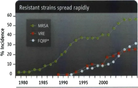
Figure 1-1: Methicillin-resistant S. aureus and vancomycin-resistant Enterococcus are two resistant pathogens spreading rapidly. Figure adapted from (12) ... 19 Figure 1-2: Rise in resistance strains. The green curve shows the increase in methicillin resistance
nosocomial Staph. infections. Less than 3% of those infections were resistant to methicillin by
1980, whereas today over 60% are resistant to methicillin and other commonly used
antibiotics. The red and silver curve shows the rise in vancomycin-resistance enterococcus infections and fluoroquinoline-resistant Pseudomonas aeruginosa. Figure adapted from (12).
... 2 0
Figure 1-3: Rise in mortality. Number of death certificates mentioning Staphylococcus aureus by methicillin resistance in England and Wales. The number of death certificates mentioning Methicillin-resistant Staphylococcus aureus (MRSA) decreased to 1,593 in 2007 and then decreased further to 1,230 in 2008, a fall of 23 per cent. The number of death certificates in England and Wales mentioning Staphylococcus auras (including those not specified as resistant) was 1,500 in 2008, a decrease of 27 per cent compared to 2007...21 Figure 1-4: Timeline of antibiotic drug discovery and new antibiotics approved by FDA. Only two new
classes of antibiotics have been discovered in the past 50 years and the number of new antibiotics approved by the FDA has been decreasing continually since the 1980s. Figure
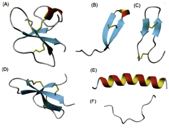
a d a p te d fro m (1 2 )...2 3 Figure 2-1: Structural classes of AmPs. (A) Mixed structure of human P-defensin-2; (B) looped thanatin
(C)
p-sheeted
polyphemusin; (D) rabbit kidney defensin-1; (E) a-helical magainin-2; (F) extended indolicidin. The disulfide bonds are indicated in yellow, and the illustrations have been prepared with use of the graphic program MolMol 2K.1. Figure adapted from Jenssen2 0 0 6 ... 2 6
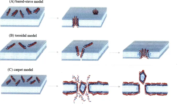
Figure 2-2: Proposed models of AmPs induced killing. (A) In the barrel-stave model, the AmPs insert perpendicularly into the membrane bilayer. (B) In the toroidal model, the AmPs induce the lipid monolayers to bend continuously through the pore. (C) In the carpet model, the peptides align parallel to the surface and disrupt the membrane. Hydrophilic regions of the peptide are shown colored red, hydrophobic regions of the peptide are shown colored blue. Figure
adapted from B rogden 2005 (22)...28 Figure 3-1: Engineered bacteriophages expressing lytic proteins. Upon initial infection, bacteriophages
multiply and express lytic agents in the bacterial host. New phages progeny and lytic agents are released upon cell lysis. The phage system then enters a continued infection cycle leading
to the complete eradication of the bacterial culture where phage resistant bacteria are su p p ressed by lytic agents...34
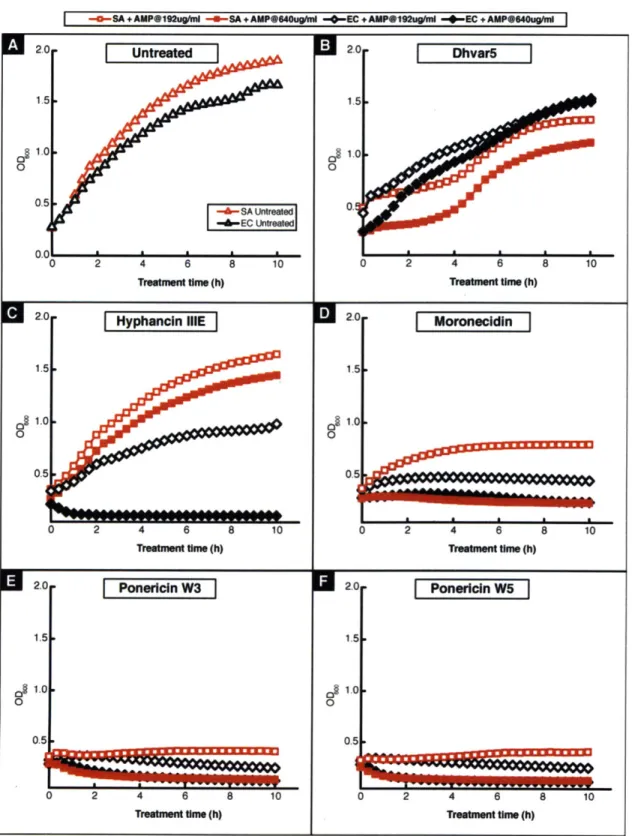
Figure 3-2: In vitro bactericidal activity of selected AmPs was measured against E. coli (EC) and S. aureus
(SA) at an intermediate concentration of 192 g/ml and a high concentration 640ug/ml and
compared to (A) the growth profile for the untreated culture. All experiments were carried o u t in ... 4 1
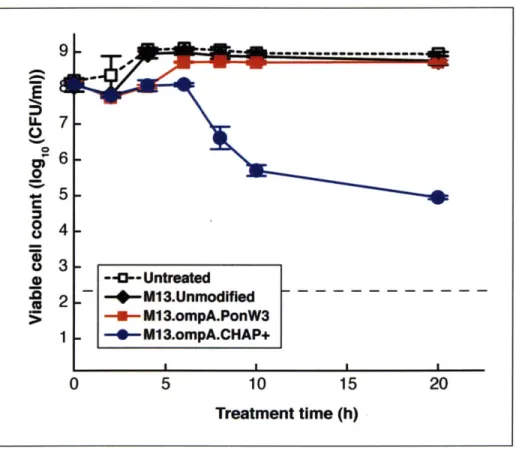
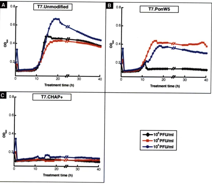
Figure 3-3: Viability and time course treatment of E. coli EMG2 cultures with 108 PFU/ml of unmodified M13 phage or engineered phage expressing Ponericin W3 or CHAP+. Treatment with phage expressing CHAP+ resulted in a long-term decrease in viable cell counts by 10,000-fold compared to treatment with the unmodified M13 phage. All experiments were carried in triplicates with the standard error shown. The horizontal dotted line denotes the detection lim it of our viable cell count assay...4 3 Figure 3-4: Optical density and time course treatment of an E. coli BL21 culture with unmodified T7
phage and engineered T7 phage expressing Ponericin W5 and CHAP+ with an initial phage count of 108, 106, and 104 PFU/ml. (A) Treatment with unmodified T7 phage results in a rapid drop in optical density resulting from the natural lytic action of the bacteriophage. However, after 10 hours of treatment the culture is able to regrow due to bacterial resistance to T7 regardless of the initial phage concentration. (B) Treatment with T7 phage expressing Ponericin W5 is able to overcome the cell regrowth for initial phage counts at 108 PFU/ml or above. (C) Treatment with T7 phage expressing CHAP+ completely suppresses regrowth of resistant cells starting at phage counts of 104 PFU/ml or above thereby resulting in long-term suppression of the bacterial culture...4 5 Figure 3-5: Viability and time course treatment of E. coli BL21 cultures with 104 PFU/ml of unmodified
T7 phage or phage expressing Ponericin W5 or CHAP+. Treatment with phage expressing CHAP+ resulted in a long-term decrease in viable cell counts by 10,000-fold compared to
treatment with the unmodified T7 phage, resulting in a viable cell count below our assay detection limit of 200 CFU/ml. All experiments were carried out in triplicates with the
standard error shown. The horizontal dotted line denotes the detection limit of our viable cell co u n t a ssay ... 4 7 Figure 4-1: Schematic of layer-by-layer films and the time-release profile of therapeutic drugs. Figure
adapted from Ch uang 2 00 7 (7)...6 0
Figure 4-2: Schematic of alternating layer-by-layer assembly to create thin films. The substrate is dipped successively in a polycation and polyanion solution resulting in the build up of a film of alternating polymer layers. Figure adapted from Chuang 2007 (7)...61 Figure 4-3: Structure of the polycations and polyanions used to build the LbL films. Three different
polyanions and four polycations with carboxyl (hyaluronice acid and algenic acid) and sulfonate groups (chondroitin sulfate and dextran sulfate) were studied to obtain large diversity in the LbL films constructs and span a range of affinity for the AmPs. ... 63
Figure 4-4: Melittin release from [(Poly1/HA)(Mel/HA)]n films (n= 20 or 70). Melittin release from the films follows a burst profile in the first 8 hours followed by a sustained linear release for the
following 8 days. The amount of melittin released from the film constructed with 70 layers is 5-tim es higher than that released from the 20-layers film ... 66 Figure 4-5: Dermaseptin and melittin release from [(Poly1/HA)(Mel/HA)]70 films. The films were able to
incorporate 160ug/cm2 of dermaseptin and 73ug/cm2 of melittin, which correspond to a constant Am P surface loading of 40-6Onm ol/cm 2 ... ... .... .... .... ... .... ... . . . 67
Figure 4-6: Co-release of dermaseptin with bFGF underlayer. The growth factor underlayer allows for diffusion of the AmP and slows down the burst release profile of the AmPs thereby extending the realease tim e from the LbL coating. ... 69
Figure 4-7: Antimicrobial activity of the released media. Ponericin G1 was released from
[(Polyl/HA)(Pon/HA)]1oo and the ponericin content was estimated using BCA. The released media prevented the growth of S. aureus when the ponericin content reached 22 ug/ml...70
Figure 4-8: MC3T3 mammalian toxicity assay. Ponericin had little effect on metabolic activity of the
MC3T3 cell up to 64ug/ml after 2 days of exposure. Metabolic activity was reduced to 50%
after 4 days of exposure to ponericin concentrations up to 128ug/ml and reduced to 20% above for concentration up to 512ug/m l... 71 Figure 5-1: Highly conserved patterns in cecropins. Alignment of cecropins from a variety of organisms
reveals a highly conserved pattern. This pattern can be represented by a motif, the bottom sequence. In the motif, bracketed terms indicate a specific set of amino acids that may...76
4.2: Representation of a gram m ar based search space... 77 Figure 5-2: Representation of a motif based search space. Sequence space for a typical 20 amino acid AmP contains approximately 1026 sequences; only hundreds of these AmPs are known. The motif-based design of AmPs focuses on peptides inside the "motif space," while allowing deviation from natural AmP sequences. This allows the design peptides that show no significant homology to any naturally occurring sequences, but have the desired antimicrobial
properties. Figure adapted from Jensen 2006 ... 77 Figure 5-3: Sample results of a Teiresias search. Patterns which meet an L/W of 6/6, 4/7, and 9/13 are
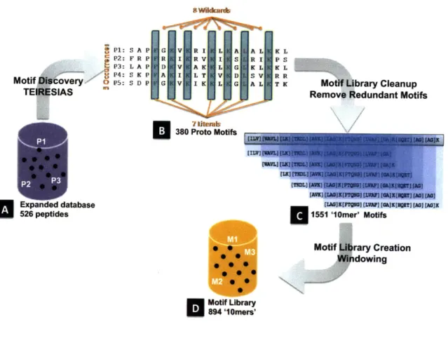
shown. An L/W of 4/7 means that in a sliding window of 7 characters, at least 4 must be uniquely specified (not brackets or wild card). Figure adapted from Jensen 2006...79 Figure 5-4: Creation the motifs library. (A) TEIRESIAS motif discovery was applied to the Nebraska
Antimicrobial Peptide Database containing 526 natural AmPs and other previous designed AmPs. (B) 380 proto-motifs with 5 occurrences in the database and containing a minimum of
7 literals and a maximum of 8 wildcards are identified. (C) The proto-motifs were then cut
into 1551 motifs of size 10 amino acids using a sliding window. (D) Duplicate entries were removed to constitute the complete motif library of 894 10-mer motifs...82
Figure 5-5: Assigning peptides activity to motifs. (A) The complete peptide and motif database was restricted to only short, linear peptides with less 40 amino acids to make up (B) the starting peptide database of 163 and motif library of 667. (C) each of these 163 peptides was synthesized and the antimicrobial activity measured against 4 different pathogenic
organisms. (D) The content matrix T relates associates peptides from the database to motif from the library. Each rows of the matrix represents one of the 667 motifs identified and each column represents one of the 163 peptides tested. A blue dot indicates that the motif is present in peptide. (E) The peptides were then sorted by decreasing activity level and partitioned in three activity zones: strong peptides in green with MIC <32ug/ml, medium peptides in yellow with MIC between 32ug/ml and 128ug/ml, and weak peptides in red with
M IC > 1 2 8 u g / m l...8 4 Figure 5-6: Motif scoring and activity zone assignment. (A) The sorted T matrix contains a total of 7
strong, 39 medium and 117 weak peptides. (B) Motif M234 for example is found in 2 of the 7 strong, 4 of the 39 medium and 6 of the 117 weak peptides. The motif's respective
representation is given by the ration where it is found. (C) The score of the motif is the adjusted ratio computed to account for the uncertainty introduced by the different size of each activity zone. The adjusted ratio accounts for sample size uncertainty in the motif score.
p is the observed unadjusted ratio, z,/2 is the (1-a/2) quantile of the standard normal distribution, and n is the total number of observations. (D) The score of the motif for each activity zone is computed and (E) the motif is assigned to the activity zone with the highest score. The motifs are then ranked according to their activity scores and the highest-ranking motifs are selected to form the motif basis function to design new peptides. 25, 75, and 150 motifs were selected to form the basis function for the strong, medium and weak zone
re sp e ctiv e ly ... 8 6
Figure 5-7: Algorithm validation. (A) The database of 163 tested peptides is divided into (B) a training set of 83 peptides used to train the algorithm and (C) assign motifs to a particular activity zone as described in Figure 5-5: .The remaining peptides constitute (D) a testing set of...88 Figure 5-8: Tiling of motifs to created parent peptide. In this example, four 6-mer motifs are tiled to form
an 8-mer parent peptide. All 72 instances of the 8-mer parent peptide are enumerated by picking one amino acid from the choice in the brackets for each location...90 Figure 5-9: MIC activity distribution for (A) database of existing peptide, (B) designed peptides, (C) their
shuffled peptides equivalent; and (D) the blind peptide designs from (143)...100 Figure 5-10: MIC activity distribution for the designed peptides against the different pathogens tested. In
green are strong peptides with MICs32ug/ml, in yellow medium peptides with...101 Figure 6-1: Kill curve for (A) bacteriostatic, (B) rebounding and (C) bactericidal ponericins against E. coli
a n d S. a u re u s...1 1 2 Figure 6-2: Kill curve for natural and amidated ponericin G1, W 3 and W 6...113
Figure 6-3: Imaging using high-speed atomic force microscopy shows E. coli cell disruption induced by th e A m P C A M ... 1 1 5
Figure 6-4: Luminscence reading of Xen14 luminescent E.coli subjected to XXug/ml of cecropin A melitin. The immediate drop in luminescence caused by disruption of the cell membrane Effect on intracellular ATP concentration and preventing the ATP driven cell luminescence...117 Figure 6-5: Imaging using a microfluidic flow chamber shows resistance to Ponericin G1 and cidal action
of Ponericin W3. Treatment with Ponericin W3 at 1oX MIC induces a membrane disruption manifested by a morphological change starting within 10min and affecting all the cells in the reactor. The cells are no longer able to divide and multiply resulting in the cidal effect of the peptide (Panel A). The morphology change is manifested through deflation of the cells and surface corrugation at the cell membrane. For cells disrupted during division, the septal ring at the centerline is even visible (Panel C). Treatment with Ponericin W3 affect some cells that become corrugated and cannot grow while others appear not affected and colonized the entire m icrofluidic cham ber (Panel B and D)...120 Figure 6-6: Optical density of culture of CpxA- knockouts challenged with 1OX MIC of PonG1, PonG5 and
PonW3. CpxA- knockouts show CpxAR system play an important role in the cell's ability to adapt and derive resistance to A m Ps...123 Figure 6-7: Survival of DpiA and DpiB knockouts challenged with 1OX MIC of PonG1, PonG5 and PonW3.
... 1 2 5
Figure 6-8: Survival of RecA knockouts mutants challenged with 1OX MIC of PonG1, PonG5 and PonW3.
... 1 2 8
Figure 6-9: Gene expression levels rebounding amidated Ponericin G1-NH2 ... 136
Figure 6-10: Gene expression m ap for cidal natural Ponericin G1...136 Figure 6-11: Synergistic effect for PonG1 and Kan. Ofloxacin resistant E.coli cells were treated with
combination of ofloxacin or kanamycin with (A) PonG1 or (B) PonG1-NH2. PonG1 and kanamycin act synergistically by both targeting the ribosome and the combination treatment leads suppression of resistant culture by over 300-fold compare to single treatment with either kanamycin or PonG1 alone. Combination of PonG1 and ofloxacin or PonG1-NH2 and kanamycin also suppresses the bacterial culture although to a lesser extend...144 Figure 7-1: Mechanism of action of enhanced bacteriophages and antibiotics. The bacteriophages (blue)
in combination with antibiotics (red and blue capsule) attack the bacterial cell. The
bacteriophages infect the cell and force production of four entities: 1) more bacteriophages; 2) repressors of bacterial DNA damage repair mechanisms (stop sign); 3) biofilm-degrading enzymes (pac-man); and 4) broad-spectrum antimicrobial peptides (screw)...158 Figure 7-2: Novophage enhanced bacteriophage prevents emergence of antibiotic resistant bacteria. Lane
number of mutants is 1600; lane 3 is natural bacteriophage treatment plus ofloxacin; and lane 4 is lexA3-enhanced bacteriophage plus ofloxacin treatment. The number of...160 Figure 7-3: Novophage enhanced bacteriophage in combination with antibiotic increases survival of mice
in a bacteremia model. Five day survival of mice in an intraperitoneal E. coli...161 Figure 7-4: Enhanced bacteriophage treatment of biofilm reduces the number of live bacteria on and
within the biofilm. A. Untreated biofilm of E. coli where only the top layers of cells are visible; the lower layers of cells are shielded. B. Biofilm treated with DispersinB -enhanced
bacteriophage. No clear and defined E. coli cells are visible and very few viable cells were recovered; the residue is cellular debris...162 Figure 7-5: M anufacturing batch process for bacteriophages...169
List of tables
Table 2-1: Selected characteristics of AmPs vs. traditional antibiotics ... 30
Table 3-1: Minimum Inhibitory Concentration (MIC) of selected panel of AmPs measured against E. coli and S. aureus. HCso denotes the 50% hemolytic concentration indicative of the peptide's to x ic ity ... 4 0 Table 4-1: Am Ps incorporated in the LbL film s... 61
T ab le 5 -1: Fin al p arent p ep tide ... 9 1 Table 5-2: Activity table of the 163 tested natural AmPs. ... 95
Table 5-3: Hemolytic activity and MIC of designed and shuffled peptides against E. coli, P. aeruginosa, S. epiderm idis, S. aureus and M R SA ... 99
Table 5-4: Therapeutic index (TI) of active designed peptides. The therapeutic index is calculated as the ratio between the MIC antimicrobial concentration and HCso hemolytic concentration.103 Table 6-1: MIC (ug/ml) and bactericidal activity of natural and amidated ponericins against E. coli (EC) a n d S . au reu s (SA ) ... 1 1 0 Table 6-2: Z-score change for amidated PonG1-NH2 ... 132
Table 6-3: Z-score change for natural PonG 1...135
Table 6-4: Z-score change of iron import related gene upon treatment with PonG1-NH2...137
Table 6-5: MNI output showing the 100 highest ranked putative gene targets. The putative targets underlying the mode of action of PonG1-NH2 and PonG1 are shown for each time point. Pathway analysis was then performed on the MNI gene list using Gene Ontology enrichement and the gene associated with the identified pathways are shown in red (refer to Section 6.7.4 for details on GO enrichm ent analysis)... ... ... 138
Table 6-6: Amidated PonG1-NH2 Gene Ontology enrichment following MNI analysis...140
Table 6-7: Natural PonG1 Gene Ontology enrichment following MNI analysis ... 142
Chapter 1. Critical need for new antibiotics
1.1. The rise of antibiotic resistant infections
Antibiotic-resistant microbes infect more than 2 million Americans and kill over 100,000 each year
(1). They are one of the biggest and fastest growing clinical problems today (2) and constitute a global
threat that has the potential to wipe out many of the major health benefits that have occurred over the last century (3).
These infections pose a risk to everyone from infants, to college athletes, to elderly in nursing homes
and hospitals. For example, the 2009 MRSA outbreak at Beth Israel Hospital maternity ward infecting
over 30 mothers and infants. In 2008, New England Patriot's quarterback Tom Brady contracted an
MRSA during a knee surgery and required several IV antibiotics treatment to eradicate the infection, which ended his football career for the year. Antibiotic resistant infections have spread beyond the
hospital settings and now threaten communities. Nationwide outbreaks have already occurred shutting
down in schools, and athletic events while claiming many lives. MRSA alone kills more Americans than
AIDS, Parkinson's disease or homicide. (4)
The populations with the highest risk of mortality following infection are infants and young children,
elderly and immuno-suppressed patients (5). These groups are at increased risk as a result of a
suppressed immune system, increased exposure to numerous pathogens, or because of restriction on the
antibiotics they can be administered. Thus, infections are the fourth most common cause of death for
children in their first year (6), are the most common cause of hospitalization for older adults, and are
responsible for 30 percent of the deaths in senior citizens (7). Patients with particular medical conditions
healthiest individual becomes exposed to antibiotic-resistant pathogens every time they enter hospitals, even for the most benign interventions (8).
Infections are not only a health problem but also an economical one. Resistance to antibiotics
significantly extent the length of hospital stay typically by 6 additional days and increases the total cost of
treatment by $24,000 per patient (9). John Paul, retired CFO of the University of Pittsburgh Medical
Center explains that the ability to control hospital-based infections would add tens of millions of dollars
straight to a medical center's bottom line (10).
Consequently, there is an unmet need to develop new antimicrobial solutions that decrease morbidity
and mortality in patients, to reduce hospital costs or to prevent and control nationwide outbreaks.
1.2. A multifaceted problem
The problem of antibiotic resistance in bacterial infections is multifaceted and the reasons for their recent rise are multiple. Firstly, the sheer number of infections has increased rapidly by 200% in the last
5 years, with over 2 million cases and 99,000 deaths per year in 2007 (11).
Figure 1-1: Methicillin-resistant S. aureus and vancomycin-resistant Enterococcus are two resistant
pathogens spreading rapidly. Figure adapted from (12).
Secondly, these infections are becoming increasingly difficult to treat. Figure 1-2 shows the rapid
spread of resistant strains. Methicillin resistance in staphyloccal infections (MRSA), one of the most . ....
common hospital pathogen, rose from just 3% in the 1980s to over 60% by 2007 (12). Over 30% of hospital infections today are due to infections resistant to all but one antibiotic, vancomycin which is
considered to be the last resort antibiotic (13). Already, cases of high-level resistance to vancomycin are
being reported worldwide. Many physicians worry that if these strains were to spread like MRSA has,
death rates would likely increase to pre-antibiotic era at greater than 50% (14).
60 50 c 40 40 $20 10 0
1980
1985
1990
1995
2000
*RuoroquinoAine-resistant Pseudomonas aeruginosa
Figure 1-2: Rise in resistance strains. The green curve shows the increase in methicillin resistance nosocomial Staph. infections. Less than 3% of those infections were resistant to methicillin by 1980, whereas today over 60% are resistant to methicillin and other commonly used antibiotics. The red and silver curve shows the rise in vancomycin-resistance enterococcus infections and
fluoroquinoline-resistant Pseudomonas aeruginosa. Figure adapted from (12).
Antibiotic resistance in bacterial infections result in increased morbidity and mortality in patients
(15). A study by the Duke University medical center shows that patients infected by methicillin resistant S. aureus were seven times more likely to die and 35 times more likely to be readmitted within 90 days
than those infected by S. aureus sensitive to methicillin (9). Figure 1-3 shows the increase in number of
death either directly caused or correlated with the methicillin resistant infections in England and Wales.
Number of deaths 1,800 1,600 1,400 1,200 1,000 800 600 400 200 0 1993 1995 1997 1999 2001 2003 2005 2007
mUnderlying
cause EMentionsFigure 1-3: Rise in mortality. Number of death certificates mentioning Staphylococcus aureus by methicillin resistance in England and Wales. The number of death certificates mentioning
Methicillin-resistant Staphylococcus aureus (MRSA) decreased to 1,593 in 2007 and then decreased further to 1,230 in 2008, a fall of 23 per cent The number of death certificates in England and Wales
mentioning Staphylococcus auras (including those not specified as resistant) was 1,500 in 2008, a decrease of 27 per cent compared to 2007.
Finally, the threat of antibiotic resistant bacteria is further accentuated by a decline in the
development of new antibiotics. Although the $25B global market for antibiotics is attractive, drugs for chronic diseases offer much greater return on investments says Steve Projan, vice president of biological technologies at Wyeth. Pharmaceutical companies developing new antibiotics are also faced with a Catch-22; the better their antibiotic, the less health experts want to see it used to avoid the development of
resistance. This led to an exodus of pharmaceutical companies from antibiotic development starting in
the 1980s. Of the 15 major pharmaceutical companies that once had flourishing antibiotic discovery
programs, only five - GlaxoSmithKline, Novartis, AstraZeneca, Merck, and Pfizer - still have antibiotic
discovery efforts. As a result there has been a decrease in the number of new antibiotics available to
clinical doctors to treat infections and many researchers also suggest that the intracellular metabolic ...
... INNIM - M ONRO E .....................
targets for antibiotic discovery are being depleted (16). Figure 1-4 shows the decrease in the number of
antibiotic products approved by the FDA over time (12). These antibiotics actually only constitute two new classes of antimicrobials discovered since the 1980s - oxazolidinones (linezolid) and cyclic
lipopeptides (daptomycin). All of these new classes of antibiotics target only Gram positive bacteria and
no new Gram-negative antibiotics have been developed since quinolone in the 1960s (17). Gram-negative
bacteria tend to be harder to kill because of their cell membrane which the antibiotics needs to penetrate
and additional defense mechanisms such as shutting down protein channels through which the antibiotic
penetrates or activating efflux pump to excrete the antibiotics.
+sdanmd G*mcidinqpedde)w~cpa4~qn~n. oepi e Peniiin (M~actangj
e Neomycn (aminog oside)
rmen c0 ecepOtosporin (04actam) (anycin) (XatM
hoe
Poos (1950-1960) & hcai atnyi
suttoam d) -cwhloamheiol(pheny pan x4 kn
.
~n
l Mntpopepide) * Eryhromycin maceoude) Vwaianide acriooprmya (qioln) ) Eoepd) 16 14 12 10 8 6 4 2 0 1983-1987 1988-1992 1993-1997 1998-2002 2003-2007 FD;A OW WFigure 1-4: Timeline of antibiotic drug discovery and new antibiotics approved by FDA. Only two new classes of antibiotics have been discovered in the past 50 years and the number of new antibiotics approved by the FDA has been decreasing continually since the 1980s. Figure adapted from (12).
1.3. Thesis objective and organization
This thesis aims at providing an advance in the development of new antimicrobial therapies. The
work explores the use and the design of novel antimicrobial peptides and engineered bacteriophages to treat bacterial infections. The thesis is organized into the following six chapters:
" Chapter 2. Antimicrobial peptide overview - provides background and review on antimicrobial peptides, the central element of this thesis and introduce them as new human antimicrobial
therapeutics.
" Chapter 3. Engineered bacteriophages expressing AmPs - offers an approach to deliver genes expressing antimicrobial peptides and other lytic antimicrobial agents using bacteriophages.
* Chapter 4. Controlled delivery of AmPs from LBL surfaces - describes an approach for the controlled release of antimicrobial peptides using layer-by-layer biodegradable polymer films
coated on the surfaces of implantable devices.
- Chapter 5. Motif-based rational design of AmPs - discusses advances made in the design of novel peptides using conserved sequences founds in natural AmPs.
* Chapter 6. Mechanism and potentiation ofAmPs - gives insight on the mechanism of antimicrobial peptides, their intra- and extra-cellular targets, and outlines an approach to potentiate AmP-based therapeutics.
* Chapter 7. Therapeutic opportunities for engineered bacteriophages (PhDCEP Capstone) - describes a market entry and expansion strategy for an antimicrobial start-up company commercializing
1.4.
References
1. B. Spellberg, Rising Plague: The Global Threat from Deadly Bacteria and Our Dwindling Arsenal to
Fight Them. (2009).
2.
J.
Katz, Oral communication, (2009).3. M. P. M. Shnayerson. The Killers Within: The Deadly Rise of Drug-Resistant Bacteria (2002). 4. R. M. Klevens et al., in Public Health Rep. (2007), vol. 122, pp. 160-6.
5. K. F. Shea, M., Barlam,
J.,
Environmental Defense, 1 (Mar 12, 2002).6. Gotoff, Infections of the Neonatal Infant. R. M. K. Behrman, and H.B. Jenson, Ed., Nelson Textbook
of Pediatrics (2000).
7. M. S. a. D. Kaye, Infectious Disease Clinics of North America, 357 (2000).
8. R. P. W. a. M. B. Edmond, New England
Journal
of Medicine 343, 1961 (2000). 9. D.J.
Anderson et al., PLoS ONE 4, e8305 (2009).10. D. L. C. Loose,
J.
Moxley, Stericoat Business Plan.11. K. Krul, Kalorama Information, (2009).
12. G. Taubes, Science 321, 356 (July 18, 2008, 2008). 13. C. Loose, MIT (2007).
14. Staph Bacteria. Science Blog (Rockefeller University, 1997), vol. June 11.
15. http://www.statistics.gov.uk/cci/nugget.asp?id=1067. (Office for National Statistics).
16. G. D. Wright, Nature Reviews Microbiology 5, 175 (2007).
17. M. S. Daniel Becker, Claudia Rollenhagen, Matthias Ballmaier, Thomas F. Meyer, Matthias Mann and Dirk Bumann, Nature, 303 (2006).
Chapter 2. Overview of antimicrobial peptides
2.1.
Executive summary
Antimicrobial peptides are part of the innate immune systems of many living organisms and serves
as a first line of protection against invading bacterial, viral or fungal infections. They represent an
exciting potential new class of antibacterial therapies as their novel mode of action prevents bacteria
from easily evolving resistance to AmPs. AmPs are small cationic amphipatic peptides between 15 and
30 amino acids that typically form alpha helices ad intercalates within the bacterial cell membrane to
form pores; eventually leading to cell lysis. In order for bacteria to evade the action of AmPs, they are
required to significantly alter their cell membrane composition, which drastically decreases their
viability. For these reason, resistance to AmPs is uncommon which constitutes one of their greatest
advantage of the tradition small-molecule antibiotics used in hospitals today.
2.2.
Antimicrobial peptides as antibiotics
Antimicrobial peptides (AmPs) are an exciting potential new class of antibiotic because their unique
mode of action is unlikely to introduce drug resistance. AmPs are typically short peptides composed of 15
to 30 amino acids that have strong antimicrobial properties. AmPs are an important component of the
innate defense mechanism of many living organisms where they constitute a first non-specific line of
defense against invading pathogens
(18). For example, many AmPsare been isolate from skin secretions
of, such as dermaseptin from the South American frogs, where they protect against pathogens in the
natural living environment. AmPs, such as histatins from human saliva or magainin from the Xenopus
laevis frog, play a role in protection from ingested pathogens. Cathelicidins, another family of AmPs, have
been isolated from many mammalian species, such as mice, rabbits, sheep, horses, and humans. Defensins
from human neutrophils are found inside the host body indicating varied biological roles in host defense.
To date, more than 800 AmPs have been reported. Their range of antimicrobial activity is unusually
broad as their primary role is non-specific. AmPs have been found to be active against Gram-positive (e.g.
S. aureus) and Gram-negative (e.g. E. coli) bacteria, viruses (e.g. HIV, Herpes virus), protozoa (e.g. T.
brucei) and fungi (e.g. C. albicans). More recently, they have been shown to bind selectively and prevent
the growth of cancer tumors (19) and also to play an important role in recruiting the innate immune
systems (20, 21).
(A)(B) (C)
(D) (E)
(F)
Figure 2-1: Structural classes ofAmPs. (A) Mixed structure of human f-defensin-2; (B) looped thanatin (C) #-sheeted polyphemusin; (D) rabbit kidney defensin-1; (E) a-helical magainin-2; (F) extended indolicidin. The disulfide bonds are indicated in yellow, and the illustrations have been prepared with use of the graphic program MolMol 2K.1. Figure adapted from Jenssen 2006.
-In contrast with their enormous diversity in activity, AmPs share several common characteristics and are generally categorized into four structural classes: a-helix,
p-sheet,
loop or extended structure (Figure2-1). These short peptides are predominately positively charged. AmPs typically assume an amphipatic
three-dimensional structure where, for a-helix peptides, the hydrophilic positively charged residues are
localized on one side of the helix and the hydrophobic residues are on the other side. This amphipatic
structure and the net positive charge give AmPs an affinity for bacterial membranes over eukaryotic
membranes. AmPs selectively coalesce and bind to bacterial membrane leading to membrane permeation
and the death of the bacteria through cell lysis. While the exact mechanism of action by which killing occurs is not clearly understood, several models have been proposed (22) and are explained below.
2.3.
Mode of action of AmPs
One model describing the killing mechanism of AmPs is the barrel-stave model shown in Figure
2-2A. The AmPs aggregate and insert perpendicularly into the membrane bilayer so that the hydrophobic
peptide regions align with the lipid core region and the hydrophilic peptide regions form the interior
region of the pore. In a second model, the toroidal model shown Figure 2-2B, the AmPs aggregate and
induce the lipid monolayers to bend continuously through the pore so that the aqueous core is lined by both the inserted peptides and the lipid head groups. In this model, the peptides are always associated
with the lipid head group even when they are perpendicularly inserted in the bilayer, which is not the case in the barrel-stave model. Finally, in the carpet model in Figure 2-2C, the peptides disrupt the membrane by orienting parallel to the surface of the lipid bilayer and forming an extensive layer or carpet. Some AmPs do not induce cell lysis but instead they permeate through the membrane and kill the
bacteria by acting on intracellular targets (23). Such AmPs have been shown to inhibit DNA, RNA and
protein synthesis. Inhibition of enzymatic activities and of the formation of structural components, such
Because the uptake of AmPs is self-promoted -- based on peptide charge-- and non-specific, bacteria are less likely to develop resistance to AmPs since it would require the bacteria to completely
change the properties and structure of their membranes (20). As a result, AmPs have been killing bacteria
for millions of years, yet bacteria have developed very little resistance and the few resistant strains have
significantly reduced viability because of the numerous changes introduced to their membranes (24).
(A) barrel-stave model
(B) toroidal model
(C) carpet model
Figure 2-2: Proposed models of AmPs induced killing. (A) In the barrel-stave model, the AmPs insert perpendicularly into the membrane bilayer. (B) In the toroidal model, the AmPs induce the lipid monolayers to bend continuously through the pore. (C) In the carpet model, the peptides align parallel to the surface and disrupt the membrane. Hydrophilic regions of the peptide are shown colored red, hydrophobic regions of the peptide are shown colored blue. Figure adapted from Brogden 2005 (22).
2.4.
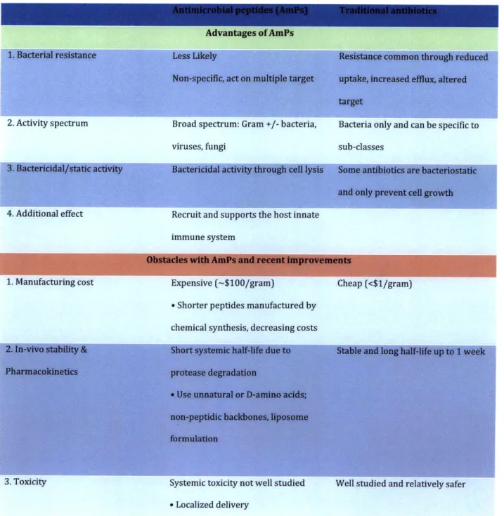
AmPs compared to traditionnal antibiotics
Several characteristics make AmPs particularly attractive compounds to develop as novel
antibiotics (20). Firstly, because AmPs are non-specific and act on multiple targets, bacteria are less ...
. ... ... .................................................. .............................. . .. .... .. .. ...
likely to develop resistance to them. On the other hand, traditional antibiotics typically act on single metabolic or intracellular targets, which the bacteria can modify to evolve antibiotic resistance. Gram
negative bacteria also posses additional defense mechanism such as the ability to modulate their protein
channel to reduce the uptake of antibiotic or to activity efflux pump to secrete and reduce intracellular
antibiotic concentration. Secondly, AmPs exhibit a broad spectrum of activities including against
Gram-negative, Gram-positive, multi-drug resistant bacteria and non-bacterial target such as viruses, fungi, or
cancerous cells. Traditional antibiotics are only efficacious on bacteria and are sometimes specific to
sub-classes of bacteria. For example, vancomycin is active only against gram-positive bacteria as it inhibits
the biosynthesis of cell wall, which gram-negative bacteria do not posses. Thirdly, AmPs kill bacteria in a
rapid and bactericidal manner through cell lysis where as some antibiotics are bacteriostatic and only
prevent the growth of bacteria with out actually killing them. Finally, AmPs have the additional effect of
boosting and recruiting the overall innate immune system of the host (25). These properties give AmPs
great advantages over traditional antibiotics.
The main obstacles for the clinical development of AmPs are their high manufacturing cost, their short in-vivo lifetime due to protease degradation, and a general lack of understanding of their systemic
toxicity. However, recent scientific and technological improvements have addressed some of these issues.
Improvement in solid phase chemical synthesis of custom peptides has driven down the manufacturing cost of a typical 20-mer peptide from $500 to $30 per peptide (26). Other synthesis methods such as recombinant production have been developed for larger size manufacturing and are continuously improved upon. Researchers were also able to improve the stability of AmPs to protease degradation by
introducing unusual or D- (rather than L-) amino acids, the use of non-peptidic backbones, and special
formulation such as in liposomes (25). Finally, the concern about systemic toxicity can be addressed by
developing topical applications for AmPs or through local drug delivery methods such the immobilization
Table 2-1: Selected characteristics of AmPs vs. traditional antibiotics
2. Activity spectrum Broad spectrum: Gram +/- bacteria, Bacteria only and can be specific to
viruses, fungi sub-classes
4. Additional effect Recruit and supports the host innate
immune system
1. Manufacturing cost Expensive (-$100/gram) Cheap (<$1/gram)
- Shorter peptides manufactured by
chemical synthesis, decreasing costs
3. Toxicity Systemic toxicity not well studied Well studied and relatively safer
- Localized delivery
2.5.
References
1. M. Zaiou, inJ Mol Med. (2007), vol. 85, pp. 317-329.
2. Lehmann et al., in European Urology. (2006), vol. 50, pp. 141-147.
3. A. Marr, W. Gooderham, R. Hancock, in Current opinion in pharmacology. (2006).
4. N. Mookherjee, R. Hancock, in Cell. Mol. Life Sci. (2007). 5. K. Brogden, in Nat Rev Micro. (2005).
6. H. Jenssen, P. Hamill, R. E. W. Hancock, in Clinical Microbiology Reviews. (2006), vol. 19, pp.
491-511.
7. H. G. Boman, injounral of Internal Medicine. (2003), pp. 197-215.
8. R. E. W. Hancock, H.-G. Sahl, in Nat Biotechnol. (2006), vol. 24, pp. 1551-1557.
Chapter 3. Engineered bacteriophages express antimicrobial peptides
3.1.
Executive summary
The rise of antibiotic resistant infections has led to a renewed interest in alternative treatments
such as bacteriophage therapy. However, the therapeutic use of bacteriophage has been contested, as bacteria are able to rapidly evolve resistance to bacteriophages. In this work, we engineered
bacteriophages to overexpress antimicrobial peptides and lytic enzymes to enhance their cidal activity.
We demonstrate that M13 lysogenic bacteriophages expressing the lytic CHAP+ enzyme result in a
10,000-fold decrease in viable cell counts relative to the unmodified phage and the effect is maintained
for over 20 hours. Engineered T7 lytic bacteriophages expressing CHAP+ lead to long term suppression of
bacteria culture and prevent the evolution to bacterial resistance to bacteriophages. This work
establishes an engineering approach to confer new functionalities to bacteriophages and enable them to eradicate difficult-to-treat bacterial infections.
3.2.
Introduction
3.2.1. Bacteriophages and their therapeutic potential
A naturally occurring countermeasures to bacteria are bacteriophages, viruses that infect and
multiply in bacteria. Phages specifically recognize and attach to bacterial membranes, inject their genomes and utilize the host replication system to multiply. This process subsequently leads to new
phage progeny that are released either through extrusion in the case of lysogenic phages or by inducing
cell lysis in the case of lytic phages. The therapeutic potential of lytic bacteriophages was recognized
early (27, 28) when they were successfully used in the 1920s to treat and control the spread of cholera
the development of bacteriophages in the West though some efforts have continued up to this day in the former Soviet Republic (29, 30). The spread of antibiotic resistance has led to a recent regained interest
in bacteriophage therapy (31) with several clinical trials carried out worldwide (32-36). However,
criticism for phage therapy still remains. Two primary concerns are their high specificity and the rapid
evolution of bacterial resistance (37). Modern molecular biology has now made it feasible to re-engineer
bacteriophages and express foreign proteins to address these concerns and confer new functionality.
We have previously shown that expression of a biofilm-degrading enzyme (DspB) from a T7
bacteriophage infecting E. coli increases the disruption of bacterial biofilms (38). In another model, we
showed that expression of a repressor protein of the SOS response (LexA3) from an M13 bacteriophage administered as an antibiotic adjuvant suppressed the evolution of bacterial resistance to antibiotics and
increased bacterial susceptibility (39). Both models demonstrated the feasibility of engineering
bacteriophages without compromising their natural infectivity, replication, packaging and lytic activity.
3.2.2. Engineered phages expressing AmPs and lytic enzymes
In this work, we address the concern of bacterial resistance to phages by overexpressing
antimicrobial peptides and lytic enzymes during infection and increase bacterial killing. Antimicrobial peptides were introduced in the Chapter 2. Lytic enzymes (lysins) are another new class of prospective
antimicrobials. These biologics range in size from 50 to several hundreds of amino acids, and are typically used by bacteriophages to lyse bacterial membranes and escape from their hosts (40, 41). Lysins act in concordance with holins, which permeabilize cell membranes (42) while lysins degrade peptidoglycan cell walls (43). When applied exogenously, phage lysins exhibit immediate and strong bacteriolytic
activity (44). One well-studied phage lysin is the CHAP+ protein, an optimized derivative of the LysK
staphylococcal phage K endolysin truncated to the first 165 amino acids of its active domain (45).
While promising, peptide and enzyme-based therapeutics suffer several practical challenges for
systemic application. They are subject to protease degradation when administered in-vivo. Their
their toxicity levels and potential immune reactions at required active physiological concentrations are undetermined (46-48). More practically, their cost of production and purification is still prohibitively
high compared to that of small molecule antibiotics.
We first determined the minimum inhibitory concentrations (49) and the bactericidal activities of
a panel of five AmPs. We selected the two most active AmPs candidates, Ponericin W3 and Ponericin WS, along with the CHAP+ lytic enzyme for expression in our bacteriophage system. To confer cidality to
lysogenic bacteriophages, we expressed these lytic proteins fused to an ompA secretion signaling peptide in an M13 phage infecting E. coli EMG2 K12. To further increase bacterial killing, we then combined the activity of these proteins with the innate lytic activity of a T7 bacteriophage (Figure 3-1). Through this work, we demonstrate that overexpression of broad-spectrum lytic agents increases the efficacy of
bacterial killing, prevents bacterial resistance to bacteriophages and enables the long-term suppression
of bacterial cultures for at least 40 hours.
Initial nfection with Bacteriophage replication Cell
lysis,
phage release, Regrowth ofunmnodified T7 phage and continued infection phage resistant Ceas
Initial infection with T7 phage Bacteriophage replication Cel
lysis,
phage andlyic
protein Protein induced lysis ofexpressing lytic protein and expression of lytic protein release, continued infection phage resistant cell
Figure 3-1: Engineered bacteriophages expressing lytic proteins. Upon initial infection,
bacteriophages
multiply and express lytic agents in the bacterial host New phages progeny and lytic
agents are released upon cell lysis. The phage system then enters a continued infection cycle leading
to the complete eradication of the bacterial culture where phage resistant bacteria are suppressed
by lytic agents.
3.3.
Materials and Methods
3.3.1. Synthesis of Cationic Antimicrobial Peptides
All antimicrobial peptides were synthesized by Fmoc (fluorenylmethoxycarbonyl) chemistry on
an Intavis Multipep Synthesizer (Intavis LLC, San Marcos, CA) at the Massachusetts Institute of
Technology's Biopolymers Lab core facility. Mass spectrometry was routinely used to confirm the
accuracy of the synthesis and typical purities obtained with the synthesizer were >85%.
3.3.2. Bacteriostatic of Antimicrobial Peptides
The Minimum Inhibitory Concentration (MIC) was measured using a standard assay based on the
NCCLS M26A and the Hancock assay for cationic peptides (50). Serial two-fold dilutions of peptides were
performed, starting with a base concentration ten times higher than the highest assay concentration, i.e.
at 2560[ig/ml in 0.2% Bovine Serum Albumin and 0.01% Acetic acid. E. coli and S. aureus were grown in
Mueller Hinton Broth (Becton-Dickinson, Franklin Lakes, NJ) to an OD60 0 = 0.1 - 0.3 and diluted down to
-5 x10⁵ CFU/ml in fresh MHB. Ten ptL of the peptide dilutions were incubated with 90 tL of the target in a 96-well plate (Corning Life Sciences, Lowell, MA) for 16-20 hrs. The MIC was defined as the minimum
concentration that prevented growth based on OD6 0 0.
3.3.3. Bacteriocidality of antimicrobial peptides
E. coli and S. aureus were inoculated from a -802C culture the day before the assay, into 3ml LB
plus any appropriate antibiotic, in a 14ml Falcon snap-cap tube (BD Biosciences), incubated overnight, shaking at 300rpm and 37"C. On the day of the assay the overnight stationary phase culture is diluted
1:5000 into 50ml LB + any appropriate antibiotic in a 250ml Erlenmeyer flask, shaking at 300rpm at
372C. The culture is monitored by taking samples and measuring the absorption of the culture at 600nm
to determine the optical density (OD600). The culture is grown until it reaches an OD6 0 0=0.2-0.4, but