1
Revised version
1Ms # JNC-2015-0803
2 3Involvement of endogenous antioxidant systems in the protective
4activity of pituitary adenylate cyclase-activating polypeptide
5against hydrogen peroxide-induced oxidative damages in cultured
6rat astrocytes
7Salma Douiri,* Seyma Bahdoudi,*,¥ Yosra Hamdi,* Roger Cubì,¥ Magali Basille,¥,Ω
8
Alain Fournier,π,§ Hubert Vaudry,¥,Ω,§ Marie-Christine Tonon,¥,Ω Mohamed
9
Amri,* David Vaudry¥,Ω,§ and Olfa Masmoudi-Kouki*
10
*Laboratory of Functional Neurophysiology and Pathology, Research Unit
11
UR/11ES09, Department of Biological Sciences, Faculty of Science of Tunis,
12
University Tunis El Manar, 2092 Tunis, Tunisia
13
¥Inserm U982, Laboratory of Neuronal and Neuroendocrine Communication and
14
Differentiation, University of Rouen, 76128 Mont-Saint-Aignan, France
15
ΩRegional Platform for Cell Imaging of Normandie (PRIMACEN), Institute for
16
Biomedical Research and Innovation, University of Rouen, Mont-Saint-Aignan,
17
France
2
πINRS - Institut Armand-Frappier, 531 boul. des Prairies, Laval, QC H7V 1B7,
1
Canada; Laboratoire International Associé Samuel de Champlain, Canada
2
§International Associated Laboratory Samuel de Champlain, University of Rouen,
3
Mont-Saint-Aignan, France
4 5
Address correspondence and reprint requests to Dr Olfa Masmoudi-Kouki, Laboratory
6
of Functional Neurophysiology and Pathology, Research Unit UR/11ES09, Faculty of
7
Science of Tunis, University Tunis El Manar, 2092 Tunisia. Tel: +216 71 872 600;
8
Fax: +216 71 871 666. E-mail address: [email protected]. and to Dr David
9
Vaudry, Inserm U982, Laboratory of Neuronal and Neuroendocrine Communication
10
and Differentiation, Institute for Biomedical Research and Innovation (IRIB),
11
University of Rouen, 76128 Mont-Saint-Aignan, France. Tel: +33 2 35 14 67 60.
E-12
mail address: [email protected]
13 14
Abbreviations used: H2O2, hydrogen peroxide; LDH, lactate dehydrogenase; MAPK, 15
mitogen-activated protein kinase; NBT, nitrotetrazolium blue chloride; O2°-, 16
superoxide anions; PACAP, pituitary adenylate cyclase-activating polypeptide; PKA,
17
protein kinase A; PLC, phospholipase C; ROS, reactive oxygen species; SOD,
18
superoxide dismutase.
19 20
3
Abstract
1 2
Astroglial cells possess an array of cellular defense mechanisms, including superoxide
3
dismutase (SOD) and catalase antioxidant enzymes, to prevent damages caused by
4
oxidative stress. Nevertheless, astroglial cell viability and functionality can be affected
5
by signifcant oxidative stress. We have previously shown that pituitary adenylate
6
cyclase-activating polypeptide (PACAP) is a potent glioprotective agent that prevents
7
hydrogen peroxide (H2O2)-induced apoptosis in cultured astrocytes. The purpose of 8
the present study was to investigate the potential protective effect of PACAP against
9
oxidative-generated alteration of astrocytic antioxidant systems. Incubation of cells
10
with subnanomolar concentrations of PACAP inhibited H2O2-evoked reactive oxygen 11
species accumulation, mitochondrial respiratory burst and caspase-3 mRNA level
12
increase. PACAP also stimulated SOD and catalase activities in a
concentration-13
dependent manner, and counteracted the inhibitory effect of H2O2 on the activity of 14
these two antioxidant enzymes. The protective action of PACAP against H2O2-evoked 15
inhibition of antioxidant systems in astrocytes was PKA, PKC and MAP-kinase
16
dependent. In the presence of H2O2, the SOD blocker NaCN and the catalase inhibitor 17
3-aminotriazole, both suppressed the protective effects of PACAP on SOD and
18
catalase activities, mitochondrial function and cell survival. Taken together, these
19
results indicate that the anti-apoptotic effect of PACAP on astroglial cells can account
20
for the activation of endogenous antioxidant enzymes and reduction of respiration rate,
21
thus preserving mitochondrial integrity and preventing caspase-3 expression provoked
22
by oxidative stress. Considering its powerful anti-apoptotic and anti-oxidative
23
properties, the PACAPergic signaling system should thus be considered for the
4
development of new therapeutical approaches to cure various pathologies involving
1 oxidative neurodegeneration. 2 3 4
Key words: PACAP; astroglial cells; apoptosis; catalase; superoxide dismutase;
5
oxidative stress
6
7
Running Title: PACAP prevents astrocytic cell death by activating antioxidant
8
systems
9 10
5
Introduction
1
Pituitary adenylate cyclase-activating polypeptide (PACAP) was first isolated from
2
ovine hypothalamus on the basis of its ability to stimulate cAMP formation in rat
3
anterior pituitary cells (Miyata et al. 1989). PACAP is a member of the vasoactive
4
intestinal peptide (VIP) / secretin / growth hormone-releasing hormone / glucagon
5
superfamily (Vaudry et al. 2009). Three PACAP receptors have been cloned, the
6
PACAP-selective PAC1-R and the PACAP/VIP mutual receptors VPAC1-R and
7
VPAC2-R (Vaudry et al. 2009). All PACAP receptors belong to the
seven-8
transmembrane domain G protein-coupled receptors superfamily (Harmar et al. 2012)
9
and modulate several signaling pathways including the cAMP / protein kinase A
10
(PKA), phospholipase C (PLC) / protein kinase C (PKC) and mitogen-activated
11
protein kinase (MAPK) cascade (Masmoudi et al. 2003, Ravni et al. 2006, Vaudry et
12
al. 2009). PACAP sequence has been highly conserved during evolution (Vaudry et al.
13
2009), and it is now established that this peptide is involved in the regulation of
14
important biological functions, such as neurotrophic and neuroprotective activities
15
(Seaborn et al. 2011). The antiapoptotic effects of PACAP have been reported on
16
several neuronal cell types in vitro and models of neurodegenerative diseases in vivo
17
(Seaborn et al. 2011). In particular, PACAP has been found to protect dopaminergic
18
neurons (Reglodi et al. 2006), cerebellar granule neurons (Vaudry et al. 2002) and
19
hippocampal neurons (Stetler et al. 2010) from oxidative stress-induced apoptosis.
20
Increasing evidence indicates that the neuroprotective effect of PACAP may be
21
indirect, via the release of neuroprotective factors by glial cells (Dejda et al. 2005,
22
Masmoudi-Kouki et al. 2007, Miyamoto et al. 2014, Yang et al. 2006). Moreover,
6
previous studies have revealed that part of the antiapoptotic effect of PACAP is
1
mediated through an inhibition of reactive oxygen species (ROS) production by
2
astrocytes (Masmoudi-Kouki et al. 2011, Miyamoto et al. 2014), highlighting their
3
importance in the neuroprotective activity of the peptide against oxidative stress. As
4
astrocytes are in close interaction with neurons, their protection from oxidative insult
5
appears essential to maintain brain function and to prevent neuronal damages (Barreto
6
et al. 2011).
7
It is well established that astrocytes play an important role in the protection of the
8
brain against oxidative stress damage in both physiological and pathological
9
conditions (Fernandez-Fernandez et al. 2012, Steele and Robinson 2010, Takuma et
10
al. 2001, 2004). Oxidative stress induces an imbalance in ROS production, damages
11
cellular biomolecules, impairs cellular antioxidant defenses and finally triggers cell
12
death by apoptosis (Dasuri et al. 2013, Sanders and Greenamyre 2013). Astrocytes
13
contain high levels of antioxidant molecules, such as reduced glutathione, and express
14
high level of antioxidant enzymes such as superoxide dismutases (SODs), catalase,
15
glutathione peroxidase or peroxiredoxin, which strongly contribute to protect neurons
16
from oxidative stress induced injury (Cabezas et al. 2012, Takuma et al. 2004, Zhu et
17
al. 2012). Despite their high antioxidative activities, astrocytes cannot survive and
18
protect neurons under substantial oxidative stress (Chen and Gibson 2008, Emerit et
19
al. 2004, Shibata and Kobayashi 2008). In particular, astrocytes are very sensitive to
20
hydrogen peroxide (H2O2; Ferrero-Gutierrez et al. 2008, Hamdi et al. 2011) and 21
astrocytic apoptosis, which is associated with a depletion of antioxidant activities that
22
occurs in brain injuries caused by excessive ROS production (Hsu et al. 2009, Ouyang
7
and Giffard 2004). Reciprocally, stimulation of the activity of the endogenous
1
antioxidant system or over-expression of SOD in astroglial cells is associated with an
2
increased resistance to oxidative injury (Chen et al. 2001, Hamdi et al. 2012).
3
Altogether, these observations suggest that antioxidant systems play a prominent role
4
in the protection of astrocytes from oxidative assault. As a matter of fact, we have
5
previously reported that PACAP rescues astroglial cells from the deleterious effects of
6
oxidative stress by attenuating H2O2-evoked ROS accumulation and glutathione 7
content reduction (Masmoudi-Kouki et al. 2011). In contrast, regarding the
8
glioprotective action of PACAP upon oxidative stress injuries, the potential
9
implication of endogenous antioxidant systems are currently unknown. Therefore, the
10
aim of the present study was to investigate the possible effects of PACAP on SOD and
11
catalase activities and to characterize the transduction pathways mediating the
12
antioxidant action of PACAP in astrocytes.
8
Materials and methods
1 2
Animals
3
Wistar rats (Pasteur Institute, Tunis, Tunisia, and Charles River Laboratories, St
4
5 Germain sur l'Arbresle, France) were kept in a temperature-controlled room (21 ±
5
1°C) 6 under an established photoperiod (lights on from 7:00 am to 7:00 pm) with free
6
access to food and water. All experiments have been performed in accordance with the
7
American Veterinary Medical Association. Approval for these experiments was
8
obtained from the Medical Ethical Committee For the Care and Use of Laboratory
9
Animals of Pasteur Institute of Tunis (approval number: FST/LNFP/Pro 152012).
10 11
Chemicals
12
Dulbecco’s modified Eagle’s medium (DMEM), F12 culture medium, D(+)-glucose,
13
L-glutamine, N-2-hydroxyethylpiperazine-N-2-ethane sulfonic acid (HEPES) buffer
14
solution, fetal bovine serum (FBS), trypsin-EDTA and the antibiotic-antimycotic
15
solution were obtained from Gibco (Invitrogen, Grand Island, NY, USA). Lactate
16
dehydrogenase (LDH) assay kit, nitrotetrazolium blue chloride (NBT), bovine liver
17
catalase, DL-epinephrine, chelerythrine, H89, U73122, Triton X-100, insulin and
18
calcein-AM and fluorescein diacetate-acetoxymethyl ester (FDA-AM) were purchased
19
from Sigma-Aldrich (St. Louis, MO, USA). U0126 was commercialized by Promega
20
(Charbonnières, France). The mitochondrial potential sensor JC-10 was obtained from
21
Molecular Probes (Eugene, Oregon, USA). Bovine serum albumin fraction V (BSA)
22
was supplied by Roche Diagnostics (Mannheim, Germany). The 38-amino acid form
9
of PACAP and PACAP6-38 were synthesized by solid-phase methodology as
1
previously described (Jolivel et al. 2009).
2
Secondary cultures of rat cortical astrocytes
3
Secondary cultures of rat cortical astrocytes were prepared from newborn Wistar rats
4
of both sexes as previously described (Masmoudi et al. 2005). Briefly, cerebral
5
hemispheres were collected in DMEM/F12 (2 : 1; v/v) culture medium supplemented
6
with 2 mM glutamine, 1% insulin, 5 mM HEPES, 0.4% glucose and 1% of the
7
antibiotic-antimycotic solution. The tissues were scattered mechanically with a syringe
8
equipped with a 1-mm gauge needle and filtered through a 100-µm sieve (Falcon,
9
Franklin Lakes, NJ, USA). Dissociated cells were resuspended in culture medium
10
supplemented with 10% FBS, plated in 75-cm2 flasks (Greiner Bio-one GmbH,
11
Frickenhausen, Germany) and incubated at 37°C in a 5% CO2/95% air. When cultures 12
were confluent, astrocytes were isolated by shaking overnight the flasks on an orbital
13
agitator. Adherent cells were detached by trypsination and pre-plated for 5 min to
14
discard contaminating microglial cells. Then, the non-adherent astrocytes were
15
harvested and plated on 35-mm Petri dishes at a density of 0.3 × 10⁶ cells/mL. All 16
experiments were performed on 5- to 7-day-old secondary cultures and cell purity was
17
controlled by immunofluorescence as reported below.
18 19
Cell immunolabeling 20
For immunolabeling studies, cells were cultured on poly-L-lysine coated coverslips.
21
After 5 days, cells were fixed with 4% paraformaldehyde in phosphate buffered saline
10
1M (PBS), and incubated for 1 h in a blocking solution containing 1:50 normal donkey
1
serum, 1% bovine serum albumin, and 10% Triton X-100 (VWR International,
2
Strasbourg, France) in PBS. Cells were then incubated overnight at 4˚C with primary
3
antibodies, i.e. rabbit anti-GFAP (astrocytes labeling; Dako Denmark A/S; at1:400),
4
rabbit CD68 (microglia labeling; Abcam, Paris, France; at 1:200) or sheep
anti-5
olig2 (oligodendrocyte labeling; Abcam, Paris, France; at 1:100). Subsequently, cells
6
were incubated with secondary antibodies for 2 h at room temperature, i.e. Alexa
488-7
conjugated donkey anti-rabbit IgG (Invitrogen, Boulogne-Billancourt, France), Alexa
8
594-conjugated donkey anti-rabbit IgG (Invitrogen, Boulogne-Billancourt, France) or
9
Alexa 594-conjugated donkey anti-Sheep IgG (Invitrogen, Boulogne-Billancourt,
10
France) at 1:400 in PBS. Cell nuclei were stained with the nuclear marker DAPI (1
11
µg/mL, 10 min at room temperature) and finally cells were cover slipped with
12
Mowiol®. Immunofluorescence was observed under an Eclipse E600 microscope
13
(Nikon Instrument, Champigny-sur-Marne, France). After counting, it appeared that in
14
our culture conditions, more than 98% of the cells were labeled with GFAP antibodies
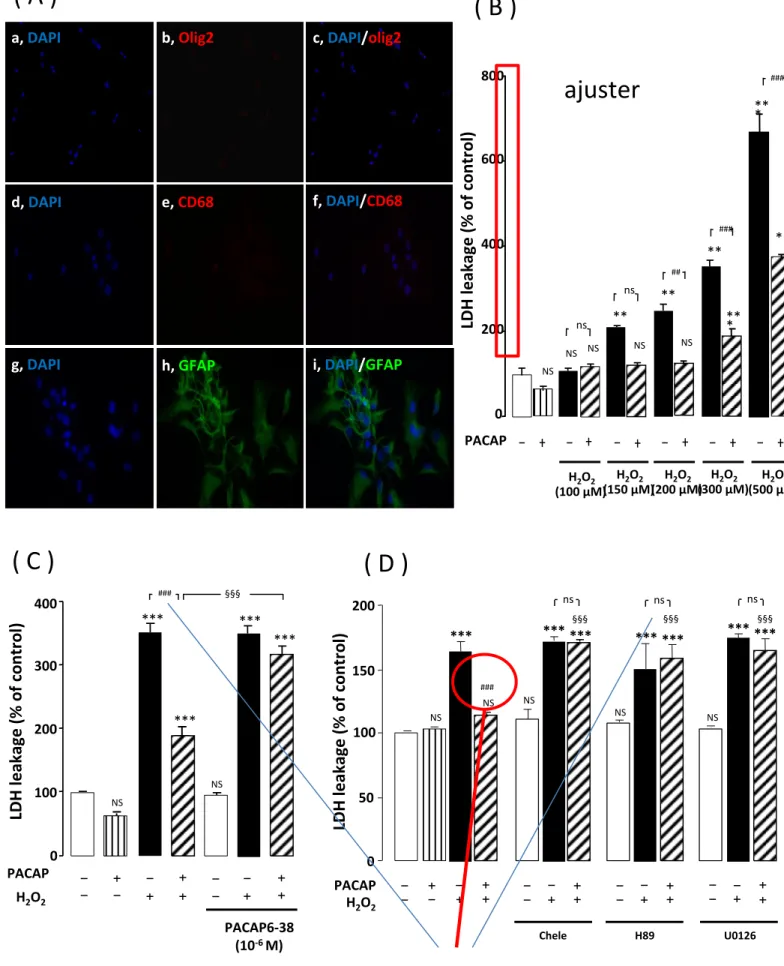
15 (Supplementary Figure 1). 16 17 Cell treatment 18
To examine the potential protective action of PACAP against H2O2-provoked 19
astrocytes assault (LDH release, cell death, caspase-3 activation and expression, SOD
20
and catalase activities.), cultured cells were treated with a control solution or PACAP
21
10 min before starting incubation with H2O2 for 1 h (Supplementary Figure 2A). When 22
11
PACAP antagonist or transduction pathway inhibitors were used, they were added 30
1
minutes before H2O2 exposure. To block catalase activity, ATZ was added 3 h before 2
H2O2. In one subset of experiments, PACAP was administered 10 min after the 3
beginning of the incubation with H2O2 (Supplementary Figure 2B). 4
5
Measurement of cell cytotoxicity
6
Cultured cells were incubated at 37°C with fresh serum-free medium in the absence or
7
presence of test substances. Membrane integrity was assessed as a function of the
8
amount of cytoplasmic LDH released into the medium. The amount of LDH released
9
into the medium was quantified using a cytotoxicity detection kit (MAK066, Sigma-
10
Aldrich) according to the manufacturer’s instructions. LDH activity was measured at
11
450 nm with a spectrophotometric microplate reader (Flexstation 3, Molecular Devices 12
Sunnyvale, CA, USA).
13 14
Assessment of cell survival
15
Cultured cells were incubated at 37°C with fresh serum-free culture medium in the
16
absence or presence of the test substances. Qualitative visualization of cell survival
17
was conducted by using the LIVE/DEAD Viability/Cytotoxicity Kit for mammalian
18
cells (Invitrogen) according to the manufacturer protocol, as previously reported
19
(Vaudry et al. 2002). Briefly, cells were incubated for 20 min at 37°C with a solution
20
of 1.2 µg/mL calcein-AM (producing green fluorescence in living cells) and 3.4
21
µg/mL ethidium homodimer-1 (EH-1, producing red fluorescence in dead cells) and
12
rinsed twice with PBS. Images of astroglial cells were randomly acquired on an
1
inverted microscope (IRE2; Leica Microsystems, Nanterre, France). For quantification
2
of surviving astrocytes, cells were incubated for 8 min with FDA-AM, rinsed twice
3
with PBS and lysed with a Tris/HCl solution containing 1% sodium dodecyld sulfate.
4
Fluorescence was measured with excitation at 485 nm and emission at 538 nm using a
5
fluorescence microplate reader FL800TBI (Bio-Tek Instruments, Winooski, VT,
6
USA). Pilot experiments have shown that the fluorescence intensity is proportional to
7
the number of cells (in the range 75 X 104 to 1 X 10⁶ cells/mL).
8 9
Quantitative PCR analysis
10
The effect of PACAP on caspase-3 mRNA levels was performed by quantitative
RT-11
PCR. Total RNA was isolated from astrocytes using the NucleoSpin kit
(Macherey-12
Nagel, Hoerd, France) and 3-4 µg were used for cDNA synthesis using ImProm II
13
Promega kit (Promega). PCR amplifications were performed with an ABI PRISM
14
7500 Sequence Detection System (Applied Biosystems) using 5 ng cDNA, 1X Fast
15
SYBR Green universal PCR Mastermix (Applied Biosystems, Courtaboeuf, France)
16
and 300 nM forward (5’-CTGACTGGAAAGCCGAAACTCT-3’) and reverse
(5’-17
CATCGTCAGTTCCACTGTCTGTCT-3’) caspase-3 primers, under standard running
18
conditions as suggested by the manufacturer. The amount of cDNA in each sample
19
was calculated by the comparative quantification cycle (Cq) method and expressed as
20
2exp(-ΔΔCq) using glyceraldehyde-3-phosphate dehydrogenase as an internal control.
21 22
13
Measurement of mitochondrial activity
1
Mitochondrial membrane potential was quantified using the JC-10 probe. Cells seeded
2
into 96-well plates were subjected to various treatments, incubated in the presence of
3
the JC-10 probe (10 µg/mL) at 37°C for 1 h and then washed twice with PBS. In
4
healthy astrocytes, the intact membrane potential allows the lipophilic dye JC-10 to
5
enter into the mitochondria where it accumulates and aggregates producing an intense
6
orange signal. In dying cells, where the mitochondrial membrane potential collapses,
7
the monomeric JC-10 remains cytosolic and stains cell cytoplasm in green.
8
Fluorescence was measured with excitations at 485 (monomer) and 510 nm
9
(aggregates), and emissions at 534 (green) and 610 nm (orange), respectively.
10 11
Superoxide radical generation assay
12
The intracellular production of superoxide anion was detected by the reduction of
13
nitroblue tetrazolium (NBT) to dark blue formazan deposits. Treated cells in 6-well
14
plates were incubated for 2 h in the dark with reaction mixture containing NBT
15
(1 mg/mL) and BSA (1 mg/mL). To visualize the formation of blue deposits, cells
16
were examined and images were acquired with an eclipse E-600 microscope (Nikon,
17
Champigny-sur-Marne, France) equipped with a 3 CCD Sony DXC950 camera
18
interfaced with the Visiolab computerized program (Biocom, Les Ulis, France). To
19
quantify cellular superoxide radical levels, the deposits were dissolved with (2 M)
20
KOH/DMSO (v:v, 1:1.15) solution and the absorbance of the mixture was measured at
21
645 nm with a spectrophotometer (Jenway, Philadelphia, USA).
14
Measurement of antioxidant enzyme activities
1
Cells were incubated at 37°C with fresh serum-free medium. At the end of the
2
incubation, cells were rinsed twice with PBS, rubber scraped and centrifuged at 3000 g
3
for 10 min at 4°C. The cell pellet was resuspended in 50 µL of ice-cold lysing buffer
4
containing 50 mM Tris–HCl (pH 8), 10 mM EDTA, 100 µM
phenylmethyl-5
sulfonylfluoride and 1% Triton X-100 before centrifugation at 16,000 g for 20 min at
6
4°C. The supernatant was stored at -20°C until enzyme activity determination.
7
SOD activity was assessed using a spectrophotometric assay, which consists of
8
measuring epinephrine autoxidation induced by superoxide anions. Samples were
9
incubated for 3 min with a mixture containing bovine catalase (0.4 U/µL),
DL-10
epinephrine (5 mg/mL) and Na2CO3/NaHCO3 buffer (62.5 mM, pH 10.2). The 11
oxidation of epinephrine was measured at 480 nm using a Bio-Rad spectrophotometer
12
(Bio-Rad Laboratories, Philadelphia, PA, USA).
13
Catalase activity was determined on the basis of the disappearance of H2O2. 14
Samples, prepared as described above, were mixed with 30 mM H2O2 in PBS. The 15
decrease of H2O2 was followed at a wavelength of 240 nm for 3 min at 30 s intervals. 16
Catalase activity was expressed using the extinction coefficient of 40 mM-1 cm−1 for
17
H2O2. 18
19 20
15
Statistical analysis
1
Data are expressed as the mean ± SEM from three independent experiments. Statistical
2
analysis of the data was performed by using ANOVA, followed by Bonferroni’s test.
3
A p value of 0.05 or less was considered as statistically significant.
16
Results
1
Effect of PACAP on H2O2-induced caspase-3 activation and astrocyte cell death
2
We have previously shown that incubation of cultured astrocytes with graded
3
concentrations of PACAP (10-14 M to 10-8 M), dose-dependently prevented cell death
4
induced by 300 µM H2O2. Here we show that the addition of PACAP (10-9 M) to the 5
culture medium almost completely abolished the effect of moderate concentrations of
6
H2O2 (150 to 300 µM) on LDH release and markedly attenuated the stimulatory effect 7
of higher concentration of H2O2 (500 µM; Fig. 1A). Pre-incubation of astrocytes for 30 8
min with the PACAP receptor antagonist PACAP6-38 (10-6 M), which had no effect
9
by itself, totally abolished the action of PACAP on H2O2-induced LDH release (Fig. 10
1B). Incubation of astrocytes with the selective protein kinase C (PKC) inhibitor
11
chelerythrine (10-7 M), the PKA inhibitor H89 (2 x 10⁻⁵ M) or the mitogen-activated
12
protein kinase kinase (MEK) inhibitor U0126 (10-6 M) also blocked the effect of 13
PACAP on H2O2-induced LDH release (Fig. 1C). Furthermore, the fact that addition 14
of PACAP (10-9 M) to the culture media 10 min after 300 µM H2O2 pre-incubation 15
rescues asrocytes from cell death (97% ; P< 0.001), provides evidence that the
16
beneficial effects of PACAP are still present when the peptide is added after oxidative
17
injuries (Supplementary figure 3A).
18
To determine whether PACAP reduced H2O2-induced cell death, astrocytes were 19
stained with calcein-AM (green color) and EH-1 (red color), markers of living and
20
dead cells, respectively. We observed very few cells labeled with EH-1 in control- and
21
PACAP-treated cells (Fig. 2Aa, 2Ac and 2B), while incubation of astrocytes with 300
17
µM H2O2 induced a significant increase in the number of EH-1-labeled cells (P < 1
0.001) which mirrored a decrease in the number of calcein-positive cells (Fig. 2Ab and
2
2B). Pretreatment of the cells with PACAP (10-9 M; 10 min) counteracted 86% (P <
3
0.001) of H2O2-induced decrease in the number of calcein-positive cells (Fig. 2Ad, 4
2B). Quantification of fluorescein diacetate-acetoxymethyl ester fluorescence intensity
5
incorporated by astrocytes with a microplate reader gave similar results (data not
6
shown).
7
When looking at GFAP labeling, cells in control conditions exhibited a flat
8
polygonal morphology (Fig. 2Ca), while in cells exposed to 300 µM H2O2, processes 9
were significantly retracted (Fig. 2Cb). Treatment of the cells with PACAP totally
10
prevented the morphological alterations induced by H2O2 (Fig. 2Cd). 11
To further explore the mechanisms involved in the glioprotective action of PACAP,
12
the effect of the peptide on caspase-3 activity and gene expression were investigated.
13
In agreement with its trophic action (Ravni et al. 2006; Vaudry et al., 2000), PACAP
14
exhibited a transient intrinsic inhibition of caspase-3 gene expression (-35.8% + 2.23;
15
P ˂ 0.05) within 5 to 10 min after peptide treatment and a concomitant decrease of
16
basal caspase-3 activity (-35% + 7.05; P ˂ 0.01). On the opposite, exposure of cultured
17
astrocytes to H2O2 for 30 min produced a significant increase of caspase-3 activity 18
(+49.9 + 8.5; P < 0.01) and mRNA levels (+40.6% + 2.8; P ˂ 0.001; Fig. 3A and 3B)
19
which lasted at least 30 min. Pretreatment with PACAP (10-9 M) of cells exposed to
20
H2O2 induced a decrease of caspase-3 activity to a similar level to the one observed in 21
cells that have not been exposed to H2O2 (Fig. 3A) and a decrease of caspase-3 mRNA 22
which remained significant for all the duration of the treatment (Fig. 3B). Addition of
18
graded concentrations of PACAP (10-14 M to 10-6 M) to the culture medium suppressed
1
dose-dependently the stimulatory effect of H2O2 (300 µM; 30 min)on caspase-3 gene 2
expression (Fig. 3C).
3 4
Effect of PACAP on H2O2-generated superoxide anions
5
Considering the major effect of oxidative stress in the induction of respiratory burst
6
and superoxide anion (O2°-) production, we examined the effect of PACAP on H2O2 -7
induced O2°- generation in astrocytes. Control and PACAP (10-9 M)-treated astrocytes 8
exhibited very few blue precipitates (reflecting reduction of NBT by O2°- production) 9
in the cell bodies (Fig. 4Aa and 4Ac). Treatment of astrocytes with 300 µM H2O2 10
resulted in the labeling of most cell bodies in blue, indicating that large amounts of
11
O2°- are produced by respiratory burst (Fig. 4Ab). When PACAP 10-9 M was added to 12
H2O2-treated cells, only a few cells were labeled in blue (Fig. 4Ad), suggesting that the 13
peptide could abolish O2°- generation. Quantitative analysis indicated that H2O2 14
induced a significant increase (+518.9 % + 14.0; P < 0.001)of O2°- production which 15
was reduced by a factor of 2 in the presence of PACAP (Fig. 4B).
16 17
Effect of PACAP on the activity of antioxidant enzymes in cultured astrocytes
18
To determine if the endogenous antioxidant system was involved in the protective
19
action of PACAP, the activity of antioxidant enzymes SOD and catalase in astrocytes
20
was monitored. Incubation of cultured astrocytes with increasing concentrations of
21
PACAP (10-14 M to 10-6 M) for 10 min, induced a dose-dependent stimulation of SOD
22
and catalase activities with EC50 values of 1.11 x 10⁻⁹ M and 5.32 x 10⁻⁹ M, 23
19
respectively (Fig. 5A and 5B). Time-course experiments revealed that PACAP (10-9
1
M) significantly enhanced SOD and catalase activities within 5 and 10 min (SOD and
2
catalase) of incubation. The stimulatory effect of PACAP on SOD and catalase
3
activities reached a maximum after 10 and 20 min, respectively (Fig. 5C and 5D).
4
Thereafter the activity of the two antioxidant enzymes gradually returned to control
5
values within 60 min of treatment.
6
Time-course experiments revealed that exposure to 300 µM H2O2 significantly 7
reduced SOD and catalase activities within 30 min, with a maximum effect after 1 h of
8
incubation (Fig 6A and 6B, inset: SOD -42.75 % + 2.08, P <0.001 and catalase
9
-40.69 % + 2.07; P <0.001). After 1 h of treatment, addition of graded concentrations
10
of PACAP (10-14 M to 10-6 M) totally blocked in a dose-dependent manner the 11
inhibitory action of H2O2. Nanomolar concentrations of PACAP even significantly 12
stimulated SOD and catalase activities above control levels (Fig. 6A and 6B).
13 14
Receptor and signal transduction pathways involved in the effects of PACAP on
15
endogenous antioxidant systems
16
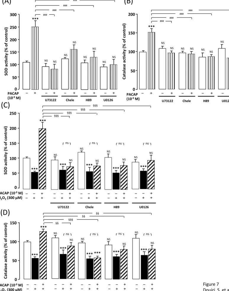
Administration of the PACAP receptor antagonist PACAP6-38 (10-6 M) to cultured
17
astrocytes did not induce any modification of SOD and catalase activities by itself, but
18
totally abolished the stimulatory effect of PACAP (10-9 M) on SOD and catalase
19
activities in the absence (Fig. 7A and 7B) or presence (Fig. 7C and 7D) of H2O2 20
exposure.
21
Incubation of astrocytes with the PLC inhibitor U73122 (10-7 M), the PKC inhibitor
22
chelerythrine (10-7 M) or the PKA inhibitor H89 (2 X 10⁻⁵ M) which had no impact by
20
themselves on antioxidant enzyme activities, totally abrogated the stimulatory effect of
1
PACAP on SOD and catalase activities. In addition, blockage of ERK phosphorylation
2
with the MEK inhibitor U0126 (1 µM) suppressed the stimulatory action of PACAP
3
on both enzyme activities in the absence of H2O2 (Fig. 8A and 8B). In the presence of 4
H2O2, incubation of astrocytes with U73122, chelerythrine, H89 or U0126 also 5
abolished the ability of PACAP to stimulate the activity of endogenous antioxidant
6
systems SOD and catalase (Fig. 8C and 8D).
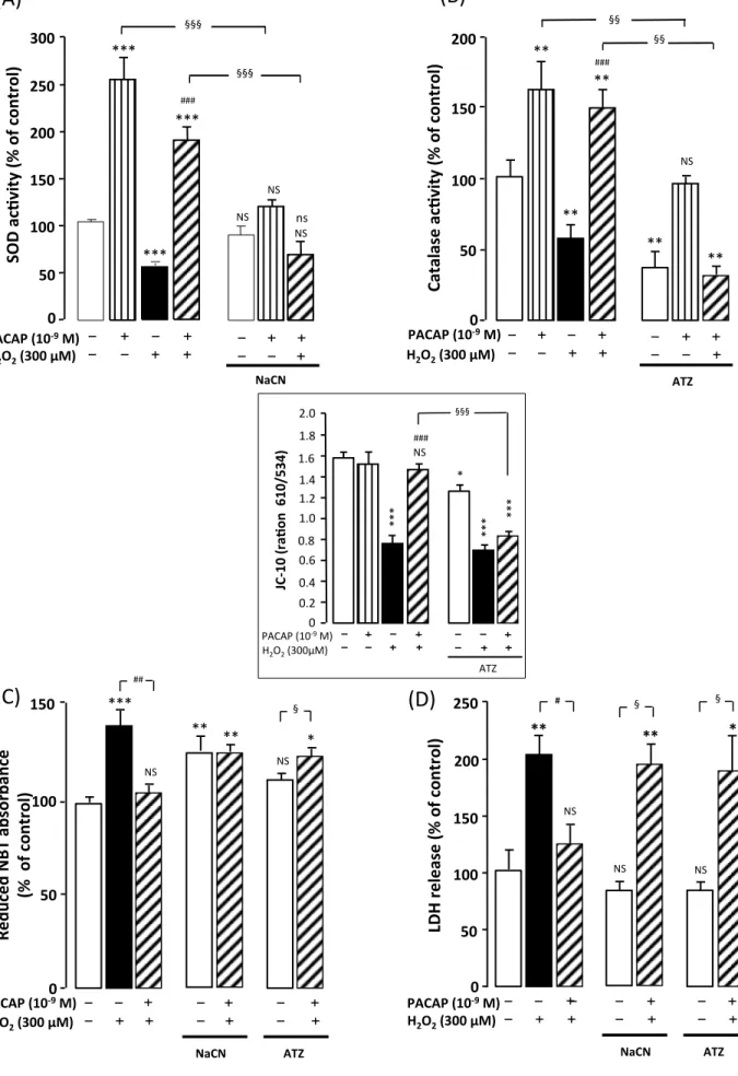
7
Addition of 2 X 10⁻² M NaCN, a SOD inhibitor, significantly reduced basal
8
enzymatic activity and fully suppressed the stimulatory effect of PACAP (Fig. 9A). In
9
a very similar manner, exposure of astrocytes to 10-2 M 3-aminotriazole, a specific 10
catalase inhibitor, strongly decreased basal and PACAP-enhanced catalase activity
11
(Fig. 9B). Exposure of the cells to 2 X 10⁻² M NaCN and 10-2 M 3-aminotriazole, 12
which did not affect cell survival by themselves, fully blocked the protective effect of
13
PACAP against H2O2-evoked alteration of SOD and catalase activities (Fig. 9A and 14
9B), mitochondrial integrity (Fig. 9C and inset) and cell death (Fig. 9D). 15
16
21
Discussion
1
It has been clearly established that oxidative stress causes apoptosis in various cell
2
types, notably in astrocytes (Sun et al. 2014, Wang et al. 2014). We have previously
3
reported that, like in neurons, PACAP protects astrocytes against H2O2-induced cell 4
death (Masmoudi-Kouki et al. 2011). Here, we show that PACAP, through the
5
activation of its receptors and the PKA, PKC and MAP-kinases signaling pathways,
6
counteracts superoxide anion accumulation and prevents the inhibition of SOD and
7
catalase activities induced by oxidative stress in cultured astrocytes. Furthermore,
8
blocking SOD and catalase activation reduces the protective effect of PACAP. Thus,
9
our results show that the glioprotective action of PACAP against H2O2-induced 10
apoptotic cell death can be accounted for activation of endogenous antioxidant systems
11
(Fig. 10).
12
In agreement with the well-known cytoprotective effect of PACAP (Bourgault et al.
13
2011), we found that nanomolar concentrations of PACAP exert a protective effect
14
against oxidative stress-induced cell death in cultured astrocytes. Indeed, PACAP was
15
able to prevent the deleterious action of graded concentrations of H2O2 (100 to 500 16
µM) on astrocytes. Visualization of living cells by calcein-AM and GFAP staining
17
revealed that the cytotoxic effect of H2O2 was associated with modifications in 18
astrocyte morphology, such as cell shrinkage and appearance of thin processes that are
19
suggestive of apoptotic cell death. These morphological changes were also prevented
20
by the addition of subnanomolar concentrations of PACAP to the culture medium. In
21
agreement with these observations, it has already been shown that H2O2-treated 22
astrocytes exhibit the characteristic features of apoptotic cells with increased caspase-3
22
activity, nuclear condensation and DNA fragmentation (Peng et al. 2013, Ramalingam
1
and Kim 2014, Zhou et al. 2015), which are all blocked by PACAP treatment.
2
Furthermore, it has been reported that in cerebellar granule cells PACAP is able to
3
prevent ROS-induced mitochondrial dysfunction, the stimulation of caspase-3 activity
4
and LDH leakage, which together induce apoptotic cell death (Tabuchi et al. 2003,
5
Vaudry et al. 2002). These data suggest that PACAP, which is released by both glial
6
cells and neurons in the brain (Vaudry et al. 2009), may act as an autocrine and/or
7
paracrine factor to enhance the resistance of astrocytes to H2O2. 8
The cytotoxic effect of H2O2 via production of highly reactive species inside the cell 9
has been well documented in numerous cell types, including astrocytes (Feeney et al.
10
2008, Hamdi et al. 2011, Liu et al. 2013). Various studies indicate that an excess of
11
H2O2 can provoke cell apoptosis via multiple mechanisms, including the stimulation of 12
some pro-apoptotic genes of the Bcl-2 family responsible for the formation of
13
mitochondrial permeability transition pores, a collapse of the mitochondrial membrane
14
potential and a decrease of ATP generation (Gyulkhandanyan et al. 2003, Kaddour et
15
al. 2013, Liu et al. 2013). Consistent with these notions, the present study shows that
16
H2O2 severely impaired mitochondrial integrity, increased respiration rate and 17
mitochondrial O2°- generation, which were significantly reduced by PACAP. 18
Measurement of mitochondria activity by means of the membrane potential-sensitive
19
probe JC-10 revealed that H2O2 decreased the proportion of active mitochondria, and 20
that PACAP prevented this deleterious action of H2O2. The inhibitory effect of 21
PACAP on H2O2-evoked burst of superoxide generation is probably a key mechanism 22
in its glioprotective action. Indeed, we have previously reported that, in H2O2-treated 23
23
astrocytes, PACAP increases the cellular content of GSH, the major intracellular free
1
radical scavenger in the brain (Masmoudi-Kouki et al. 2011), and enhances cell
2
resistance to oxidative injury. The involvement of GSH in the protective effect of
3
PACAP is supported by recent data showing that PACAP increases uptake of the GSH
4
precursor, cysteine, in cultured astrocytes (Resch et al. 2014). Furthermore, inhibition
5
of GSH synthesis in neuroblastoma cells enhances intracellular ROS levels and
6
increases cell sensitivity to oxidative damages (Miyama et al. 2011).
7
It has been reported that intracellular ROS overproduction and GSH depletion
8
impair endogenous antioxidant defences, notably by decreasing SOD and catalase
9
activities (Dokic et al. 2012, Lopez et al. 2007). The present study reveals that PACAP
10
at subnanomolar concentrations induced a dose-dependent increase of both SOD and
11
catalase activities in astrocytes and simultaneously block H2O2-evoked inhibition of 12
the activity of these two enzymes. We have previously shown that, in cultured
13
astrocytes, PACAP in the same range of concentrations protects cells against apoptosis
14
provoked by oxidative assault (Masmoudi-Kouki et al. 2011). The fact that PACAP
15
increases Mn-SOD and peroxiredoxins (Prx-1 and Prx-6) mRNA levels to promote
16
neuronal survival after spinal cord injury (Fang et al. 2010), provides evidence for the
17
implication of antioxidant enzymes in the antiapoptotic action of PACAP. Consistent
18
with these observations, it has already been reported that a reduction in the activities of
19
the antioxidant enzyme suppresses the beneficial effect of protective molecules against
20
oxidative-stress induced astrocyte apoptosis (Gaspar et al. 2008, Hamdi et al. 2011,
21
Smith et al. 2007). Besides, in vivo studies have demonstrated that Mn-SOD
22
overexpression improves resistance to oxidative injuries in mouse models of ischemia
24
or Alzheimer’s disease (Dumont et al. 2009, Saito et al. 2003) and deficiency of SOD
1
enhances oxidative damages, in a mouse model of Alzheimer’s disease (Schuessel et
2
al. 2005). Together, these data indicate that the protective effect of PACAP against
3
H2O2-provoked oxidative stress and cell death in astrocytes is attributable to the 4
activation of the antioxidant enzymes that act as H2O2 and ROS scavengers. It is 5
noteworthy that in H2O2-treated astrocytes PACAP was still effective in maintaining 6
SOD and catalase activities above control values after 1 h of treatment. This indicates
7
that the neuropeptide exhibits a sustained antioxidant action under stress situations.
8
The increase of PACAP receptors in cultured astrocytes under stress assault (Suzuki et
9
al. 2003, Stum et al., 2007) may contribute to the extended activation of the
10
endogenous antioxidant system and to prolonged cell protection.
11
Previous studies have shown that the glioprotective action of PACAP upon H2O2 -12
evoked cell death is mediated through activation of PAC1 receptor coupled to the
13
PKA, PKC and MAP-kinase extracellular signal-regulated kinase (ERK) transduction
14
pathways (Masmoudi-Kouki et al. 2011). Indeed, blocking the activity of these kinases
15
abrogated the cell survival-promoting effect of PACAP under oxidative stress.
16
Furthermore, we demonstrated that like cell survival, activation of SOD and catalase
17
activities were PKA and PLC / PKC dependent. It has also been shown that
18
stimulation of SOD and catalase activities or expression of the enzymes in a mouse
19
model of Alzheimer’s disease (Leem et al. 2009), in the rat inferior colliculus (Mei et
20
al. 1999) and in cultured glial cells (Huang et al. 2001) depend on PKA and/or PKC
21
activation (Kupershmidt et al. 2011, Leem et al. 2009, Mao et al. 2014, Priyanka et al.
22
2013). Downstream of PKA and PKC, PACAP stimulates phosphorylation of ERK
25
(Seaborn et al. 2011), a kinase which alone or together with PKA induces c-fos gene
1
expression (Mullenbrock et al. 2011, Vaudry et al. 1998) an immediate early gene
2
known to stimulates SOD and catalase protein expression (Radjendirane V. and Anil,
3
Mikawa et al. 1995). Concomitantly, PACAP promotes the expression and activity of
4
the antioxidant protein peroxyredoxin-2 and glutathione peroxidase 1 through the
5
AC/PKA, PLC/PKC and MAPK/ERK pathways (Miyamoto et al. 2014) and inhibition
6
of peroxyredoxin-2 with siRNA reduces the antiapoptotic activity of the peptide (Botia
7
et al., 2008). Taken together, these data highlight the importance of the
8
AC/PKA/PKC/MAPK/ERK pathways in the antioxidant action of PACAP. However,
9
further investigations are needed to definitely establish the functional contribution of
10
PKA/PKC/ERK transduction pathways in the glioprotective action of PACAP, for
11
example by using siRNA against PKA, PKC and/or MAPK/ERK, as previously done
12
with SH-SY5Y cell lines (Quesada et al. 2011) or granule neurons (Botia et al., 2008).
13
The protective effect of PACAP against H2O2-reduced antioxidant enzyme activities 14
might have pathophysiological significance in the context of brain injuries i.e. in
15
neurodegenerative diseases or stroke. The central nervous system is very vulnerable to
16
oxidative damage due to its high metabolic rate and high levels of unsaturated lipids.
17
Furthermore, the up-regulation of antioxidant enzymes in astroglial cells would prove
18
beneficial against cellular dysfunction, biomolecule oxidation and neuronal apoptosis
19
observed in brain injuries (Baraibar et al. 2012, Negre-Salvayre et al. 2010). In
20
agreement with this hypothesis, it has been shown that PACAP exerts potent
21
glioprotective and neuroprotective effects against oxidative stress-provoked apoptosis
22
(Seaborn et al. 2011). The fact that the anti-apoptotic action of PACAP is likely
26
mediated through PAC1 receptor is of particular interest. Recent data indicate that a
1
synthetic PACAP analog, Ac-[Phe(pI)6, Nle17]PACAP(1-27), mimics the
2
neuroprotective action of PACAP against 1-methyl-4-phenylpyridinium (MPP+)
3
toxicity on SH-SY5Y neuroblastoma cells (Lamine et al. 2015). Since intravenous
4
injections of the analog induce potent neuroprotection in a mouse model of
5
Parkinson’s disease (Lamine et al. 2015), the development of specific PACAP
6
analogs, that would selectively promote the glioprotective and anti-oxidative
7
proprieties of PACAP, might prove useful for the treatment of ischemia and
8
neurodegenerative diseases.
9
In conclusion, the present study demonstrates that the neuropeptide PACAP
10
rescues cultured astrocytes against cell death provoked by oxidative stress. This
11
glioprotective action of PACAP is attributable, at least in part, to the activation of
12
endogenous antioxidant enzymes and reduction of ROS formation, which preserve
13
mitochondrial membrane integrity and prevent caspase-3 activation.
14 15 16 17 18 19 20 21 22 23
27 1 2 3 4 5 Acknowledgments 6
Salma Douiri and Seyma Bahdoudi were recipients of fellowships from the University
7
of Tunis El Manar and a France-Tunisia exchange program Inserm-DGRS. This study
8
was supported by the Research Unit UR/11ES09, a CMCU-Utique program (to
9
Mohamed Amri and Marie-Christine Tonon; grant number 07G0822), an
Inserm-10
DGRS program (to Mohamed Amri and Marie-Christine Tonon; grant number
11
M10/M), Inserm (U982), the Institute for Medical Research and Innovation (IRIB) and
12
the Normandy region, the Normandy region and the PeReNE Interreg project. Authors
13
declare that they have no conflict of interest.
14 15
28
References
1
Baraibar, M. A., Liu, L., Ahmed, E. K. and Friguet, B. (2012) Protein oxidative
2
damage at the crossroads of cellular senescence, aging, and age-related diseases.
3
Oxid. Med. Cell. Longev. 2012, 919832.
4
Barreto, G. E., Gonzalez, J., Torres, Y. and Morales, L. (2011) Astrocytic-neuronal
5
crosstalk: implications for neuroprotection from brain injury. Neurosci. Res. 71,
6
107–113.
7
Botia, B., Seyer, D., Ravni, A. et al. (2008) Peroxiredoxin 2 is involved in the
8
neuroprotective effects of PACAP in cultured cerebellar granule neurons. J. Mol.
9
Neurosci. 36, 61–72
10
Bourgault, S., Chatenet, D., Wurtz, O., Doan, N. D., Leprince, J., Vaudry, H.,
11
Fournier, A. and Vaudry, D. (2011) Strategies to convert PACAP from a
12
hypophysiotropic neurohormone into a neuroprotective drug. Curr. Pharm. Des.
13
17, 1002–1024.
14
Cabezas, R., El-Bacha, R. S., Gonzalez, J. and Barreto, G. E. (2012) Mitochondrial
15
functions in astrocytes: neuroprotective implications from oxidative damage by
16
rotenone. Neurosci. Res. 74, 80–90.
17
Castel, H., Diallo, M., Chatenet, D. et al. (2006) Biochemical and functional
18
characterization of high-affinity urotensin II receptors in rat cortical astrocytes. J.
19
Neurochem. 99, 582–595.
29
Chen, Y., Chan, P. H. and Swanson, R. A. (2001) Astrocytes overexpressing Cu,Zn
1
superoxide dismutase have increased resistance to oxidative injury. Glia 33, 343–
2
347.
3
Chen, Y. and Gibson, S. B. (2008) Is mitochondrial generation of reactive oxygen
4
species a trigger for autophagy? Autophagy 4, 246–248.
5
Dasuri, K., Zhang, L. and Keller, J. N. (2013) Oxidative stress, neurodegeneration, and
6
the balance of protein degradation and protein synthesis. Free Radic. Biol.
7
Med.62, 170–185.
8
Dejda, A., Sokolowska, P. and Nowak, J. Z. (2005) Neuroprotective potential of three
9
neuropeptides PACAP, VIP and PHI. Pharmacol. Rep. 57, 307–320.
10
Dokic, I., Hartmann, C., Herold-Mende, C. and Regnier-Vigouroux, A. (2012)
11
Glutathione peroxidase 1 activity dictates the sensitivity of glioblastoma cells to
12
oxidative stress. Glia 60, 1785–1800.
13
Dumont, M., Wille, E., Stack, C., Calingasan, N. Y., Beal, M. F. and Lin, M. T. (2009)
14
Reduction of oxidative stress, amyloid deposition, and memory deficit by
15
manganese superoxide dismutase overexpression in a transgenic mouse model of
16
Alzheimer's disease. FASEB J. 23, 2459–2466.
17
Emerit, J., Edeas, M. and Bricaire, F. (2004) Neurodegenerative diseases and oxidative
18
stress. Biomed. Pharmacother. 58, 39–46.
19
Fang, K. M., Chen, J. K., Hung, S. C. et al. (2010) Effects of combinatorial treatment
20
with pituitary adenylate cyclase activating peptide and human mesenchymal stem
21
cells on spinal cord tissue repair. PLoS One 5, e15299.
30
Feeney, C. J., Frantseva, M. V., Carlen, P. L., Pennefather, P. S., Shulyakova, N.,
1
Shniffer, C. and Mills, L. R. (2008) Vulnerability of glial cells to hydrogen
2
peroxide in cultured hippocampal slices. Brain Res. 1198, 1–15.
3
Fernandez-Fernandez, S., Almeida, A. and Bolanos, J. P. (2012) Antioxidant and
4
bioenergetic coupling between neurons and astrocytes. Biochem. J. 443, 3–11.
5
Ferrero-Gutierrez, A., Perez-Gomez, A., Novelli, A. and Fernandez-Sanchez, M. T.
6
(2008) Inhibition of protein phosphatases impairs the ability of astrocytes to
7
detoxify hydrogen peroxide. Free Radic. Biol. Med. 44, 1806–1816.
8
Gaspar, T., Snipes, J. A., Busija, A. R., Kis, B., Domoki, F., Bari, F. and Busija, D. W.
9
(2008) ROS-independent preconditioning in neurons via activation of
10
mitoK(ATP) channels by BMS-191095. J. Cereb. Blood Flow. Metab. 28, 1090–
11
1103.
12
Gyulkhandanyan, A. V., Feeney, C. J. and Pennefather, P. S. (2003) Modulation of
13
mitochondrial membrane potential and reactive oxygen species production by
14
copper in astrocytes. J. Neurochem. 87, 448–460.
15
Hamdi, Y., Kaddour, H., Vaudry, D. et al. (2012) The stimulatory effect of the
16
octadecaneuropeptide ODN on astroglial antioxidant enzyme systems is
17
mediated through a GPCR. Front. Endocrinol. 3, 138.
18
Hamdi, Y., Kaddour, H., Vaudry, D. et al. (2015) Octadecaneuropeptide ODN
19
prevents hydrogen peroxide-induced oxidative damage of biomolecules in
20
cultured rat astrocytes. Peptides 71, 56–65.
31
Hamdi, Y., Masmoudi-Kouki, O., Kaddour, H. et al. (2011) Protective effect of the
1
octadecaneuropeptide on hydrogen peroxide-induced oxidative stress and cell
2
death in cultured rat astrocytes. J. Neurochem. 118, 416-428.
3
Harmar, A. J., Fahrenkrug, J., Gozes, I. et al. (2012) Pharmacology and functions of
4
receptors for vasoactive intestinal peptide and pituitary adenylate
cyclase-5
activating polypeptide: IUPHAR review 1. Br. J. Pharmacol. 166, 4–17.
6
Hsu, M. J., Sheu, J. R., Lin, C. H., Shen, M. Y. and Hsu, C. Y. (2009) Mitochondrial
7
mechanisms in amyloid beta peptide-induced cerebrovascular degeneration.
8
Biochim. Biophys. Acta. 1800, 290–296.
9
Huang, W. C., Chen, P. C., Jou, S. B. and Cheng, J. T. (2001) Protein kinase C and
10
changes in manganese superoxide dismutase gene expression in cultured glial
11
cells. Clin. Exp. Pharmacol. Physiol. 28, 822–825.
12
Jolivel, V., Basille, M., Aubert, N. et al. (2009) Distribution and functional
13
characterization of pituitary adenylate cyclase-activating polypeptide receptors in
14
the brain of non-human primates. Neuroscience 160, 434–451.
15
Kaddour H., Hamdi Y., Vaudry D. et al. (2013) The octadecaneuropeptide ODN
16
prevents 6-hydroxydopamine-induced apoptosis of cerebellar granule neurons
17
through a PKC-MAPK-dependent pathway. J. Neurochem. 125, 620–633.
18
Kupershmidt, L., Weinreb, O., Amit, T., Mandel, S., Bar-Am, O. and Youdim, M. B.
19
(2011) Novel molecular targets of the neuroprotective/neurorescue multimodal
20
iron chelating drug M30 in the mouse brain. Neuroscience 189, 345–358.
21
Lamine, A., Letourneau, M., Doan, N. D., Maucotel, J., Couvineau, A., Vaudry, H.,
22
Chatenet, D., Vaudry, D. and Fournier, A. (2015) Characterizations of a synthetic
32
pituitary adenylate cyclase-activating polypeptide analog displaying potent
1
neuroprotective activity and reduced in vivo cardiovascular side effects in a
2
Parkinson's disease model. Neuropharmacology doi: 10.1016.
3
Leem, Y. H., Lim, H. J., Shim, S. B., Cho, J. Y., Kim, B. S. and Han, P. L. (2009)
4
Repression of tau hyperphosphorylation by chronic endurance exercise in aged
5
transgenic mouse model of tauopathies. J. Neurosci. Res. 87, 2561–2570.
6
Liu, J., Qiu, J., Xiong, Y., Liu, Z. and Gao, J. (2013) The mitochondrial protective
7
mechanism of olfactory ensheathing cells conditioned medium protects against
8
H2O2-induced injury in astrocytes. Neurosci. Lett. 555, 91–96. 9
Lopez, M. V., Cuadrado, M. P., Ruiz-Poveda, O. M., Del Fresno, A. M. and Accame,
10
M. E. (2007) Neuroprotective effect of individual ginsenosides on astrocytes
11
primary culture. Biochim. Biophys. Acta 1770, 1308–1316.
12
Mao, X., Cao, Y., Li, X. et al. (2014) Baicalein ameliorates cognitive deficits in
13
epilepsy-like tremor rat. Neurol. Sci. 35, 1261–1268.
14
Masmoudi-Kouki, O., Douiri, S., Hamdi, Y. et al. (2011) Pituitary adenylate
cyclase-15
activating polypeptide protects astroglial cells against oxidative stress-induced
16
apoptosis. J. Neurochem. 117, 403–411.
17
Masmoudi-Kouki, O., Gandolfo, P., Castel, H., Leprince, J., Fournier, A., Dejda, A.,
18
Vaudry, H. and Tonon, M. C. (2007) Role of PACAP and VIP in astroglial
19
functions. Peptides 28, 1753–1760.
20
Masmoudi, O., Gandolfo, P., Leprince, J., Vaudry, D., Fournier, A., Patte-Mensah, C.,
21
Vaudry, H. and Tonon, M. C. (2003) Pituitary adenylate cyclase-activating
33
polypeptide (PACAP) stimulates endozepine release from cultured rat astrocytes
1
via a PKA-dependent mechanism. FASEB J. 17, 17–27.
2
Masmoudi, O., Gandolfo, P., Tokay, T., Leprince, J., Ravni, A., Vaudry, H. and
3
Tonon, M. C. (2005) Somatostatin down-regulates the expression and release of
4
endozepines from cultured rat astrocytes via distinct receptor subtypes. J.
5
Neurochem. 94, 561–571.
6
Mei, Y., Gawai, K. R., Nie, Z., Ramkumar, V. and Helfert, R. H. (1999) Age-related
7
reductions in the activities of antioxidant enzymes in the rat inferior colliculus.
8
Hear Res. 135, 169–180.
9
Mikawa, S., Sharp, F. R., Kamii H., Kinouchi H., Epstein C. J. and Chan P. H. (1195)
10
Expression of c-los and hsp70 mRNA after traumatic brain injury in transgenic
11
mice overexpressing CuZn-superoxide dismutase. Molecular Brain Research 33,
12
288–294
13
Miyama, A., Saito, Y., Yamanaka, K., Hayashi, K., Hamakubo, T. and Noguchi, N.
14
(2011) Oxidation of DJ-1 induced by 6-hydroxydopamine decreasing
15
intracellular glutathione. PLoS One 6, e27883.
16
Miyamoto, K., Tsumuraya, T., Ohtaki, H. et al. (2014) PACAP38 suppresses cortical
17
damage in mice with traumatic brain injury by enhancing antioxidant activity. J.
18
Mol. Neurosci. 54, 370–379.
19
Miyata, A., Arimura, A., Dahl, R. R., Minamino, N., Uehara, A., Jiang, L., Culler, M.
20
D. and Coy, D. H. (1989) Isolation of a novel 38 residue-hypothalamic
21
polypeptide which stimulates adenylate cyclase in pituitary cells. Biochem.
22
Biophys. Res. Commun. 164, 567–574.
34
Mullenbrock, S., Shah, J. and Cooper, G. M. (2011) Global expression analysis
1
identified a preferentially nerve growth factor-induced transcriptional program
2
regulated by sustained mitogen-activated protein kinase/extracellular
signal-3
regulated kinase (ERK) and AP-1 protein activation during PC12 cell
4
differentiation. J. Biol. Chem. 286, 45131–45145.
5
Negre-Salvayre, A., Auge, N., Ayala, V. et al. (2010) Pathological aspects of lipid
6
peroxidation. Free Radic. Res. 44, 1125–1171.
7
Ouyang, Y. B. and Giffard, R. G. (2004) Changes in astrocyte mitochondrial function
8
with stress: effects of Bcl-2 family proteins. Neurochem. Int. 45, 371–379.
9
Peng, T. I., Lin, M. S. and Jou, M. J. (2013) Dual phases of respiration chain
defect-10
augmented mROS-mediated mCa2+ stress during oxidative insult in normal and
11
rho 0 RBA1 astrocytes. Oxid. Med. Cell Longev. 2013, 159567.
12
Priyanka, H. P., Bala, P., Ankisettipalle, S. and ThyagaRajan, S. (2013) Bacopa
13
monnieri and L-deprenyl differentially enhance the activities of antioxidant
14
enzymes and the expression of tyrosine hydroxylase and nerve growth factor via
15
ERK 1/2 and NF-kappaB pathways in the spleen of female wistar rats.
16
Neurochem. Res. 38, 141–152.
17
Quesada A., Ogi J., Schultz J. and Handforth A. (2011) C-terminal mechano-growth
18
factor induces heme oxygenase-1-mediated neuroprotection of SH-SY5Y cells
19
via the protein kinase Cϵ/Nrf2 pathway. J. Neurosci. Res. 89, 394–405
20
Radjendirane V. and Anil K.J. (1998) Nrf2 and Nrf1 in association with Jun proteins
21
regulate antioxidant response element-mediated expression and coordinated
22
induction of genes encoding detoxifying enzymes. Oncogene 17, 3145 –3156
35
1
Ramalingam, M. and Kim, S. J. (2014) Insulin on hydrogen peroxide-induced
2
oxidative stress involves ROS/Ca2+ and Akt/Bcl-2 signaling pathways. Free
3
Radic. Res. 48, 347–356.
4
Ravni, A., Bourgault, S., Lebon, A. et al. (2006) The neurotrophic effects of PACAP
5
in PC12 cells: control by multiple transduction pathways. J. Neurochem. 98,
6
321–329.
7
Reglodi, D., Lubics, A., Kiss, P., Lengvari, I., Gaszner, B., Toth, G., Hegyi, O. and
8
Tamas, A. (2006) Effect of PACAP in 6-OHDA-induced injury of the substantia
9
nigra in intact young and ovariectomized female rats. Neuropeptides 40, 265–
10
274.
11
Resch, J. M., Albano, R., Liu, X., Hjelmhaug, J., Lobner, D., Baker, D. A. and Choi,
12
S. (2014) Augmented cystine-glutamate exchange by pituitary adenylate
cyclase-13
activating polypeptide signaling via the VPAC1 receptor. Synapse doi:
14
10.1002/syn.21772
15
Saito A., Hayashi T., Okuno S., Ferrand-Drake M. and Chan P.H. (2003)
16
Overexpression of copper/zinc superoxide dismutase in transgenic mice protects
17
against neuronal cell death after transient focal ischemia by blocking activation
18
of the Bad cell death signaling pathway. J. Neurosci. 23, 1710–1718.
19
Sanders, L. H. and Greenamyre, J. T. (2013) Oxidative damage to macromolecules in
20
human Parkinson disease and the rotenone model. Free Radic. Biol. Med. 62,
21
111–120.
36
Schuessel, K., Schafer, S., Bayer, T. A., Czech, C., Pradier, L., Muller-Spahn, F.,
1
Muller, W. E. and Eckert, A. (2005) Impaired Cu/Zn-SOD activity contributes to
2
increased oxidative damage in APP transgenic mice. Neurobiol. Dis. 18, 89–99.
3
Seaborn, T., Masmoudi-Kouki, O., Fournier, A., Vaudry, H. and Vaudry, D. (2011)
4
Protective effects of pituitary adenylate cyclase-activating polypeptide (PACAP)
5
against apoptosis. Curr. Pharm. Des. 17, 204–214.
6
Shibata, N. and Kobayashi, M. (2008) [The role for oxidative stress in
7
neurodegenerative diseases]. Brain Nerve 60, 157–170.
8
Smith, P. S., Zhao, W., Spitz, D. R. and Robbins, M. E. (2007) Inhibiting catalase
9
activity sensitizes 36B10 rat glioma cells to oxidative stress. Free Radic. Biol.
10
Med. 42, 787–797.
11
Steele, M. L. and Robinson, S. R. (2010) Reactive astrocytes give neurons less
12
support: implications for Alzheimer's disease. Neurobiol. Aging 33, 423
e421-13
413.
14
Stetler, R. A., Gao, Y., Zukin, R. S., Vosler, P. S., Zhang, L., Zhang, F., Cao, G.,
15
Bennett, M. V. and Chen, J. (2010) Apurinic/apyrimidinic endonuclease APE1 is
16
required for PACAP-induced neuroprotection against global cerebral ischemia.
17
Proc. Natl. Acad. Sci. U.S.A. 107, 3204–3209.
18
Stumm R., Kolodziej A., Prinz V., Endres M., Wu D. F. and Höllt V. (2007) Pituitary
19
adenylate cyclase-activating polypeptide is up-regulated in cortical pyramidal
20
cells after focal ischemia and protects neurons from mild hypoxic/ischemic
21
damage. J. Neurochem. 103, 1666–1681.
37
Sun, G. Y., Chuang, D. Y., Zong, Y., Jiang, J., Lee, J. C., Gu, Z. and Simonyi, A.
1
(2014) Role of cytosolic phospholipase A2 in oxidative and inflammatory
2
signaling pathways in different cell types in the central nervous system. Mol.
3
Neurobiol. 50, 6–14.
4
Suzuki R., Arata S., Nakajo S., Ikenaka K., Kikuyama S. and Shioda S. (2003)
5
Expression of the receptor for pituitary adenylate cyclase-activating polypeptide
6
(PAC1-R) in reactive astrocytes. Brain Res. Mol. Brain Res. 115, 10–20.
7
Tabuchi, A., Funaji, K., Nakatsubo, J., Fukuchi, M., Tsuchiya, T. and Tsuda, M.
8
(2003) Inactivation of aconitase during the apoptosis of mouse cerebellar granule
9
neurons induced by a deprivation of membrane depolarization. J. Neurosci. Res.
10
71, 504–515.
11
Takuma, K. (2001) [Delayed apoptosis and its regulation in astrocytes]. Yakugaku.
12
Zasshi. 121, 663–669.
13
Takuma, K., Baba, A. and Matsuda, T. (2004) Astrocyte apoptosis: implications for
14
neuroprotection. Prog. Neurobiol. 72, 111–127.
15
Vaudry, D., Falluel-Morel, A., Bourgault, S. et al. (2009) Pituitary adenylate
cyclase-16
activating polypeptide and its receptors: 20 years after the discovery. Pharmacol.
17
Rev. 61, 283–357.
18
Vaudry, D., Gonzalez, B. J., Basille, M., Anouar, Y., Fournier, A. and Vaudry, H.
19
(1998) Pituitary adenylate cyclase-activating polypeptide stimulates both c-fos
20
gene expression and cell survival in rat cerebellar granule neurons through
21
activation of the protein kinase A pathway. Neuroscience 84, 801–812.
38
Vaudry, D., Pamantung, T. F., Basille, M., Rousselle, C., Fournier, A., Vaudry, H.,
1
Beauvillain, J. C. and Gonzalez, B. J. (2002) PACAP protects cerebellar granule
2
neurons against oxidative stress-induced apoptosis. Eur. J. Neurosci. 15, 1451–
3
1460.
4
Vaudry, D., Gonzalez B.J., Basille M., Pamantung T.F., Fontaine M., Fournier A. and
5
Vaudry H. (2000) The neuroprotective effect of pituitary adenylate
cyclase-6
activating polypeptide on cerebellar granule cells is mediated through inhibition
7
of the CED3-related cysteine protease caspase-3/CPP32. Proc. Natl. Acad. Sci.
8
U.S.A. 97, 13390–13395.
9
Wang, J. F., Li, Y., Song, J. N. and Pang, H. G. (2014) Role of hydrogen sulfide in
10
secondary neuronal injury. Neurochem. Int. 64, 37–47.
11
Yang, S., Yang, J., Yang, Z. et al. (2006) Pituitary adenylate cyclase-activating
12
polypeptide (PACAP) 38 and PACAP4-6 are neuroprotective through inhibition
13
of NADPH oxidase: potent regulators of microglia-mediated oxidative stress. J.
14
Pharmacol. Exp. Ther. 319, 595–603.
15
Zhou, Y., Yu, S., Wu, J., Chen, Y. and Zhao, Y. (2015) Sulfiredoxin-1 exerts
anti-16
apoptotic and neuroprotective effects against oxidative stress-induced injury in
17
rat cortical astrocytes following exposure to oxygen-glucose deprivation and
18
hydrogen peroxide. Int. J. Mol. Med. 36, 43–52.
19
Zhu, H., Santo, A. and Li, Y. (2012) The antioxidant enzyme peroxiredoxin and its
20
protective role in neurological disorders. Exp. Biol. Med. 237, 143–149.
21
39
Legends to figures
1
Fig. 1 Glioprotective effect of PACAP on H2O2-induced cell death. (A) Cultured rat 2
astrocytes were pre-incubated for 10 min in the absence or presence of 10-9 M PACAP
3
and then incubated for 1 h with medium alone (£) or with graded concentrations of
4
H2O2 (100 – 500 µM) in the absence (n) or presence of 10-9 M PACAP ( ). (B) Cells 5
were pre-incubated for 20 min in the absence or presence of PACAP6-38 (10-6 M) and
6
then incubated for 1 h with medium alone (£) or with H2O2 (300 µM) in the absence 7
(n) or presence of 10-9 M PACAP ( ). (C) Cells were pre-incubated for 30 min in the
8
absence or presence of chelerythrine (10-7 M; Chel), H89 (2 x 10⁻⁵ M), or U0126 (10-6
9
M), and then incubated for 1 h with medium alone (£) or with 300 µM H2O2 in the 10
absence (n) or presence of PACAP ( ). Membrane integrity was assessed by
11
measuring LDH activity in culture media. . Each value is the mean (± SEM) of at least
12
12 different wells from three independent cultures. ANOVA followed by the
13
Bonferroni’s test. ** P < 0.01, *** P < 0.001, NS, not statistically different vs.
14
untreated cells (absence of PACAP and absence of H2O2, open bars). ## P < 0.01, ### P 15
< 0.001, ns, not statistically different vs. cells treated with H2O2 in the absence or 16
presence of transduction pathway inhibitors (black bars). §§§ P < 0.001 vs. cells treated
17
with PACAP plus H2O2 (vertical hached bars). 18
19
Fig. 2 Representative images illustrating the effect of PACAP on H2O2-induced cell 20
death. (A) Cells were pre-incubated for 10 min in the absence or presence of 10-9 M
21
PACAP, and then incubated for 1 h with medium alone (a), PACAP (c), 300 µM
22