UNIVERSITÉ DU QUÉBEC À MONTRÉAL
BODY COMPOSITION OF CANCER SURVIVORS AND
RISK OF SHOULDER DYSFUNCTION
IN PATIENTS PREPARING FOR BREAST SURGERY
THESIS PRESENTED
AS PARTIAL REQUIREMENT FOR THE DOCTORATE IN BIOLOGY
BY DAVID JONES
Avertissement
La diffusion de ce mémoire se fait dans le respect des droits de son auteur, qui a signé le formulaire Autorisation de reproduire et de diffuser un travail de recherche de cycles supérieurs (SDU-522 - Rév.07-2011 ). Cette autorisation stipule que «conformément à l'article 11 du Règlement no 8 des études de cycles supérieurs, [l'auteur] concède à
l'Université du Québec à Montréal une licence non exclusive d'utilisation et de publication de la totalité ou d'une partie importante de [son] travail de recherche pour des fins pédagogiques et non commerciales. Plus précisément, [l'auteur] autorise l'Université du Québec à Montréal à reproduire, diffuser, prêter, distribuer ou vendre des copies de [son] travail de recherche à des fins non commerciales sur quelque support que ce soit, y compris l'Internet. Cette licence et cette autorisation n'entraînent pas une renonciation de [la] part [de l'auteur] à [ses] droits moraux ni à [ses] droits de propriété intellectuelle. Sauf entente contraire, [l'auteur] conserve la liberté de diffuser et de commercialiser ou non ce travail dont [il] possède un exemplaire.»
UNIVERSITÉ DU QUÉBEC À MONTRÉAL
COMPOSITION CORPORELLE DES SURVIVANTES DU CANCER ET
RISQUE DE DYSFONCTION DE L’ÉPAULE
CHEZ LES PATIENTES QUI SE PRÉPARENT POUR LA CHIRURGIE DU SEIN
THЀSE PRÉSENTÉE
COMME EXIGENCE PARTIELLE DU DOCTORAT EN BIOLOGIE
PAR DAVID JONES
FOREWARD
Over the last fifteen years there has been an increased emphasis on taking a preventative approach to reduce the risk of developing different chronic diseases including certain types of cancers. Between 2020 and 2030 it is expected that cancer will overtake cardiovascular disease as the number one cause of death in the world. The literature has shown that some types of cancers and some of the side effects associated with cancer treatment may be diminished by adopting the following recommendations: 1) leading a physically active lifestyle; 2) maintaining a healthy weight throughout one’s lifetime; 3) avoiding the use of tobacco; 4) limiting the use of alcohol. These guidelines have been around for the past decade and were endorsed at the Montréal 2012 World Cancer Congress.
In January 2008 a link was established with the Ville-Marie Medical Centre (VM Medical) where these preventative approaches could be implemented. A two-year process was undertaken to create an integrative health and wellness centre at VM Medical. Tasks included carrying out an in-house survey, defining the mission statement for the wellness center, designing the layout of the facility and finally, outlining the services that would be provided to patients. In the Fall of 2009 the VM Medical Integrative Health and Wellness Centre (WC) opened its doors; however, it would take another half-year before protocols would be developed and integrated with the medical team, including how the outcomes measures would be entered in to the electronic database. After 2 ½ years of work, we were ready to begin the initial data collection process. The goal was to standardize the evaluation process in regards to body composition and blood pressure values, implement preventative protocols to address treatment, and advise women on healthy lifestyle choices. The importance of standardizing this intake evaluation and integrating it with the patient’s visit to the physician served to minimize the patient’s stress during the testing process and delivered
important information to the medical team. The thesis presented here includes a portion of the information that has been collected thus far at the WC.
ACKNOWLEDGEMENTS
I have been fortunate to have worked with a wonderful team at the Ville-Marie Medical Centre (VM Medical) starting with Melissa Nestore, Director of the Ville-Marie Integrative Health and Wellness Centre (WC) and Dr. John R. Keyserlingk, Medical Director and Surgical Oncologist at VM Medical. Both made it possible to implement protocols with the patients in order to obtain the critical data for this project. Thank you to Melisa’s team, which included Sara Henophy and Julia Cousin, who worked extremely hard at evaluating and educating the patients at the Centre.
I would like to express my appreciation to Sandra Parker who has done a wonderful job of editing various drafts of my thesis for syntax as well as formatting.
Many thanks to Dr. Alain-Steve Comtois, my doctoral supervisor, for his direction, guidance and encouragement, as well as for keeping me focused on the completion of this project.
A special thank you to all of the women at VM Medical for their important contribution to the success of this project.
I would not have been able to follow through with this project without the tremendous love and support I received every day from my wife, Carol, and our sons Matthew and Ben.
On a final note, I would like to acknowledge the individuals who were important in my life that passed away since the time I started the doctoral program in biology in January 2010. I have had to say goodbye to my Mom, Aunt Rachel, as well as Roger, Maurice and Molly.
TABLE OF CONTENTS FOREWARD ... iii ACKNOWLEDGEMENTS ... v LIST OF FIGURES ... ix LIST OF TABLES ... x RÉSUMÉ ... xii ABSTRACT ... xiv CHAPTER I INTRODUCTION - REVIEW OF THE CURRENT LITERATURE ... 1
1.1 Breast Cancer... 1
1.2 Shoulder problems associated with treatment for breast cancer ... 3
1.3 Summary of Introduction ... 8
1.4 Hypotheses ... 10
CHAPTER II METHODOLOGY ... 12
2.1 Primary screening of VM Medical patient database ... 12
2.2 Stage 1: Preliminary evaluation ... 12
2.3 Stage 2: Inclusion of the bioimpedance unit to measure body fat ... 14
2.4 Stage 3: Stratifying by surgery and stratifying by treatment ... 14
2.5 Stage 4: Pre-surgery shoulder assessment ... 15
2.6 Stage 5: Evaluation of the fitness level of patients ... 18
2.7 Stage 6: Validation of the Jones template on breast cancer patients ... 18
2.8 Statistical analyses performed in projects ... 23
CHAPTER III MANUSCRIPT NUMBER 1 ... 25
3.1 Overview of the first manuscript ... 25
3.2 Manuscript 1: Increased cardiovascular risk factors in breast cancer survivors identified by routine measurements of body composition, resting heart rate and arterial blood pressure ... 25
CHAPTER IV
MANUSCRIPT NUMBER 2 ... 40 4.1 Overview of the second manuscript ... 40 4.2 Manuscript 2: Fundamental measurements of body composition
and resting heart rate as indicators for providing women with
healthy life style choices ... 40 CHAPTER V
MANUSCRIPT NUMBER 3 ... 58 5.1 Overview of the third manuscript ... 58 5.2 Manuscript 3: Impact of different surgical interventions on
anthropometric and vital sign measurements in breast
cancer survivors ... 58 CHAPTER VI
MANUSCRIPT NUMBER 4 ... 75 6.1 Overview of the fourth manuscript ... 75 6.2 Manuscript 4: Risk of shoulder dysfunction in women waiting
for breast surgery ... 75 CHAPTER VII
RESULTS PERTAINING TO ADDITIONAL QUESTIONS ... 92 7.1 Question 1: Relationship between hormone therapy and body
Composition ... 92 7.2 Question 2: Fitness levels of the patients at VM Medical ... 95 7.3 Question 3: Use of the hand-held algometer and the Jones
Template ... 98 CHAPTER VIII
SUMMARY AND FUTURE DIRECTION ... 99 8.1 Summary of manuscripts ... 99 8.2 Future Direction ... 99 ANNEX A
FORMULAIRE DE CONSENTEMENT POUR PARTICIPER DANS UN
ANNEX B
CONSENT FORM TO PARTICIPATE IN A RESEARCH PROJECT ... 106 ANNEX C
MANUSCRIPT: ANTHROPOMETRIC AND VITAL SIGN MEASUREMENTS OF CANCER SURVIVORS STRATIFIED ACCORDING TO TREATMENT ... 110 BIBLIOGRAPHY... 122
LIST OF FIGURES
Figure Page
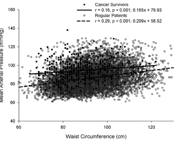
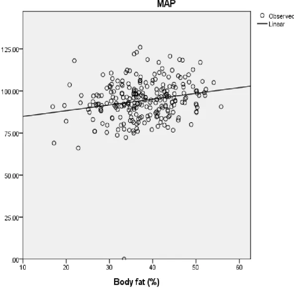
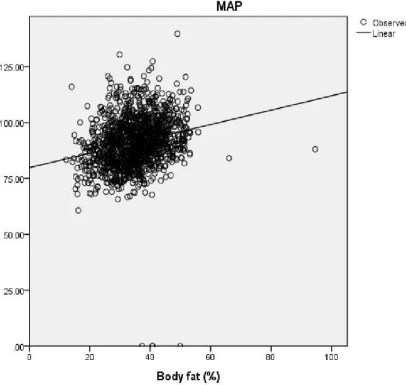
3.1 Relationship between mean arterial pressure and waist circumference
in regular (R) patients and cancer survivors (CS) ... 39 4.1 Physiologic Auto-memory 90 (Measures resting heart rate and blood
pressure) ...……… …….………..…….53 4.2 In-Body 230 (Body Composition Analyzer)...……… ………….….…53 4.3 Regression analysis of mean arterial pressure and body fat in cancer
survivors (CS)……… ………...56 4.4 Regression analysis of mean arterial pressure and body fat in regular (R)
LIST OF TABLES
Table Page
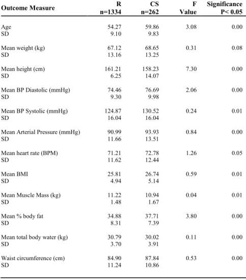
3.1 Summary table comparing all regular (R) patients to all cancer survivors
(CS) ... 36
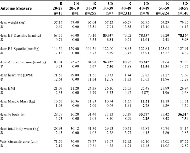
3.2 Anthropometric and vital signs measures stratified by 10-yr age groups (20-59 years) for regular (R) patients and cancer survivors (CS) ... 37
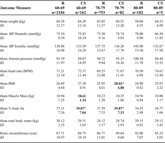
3.3 Anthropometric and vital signs measures stratified by 10-yr age groups (60-89 years) for regular (R) patients and cancer survivors (CS) ... 38
4.1 Group Statistics for all patients ... 52
4.2 Outcome measures for regular (R) patients and cancer survivors (CS) (Ages 20-59)……… ... 54
4.3 Outcome measures for regular (R) patients and cancer survivors (CS) (Ages 60-89) ... 55
5.1 All patients stratified by type of surgery... 70
5.2 Patients aged 40-49 stratified by type of surgery ... 71
5.3 Patients aged 50-59 stratified by type of surgery ... 72
5.4 Patients aged 60-69 stratified by type of surgery ... 73
5.5 Patients aged 70-79 stratified by type of surgery ... 74
6.1 Impingement and thoracic outlet testing……… ... 89
6.2 Patients stratified according to surgical intervention ... 89
6.3 Analysis of variance (ANOVA) of pre-surgery and post-surgery measures ... 90
6.4 Post-hoc analysis of shoulder flexion after surgery ... 90
6.5 Perceived activity level of patients ... 91
7.1 Treatment of patients stratified according to type of hormone therapy ... 94 7.2 Fitness levels of women at the VM Medical Integrative Health and
Wellness Centre (WC) ... 96 7.3 Mean values from the Canadian Physical Activity, Fitness and Lifestyle
RÉSUMÉ
OBJECTIFS
Cette thèse visait trois objectifs. Le premier consistait à créer un ensemble de tests normalisés à intégrer au processus de dépistage courant au Centre médical Ville-Marie (VM Médical), spécialisé dans la prévention, la détection, le diagnostic et le traitement du cancer du sein. Le deuxième objectif était de repérer les femmes risquant de développer diverses maladies cardiovasculaires et chroniques, et certains types de cancer. Le troisième objectif visait à déterminer quelles femmes risquent de souffrir de problèmes d’épaules à la suite d’une chirurgie.
MÉTHODOLOGIE
Les patientes de VM Médical ont été soumises à l’une des deux évaluations. Chez celles évaluées pendant leur bilan de santé annuel, les valeurs de la composition corporelle et de la pression sanguine ont été mesurées, puisqu’elles contribuent à déterminer le risque de maladies cardiovasculaires. Les patientes devant subir une chirurgie ont été soumises à un ensemble de tests sur la mobilité de l’épaule avant et après la chirurgie. Elles ont également reçu de l’information sur la chirurgie mammaire et les mesures à prendre pour minimiser les problèmes à la suite de la chirurgie.
RÉSULTATS
Selon l’analyse préliminaire des résultats de 9 315 patientes, les femmes ayant survécu à un cancer (C), qui ont été traitées contre cette maladie, étaient plus susceptibles de présenter des résultats défavorables concernant la circonférence de la taille, l’indice de masse corporelle (IMC) et la pression sanguine que les patientes régulières (R) qui n’ont reçu aucun traitement. Dans le groupe C, une corrélation faible a été constatée entre la circonférence de la taille et la pression sanguine.
Un sous-ensemble de 1 596 patientes, regroupant 262 patientes C et 1 334 patientes R, a également été évalué. Dans ce sous-ensemble, les valeurs de la pression sanguine, de la masse grasse et de la masse maigre ont été comparées. D’importantes différences entre les patientes C et R ont été relevées dans la plupart des résultats. De plus, une forte corrélation a été établie entre les valeurs de la masse grasse et de la pression sanguine dans le groupe C.
Dans un deuxième ensemble de données recueillies auprès de 4 300 patientes, une analyse a été réalisée pour déterminer les répercussions de la chirurgie et des différentes formes de traitement du cancer sur la composition corporelle et la pression sanguine. Les 747 femmes du groupe C ont été stratifiées selon les interventions chirurgicales mammaires courantes qu’elles ont subies. Les résultats de l’analyse indiquent que plus
les interventions n’étaient invasives, plus les patientes présentaient des résultats défavorables.
Ces données ont aussi été analysées en fonction des interventions thérapeutiques. L’analyse a démontré que les patientes ayant subi le plus de traitements présentaient les facteurs de risque de maladies cardiovasculaires les plus élevés, surtout parmi celles âgées de 40 à 49 ans. Un groupe constituait une anomalie par rapport à cette tendance : les femmes ayant subi une mastectomie, en particulier celles ayant subi une chirurgie seulement, sans aucun autre traitement. Ce groupe présentait aussi un risque accru de maladie cardiovasculaire comparativement aux patientes du groupe R.
Dans une autre analyse, 102 patientes ont été soumises à des tests avant et après la chirurgie. Selon les résultats des tests pré-chirurgicaux, un nombre élevé de femmes présentaient un conflit sous-acromial et un syndrome de la traversée thoracobrachiale. Les résultats des tests post-chirurgicaux ont démontré une diminution de la flexion de l’épaule, dont l’importance dépendait du type de chirurgie. La perte était supérieure chez les patientes ayant subi une mastectomie complète comparativement à celles ayant subi une mastectomie partielle.
CONCLUSION
Les femmes ayant reçu un traitement contre le cancer affichaient des valeurs défavorables par rapport aux autres en ce qui concerne la composition corporelle et la pression sanguine, et courraient donc un risque plus élevé de maladies cardiovasculaires. Il convient de souligner que ce risque accru était peut-être présent avant le traitement ou peut avoir été exacerbé par le traitement.
Le traitement vise principalement à atténuer les conséquences du cancer. Malheureusement, il peut entraîner certains problèmes secondaires, par exemple un risque accru de maladies cardiovasculaires et métaboliques. Comprendre ces risques contribuera à améliorer la qualité de vie des personnes survivant au cancer.
Mots-clés: cancer du sein, pression sanguine, composition corporelle, conflit sous-acromial
ABSTRACT
OBJECTIVES
The objectives of this thesis were three-fold. The first objective was to establish a series of standardized testing procedures to be included in the regular screening process at the Ville-Marie Medical Centre (VM Medical), a centre that specializes in the prevention, detection, diagnosis and treatment of breast cancer. The second objective was to identify women who may be at risk for various cardiovascular and chronic diseases, as well as at risk of developing certain types of cancers. The third objective was to identify women who may be at risk for shoulder problems associated with a surgical intervention. METHODOLOGY
Patients at VM Medical underwent one of two different evaluations. Patients who were being evaluated as part of their annual medical examination, had body composition and blood pressure values measured to help assess their level of risk for cardiovascular disease. Patients who were scheduled for surgery, underwent a series of pre and post-surgery shoulder mobility tests, and were also provided with information on what to expect with breast surgery and what to do to minimize post-surgical problems.
RESULTS
Preliminary analysis of 9,315 female patients found that cancer survivors (CS), who received cancer treatment, were more likely to have poorer outcome measures related to waist circumference, body mass index (BMI) and blood pressure compared to regular (R) patients who had not received any treatment. A weak correlation was found between waist circumference and blood pressure in the CS group.
A subset group of 1,596 patients made up of 262 CS and 1,334 R patients were also evaluated. In this analysis, blood pressure, body fat and lean muscle mass were compared. Significant differences were observed between CS and R patients in most measurements and a good correlation was seen between body fat and blood pressure values in the CS group.
In a second set of data collected on 4,300 patients, an analysis was conducted to determine how surgery and different forms of cancer treatment affected body composition and blood pressure values. The 747 women in the CS group were stratified according to common surgical interventions for breast surgery. The analysis showed that the more invasive the intervention the women received, the poorer were the outcome measures.
The same data was analyzed based on treatment intervention. The more treatment the patient received, the greater the impact was on cardiovascular risk factors especially,
in the 40-49 age group. The one group that was an anomaly from this trend was the mastectomy group, in particular those who had surgery alone and no other treatment. This group also exhibited increased risk of cardiovascular disease compared to the R patients group.
In another analysis, 102 patients were tested before and after surgery. Pre-surgery testing demonstrated that a significant number of the women had positive tests for shoulder impingement and thoracic outlet problems. Post-surgery testing showed a decrease in shoulder flexion, the significance of which, depended on the type of surgery performed. Patients that underwent a full mastectomy were affected more severely than patients who underwent a partial mastectomy.
CONCLUSION
Women who received cancer treatment displayed poor body composition and blood pressure values, creating a risk for cardiovascular disease. Note that this risk may have been present before treatment and/or exacerbated by treatment.
The primary goal of treatment is to address the underlying problems associated with cancer. Unfortunately, treatment can lead to some secondary problems such as increased risk for cardiovascular disease as well as metabolic diseases. Understanding these risks could help improve the quality of life for cancer survivors.
CHAPTER I
INTRODUCTION - REVIEW OF THE CURRENT LITERATURE
1.1 Breast Cancer
Over the past 15 years there has been a growing body of evidence to indicate that people who are diagnosed with cancer should utilize exercise to diminish the negative effects of cancer therapy (Kruijsen-Jaarsma et al., 2013). The idea of exercise having a positive impact on someone’s life has even gone one step further. Major health organizations have started to take the position that people can reduce their risk of developing certain types of illnesses and cancers if they include exercise as part of their daily activities (World Cancer Research Fund, 2007). These same health organizations have also indicated that once someone has had cancer and undergone adjuvant therapy, they should include regular physical activity as a way to reduce their risk of the cancer re-occurring (World Cancer Congress, 2012).
There are over 200 different types of cancers (MacDonald, Ford and Casson, 2004).This research project focused on one type of cancer, that is, breast cancer. Breast cancer is the most common cancer affecting women around the world, accounting for 23% of all cancers worldwide. According to Statistics Canada, it is estimated there will be approximately 22,800 new cases and 5, 000 deaths from this disease in Canadian 2013 (Canadian Cancer Society/Statistics Canada, 2013). Breast cancer in Canada accounts for 26% of all women’s cancers. About 1 in 9 women will develop breast cancer in their life time. Due to advances in prevention, early detection and treatment, however, the number of women surviving breast cancer has increased dramatically in recent years.
Epidemiologic studies have linked obesity and low levels of physical activity with an increased risk of breast cancer (Brown et al., 2003; Thune and Furberg, 2001). Clinical and epidemiologic studies have also identified obesity and weight gain as important negative prognostic factors for survival among women with this disease (Chleboski, et al., 2002). Physical activity has been associated with weight loss and weight maintenance among healthy individuals (Irwin et al., 2004; Wing, 1999). Studies have shown a favourable effect of exercise on body weight among breast cancer survivors (Rock and Demark-Wahnefried, 2002; Irvin et al., 2004). Despite the evidence indicating that regular physical activity can protect against weight gain, decrease breast cancer risk, and potentially improve breast cancer prognosis, efforts to encourage physical activity are not a routine part of the cancer treatment or rehabilitation process. Rarely are the topics of exercise and levels of activity addressed or even considered when patients are consulting with their physicians. The evidence is conclusive that physical activity for women before, during and after breast cancer therapy has a positive influence on the women’s overall health. Physical activity intervention researchers have been faced with similar challenges when evaluating activity levels and developing effective strategies aimed at physical activity behaviour (Vallance et al., 2007). Unfortunately despite the reported benefits of physical activity, the majority of breast cancer survivors are not meeting public health guidelines (at least 150 min/wk. of moderate-to-vigorous intense physical activity) (Irvin et al., 2004). This difficulty to modify breast cancer survivors’ physical activity behaviour seems to extend to the general population.
Several questions need to be asked: 1) Are women interested in finding out about exercise? 2) Are women utilizing the available resources to learn about exercise? 3) Are women actually changing their activity habits? 4) Are these changes in physical activity habits being reflected in women’s body compositions and levels of fitness?
The first question was addressed in a preliminary survey that was performed at VM Medical in 2007. The survey was performed to gain a better understanding of the interest
patients of the clinic would have in receiving information and guidance on exercise (Jones et al., 2008). The in-house survey helped to evaluate the interest in establishing an onsite fitness center for the women at VM Medical. Over a one-month period, 606 women at the clinic were asked about their level of activity, their interest in receiving direction on exercise and exercise rehabilitation, and whether they would make use of a local facility to exercise (Jones et al., 2008). The survey indicated that 45.4% (276) of the women considered themselves regular exercisers, 43% (263) of the women would be interested in receiving individual instruction on exercise and 34% (203) of the women would like the service to be available at VM Medical.
The results of the survey led to the opening of a fitness centre at VM Medical in November 2008. This led to the following questions: Has the establishment of a fitness program at VM Medical had an impact on the fitness level and body composition of the clientele? Is there a difference in cardiovascular risk factors between women who have been treated for breast cancer and women who are part of the regular patient clientele?
1.2 Shoulder problems associated with treatment for breast cancer
A number of studies have looked at the impact exercise has on people undergoing cancer therapy (Kruijsen-Jaarsma et al., 2013). Some studies have evaluated women before and after chemotherapy. However, many studies are done in a post-hoc manner, often after the completion of adjuvant therapy. The physical condition of the women before any type of exercise / physical activity interventions has gone relatively unstudied. For example, it is unclear how many women present with underlying shoulder pathology since current fitness levels and physical activity histories are seldom reported. When these measures are obtained, it is often after the initial intervention of cancer treatment; many times the measures are never taken. This is why it has been recommended that screening for pain and range of motion (ROM) restrictions has become an integral part of breast cancer follow-up care (Thomas-Maclean et al., 2008).
Aside from the lack of physical activity, survivors of breast cancer must deal with a host of physical, psychological and sociological problems that can occur with treatment. At this point it is appropriate to review some of the expected challenges women may encounter while undergoing therapy.
Breast cancer therapy may cause unfavourable changes in physical functioning, body composition, psychosocial functioning, and quality of life (QOL) (Courneya et al., 2007). Some of the limitations may include: 1) Weight gain, a commonly reported side effect of adjuvant chemotherapy (Campbell et al., 2007); 2) Breast cancer-related lymphoedema (BCRL) - a chronic swelling that can occur in the ipsilateral hand or arm of women treated for breast cancer (Lane et al., 2005); 3) Pain and ROM (Thomas-Maclean et al., 2008); 4) Overall body fatigue during and after treatment (Dimeo et al., 2008); and 5) Decrease in physical activity level leading to changes in body composition that may have important health implications for survivors (Campbell et al., 2007). It is clear that the need to regain shoulder mobility exists for this cohort of women, to avoid prolonged shoulder problems. One approach that maybe utilized to address this problem is to control the amount of post-operative pain the women are enduring so that they may begin to move their arm. Research projects by different groups (Kilgour et al., 2007) try to minimize the amount of post-operative shoulder dysfunction that does take place through the use of a home-based exercise program, but there is still much work to be done.
Certainly having a portion of one’s body removed is a very traumatic event but in comparison to many types of orthopedic surgery in and around the shoulder, this intervention is considered minor surgery (Leidenius et al., 2003). Oncology physicians are still not certain why so many women run into problems, yet many of the women who do undergo this regime of treatment are at risk for developing chronic arm, shoulder and neck problems (Leidenius et al., 2003).
Delay in, or lack of, immediate post-operative exercise may result in adverse outcomes such as spasm of the musculature surrounding the joint, muscle atrophy, tightening of the shoulder capsule and decreased short-and long-term functional mobility, leading to chronic immobility and pain (Kilgour et al., 2008).
Concerning shoulder function, two comparative studies have described significantly less impaired function after Breast Cancer Therapy (BCT) compared to ROM and moderately impaired shoulder mobility reported after BCT. Other studies have found no significant differences in such impairment between patients treated for BCT or ROM (Enrst et al., 2002; Kuehn et al., 2000).
It is possible that women may be predisposed to developing problems in the upper body. Thus, it may also be important to look at other areas of daily living where women may be at risk for developing pain in the neck, shoulder and arm regions. These predispositions to developing upper body complications are outlined in the following section.
Women who have a pre-existing shoulder problem:
A number of studies have shown that workers, performing the same movements over prolonged periods of time, are prone to be at risk of developing chronic musculoskeletal dysfunction and possibly neurological problems. These workers fall into a number of occupational groups, from manual labourers, workers in jobs that are considered hazardous (e.g. forestry), assembly line workers, professional support staff, and office workers, to name a few. In this project we focused on office workers whose jobs require them to spend a good portion of their workday at computer terminals. Over time this group demonstrated that they were at risk of developing problems in the neck, shoulder and arm regions (Johansson et al., 2003; Luime et al., 2004). These problems may include pain in the neck due to muscle tension, possibly leading to tension headaches. The pain in the shoulder region may be due to tendonitis (Lundberg et al., 1999), possible nerve
entrapment, causing periodic numbness (Pascarelli and Hsu, 2001), thoracic outlet, or brachial plexopathies (Mense and Simons, 2001). The pain in the elbow/forearm region may be related to lateral or medial epicondylitis (Johansson et al., 2003). Finally pain in the wrist, hand and finger may be due to carpal tunnel syndrome and/or Dequarvian syndrome (Rice et al., 1996).
A common thread among workers who experience pain in the neck, upper torso and arm regions, is that they are often asked to perform a task(s) that requires a muscle or group of muscles to maintain a static position for a prolonged period of time (Mense and Simons, 2001). This may be somewhat contrary to what many people may think leads to an increase in the risk of problems. The general population may believe that only workers who perform tasks that involve large muscle groups, like manual labourers, would be at risk. This perception has led to a delay in understanding and appreciating the impact of low intensity and repetitive movement have on the worker’s body (Mense and Simons, 2001).
Women seem to be at greater risk for developing pain in the neck, shoulder and arm regions in comparison to men (Chesterton et al., 2003). Women often comprise a significant portion of different occupations (Sorock and Courtney, 1996) that are most at risk to upper body injuries. Men have also been shown to have repetitive injury syndrome, but not to the same extent as women. When evaluating the work of men and women who work on production lines, there is a similarity in the type of chronic injuries that occur (Westgaard and Winkel, 1996), although women seem to be affected to a greater extent. There is growing consensus that musculoskeletal disorders may be related to the occupational activities of the individuals (Punnett and Wegman, 2004) but it is still unclear how much predisposing factors play a role in a worker developing musculoskeletal disorders. A common trend that is seen in many of these studies is the high prevalence of neck–shoulder pain in females (Chesterton et al., 2003).
An evaluation of muscle fiber composition in the trapezius muscle of the neck and shoulder region, showed a significant difference between men and women (Lindman et al; 1990, and 1991). This study led to speculation that women maybe at greater risk for developing neck, shoulder and arm problems. Through the use of muscle biopsies, a significantly greater proportion of type 1 muscle fibers were found in the trapezius muscle of women in comparison to men (Lindman et al., 1990; 1991). Thus, having a greater composition of type 1 (slow twitch fatigue resistant) fibers in the trapezius muscle would perhaps allow a worker not to fatigue as quickly when performing low intensity tasks, in comparison to type 2 fibers. On the other hand, having a greater composition of type 2 fibers (fast twitch non-resistant to fatigue) in the trapezius muscle, a worker would be able to generate greater explosive contractions/force, but would fatigue quicker. This initial investigation led Lindeman et al., (1991) to evaluate the way women and men perform the same activity, specifically data entry. The women involved in the study were shown to use shoulder, arm and wrist motions in a different manner compared to men and were expending significantly more energy. The women were also often found to be much more effective at entering large volumes of data compared to men. Entering more data and expending more energy may also be two factors that have contributed to women experiencing more pain while performing this task in comparison to men, who used less energy as well as entered less data (Lindman, Eriksson and Thornell, 1990).
Karlqvist et al. (2003) believe that workers that are in poor physical condition are at greater risk for developing pain and dysfunction in different regions of the body. Their work has shown that workers, both men and women, but in particular women, are not in good physical condition and that the jobs they perform on a regular basis do not improve their fitness level. If anything, the jobs tend to be deleterious to workers’ health (Chesterton et al., 2003).
It has been shown that many women are at risk of developing musculoskeletal pain in the upper body particularly the neck and shoulder regions. Often the jobs that they
perform along with the muscle fiber composition in their neck and shoulder region place them at greater risk of developing pain in these two locations compared to men (Chesterton et al., 2003; Johansson et al., 2003).
It would be expected that any pain a woman experiences in the neck and shoulder regions would have a lower priority once a lump in the breast has been discovered and surgery is recommended. It is unclear how many women who begin adjuvant therapy are dealing with musculoskeletal restrictions in these areas. Thus the above discussion leads to the following question: Does the presence of pre-existing upper body dysfunction prolong the recovery time for women who are undergoing breast sparing surgery?
Research has indicated that women who work outside of the home appear to be at greater risk of developing orthopaedic problems in the neck, shoulder and arms. Furthermore, women in western society also appear to be at greater risk for developing breast cancer compared to their eastern counterparts. The goal of this project is to evaluate the possible link between breast cancer therapy and the risk of developing upper body dysfunction in women who are undergoing adjuvant therapy. We believe that a number of women may already be at risk for developing problems in the upper body and that breast cancer therapy further increases their risk of dysfunction. Understanding the risk and implementing preventative strategies may help these women better tolerate the treatment protocols.
It seems apparent from the above discussion that the need to regain shoulder mobility exists for this cohort of women in order to avoid prolonged shoulder problems. One approach that may be utilized to address this problem is to control the amount of post-operative pain the women are enduring so that they may begin to move the afflicted ipsilateral arm. Exercise is a popular method to address limited shoulder and arm ROM (Kilgour et al., 2007).
Consistent with the requirements of a doctoral thesis, this was a multi-staged project made up of the following five stages;
Stage 1
In stage 1 of the project, I began with are view of the patient file database at VM Medical. The initial objective was to determine the cardiovascular risk of the women at the Centre, which led to evaluating the possibility of implementing standardize procedures for obtaining the anthropometric measurements, and blood pressure and resting heart rate values of the women. Also, testing of cancer survivors (CS) and regular (R) patients was done to see whether there were any differences between the two groups. We note a relationship between blood pressure and waist circumference in both of the groups.
Stage 2
In stage 2 of the project, we investigated the use of bioimpedance to measure body composition in CS and R patients. The additional information provided from bioimpedance allowed us the opportunity to evaluate the relationship between body fat and blood pressure.
Stage 3
In stage 3 of the project we utilized the standardized measurements of stage 1 and took into consideration the type of surgery and treatment that the women underwent. We then stratified the women according to the type of surgery and treatment they received.
Stage 4
In stage 4 of the project we looked at the incidence of pre-existing shoulder dysfunction and shoulder range of motion ROM in women preparing for breast surgery.
Stage 5
In stage 5 of the project we implemented the Jones template (Jones et al, 2007) to evaluate women who were about to undergo surgery associated with breast cancer. The template evaluation was included in the regular pre-surgery and post-surgery evaluation process.
At this stage the evaluator used the Jones template along with orthopaedic testing to evaluate if the women presented with upper body problems in addition to a diagnosis of a suspicious breast mass. The expectation was that the women who already had problems in the upper body before surgery were much more likely to continue to have problems after surgery.
1.4 Hypotheses
The working hypothesis was that cardiovascular risk factors were related to deleterious changes in body composition. The specific hypothesis for each stage of the project are as follows;
Stage 1
The hypothesis for this stage was that anthropometric measurements are directly proportional to deleterious cardiovascular risk factors.
Stage 2
For stage 2 the hypothesis was that an increase in resolution of body composition provided by bioimpedance analysis discriminates cardiovascular risk factors between cancer survivors (CS) and regular (R) patients.
Stage 3
The hypothesis for stage 3 was that cancer surgery and treatment type has a negative effect on both body composition and cardiovascular risk factors in CS when compared to R patients.
Stage 4
In this stage the hypothesis was two-fold. First, we expected that a number of women would be at risk for developing shoulder problems before breast surgery. Secondly, we suspected that some of the women were not meeting recommended fitness standards and that there would be a difference between CS and R patients.
Stage 5
We expected that certain locations on the Jones template would be more sensitive than other locations for breast cancer patients.
CHAPTER II
METHODOLOGY
2.1 Primary screening of VM Medical patient database 2.2 Stage 1: Preliminary evaluation
As part of the standard intake evaluation, patients waiting to meet with one of the physicians at the WC underwent a series of tests administered by one of the Centre’s kinesiologists. Results from tests, which included vital signs and body composition measurements, were entered into VM Medical’s electronic medical records. Each patient that came to VM Medical was asked to review and consider signing a consent form that allowed for their medical information to be entered into the electronic database. The information that was extracted from the electronic database and which was used for this study, was stripped of personal information so that no individual could be identified. The electronic database was used by the medical team to monitor and address patient needs. This study was approved by VM Medical Ethics Board and conformed to the World Medical Association (WMA) Declaration of Helsinki, Ethical Principles for Medical Research Involving Human Subjects.
As part of the preparation for testing, patients were asked to refrain from smoking or drinking caffeine products for at least 4 hours prior to testing. Before testing began, patients were also asked if they needed to use the washroom to void any fluids. Testing of the patient took place in a medical evaluation room, away from the main clinic area. The kinesiologist explained to the patient the purpose for the testing. After approximately 5 minutes the patient was then given the opportunity to relax and ask questions before the testing began.
Resting heart rate and blood pressure values were obtained through the use of an automated sphygmomanometer (Physiologic Auto-memory 90; AMG Medical Inc., Montréal Canada). The patient’s left arm was used for testing. If the patient had breast surgery on the left side of their torso with lymph node(s) removed, the right arm was used.
The cuff was wrapped around the upper arm and was supported at the level of the heart. The cuff was aligned with the brachial artery. If measurements obtained on the patient were outside of expected values for the patient’s age group, the test was repeated after a 5-minute waiting period.
The patient’s height was taken using a stadiometer (equipment used for measuring height, consisting of a vertical ruler with a sliding horizontal rod that is adjusted so that it rests on the top of the head. Also, the patient’s weight was taken using a weight scale. Waist circumference was the last measurement taken. The American College of Sports Medicine (ACSM) 2013, guidelines were followed to measure waist circumference and was measured at the level of the greater trochanter of the hip and the umbilicus using a Gulick anthropometric tape (Mississagua, Ontario Canada). Waist measurements were taken with the patient standing upright, feet together, and arms at their side while maintaining a relaxed breathing pattern. A horizontal measure was taken at the narrowest location between the umbilicus and the sternum. The test was repeated twice and the average was taken. If there was a difference of more than 5 millimeters between measurements, a third measurement was taken.
Patients were classified into two groups; cancer survivors (CS) or regular (R) patients. All of the women classified as CS underwent either a partial or a full mastectomy and may also have received adjuvant therapy such as chemotherapy, radiotherapy and hormonal therapy. Some patients may have received only one adjuvant therapy while others may have received up to three. Patients classified, as R patients may not have had
any of the adjuvant therapies mentioned above. Both the CS and the R patients may also have had other health issues for which they were taking additional medication that may have affected outcome measurements.
2.3 Stage 2: Inclusion of the bioimpedance unit to measure body fat
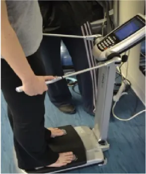
The recently validated (Karelis et al 2013) body composition measurement by bioimpedance was used to determine body weight, percent body fat, lean muscle mass, and total body water. Before standing on the foot pads of the unit (In-Body 230; Seoul, Korea), the patient was asked to remove her shoes and socks as well as any external metal objects (e.g. watches, rings) that might affect the results. The patient was asked to hold on to the external handles of the unit allowing data to be collected and documented from the display by the kinesiologist.
2.4 Stage 3: Stratifying by surgery and stratifying by treatment
CS were stratified according to one of five common surgical interventions that they underwent: 1) A partial mastectomy; 2) A full mastectomy; 3) Bilateral surgery; 4) Bilateral full mastectomy; and 5) Reconstruction, either a unilateral reconstruction or bilateral reconstruction that may have occurred on the same day as the full mastectomy or on a different day.
CS were also stratified according to the treatment intervention they underwent. The eight different categories included: 1) Surgery only; 2) Surgery and chemotherapy; 3) Surgery and radiotherapy; 4) Surgery and hormone therapy; 5) Surgery, chemotherapy, radiotherapy and hormone therapy; 6) Surgery, radiotherapy and hormone therapy; 7) Surgery, chemotherapy and radiotherapy; and 8) Surgery, chemotherapy and hormone therapy.
2.5 Stage 4: Pre-surgery shoulder assessment
Patients who were scheduled for breast surgery were asked to undergo a preoperative evaluation by the kinesiology team. The preoperative session had five main components that included; 1) Outlining to the patient what she could expect post-operatively; 2) Reiterating the surgeon’s preoperative instructions; 3) Outlining the post-operative exercises that needed to be performed; 4) Performing base line testing of patients, including shoulder mobility, arm strength, present activity level, body composition, shoulder impingement, and testing to evaluate neurovascular occlusion; 5) Addressing additional questions that the patient had concerning treatment.
Kinesiologists met with each patient for approximately 90 minutes. Recognizing the amount of stress that the patient was under and given the recent diagnosis of a suspicious lump in their breast, it was left to the discretion of the kinesiologist working in consultation with the patient to determine what information and testing was most important for the patient at that particular stage.
A series of tests were performed to evaluate the patient’s shoulder range of motion (ROM), forearm strength, and the presence of shoulder pain or reduced upper extremity circulation before surgery. The breast surgery performed was either a partial or full mastectomy with sentinel node or axillary node sampling.
Before their surgery the women were asked to undergo testing (methodology described below) to evaluate their current level of shoulder ROM and their current level of physical activity. During the testing session, the kinesiologists advised the women on the appropriate exercises to be performed post-operatively. Also, the women were provided an opportunity to clarify any questions they had concerning potential post-operative problems. Again, recognizing the amount of stress that the patient was under with a recent diagnosis of a suspicious lump in their breast, it was left to the discretion of the kinesiologists working in consultation with the patient to determine what information
and testing was most important for the patient at this stage. The following tests were performed;
Shoulder Flexion
Shoulder flexion was assessed with the use of a handheld goniometer, an instrument used to measure range of motion (ROM). The subject was assessed in a standing position, with the axis of the goniometer positioned at the acromion process of the scapula, through the head of the humerus. The stationary arm of the goniometer was placed along the mid axillary line of the trunk, while the moving arm was placed along the midline of the humerus in line with the lateral epicondyle.
Forearm Strength
A Jamar handgrip dynamometer (Lafayette Instruments, Lafayette, IN USA) was used to assess forearm strength. The adjustable handle was fit to the hand size of the patient so that the proximal interphalangeal (PIP) joints of digits 2 through 5 were positioned at a 90 degree angle of finger flexion as a starting position. The subject kept their shoulder in a neutral position and their elbow extended by their side while performing the test. The subject squeezed the dynamometer with maximum isometric effort, and maintained the effort for 5 seconds.
Shoulder Dysfunction
Shoulder impingement was assessed with the use of the Neer test and the Hawkins Kennedy. With the Neer test, the patient’s arm was moved into end range of flexion at the same time as the patient’s scapula was stabilized. The test was considered positive if it elicited pain, typically at the end of the range of flexion. If the patient had full shoulder flexion with overpressure applied at the end of the range without experiencing pain, then the test was considered negative.
The Hawkins Kennedy test was performed by flexing the elbow to 90 degrees and then elevating the patient's arm to 90 degrees of shoulder flexion and then internally rotating
the shoulder. The patient either experienced pain with this manoeuvre or tried to elevate the ipsilateral shoulder to increase the subacromial space and prevent the impingement. When pain occurred with either of these tests, it was considered a positive sign for sub-acromial impingement. The goal behind these tests was to transiently aggravate inflamed or impinged structures as they passed under the coracoacromial arch. The inflamed structures may have included the long head of biceps tendon, the supraspinatus tendon and/or sub acromial bursa.
Thoracic outlet test using the Roos test
Testing was performed to assess for the possibility of neurovascular occlusion often associated with thoracic outlet syndrome through the use of a Roos Test (Howard, Lee and Dell 2003).The patients were asked to abduct both shoulders to 90 degrees and then flex both of their elbows to 90 degrees. Patients were then asked to open and close their hands repeatedly for up to 3 minutes. During that time if the patient started to experience numbness and/or a pins and needles sensation in the hands, the test was stopped and a positive sign noted. If there was no presence of numbness and/or a pins and needles sensation, then a negative sign was recorded.
Level of activity
To gain an understanding of the patient’s typical level of physical activity, patients were asked to indicate what best represented their current level of activity. The scale was: 1) Active, 1 day per week 2) Active 3-5, days a week, and 3) Active, every day of the week. Patient’s height and weight were recorded and classified according to BMI (Body Mass Index) based on guidelines from the Canadian Physical Activity, Fitness and Lifestyle Approach (CPAFLA, CSEP, 2003). Patients were ranked as follows; Mild Thinness (BMI of less than 17.9), Normal (BMI 18.0-24.9), Overweight (BMI 25.0-29.9), Obese level 1 (BMI 30.0-34.9), Obese level 2 (BMI 35.0-39.9), Obese level 3 - morbidly obese (BMI greater than 40.0).
Data analysis
Statistical analysis was performed using IBM SPSS version 19.0. A one-way nova with Tukey post-hoc analysis was performed on shoulder ROM measures with significance assumed at P<0.05. Simple t-tests were performed on handgrip strength measures. Significance was set at P< 0.05. Pearson correlation was used to evaluate relationships between outcomes measures. Significance was set at P< 0.05.
2.6. Stage 5: Evaluation of the fitness level of patients
An evaluation of the fitness level of patients was performed on both R patients and CS at the WC. A battery of tests, proposed by the Canadian Society of Exercise Physiology, namely the Canadian Physical Activity Fitness & Lifestyle Approach (CPAFLA, CSEP 2003), was utilized to evaluate the patients. The tests are based on age-matched data to determine how each individual ranked in comparison to people in the same age-matched group. The tests included assessment of low back and abdominal strength, handgrip strength, lower body flexibility as well as aerobic testing with the use of a treadmill and the modified Bruce protocol. A screening process was done to identify women that were at risk; either a Physical Activity Readiness Questionnaire (Par-Q) or a medical Par-Q form was completed for each patient that was tested.
2.7 Stage 6: Validation of the Jones template on breast cancer patients
In this stage the subjects underwent primary and secondary measures that are outlined in the following protocol.
Pre and post-surgical measurements in both Stage 4 objective measures and self-reported data were collected. The outcome measures were repeated for both stages. Primary outcome measures were: 1) The assessment of arm/shoulder mobility using the “arm-shoulder/upper torso grid” developed by Jones et al. (2007) to measure pressure pain threshold (PPT) with a handheld algometer. The upper body grid
evaluates the PPT of eight different locations in the upper arm and torso. The specific locations were selected for the following reasons; 1) Several of the locations such as the trapezius, deltoid and infraspinatus have been used in previous studies. 2) Locations such as the rhomboid and supraspinatus have often been identified in a clinical setting as being problematic. 3) The biceps and triceps were selected because these regions have been found to be frequently painful especially in women who have had breast surgery with an axillary node dissection for the determination of breast cancer. Sensitivity of locations and possible trigger points were noted and recorded. This template is important when considering the fact that many breast cancer patients develop myofascial dysfunction characterized by the presence of palpable nodules ”trigger points” (Cheville 2007). (Evaluation time required, 25 minutes)
Twenty one (21) female subjects were recruited from VM Medical. Demographic information including age, height, weight and mean body mass was collected.
As part of their job, subjects typically spent at least 2 hours a day at a computer terminal. Some subjects experienced shoulder pain or significant musculoskeletal pain at the time but were not receiving any therapeutic treatments to address their existing problems. Subjects were recruited for this study following approval of the l’Université du Québec à Montréal (UQAM) Human Research Ethics Committee. Each subject had all of the risks of participating in this study explained to them and were asked to voluntarily give their written informed consent. (Annexes 1 &2)
The subjects were asked not to undertake any new physical activity in the two days preceding the testing. New physical activities were defined as activities performed routinely for an extended period of time (for example, weight training after a prolonged period of inactivity). The ingestion of caffeine has been found to alter a person’s perception of pain so subjects were asked to avoid ingesting any caffeine products (Galeotti et al., 2002) for at least 4 hours prior to each testing period. Subjects were also asked to avoid any analgesic medication for 12 hours prior to the testing time.
Women participating in this project underwent repeated measures at 3 different intervals during a 2-month period beginning from the time of their breast cancer diagnosis. The objective measures were repeated (Maastricht questionnaires were only completed at the pre-surgery evaluation).
All participants for this project were recruited through VM Medical. The women in the project, aged 25-60, were in relatively good health, and without multiple health related problems with the exception of being diagnosed with a suspicious breast mass. The attending physician/medical team recommended a course of treatment which either began with surgery or neo-adjuvant therapy. The women participating in the project did not have any psychological or emotional factors which would have limited their ability to understand the project. The women were then referred to the VM Medical Integrative Health and Wellness Centre (WC) where baseline measurements were taken. As with most research we wanted to have a positive impact on the lives of the women dealing with breast cancer. Our goal was not to establish a program that would end when the research project ended. Our goal was to implement changes in the typical protocols for breast cancer patients so as to diminish the negative signs and symptoms associated with treatment. A number of the tests that were used in this protocol could easily be carried over to clinical situations, and utilized as part of the standard protocol/procedure for patients.
Materials
The testing protocol outlined in detail below was initially utilized and validated in the 2008 Masters project by D.H. Jones. For consistency the same calibration of the algometer, subject preparation, testing locations on the subject, and application of the algometer, were utilized for all projects.
Calibration: The algometer was calibrated at the onset of each day of testing before
being applied to the subject. The algometer (Somedic Algometer Type II; Somedic Production AB, Sollentuna, Sweden) was calibrated using a standard protocol recommended by the manufacturer. The acceptable calibration values ranged from 98 to 102 kilopascals. This meant a potential 4% error in the recorded values. The calibration process was repeated 3 times to make sure that calibration values fell within the expected range. When the values did not fall within the acceptable, ± 2 kilopascal range, the algometer was restarted and the calibration process repeated.
Application: Following the manufacturer’s calibration guidelines, the 0.5 cm applicator
head was replaced with a 2 cm applicator head to measure PPT’s. The 2 cm applicator head reduced the risk of causing excessive pain, and bruising. Using an applicator head smaller than 2 cm on subjects who were post-surgery may have placed them at an increased risk of bruising and localized tissue trauma. This may have heightened their concern about developing lymphedema, a significant problem for this population (McCredie et al., 2001). The 2 cm applicator appears to have been more effective at indicating local pain sensation when compared to smaller applicator heads (Takahashi et al., 2005).
Patient Position
The subject was placed in a supine position on a portable treatment table and then in a prone position on the same table. The two different positions were used so that the algometer could be safely applied and allowed for better stabilization of shoulder and torso compared to a seated position. With the subject in the supine position, the kinesiologist applied the algometer to the anterior aspect of the shoulder and arm for the PPT measurement over the marks located on the anterior deltoid and bicep. With the subject in a prone position, the kinesiologist then applied the algometer to the posterior aspect of the arm (triceps), shoulder (posterior deltoid) and torso (infraspinatus, supraspinatus, rhomboids and the trapezius) for the measurement of PPTs. Pillows and
padding were used to support the subject’s shoulder and arm when appropriate. The total time required to collect 3 complete sets of data for each subject (4 trials total) was approximately 25 minutes. This included the time to change the position of the subject (8 times total; supine-prone-supine) as well as the time to set up and stabilize the shoulder. This worked out to approximately a 5-minute rest time between applications of the algometer at each location.
Testing
An initial trial was performed at the outset of each testing session to familiarize the subject with the testing procedure. The data from this trial was discarded. Thereafter, three complete sets of data were collected on the subject. A complete set of data included applying the algometer to each location (1 through 8 in sequence) and recording the values obtained from the location. This order of measurement was done two more times. Each trial (of all 8 locations) were recorded on a single data sheet. Three complete sets of data were collected on each subject and at each testing session. When the data sheet was completed, the sheet was removed from view of the kinesiologist so that subsequent data sets could be collected without bias.
Determination of the PPTs
At the beginning of each testing session, the subject was told that a PPT is defined as “the instant or moment that the pressure on the skin surface changes from the sensation of pressure to the sensation/perception of pain”. The kinesiologist explained to each subject that they would feel a gradual increase in pressure on the skin. The pressure would continue to increase until the subject would experience a sensory transition from pressure to pain. The kinesiologist explained to each subject that the trial at a specific location would end if the subject pressed the handheld switch or if the readings went beyond 400 kilopascals. This precautionary measure was installed to avoid any unnecessary pain or damage to the skin and underlying structures. The kinesiologist applied the nozzle of the handheld algometer to each landmark location at an
approximate rate of 30 kilopascals per second. The increase in the force applied to the skin surface was viewed by the experimenter via the digital output display on the algometer. The kinesiologist continued to apply pressure until the subject pressed the button on the handheld switch indicating that they perceived a sensory transition from pressure to pain. The subject never viewed the recorded values.
Reference points - Bony landmarks
The use of bony landmarks as anatomical reference points (McGee, 2002; Kendall et al., 2005) was thought to be the most appropriate method to identify the locations in the upper extremity and torso region. The use of a bony reference point is a common technique used by clinicians to locate anatomical structures due to the tremendous variability in body composition. For this project it would have been impractical to measure a specific distance with a tape measure because of the number of locations in and around the scapula and the variability of the scapula’s position relative to the spinal column.
2.8 Statistical analyses performed in projects
Statistical analyses were performed using IBM SPSS version 19.0.
Projects 1 and 2
Simple t-tests were performed between the groups and the significance was established at P< 0.05. Regression analysis was performed looking at the relationship between mean arterial pressure and waist circumference, as well as mean arterial pressure and body fat.
Projects 3 and 4
A one-way ANOVA and Dunnett post-hoc analysis with significance set at P< 0.05. R patients were used as the control group. CS were stratified according to age, in groups
of 10 years. The same one-way ANOVA with Dunnett post-hoc analysis was performed.
Project 5
A one-way nova with Tukey post-hoc analysis was performed on shoulder ROM with significance established at P<0.05. Simple t-tests were performed on handgrip strength measures. Significance was fixed at P< 0.05. Pearson correlation test was used to evaluate relationships between outcomes measures. Significance level was set at P< 0.05.
Individual and combined effects of the variables on the risk of neck and shoulder pain were examined using regression analysis. The 0.05 significance level was used as a guide to determine the statistical significance of the relationship between the variable time at the computer, previous pathology in the model, and the dependent variable, shoulder dysfunction.
CHAPTER III
MANUSCRIPT NUMBER 1
3.1 Overview of the first manuscript
This manuscript was presented in part as a poster at the annual conference of the National Consortium of Breast Centers 2012.The primary goal of the project was to investigate the potential relationship between cardiovascular risk and body composition in both R patients and CS and to determine if any differences in cardiovascular risk factors existed between both groups.
The main finding of the project was the significant differences across a number of outcome measures when comparing CS to R patients. Also evident was that waist circumference changes do not reflect changes in mean arterial pressure in CS. After this initial study it was proposed that additional information on body composition, specifically, body fat, and muscle mass be gathered to allow for a potentially more robust evaluation of cardiovascular risk factors.
3.2 Manuscript 1: Increased cardiovascular risk factors in breast cancer survivors identified by routine measurements of body composition, resting heart rate and arterial blood pressure
David H. Jones M.Sc., CAT (C), CSCS1,2,3, Melisa Nestore M.Sc.3, Sara Henophy
M.Sc.3, Julia Cousin B.Sc.3, Alain S. Comtois Ph.D., FACSM 2.
1Department of Exercise Science 2Département de kinanthropologie
The Richard J. Renaud Science Complex Université du Québec à Montréal
Concordia University CP 8888, Succursale centre-ville
7141 Sherbrooke Street West Montréal, QC H3C 3P8
3Ville-Marie Medical Centre
Ville-Marie Medical
Integrative Health & Wellness
1538 Sherbrooke West, 8th floor, suite 810
Montréal, QC H3G 1L5
Abstract
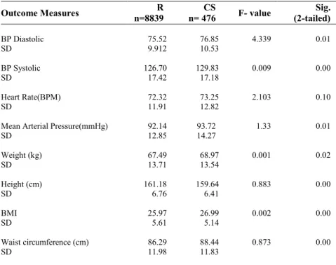
Fundamental body composition measurements is important information that can be utilized to determine the risk for various diseases. At the Ville-Marie Medical Centre (VM Medical), kinesiologists worked side by side with the medical/oncology team to collect a number of base-line measurements on body composition, resting heart rate, and blood pressure, as part of the standard intake evaluation when female patients visited their physician for their annual checkup. A total of 9,315 patients were evaluated: 476 cancer survivors (CS) and 8,839 regular (R) patients. CS were more likely to have a higher BMI (P = 0.001) and a larger waist circumference (P = 0.001) than R patients. CS were also shown to have higher blood pressure values: diastolic pressure of 76.9 mmHg ± 10.5 vs 75.5 mmHg ± 9.9, (P = 0.01) and systolic pressure of 129.8 mmHg ± 17.2 vs 126.7 mmHg ±17.4 (P = 0.001) compared to R patients, respectively.
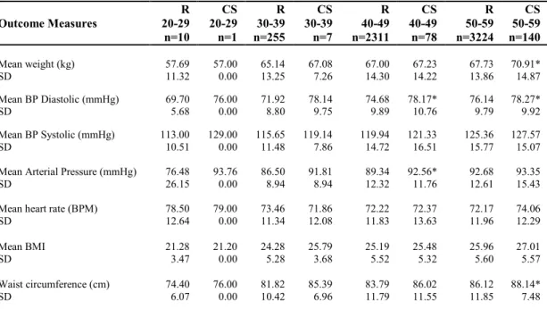
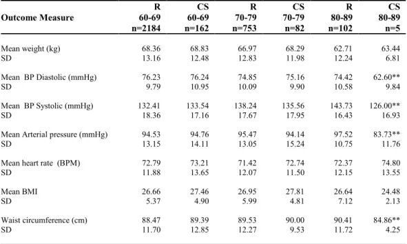
Data was also stratified according to ten-year age segments. The same trends were seen in many of the age groups. Regression analysis looking at the relationship between mean arterial pressure and waist circumference did not show any difference between the two groups that is CS vs R. Patients who had a higher BMI, a larger waist circumference, and higher blood pressure levels, were also shown to be at greater risk for developing cardiovascular disease, diabetes and various musculoskeletal problems as well as an increased risk for various forms of cancers including reoccurrence of previously treated cancer.
Changes in body composition should be considered by the medical team when planning preventative healthcare strategies for their patients.
Introduction
It has been well established that as body composition changes with an increase in waist circumference and an overall increase in body mass index (BMI), there is a greater risk for individuals to develop various metabolic diseases such as diabetes, heart disease, as well as an increased risk of developing cancer or having a reoccurrence of a previously treated cancer (Han et al 2006). Major health organizations, such as the World Health Organization (WHO), have recognized the importance of physical activity, maintaining ideal body size, improving dietary habits and ceasing smoking to reduce the risk of developing various metabolic diseases (Harvie et al 2005). Unfortunately even with these guidelines, many people still present with increases in total body fat, increases in BMI, as well as reduced fitness levels (Flegal et al 2010).
A number of studies have looked at the relationship between changes in body composition and vital sign measures and the increase in risk for developing various diseases. Many studies have had to rely on patients self-reporting these measures or have had to rely on data extraction from records kept at multiple locations, which tend to result in underestimated patient body composition values (Battaglini et al 2011, Blair et al 2011).
Some researchers have used meta-analysis to evaluate the impact that body composition has on one’s health. (Carmichael and Bates 2004).
In our study we hypothesized that by measuring a woman’s body composition and vital signs as part of their regular follow-up care and compared the CS to the R patients that we would obtain a better understanding of the potential health risks that cancer patients face after undergoing cancer therapy. Thus, the main objective of this prospective study
was to obtain a better understanding of the body composition and vital sign measures of CS compared to R patients.
Methodology
Participants were recruited at VM Medical. Patients waiting to meet a physician as part of their routine checkup or de novo consult were invited to go to the adjacent VM Medical Integrative Health and Wellness Centre (WC) to undergo a series of tests administered by one of the resident kinesiologists. Results from tests, which included vital signs and body composition measurements, were entered into VM Medical’s electronic medical records. The information that was extracted from the electronic database and that was used for this study had personal information removed so that no individual could be identified. Each patient that came to VM Medical was asked to review and sign a consent form that allowed for their medical information to be entered into the electronic database. The electronic database was used by the medical team to monitor and address patient needs. This study was approved by VM Medical’s Ethics Board and conformed to the World Medical Association (WMA) Declaration of Helsinki, Ethical Principles for Medical Research Involving Human Subjects.
Testing of the patient took place in a medical evaluation room, away from the main clinic area. The kinesiologist explained to the patient the purpose of the testing. After approximately 5 minutes the patient was given the opportunity to relax and ask questions before the testing began.
Resting heart rate and blood pressure values were obtained with the use of an automatic blood pressure monitor (Physiologic Auto-memory 90; AMG Medical Inc., Montréal Canada). This monitor contains a technology called Fuzzy Logic that enables personalized inflation levels resulting in accurate and more comfortable readings. The patient’s left arm was used for testing. If the patient had breast surgery on the left side of their torso with lymph node(s) removed, the right arm would be used. The cuff was wrapped around the upper arm and the arm was supported at the level of the heart. The