HAL Id: hal-03211880
https://hal.inrae.fr/hal-03211880
Submitted on 29 Apr 2021
HAL is a multi-disciplinary open access
archive for the deposit and dissemination of
sci-entific research documents, whether they are
pub-lished or not. The documents may come from
teaching and research institutions in France or
abroad, or from public or private research centers.
L’archive ouverte pluridisciplinaire HAL, est
destinée au dépôt et à la diffusion de documents
scientifiques de niveau recherche, publiés ou non,
émanant des établissements d’enseignement et de
recherche français ou étrangers, des laboratoires
publics ou privés.
Distributed under a Creative Commons Attribution| 4.0 International License
Effect of Aqueous Extract and Polyphenol Fraction
Derived from Thymus atlanticus Leaves on Acute
Hyperlipidemia in the Syrian Golden Hamsters
Mhamed Ramchoun, Tarik Khouya, Hicham Harnafi, Souliman Amrani,
Chakib Alem, Mohamed Benlyas, Fatima Kasbi Chadli, El-Hassane Nazih,
Patrick Nguyen, Khadija Ouguerram
To cite this version:
Mhamed Ramchoun, Tarik Khouya, Hicham Harnafi, Souliman Amrani, Chakib Alem, et al.. Effect
of Aqueous Extract and Polyphenol Fraction Derived from Thymus atlanticus Leaves on Acute
Hyper-lipidemia in the Syrian Golden Hamsters. Evidence-Based Complementary and Alternative Medicine,
Hindawi Publishing Corporation, 2020, 2020, pp.1-9. �10.1155/2020/3282596�. �hal-03211880�
Research Article
Effect of Aqueous Extract and Polyphenol Fraction Derived from
Thymus atlanticus Leaves on Acute Hyperlipidemia in the Syrian
Golden Hamsters
Mhamed Ramchoun ,
1,2,3Tarik Khouya ,
2Hicham Harnafi,
3Souliman Amrani,
3Chakib Alem,
2Mohamed Benlyas,
2Fatima Kasbi Chadli,
4,5El-Hassane Nazih,
6Patrick Nguyen,
7and Khadija Ouguerram
4,51Laboratory of Biotechnology & Sustainable Development of Natural Resources, Polydisciplinary Faculty,
University of Sultan Moulay Slimane, Beni Mellal 23000, Morocco
2Biochemistry and Natural Substances Team, Department of Biology, Faculty of Sciences & Techniques,
University of Moulay Ismail, Errachidia 52000, Morocco
3Laboratory of Biochemistry, Department of Biology, Faculty of Sciences, University of Mohammed I, Oujda 60000, Morocco 4INRA, UMR 1280, Physiology of Nutritional Adaptations, Nantes, France
5CRNH, West Human Nutrition Research Center, CHU, Nantes, France
6EA 2160 MMS Laboratory of Biochemistry, Faculty of Pharmacy, Nantes, France 7ONIRIS, Nantes-Atlantic National College of Veterinary Medicine, UNE, Nantes, France
Correspondence should be addressed to Mhamed Ramchoun; mmhamed92@yahoo.com
Received 9 November 2019; Revised 1 February 2020; Accepted 3 March 2020; Published 28 March 2020 Academic Editor: Vincenzo De Feo
Copyright © 2020 Mhamed Ramchoun et al. This is an open access article distributed under the Creative Commons Attribution License, which permits unrestricted use, distribution, and reproduction in any medium, provided the original work is properly cited.
Thymus atlanticus, an endemic plant of Morocco, is traditionally used as a liniment or a drink to treat various diseases. However,
there are few available scientific data regarding its biological effects. In this connection, the present study aimed to investigate the hypolipidemic and antioxidant effects of aqueous extract and polyphenol fraction of Thymus atlanticus in Syrian golden hamsters treated with Triton WR-1339 (triton, 20 mg/100 g body weight). The hamsters orally received the extracts (400 mg/kg), and blood samples were collected after 24 h of treatment to determine plasma lipid, insulin, and fasting blood glucose levels. Plasma malondialdehyde level and plasma total antioxidant (TAS) were also evaluated. The T. atlanticus extracts significantly decreased triglycerides, total cholesterol, VLDL-C, and LDL-C and increased HDL-C when compared with the hyperlipidemic group. Both extracts suppressed the effect of the triton injection on TAS and reduced the level of plasma malondialdehyde. The extracts produced no significant change in the blood glucose level but effectively prevented the mild hyperinsulinemia induced by triton. These findings suggest that T. atlanticus may be a useful alternative treatment for the control of hyperlipidemia and its related diseases.
1. Introduction
Hyperlipidemia is one of the major risk factors of athero-sclerosis diseases [1]. It is defined as high plasma concen-trations of total cholesterol (TC), triglycerides (TGs), and low-density lipoprotein cholesterol (LDL-C). Hyperlipid-emia is associated with increased incidence of
atherosclerosis, which is the main cause of cardiovascular diseases such as stroke, coronary artery disease, and pe-ripheral artery disease [2]. Therefore, the reduction in the levels of plasma lipid is the main strategy for the control of the progressivity of cardiovascular diseases [3]. However, the current hypolipidemic drugs such as statins have many serious side effects [4]. Some plants have been used to reduce
Volume 2020, Article ID 3282596, 9 pages https://doi.org/10.1155/2020/3282596
plasma levels of TC, TGs, and LDL-C in animal models [5, 6].
On the other hand, oxidative stress is involved in the pathogenesis of various diseases including atherosclerotic diseases [7]. The human body can generate numerous en-zymatic and nonenen-zymatic antioxidants, but their amount could be insufficient to effectively neutralize the free radicals particularly under oxidative stress and inflammation con-ditions [8]. Hence, the supplementation of human diet with additional antioxidants derived from plants may have beneficial effects in atherosclerosis by preventing the ac-cumulation of the free radicals and improving the antiox-idant defense system [9].
In the traditional medicine, some subjects suffering from hyperlipidemia utilize herbal drinks as an alternative re-medial tool to treat lipid metabolism disorders [10]. Moreover, the search for hypolipidemic drugs from me-dicinal plants has been intensified in recent years [11].
Thymus atlanticus (local Moroccan name: Z’itra or
Azukni; English name: thyme) is an endemic medicinal herb of Morocco. Its leaves are used to prepare liniments or decoctions for treating several diseases including skin in-fections, rheumatism, and bronchitis and, generally, for its potentialities as an anti-inflammatory agent [10]. Moreover, it was reported that the aqueous extract of T. atlanticus possesses high total phenolic content and exhibited a potent anti-inflammatory effect in animal models of inflammation and inhibits blood coagulation ex vivo and in vivo [12, 13]. In addition, the aqueous extract of this plant showed potent in
vitro antioxidant activity, but, to our knowledge, its
anti-oxidant activity in vivo has not yet been studied [5, 13, 14]. A preliminary screening of some Thymus species from Mo-rocco for hypolipidemic activity indicated that T. atlanticus showed possible hypolipidemic effects in a rat model [5]. However, in this latter study, the rats were treated with a high dose of aqueous extracts of the selected plants and the rat model is not an appropriate model for evaluating the hypo-lipidemic drugs because it has very low level of LDL-C and high level of high-density lipoprotein cholesterol (HDL-C) [15].
The hamster model is widely used for studying the lipid metabolism disorders and screening the new lipid-lowering drugs as the lipid metabolism of the hamster is similar to that of humans. Similar to humans, the hamster is endowed with cholesterol ester transfer protein (CETP) and all the enzy-matic pathways of the metabolism of lipoproteins and bile [16, 17]. Therefore, this study was conducted to evaluate the effects of the aqueous extract and the polyphenol fraction of
T. atlanticus on the plasma lipid concentrations and the
antioxidant status in acute hypolipidemic hamsters.
2. Materials and Methods
2.1. Plant Material. Leaves of T. atlanticus (Ball) Roussine
(synonym: Thymus dreatensis) were collected from the High Atlas Mountains (32°15′ N, 5°25′ E, 1995–2012 m),
Erra-chidia, Morocco, in the month of April. The plant was authenticated by Dr. Ibn Tatou. A voucher specimen (no. RAB77496) was deposited in the herbarium of the Scientific Institute, Mohammed V University, Rabat, Morocco.
2.2. Preparation of Plant Extracts. The preparation of the
aqueous extract (AE) was carried out in accordance with the method described in [18]. The extraction of polyphenol fraction (Pp) was performed based on the method of Jordan et al. [19].
2.3. Total Phenolic Content (TPC) Assay. The TPC assay was
performed based on the Folin–Ciocalteu method [20] de-scribed in [18]. The TPC was expressed as mg caffeic acid equivalent (CAE) per g of dried extract.
2.4. In Vitro Antioxidant Activity. The 2,2 diphenyl
pic-rylhydrazyl (DPPH) scavenging assay was performed based on the method described in [21] with minor modifications [18]. Antioxidant capacity was evaluated as the capacity of extracts to reduce Fe3+ to Fe2+ by the Benzie and Strain method [22]. The inhibition of β-carotene-linoleic acid bleaching by T. atlanticus extracts was evaluated according to Elzaawely et al. [23].
2.5. Animals and Experimental Design. Animal procedures
were carried out in strict accordance with the recom-mendations in the Guide for the Care and Use of Labo-ratory Animals of the European Union “European directive 2010/63/EU” and of the Ethical Principles of Animal Experimentation of the Ministry for Food, Agri-culture, and Fisheries (France). The protocol was approved by the local committee (Bretagne-Pays de la Loire Com-mittee). All surgery were performed under sodium pen-tobarbital anesthesia, and all efforts were made to minimize suffering.
Forty adult male golden Syrian hamsters (70–90 g, 8weeks old, Animal Experimental Station, Nantes, France) were used as experimental animals in the current study. They were maintained in standard cages (3 hamsters per cage) under a 12 h light/dark cycle at a temperature of 22 ± 3°C.
The experimental hamsters were divided into four groups of 10 animals each. Hamsters were rendered hyperlipidemic by an intraperitoneal injection of Triton WR-1339 (triton, 20 mg/100 g BW) one hour after the administration by oral route of distilled water (hyperlipidemic group, HG), AE, or Pp at a dose of 400 mg/kg BW. The control (normolipidemic group, NG) was not injected with triton and received dis-tilled water only. After 24 h of treatment, blood was taken from the retro-orbital sinus, after a slight anesthesia with isoflurane. The blood samples were centrifuged (10 min, 4°C,
3000 g), and the obtained plasma was used for the analysis of glucose, insulin, and lipids. The hamsters were euthanized at the end of the experiment with an intraperitoneal injection of sodium pentobarbital (60 mg/kg BW).
2.5.1. Dosage of Plasma Total Cholesterol (TC) and Triglyc-erides (TGs). The TC and TGs in plasma were quantified
using enzymatic kits (Bio-Merieux, France), according to the manufacturer’s instructions.
2.5.2. FPLC Analysis. The cholesterol and TG profiles in
lipoproteins were determined using fast protein liquid chromatography (FPLC) (AKTA FPLC SYSTEM, GE Healthcare, USA). The detailed procedure is available in a previous publication [24].
2.5.3. Glucose and Insulin Measurement. The blood glucose
level was measured in a drop of blood immediately after sampling using a glucometer (Roche Diagnostics, France). Insulin was analyzed in plasma prepared from the blood collected via the retro-orbital sinus of food-deprived ham-sters using the rat insulin ELISA kit (Shibayagi, Japan).
2.5.4. In Vivo Antioxidant Activities. The level of
malon-dialdehyde (MDA) in plasma was measured based on the method described in [24]. Plasma total antioxidant status (TAS) was determined according to the manufacturer’s protocol of the rat TAS reagent kit (TAS, RANDOX Lab-oratories Ltd, United Kingdom).
2.6. Statistical Analysis. Data were represented as
mean-± standard deviation (SD) (n � 10). Statistical analysis was performed using Statview software (SAS Institute Inc., 100 SAS Campus Drive, Cary, NC 27513-2414, USA). All comparisons between groups were made by means of one-way ANOVA test followed by post hoc analysis (Tukey’s or Dunnett’s test). Differences were statistically significant at
p< 0.05.
3. Results
3.1. Total Phenolic Content of T. atlanticus Extracts. The yield
of extraction of AE and Pp was 15% (w/w) and 9 % (w/w), respectively. The AE and Pp had the TPC values of 481.1 ± 23.42 and 589.34 ± 43.56 mg CAE/g of dried extract, respectively (Table 1).
3.2. In Vitro Antioxidant Activity. The in vitro antioxidant
activity of AE and Pp was evaluated using three different assays.
Results are presented as mean ± SD, n � 6.†Total phe-nolic content is expressed as mg caffeic acid equivalent per g of dried extract.
The results are presented in Table 2. The IC50values of AE and Pp in DPPH scavenging assay were 0.44 ± 0.02 mg/ mL and 0.012 ± 0.005 mg/mL, respectively. Trolox had the IC50 value of 0.51 ± 0.01 mg/mL. The DPPH scavenging activity of Pp and AE was significantly higher than that of Trolox (p < 0.05).
The FRAP values of AE and Pp were 50.31 ± 1.65 and 67.96 ± 4.12 mmol Trolox/g of dried extract, respectively (Table 2). The β-carotene bleaching results of AE and Pp present the IC50values of 46.8 ± 4.15 and 13.05 ± 1.35 mg/ mL, respectively. BHT had an IC50value of 0.12 ± 0.03 mg/ mL.
3.3. Effects of AE and Pp on Hyperlipidemia in Hamsters 3.3.1. Plasma TC and TGs. Figure 1 summarizes the level of
plasma TC and TGs. The injection of triton resulted in a significant increase in the level of plasma TC (72%; p < 0.01) and TGs (89.8%; p < 0.001) in HG when compared with hamsters not treated with triton (NG).
The plasma TC and TGs registered a significant decline in the AE-treated group by about 41% (p < 0.05) and 19.03% (p < 0.01), respectively, when compared with HG (Figure 1(a)). The TC and TG levels were also significantly decreased in the Pp-treated group by about 55% (p < 0.01) and 57% (p < 0.001), respectively (Figure 1(b)).
The plasma TC and TGs registered a significant decline in the AE-treated group by about 41% (p < 0.05) and 19.03% (p < 0.01), respectively, when compared with HG (Figure 1(a)). The TC and TG levels were also significantly decreased in the Pp-treated group by about 55% (p < 0.01) and 57% (p < 0.001), respectively (Figure 1(b)).
3.3.2. FPLC Profiles of TC and TGs in VLDL, LDL, and HDL.
The plasma lipoprotein profiles obtained by the FLPC analysis are shown in Figure 2. The results showed that the intraperitoneal injection of triton significantly increased the levels of VLDL-TGs and VLDL-C by about 92% and 97%, respectively, and effectively decreased HDL-C by about 84%, in comparison with the NG (Figures 2(a) and 2(b)).
The levels of VLDL-TGs and VLDL-C were seen to be significantly decreased in the AE-treated group by about 2% and 72% and in the Pp-treated group by 40% and 80%, respectively (Figures 2(c) and 2(d)). In addition, AE and Pp significantly increased HDL-C by about 88% and 90%, re-spectively (Figures 2(c) and 2(d)). Plasma C and LDL-TGs concentrations were very low (almost zero g/L) in AE-treated and Pp-AE-treated groups (Figures 2(c) and 2(d)).
3.3.3. Plasma Insulin and Blood Glucose. There were no
significant differences in blood glucose levels between the groups (Figure 3(a)). The injection of triton resulted in a significant (p < 0.001) increase of plasma insulin level by about 89% when compared with theNG. The levels of plasma insulin were significantly decreased by about 69% (p < 0.01) and 40% (p < 0.05) in AE and Pp groups, respectively, when compared with the NG (Figure 3(b)).
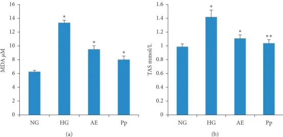
3.3.4. In Vivo Antioxidant Activity. Figure 4 shows the
plasma level of MDA Figure 4(a) and the plasma TAS Figure 4(b). The results indicate that the MDA level sig-nificantly increased by about 53% (p < 0.001) in the HG when compared with the NG. The AE and Pp significantly
Table 1: Total phenolic content of the aqueous extract and the polyphenol fraction.
Aqueous extract Polyphenol fraction Total phenolic content† 481.10 ± 23.42 589.34 ± 43.56
(p < 0.001) decreased the MDA level by about 29% and 40%, respectively, when compared with the HG (Figure 4(a)).
The plasma level of TAS was significantly increased in the HG when compared with the NG (1.42 ± 0.04 mmol/L vs. 0.99 ± 0.05 mmol/L; p < 0.01) (Figure 4(b)). The T. atlanticus extracts effectively suppressed the effect of triton on TAS. The plasma level of TAS in AE-treated and Pp-treated groups was similar to that in theNG (p > 0.05).
4. Discussion
In the present study, hamsters were rendered hyperlipidemic by an intraperitoneal injection of triton to evaluate the hypolipidemic and antioxidant effects of two polyphenol-rich extracts of T. atlanticus. The hamster is the best model to study the lipid metabolism disorders and for screening new lipid-lowering drugs as the lipid metabolism of ham-sters is similar to that of humans. Like humans, the hamster is endowed with cholesterol ester transfer protein (CETP) and all the enzymatic pathways in the metabolism of lipo-proteins and bile [16, 17]. In addition, triton is known to cause hyperlipidemia through several mechanisms, mainly by inhibiting lipoprotein lipase activity, which can result in the blockage of TG-rich lipoproteins clearance [25].
In the present study, administration of a single dose of AE or Pp of T. atlanticus ameliorated acute hyperlipidemia in hamsters, evidenced by a significant decrease in the levels
of plasma TC and TGs. We also found that the T. atlanticus extracts suppressed the effect of triton on antioxidant status and effectively prevented the increase of plasma MDA. In addition, these extracts also prevented the changes induced by triton in lipoprotein profiles. Moreover, the injection of triton produced a marked increase in VTGs and LDL-C levels and a significant decrease in the HDL-LDL-C level.
Our results are in accordance with the results of Ram-choun et al. (2012) who have reported that the crude AE of T.
atlanticus prevented the increase of plasma TC, TGs, and
LDL-C and effectively increased the HDL-C level in the rat model of acute hyperlipidemia. The effects of T. atlanticus extracts on lipid profiles in rat were similar to those of fibrates (fenofibrate 65 mg/kg BW), which are one of the major classes of the current hypolipidemic drugs, mainly used to reduce plasma TGs concentration [5].
Elevated plasma levels of TC, LDL-C, and TGs are the major risk factors for heart disease and stroke in the world [26]. The reduction of circulating levels of these particles result in a significant reduction in the incidence and the severity of heart diseases [3]. Therefore, the hypolipidemic drugs such as statins and fibrates are the main strategy for controlling and preventing cardiovascular diseases [4, 27]. However, the statins and fibrates have many side effects. This has attracted a great attention for the discovery of new hypolipidemic drugs with fewer side effects [4, 28]. In this context, some extracts derived from medicinal plants have
Table 2: Antioxidant activity (DPPH, FRAP, and β-carotene assay) of the aqueous extract and polyphenol fraction. Extracts DPPH (IC50mg/mL) FRAP (mmol Trolox/g DR) β-carotene (IC50mg/mL)
Aqueous extract 0.44 ± 0.02∗ 50.31 ± 1.65ns 46.8 ± 4.15∗∗∗
Polyphenol fraction 0.012 ± 0.005∗∗∗ 67.96 ± 4.12∗∗ 13.05 ± 1.35∗∗∗
Trolox 0.51 ± 0.01 — —
BHT — — 0.21 ± 0.03
BHT, butylated hydroxytoluene; DPPH, DPPH scavenging activity; DR, dry extract; FRAP, ferric reducing antioxidant power. Results are presented as mean ± SD and analyzed with ANOVA and Dunnett’s test, n � 6.∗p< 0.05,∗∗p< 0.01, and∗∗∗p< 0.001; ns, no significance versus Trolox or BHT.
∗∗ ∗ ∗∗ NG HG AE Pp 0 0.5 1 1.5 2 2.5 3 3.5 4 4.5 TC le vel (g /L ) (a) ∗∗∗ ∗∗ ∗∗∗ NG HG AE Pp 0 20 40 60 80 100 120 140 T Gs le ve l (g/L) (b)
Figure 1: Effect of AE and Pp of Thymus atlanticus on lipid concentrations. (a) TC. (b) TGs. AE, aqueous extract-treated hamsters; HG, hyperlipidemic hamsters; NG, normolipidemic hamsters; Pp, polyphenol fraction-treated hamsters; TC, total cholesterol; TGs, triglycerides. Results are expressed as mean ± SD and analyzed with ANOVA followed by Tukey’s test, n � 10.∗p< 0.05,∗∗p< 0.01, and∗∗∗p< 0.001; HG versus NG, AE, and Pp versus HG.
HDL VLDL LDL 1 10 19 28 37 46 TC TG 0 0.2 0.4 0.6 0.8 1 1.2 Le ve ls (g/L) (a) HDL LDL VLDL 1 10 19 28 37 46 [TC] [TG] 0 2 4 6 8 10 12 14 Le ve ls (g/L) (b) VLDL LDL HDL 1 10 19 28 37 46 [TC] [TG] 0 2 4 6 8 10 12 14 Le ve ls (g/L) (c) LDL VLDL HDL 1 10 19 28 37 46 [TC] [TG] 0 2 4 6 8 10 Le ve ls (g/L) (d)
Figure 2: Lipoprotein profiles determined by FPLC. (a) Normolipidemic group. (b) Hyperlipidemic group. (c) AE-treated hamsters. (d) Pp-treated hamsters. HDL, high-density lipoprotein; LDL, low-density lipoprotein; TC, total cholesterol; TG,triglycerides; VLDL, very LDL.
ns ns ns NG HG AE Pp 0 10 20 30 40 50 60 70 80 90 Blo od g lucos e (m g/dL) (a) ∗∗∗ ∗∗ ∗ NG HG AE Pp 0 2 4 6 8 10 12 14 16 18 20 Pl asma in su lin ( µU/mL) (b)
Figure 3: Levels of blood glucose (a) and plasma insulin (b). AE, aqueous extract-treated hamsters; HG, hyperlipidemic group; NG, normolipidemic group; Pp, polyphenol fraction-treated hamsters. Results are presented as mean ± SD and analyzed with ANOVA followed by Tukey’s test, n � 10.∗p< 0.05,∗∗p< 0.01, and∗∗∗p< 0.001; ns, not significant; HG versus NG, AE, and Pp versus HG.
been found to reduce atherosclerotic lesions and attenuate atherosclerosis development, along with a reduction of plasma lipid concentrations (TC, TGs, and LDL-C) and atherogenic indexes, in animal models for atherosclerosis [29, 30]. In the present study, the AE and Pp of T. atlanticus effectively reduced TC, LDL-C, and TGs levels, which suggests beneficial effects of these extracts in the control of cardiovascular diseases. On the other hand, the plasma HDL-C level is inversely associated with the prevalence of heart diseases [31]. We found that the HDL-C level was considerably increased upon treatment with a single dose of AE or Pp. The atherogenic effects of HDL-C are due to its role in reverse cholesterol transport and to its associated antioxidant enzymes such as paraoxonase 1 (PON1), which can degrade various proatherogenic compounds resulted from lipid oxidation [32]. Among these oxidation products, MDA, a marker of lipid oxidation and cardiovascular dis-ease, is a toxic compound that can interact with DNA and proteins, which results in the dysfunction of the endothe-lium and other atherogenic events [33]. The reduction of plasma level of MDA in the hyperlipidemic hamsters re-ceiving the T. atlanticus extracts suggests a preventive effect of these extracts against lipid oxidation and atherosclerosis development. Like hyperlipidemia, oxidative stress is a se-rious risk factor for many diseases such as heart diseases [34]. The lipid oxidation results in the generation of other atherogenic products such as oxidized LDL-C, which can induce endothelium dysfunction and activate inflammation and coagulation systems, leading to the development and progression of the atherosclerotic plaque [35]. In the present study, the T. atlanticus extracts suppressed the effect of triton on TAS and showed potent antioxidant activity, evidenced by a greater capacity to donate electrons (FRAP assay) and scavenge free radicals (DPPH assay) in com-parison with standard antioxidant, Trolox. These findings are in accordance with our previous studies, which have demonstrated that the T. atlanticus extracts protect the blood red cell membranes against 2,2′-azobis
(2-amidinopropane) dihydrochloride-mediated oxidation and plasma lipids against CuSO2-mediated oxidation [13]. Most studies have confirmed that the antioxidants can prevent lipid oxidation mediated by the free radicals and alleviate atherosclerosis and its related diseases [36]. The antioxidant proprieties of the plant are due to its secondary metabolites, mainly polyphenols [37]. Moreover, Thymus species have been reported to have high amount of phenolic acids and flavonoids [18, 38]. Based on previous phytochemical ana-lyses [5, 12, 13], the HPLC chromatograms of the T.
atlanticus AE and Pp showed the presence of various
phenolic compounds with predominance of rosmarinic acid, caffeic acid, and quercetin. As mentioned previously [13], T.
atlanticus showed highest total phenolic and flavonoid
contents than other Thymus species.
Concerning the mechanism of the hypolipidemic effects of the T. atlanticus extracts, at this time, we did not examine further on this issue. It has been reported that polyphenols inhibit the activity of 3-hydroxy-3-methyl-glutaryl-CoA reductase in vitro [39], reduce intestinal cholesterol ab-sorption [40], and inhibit the activity of CETP [41]. Moreover, the pharmacological inhibition of CETP has become a strategy for increasing HDL-C and decreasing LDL-C levels and has proven to be effective in the treatment of atherosclerosis and cardiovascular diseases [42]. The effect of T. atlanticus on decreasing LDL-C and increasing HDL-C may be partly due to the inhibition of CETP. Furthermore, the simultaneous decline in TC and LDL-C in plant-treated groups upon extract administration allows us to suggest that the T. atlanticus extracts decreased TC by enhancing the elimination of the LDL-C particles through their hepatic receptors.
Epidemiologic studies have shown that the intake of dietary polyphenols could regulate plasma lipid concen-trations and decrease the prevalence of cardiovascular dis-eases [43, 44]. Polyphenols have beneficial effects on the regulation of the PNO1 expression and the stabilization of lipoproteins [45]. In addition, polyphenols decrease
∗ ∗ ∗ NG HG AE Pp 0 2 4 6 8 10 12 14 16 MD A µM (a) ∗ ∗ ∗∗ NG HG AE Pp 0 0.2 0.4 0.6 0.8 1 1.2 1.4 1.6 TA S mmo l/L (b)
Figure 4: Plasma malondialdehyde levels (a) and plasma total antioxidant status (TAS) (b). AE, aqueous extract-treated hamsters; HG, hyperlipidemic hamster; NG, normolipidemic hamsters; Pp, polyphenol fraction-treated hamsters. Results are presented as mean ± SD and analyzed with ANOVA and Tukey’s test, n � 10.∗p< 0.01 and∗∗p< 0.001; HG versus NG, AE, and Pp versus HG.
oxidative stress in macrophages and enhance their efficacy in cholesterol efflux, ameliorate antioxidant status in athero-sclerotic lesions, and attenuate atheroathero-sclerotic plaque pro-gression [43,44]. Atherosclerosis is a multifactorial process that implicates many systems such as inflammation and coagulation. Thus, the antiatherogenic effect of polyphenols could also be due to the inhibition of proinflammatory enzymes, cytokines secretion, and coagulation factors [9]. In this connection, a previous study has reported that the T.
atlanticus’ AE suppressed inflammation induced by
carra-geenan and croton oil in animal models and effectively inhibited blood coagulation in vitro [13]. This suggests additional therapeutic effects of the active compounds of T.
atlanticus in cardiovascular diseases.
In addition, the injection of triton significantly in-creased the plasma insulin level without affecting the blood glucose level in hamsters. Both AE and Pp produced no significant change in the glucose level, but they effectively suppressed the effect of triton on the insulin level and prevented against mild hyperinsulinemia. Diabetes is as-sociated with lipid level abnormalities and accelerates atherosclerotic cardiovascular diseases [46]. Moreover, insulin regulates lipid metabolism at several levels such as the expression and the synthesis of lipoprotein lipase and the synthesis and the clearance of VLDL and chylomicrons [47]. The increase in plasma lipids after the injection of triton may be due to the increase in plasma insulin in hamsters. In fact, the beneficial effect of the T. atlanticus extracts on lipid metabolism may be partly attributed to their action on the insulin level. Clinical trials have been reported that the intake of polyphenols alleviates cardio-vascular risk in people with diabetes [48]. Most studies verified that the benefits of rosmarinic acid, which was the most abundant phenolic compound in T. atlanticus[13], are partly due to its antidiabetic effects. Rosmarinic acid has been found to improve insulin sensitivity and glucose uptake in cell culture and in rodents. It exhibited potent inhibitory effect on α-glucosidase and α-amylase [49]. The inhibition of these enzymes is widely used as a strategy to manage hyperglycemia associated with type 2 diabetes [50]. A recent study has demonstrated that the aqueous ex-tract of T. atlanticus can be classified as a low-toxicity exex-tract according to the Organization for Economic Co-operation and Development [12].
There are few limitations in this study that could be addressed in future works. Chronic hyperlipidemia induced by high-fat diet was not studied because we focused only on acute hyperlipidemia and the mechanisms of hypolipidemic action of T. atlanticus have not been explored.
5. Conclusion
Oral administration of the T. atlanticus extracts ameliorates the lipidemic state in triton-induced hyperlipidemic ham-sters via decreasing plasma cholesterol and LDL-C levels, increasing HDL-C, improving antioxidant status, and pre-venting mild hyperinsulinemia. The active compounds in the T. atlanticus extracts could be used in the treatment of hyperlipidemia.
Data Availability
The data used to support the findings of this study are available from the corresponding author upon request.
Conflicts of Interest
The authors declare that they have no conflicts of interest.
Acknowledgments
The authors gratefully acknowledge Mr. Karim Ramdaoui and Mr. El Mostapha Bedraoui for helping in animal care and particularly thank Dr. Mustapha Rouis for his assistance.
References
[1] Cardiovascular diseases (CVDs). https://www.who.int/news-room/fact-sheets/detail/cardiovascular-diseases-(cvds). [2] A. Saeed, E. Feofanova, B. Yu et al., “Association of elevated
triglycerides and atherogenic lipoproteins with incident cardiovascular diseases: insights from genetic data in the atherosclerosis risk in communities study,” Journal of Clinical
Lipidology, vol. 11, no. 3, p. 788, 2017.
[3] WHO, Raised Cholesterol, WHO, Geneva, Switzerland, 2015. [4] S. Sultan, A. D’Souza, I. Zabetakis et al., “Statins: rationale, mode of action, and side effects,” in The Impact of Nutrition
and Statins on Cardiovascular Diseases, pp. 171–200, Elsevier,
Amsterdam, Netherlands, 2019.
[5] M. Ramchoun, H. Harnafi, C. Alem et al., “Hypolipidemic and antioxidant effect of polyphenol-rich extracts from Moroccan thyme varieties,” e-SPEN Journal, vol. 7, no. 3, pp. e119–e124, 2012.
[6] D. Iyer and U. K. Patil, “Assessment of antihyperlipidemic and antitumor effect of isolated active phytoconstituents from
Apium graveolens L. through bioassay-guided procedures,” Journal of Dietary SupplementsJournal of Dietary Supple-ments, vol. 16, no. 2, pp. 193–206, 2019.
[7] P. Mury, E. N. Chirico, M. Mura, A. Millon, E. Canet-Soulas, and V. Pialoux, “Oxidative stress and inflammation, key targets of atherosclerotic plaque progression and vulnera-bility: potential impact of physical activity,” Sports Medicine, vol. 48, no. 12, pp. 2725–2741, 2018.
[8] I. Miro´nczuk-Chodakowska, A. M. Witkowska, and M. E. Zujko, “Endogenous non-enzymatic antioxidants in the human body,” Advances in Medical Sciences, vol. 63, no. 1, pp. 68–78, 2018.
[9] A. B. Santhakumar, M. Battino, and J. M. Alvarez-Suarez, “Dietary polyphenols: structures, bioavailability and protec-tive effects against atherosclerosis,” Food and Chemical
Toxicology, vol. 113, pp. 49–65, 2018.
[10] J. Bellakhdar, R. Claisse, J. Fleurentin, and C. Younos, “Repertory of standard herbal drugs in the Moroccan phar-macopoea,” Journal of Ethnopharmacology, vol. 35, no. 2, pp. 123–143, 1991.
[11] I. Touiss, S. Khatib, O. Bekkouch, S. Amrani, and H. Harnafi, “Phenolic extract from Ocimum Basilicum restores lipid metabolism in triton WR-1339-induced hyperlipidemic mice and prevents lipoprotein-rich plasma oxidation,” Food Science
and Human Wellness, vol. 6, no. 1, pp. 28–33, 2017.
[12] T. Khouya, M. Ramchoun, S. Amrani et al., “Anti-inflam-matory and anticoagulant effects of polyphenol-rich extracts
from Thymus atlanticus: an in vitro and in vivo study,” Journal
of Ethnopharmacology, vol. 252, 2019.
[13] T. Khouya, M. Ramchoun, A. Hmidani et al., “Anti-inflam-matory, anticoagulant and antioxidant effects of aqueous extracts from Moroccan thyme varieties,” Asian Pacific
Journal of Tropical Biomedicine, vol. 5, no. 8, 2015.
[14] A. Hmidani, E. Dine, T. Bouhlali, T. Khouya, and M. Ramchoun, “Anticoagulant activities of three thymus species grown in southeastern Morocco,” Future Journal of
Pharmaceutical Sciences, vol. 5, no. 1, 2019.
[15] R. C. Gupta, Biomarkers in Toxicology, Elsevier, Amsterdam, Netherlands, 2014.
[16] M. P. Sullivan, J. J. Cerda, F. L. Robbins, C. W. Burgin, and R. J. Beatty, “The gerbil, hamster, and Guinea pig as rodent models for hyperlipidemia,” Laboratory Animal Science, vol. 43, no. 6, pp. 575–578, 1993.
[17] C. S. Stancu, G. M. Sanda, M. Deleanu, and A. V. Sima, “Probiotics determine hypolipidemic and antioxidant effects in hyperlipidemic hamsters,” Molecular Nutrition & Food
Research, vol. 58, no. 3, pp. 559–568, 2014.
[18] T. Khouya, M. Ramchoun, A. Hmidani et al., “Acute toxicity and antiproliferative and procoagulant activities of fractions derived from Thymus satureioides of the Moroccan high at-las,” South African Journal of Botany, vol. 121, pp. 568–576, 2019.
[19] M. J. Jord´an, R. M. Mart´ınez, C. Mart´ınez, I. Moñino, and J. A. Sotomayor, “Polyphenolic extract and essential oil quality of Thymus zygis ssp. gracilis shrubs cultivated under different watering levels,” Industrial Crops and Products, vol. 29, no. 1, pp. 145–153, 2009.
[20] V. L. Singleton, R. Orthofer, and R. M. Lamuela-Ravent´os, “[14] Analysis of total phenols and other oxidation substrates and antioxidants by means of folin-ciocalteu reagent,”
Oxi-dants and AntioxiOxi-dants Part A, vol. 299, pp. 152–178, 1999.
[21] L. Barros, S. Falcão, P. Baptista, C. Freire, M. Vilas-Boas, and I. C. F. R. Ferreira, “Antioxidant activity of Agaricus sp. mushrooms by chemical, biochemical and electrochemical assays,” Food Chemistry, vol. 111, no. 1, pp. 61–66, 2008. [22] I. F. F. Benzie and J. J. Strain, “The ferric reducing ability of
plasma (FRAP) as a measure of “antioxidant power”: the FRAP assay,” Analytical Biochemistry, vol. 239, no. 1, pp. 70–76, 1996.
[23] A. Elzaawely, T. Xuan, H. Koyama, and S. Tawata, “Anti-oxidant activity and contents of essential oil and phenolic compounds in flowers and seeds of Alpinia zerumbet (pers.) B.L. Burtt. & R.M. Sm,” Food Chemistry, vol. 104, no. 4, pp. 1648–1653, 2007.
[24] F. Kasbi Chadli, A. Andre, X. Prieur et al., “n-3 PUFA prevent metabolic disturbances associated with obesity and improve endothelial function in golden Syrian hamsters fed with a high-fat diet,” British Journal of Nutrition, vol. 107, no. 9, pp. 1305–1315, 2012.
[25] P. E. Schurr, J. R. Schultz, and T. M. Parkinson, “Triton-induced hyperlipidemia in rats as an animal model for screening hypolipidemic drugs,” Lipids, vol. 7, no. 1, pp. 68–74, 1972.
[26] B. A. Ference, J. J. P. Kastelein, A. D. Sniderman, M. S. Sabatine, and A. L. Catapano, “A mendelian random-ization study comparing the effect of low-density lipoproteins and triglyceride-rich very low-density lipoproteins on the risk of coronary heart disease,” Atherosclerosis, vol. 275, pp. e78–e79, 2018.
[27] M. F. La Fountaine, C. M. Cirnigliaro, J. C. Hobson et al., “Fenofibrate therapy to lower serum triglyceride
concentrations in persons with spinal cord injury: a pre-liminary analysis of its safety profile,” The Journal of Spinal
Cord Medicine, vol. 44, pp. 1–6, 2019.
[28] R. L. Attridge, C. R. Frei, L. Ryan, J. I. M. Koeller, and W. D. Linn, “Fenofibrate-associated nephrotoxicity: a review of current evidence,” American Journal of Health-System
Pharmacy, vol. 70, no. 14, pp. 1219–1225, 2013.
[29] M. Sedighi, M. Bahmani, S. Asgary, F. Beyranvand, and M. Rafieian-Kopaei, “A review of plant-based compounds and medicinal plants effective on atherosclerosis,” Journal of
Research in Medical Sciences, vol. 22, no. 1, 2017.
[30] L.-C. Lee, L. Wei, W.-C. Huang, Y.-J. Hsu, Y.-M. Chen, and C.-C. Huang, “Hypolipidemic effect of tomato juice in hamsters in high cholesterol diet-induced hyperlipidemia,”
Nutrients, vol. 7, no. 12, pp. 10525–10537, 2015.
[31] R. K. Mutharasan, C. S. Thaxton, J. Berry et al., “HDL efflux capacity, HDL particle size, and high-risk carotid athero-sclerosis in a cohort of asymptomatic older adults: the Chi-cago healthy aging study,” Journal of Lipid Research, vol. 58, no. 3, pp. 600–606, 2017.
[32] D. A. Chistiakov, A. A. Melnichenko, A. N. Orekhov, and Y. V. Bobryshev, “Paraoxonase and atherosclerosis-related cardiovascular diseases,” Biochimie, vol. 132, pp. 19–27, 2017.
[33] D. Wadhwa, V. K. Mahajan, K. S. Mehta et al., “Malon-dialdehyde, lipoprotein-a, lipoprotein ratios, comprehensive lipid tetrad index and atherogenic index as surrogate markers for cardiovascular disease in patients with psoriasis: a case-control study,” Archives of Dermatological Research, vol. 311, no. 4, pp. 287–297, 2019.
[34] P. Poprac, K. Jomova, M. Simunkova et al., “Targeting free radicals in oxidative stress-related human diseases,” Trends in
Pharmacological Sciences, vol. 38, no. 7, 2017.
[35] A. J. Kattoor, S. H. Kanuri, and J. L. Mehta, “Role of ox-LDL and LOX-1 in atherogenesis,” Current Medicinal Chemistry, vol. 26, no. 9, pp. 1693–1700, 2018.
[36] D. Tousoulis, T. Psaltopoulou, E. Androulakis et al., “Oxi-dative stress and early atherosclerosis: novel antioxidant treatment,” Cardiovascular Drugs and Therapy, vol. 29, no. 1, pp. 75–88, 2015.
[37] H.-Y. Lin, T.-C. Chang, and S.-T. Chang, “A review of an-tioxidant and pharmacological properties of phenolic com-pounds in Acacia confusa,” Journal of Traditional and
Complementary Medicine, vol. 8, no. 4, pp. 443–450, 2018.
[38] B. Salehi, M. S. Abu-Darwish, A. H. Tarawneh et al., “Thymus spp. plants - food applications and phytopharmacy proper-ties,” Trends in Food Science & Technology, vol. 85, pp. 287– 306, 2019.
[39] M. S. Costamagna, I. C. Zampini, M. R. Alberto et al., “Polyphenols rich fraction from Geoffroea decorticans fruits flour affects key enzymes involved in metabolic syndrome, oxidative stress and inflammatory process,” Food Chemistry, vol. 190, pp. 392–402, 2016.
[40] M.-L. Ricketts and B. S. Ferguson, “Polyphenols: novel sig-naling pathways,” Current Pharmaceutical Design, vol. 24, no. 2, pp. 158–170, 2018.
[41] K. L. Cheuk, Z. Zhang, H. Yu et al., “Apple polyphenols inhibit plasma CETP activity and reduce the ratio of non-HDL to HDL cholesterol,” Molecular Nutrition & Food Research, vol. 52, no. 8, pp. 950–958, 2008.
[42] J. Armitage, C. Baigent, E. Barnes et al., “Efficacy and safety of statin therapy in older people: a meta-analysis of individual participant data from 28 randomised controlled trials,” The
Lancet, vol. 393, no. 10170, pp. 407–415, 2019.
[43] R. D. Mendonça, N. C. Carvalho, J. M. Martin-Moreno et al., “Total polyphenol intake, polyphenol subtypes and incidence of cardiovascular disease: the sun cohort study,” Nutrition,
Metabolism and Cardiovascular Diseases, vol. 29, no. 1,
pp. 69–78, 2019.
[44] J. Sakaki, M. Melough, S. G. Lee, G. Pounis, and O. K. Chun,
Polyphenol-Rich Diets in Cardiovascular Disease Prevention,
Elsevier, Amsterdam, Netherslands, 2018.
[45] C. Moya and S. M´añez, “Paraoxonases: metabolic role and pharmacological projection,” Naunyn-Schmiedeberg’s
Ar-chives of Pharmacology, vol. 391, no. 4, pp. 349–359, 2018.
[46] J. R. Petrie, T. J. Guzik, and R. M. Touyz, “Diabetes, hyper-tension, and cardiovascular disease: clinical insights and vascular mechanisms,” Canadian Journal of Cardiology, vol. 34, no. 5, pp. 575–584, 2018.
[47] H. N. Ginsberg, Y.-L. Zhang, and A. Hernandez-Ono, “Regulation of plasma triglycerides in insulin resistance and diabetes,” Archives of Medical Research, vol. 36, no. 3, pp. 232–240, 2005.
[48] M. Vitale, O. Vaccaro, M. Masulli et al., “Polyphenol intake and cardiovascular risk factors in a population with type 2 diabetes: the TOSCA.IT study,” Clinical Nutrition, vol. 36, no. 6, pp. 1686–1692, 2017.
[49] Y. L. Ngo, C. H. Lau, and L. S. Chua, “Review on rosmarinic acid extraction, fractionation and its anti-diabetic potential,”
Food and Chemical Toxicology, vol. 121, pp. 687–700, 2018.
[50] S. Rocha, A. Sousa, D. Ribeiro et al., “A study towards drug discovery for the management of type 2 diabetes mellitus through inhibition of the carbohydrate-hydrolyzing enzymes
α-amylase and α-glucosidase by chalcone derivatives,” Food & Function, vol. 10, no. 9, pp. 5510–5520, 2019.