OOCYTE MITOCHONDRIA: POTENTIAL
MEDIATORS 0F LIFE AND DEATH
par Paolete Soto
Départment de Biomédecine Vétérinaire faculté de médecine vétérinaire
Thèse présentée à la faculté des études supérieures en vue de l’obtention du grade de Philosophiae Doctor (Ph.D.)
en sciences vétérinaires option reproduction
Octobre 2007
D
Ç)Direction des bïbothèques
AVIS
L’auteur a autorisé l’Université de Montréal à reproduite et diffuser, en totalité ou en partie, par quelque moyen que ce soit et sur quelque support que ce soit, et exclusivement à des fins non lucratives d’enseignement et de recherche, des copies de ce mémoire ou de cette thèse.
L’auteur et les coauteurs le cas échéant conservent la propriété du droit d’auteur et des droits moraux qui protègent ce document. Ni la thèse ou le mémoire, ni des extraits substantiels de ce document, ne doivent être imprimés ou autrement reproduits sans l’autorisation de l’auteur.
Afin de se conformer à la Loi canadienne sur la protection des renseignements personnels, quelques formulaires secondaires, coordonnées ou signatures intégrées au texte ont pu être enlevés de ce document. Bien que cela ait pu affecter ta pagination, il n’y a aucun contenu manquant. NOTICE
The author of this thesis or dissertation has granted a nonexclusive license allowing Université de Montréal to reproduce and publish the document, in part or in whole, and in any format, solely for noncommercial educational and research purposes.
The author and co-authors if applicable retain copyright ownership and moral rights in this document. Neither the whole thesis or dissertation, flot substantial extracts from it, may be printed or otherwise reproduced without the author’s permission.
In compliance with the Canadian Privacy Act some supporting forms, contact information or signatures may have been removed from the document. While this may affect the document page count, it does flot represent any loss of content from the document.
Cette thèse intitulée:
OOCYTE MITOCHONDifiA: POTENTIAL MEDIATORS 0F LIFE AND DEATH
présentée par: PAOLETE SOTO
a été évaluéepar unjury composé des personnes suivantes: Alan K Goff, président-rapporteur
Lawrence C Smith, directeur de recherche Bruce D Murphy, membre dujury Benjamin Tsang, examinateur externe
Résumé
La mortalité embryonnaire avant son implantation est l’une des plus grandes causes de pertes de grossesses. Les mitochondries de l’ovocyte sont des régulateurs physiologiques des premières étapes du développement embryonnaire. Lorsque les mitochondries subissent un stress, ce stress perturbe l’ovocyte en induisant l’apoptose, ce qui affecte grandement la viabilité de l’embryon. Les objectifs de cette thèse étaient de: 1) déterminer si l’exposition d’ovocytes à un traitement de chaleur (Heat shock, HS) ou à un analogue au cyanure [carbonyl cyanide 4-(trifluoromethoxy) phenylhydrazone (FCCP)] qui agit sur la dépolarisation de la membrane mitochondriale, réduit la compétence au développement de l’ovocyte, induit l’apoptose et altère l’expression de gènes apoptotique et mitochondriale. Aussi, si l’exposition d’ovocytes (HS) à la cyclosporine A (CsA), un inhibiteur des pores de la membrane imperméable mitochondriale, prévient la réduction du potentiel au développement et l’apoptose des ovocytes causé par le ilS; 2) déterminer si l’apoptose des ovocytes matures observée suite au HS est en fait induite par la caspase-9 et/ou la caspase-3/7 et aussi d’évaluer si l’inhibition de cette apoptose pouvait redonner aux ovocytes leur potentiel de développement; et: 3) déterminer si les peptides anti apoptotiques de la famille de Bd-2 réduisent le traumatisme subit par les mitochondries des ovocytes bovins suite au traitement HS.
Des études ont été effectuées pour déterminer le rôle des mitochondries chez les ovocytes bovins devenus apoptotiques suite à un stress. L’exposition au HS ou au FCCP durant la maturation du complexe cumulus-ovocyte (COCs) a induit l’apoptose des
ovocytes et des blastocystes et était associée à la réduction de la capacité de développement. L’inhibition de l’ouverture des pores de la membrane des mitochondries par la CsA a bloqué l’apoptose habituellement induite par le HS démontrant bien que l’intégrité des mitochondries est essentielle durant les premiers stades de développement. Ces traitements ont également altérés l’expression des ARNm de gènes associés aux mitochondries et à l’apoptose chez les ovocytes matures et les blastocystes. Les résultats démontrent que les ovocytes sont sensibles au stress environnemental et pharmacologique et que leurs effets peuvent être observés au stade de blastocyste.
Nous avons établi que les effets néfastes du HS sur l’ovocyte étaient dus à l’activation des caspases. Ceci fut démontré à l’aide de plusieurs expériences où l’apoptose induite par le HS est le résultat de l’activation des caspases-9 et -3/7 dans l’ovocyte et qu’à long terme, ce stress a un effet négatif sur le blastocyste. Nous avons aussi constaté que l’ajout d’inhibiteurs de caspase (z-LEHD-fmk pour la caspase-9, z-DEVD-fmk pour la caspase-3/7 et z-VAD-fmk pour les caspases en général) élimine les effets néfastes du HS sur l’ovocyte et réduit donc la fréquence d’apoptose chez les ovocytes et chez les blastocystes. Il fut ainsi démontré pour la première fois qu’une voie intrinsèque dépendante de la caspase-9 est exprimée chez les ovocytes et activée suite à des conditions HS.
Le maintien de l’intégrité des mitochondries à l’aide de peptides anti-apoptotiques de la famille des Bel-2 a été étudié comme alternative possible pour améliorer la fertilité chez les femelles en stress dû à une hausse de température. Les effets négatifs d’apoptose et de diminution de la capacité au développement observés sur les ovocytes en HS sont
inhibés suite à l’exposition des ovocytes à un peptide inhibiteur de Bax (BIP). Étonnement, le domaine BH4 de Bd-xi (TAT-3H4) diminue la fréquence d’apoptose, mais n’arrive pas à rétablir le développement au stade de blastocyste. Le traitement au HS des ovocytes en combinaison avec les 2 peptides amènent une réduction du pourcentage des ovocytes et blastomères-TUNEL positifs et augmentent le développement embryonnaire. Ces données mettent en évidence la voie dépendante des domaines Bax et BH4 (Bax and BH4 dependant-paffiway) sur l’induction de l’apoptose et la mortalité cellulaire chez les ovocytes traités HS. Elles semblent suggérer que l’avenir de l’ovocyte serait ultimement déterminé par le résultat final des interactions entre les membres de la famille des Bd-2.
En résumé, nos résultats ont démontré que la dysfonction mitochondriale est un facteur déterminant dans l’amplitude des effets négatifs d’un stress sur la compétence au développement des ovocytes et l’apoptose. La démonstration de l’existence de mécanismes d’échange des signaux caspase-9, Bax et BH4 dans l’ovocyte va nous permettre de mieux comprendre les mécanismes par lesquels le HS et d’autres stress induisent l’apoptose chez l’ovocyte. De toute évidence, une meilleure compréhension de la régulation moléculaire, qui détermine l’avenir d’un ovocyte, nous donnera l’opportunité de développer des thérapies contre l’apoptose afin d’améliorer la fertilité même en condition de stress.
Abstract
Preimplantation embryonic mortality is a major cause of pregnancy loss. Oocyte mitochondria are physiological regulators of early embryonic development and potential sites of insuit that may perturb oocyte by inducing apoptosis and reducing subsequent embryonic viability. The objectives of this dissertation were to; 1) determine whether exposure of oocytes to heat shock (HS) or to a cyanid analog that reduces the mitochondrial membrane potential, carbonyl cyanide 4-(triftuoromethoxy) phenylhydrazone (FCCP) reduce oocyte developmental competence, induce apoptosis, alter the expression of apoptotic and mitochondrial genes. Also, if HS-oocytes exposed to cyclosporin A (CsA), an inhibitor of mitochondrial permeability transition pores, reverses the compromised developmental potential and apoptosis caused by HS; 2) examine whether HS-induced apoptosis in maturing oocytes is mediated by caspase-9 andJor caspase-3/7, and whether inhibition of apoptosis in HS-oocytes rescue oocyte developmental capacity and; 3) determine whether Bd-2 family anti-apoptotic peptides reduces HS mitochondrial injury in bovine oocytes.
Studies were performed to determine the role of mitochondrial stress-induced apoptosis in bovine oocytes. Exposure of cumulus-oocyte complexes (COCs) to HS or fCCP during maturation induced apoptosis in both oocytes and blastocysts and was associated with reduced developmental capacity. Inhibition of the opening of the mitochondrial pore by CsA blocked HS-induced apoptosis demonstrating that mitochondrial integrity is crucial for the response to stress early in development. These
treatments also altered mRNA expression of candidate mitochondrial and apoptotic related genes in matured oocytes and blastocysts. Resuits demonstrate that oocytes are sentive to environmental or pharmacological insuits and this response to stress can be observed later at the blastocyst stage.
The deleterious effects of HS in oocytes were shown to be affected by caspase activation. This was demonstrated in a series of experiments where HS-induced apoptosis resulted in activation of caspase-9 and -3/7 in oocytes, and this stress had a negative long term effect on the resulting blastocysts by increased capase-9 and -3/7 activity. Also addition of specific caspase inhibitors (z-LEHD-fmk, a caspase-9 inhibitor and z-DEVD fmk, a caspase-3/7 inhibitor) or a broad caspase inhibitor (z-VAD-fmk) suppressed the deleterious effects of HS on oocyte developmental capacity and reduced the frequency of apoptosis in both oocytes and blastocysts. For the first time it was demonstrated that the intrinsic (caspase-9-dependent) pathway is expressed in oocytes and activated under HS conditions.
Preservation of mitochondrial integrity by the anti-apoptotic peptides of Bd-2 family was investigated as a possible way of improving fertility in thermal stressed females. Exposure of 11$-oocytes to a Bax inhibiting peptide (BIP) abrogated the negative effect of 11$ on embryo developmental capacity and apoptosis. Surprisingly, the BH4 domain of Bcl-xL (TAT-BH4) did flot restore development to the blastocyst stage and, in addition, reduced the frequency of apoptosis. Treating HS-oocytes with a combination of both peptides resulted in reduced percentage of TUNEL-positive oocytes and blastomeres and
increased embryonic development. These data provide evidence of Bax and BH4 domain dependent pathway on heat-induced oocyte apoptotic ccli death and suggest that the fate of oocyte is ultimately determined by the end-resuit of a complex interaction between Bd-2 family members.
In summary these resuits demonstrate that mitochondrial dysfiinction is an important factor that determines the magnitude of negative effects of stresses in oocyte developmental competence and apoptosis. The demonstration of existence of a caspase-9, Bax, and BH4 signaling mechanism in the oocyte may help to further understand the mechanism by which H$ and other stresses induce apoptosis in oocytes. A better understanding of the molecular mechanisms dictating oocyte fate wiil in ail likelihood open opportunities for development of apoptosis-based therapeutic strategies designed to improve fertility health even under stress conditions.
Table of Contents
IDENTIFICATION DU JURY ii
RÉSUMÉ
ABSTRACT vi
TABLE 0f CONTENTS ix
LIST 0f TABLES xii
LIST 0f FIGURES xiii
LIST 0F ABBREVIATIONS xv
DEDICATION xx
ACKNOWLEDGEMENT$ xxi
INTRODUCTION 1
CHAPTER I-LITERATURE REVIEW 4
The role ofmitochondria during early embryonic development 4
Mitochondrial genetic system 5
Oogenesis 9
Embryogenesis 11
Mitochondria and apoptotis 14
Apoptosis and necrosis 15
Mitochondria outer membrane permeabilization (MOMP) 16
Bd-2 related proteins 20
Mitochondria membrane potential (APm) 23
Cysteine-dependent aspartate-specific proteinases
(Caspases) 25
Intracellular caspase-dependent and caspase-independent
pathways 32
Apoptosis andheat shock in oocytes and preimplantation
embryos 36
Effect of heat shock on oocyte competence 44 Effect of heat shock on development of preimplantation
embryos 46
Hypothesis and objectives 48
CHAPTER II -HEAT STRESS AND CARBONYL CYANIDE
4-(TRIFLUOROMETHOXY) PHENYLHYDRAZONE (FCCP) DURING OOCYTE MATURATION REDUCE DEVELOPMENT AND INDUCE APOPTOSIS BY
ALTERING MITOCHONDRIAL AND APOPTOTIC GENE EXPRESSION 50
Abstract 5 1
Introduction 52
Materials and methods 55
Resuits 65
Discussion 68
Acknowledgments 75
References 76
CHAPTER III - INDUCTION 0f CASPASE ACTIVATION N OOCYTES AND
BLASTOCY$TS BY HEAT SHOCK DURING MATURATION 101
Abstract 102
Introduction 103
Materials and methods 106
Resuits 112
Discussion 114
AcknowÏedgments 118
References 118
CHAPTER IV-BH4 PEPTIDE DERIVED FROM BCL-xL AND BAX ENHIBIING PEPTIDE SUPPRESSES APOPTOTIC MITOCHONDRIAL
CHANGES IN HEAT SHOCKED OOCYTES 131
Introduction 133
Material and methods 135
Resuits 141
Discussion 142
Acknowledgments 148
References 148
CHAPTER V - GENERAL DISCUSSION 169
CHAPTER VI- GENERAL CONCLUSION 182
List of Tables
CHAPTER II
Table 1. Primer sequences used for RT-PCR 87
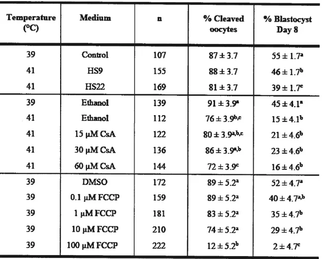
Table 2. Effects ofheat shock, cyclosporin A, and carbonyl-cyanide 4-(trifluoromethoxy) phenylhydrazone during maturation
List of figures
CHAPTER I
Figure 1. Mammalin caspase family 27
Figure 2. Heat-induced apoptosis 41
CHAPTER II
Figure 1. Representative confocal images of TUNEL labeling 90
Figure 2. Apoptosis afier heat shock 92
Figure 3. Abundance of mitochondrial and apoptotic transcripts after
heat shock 94
figure 4. The occurrence of apoptosis afier combination of
cyclosporin A and heat shock during maturation 96
Figure 5. Apoptosis afier FCCP 98
Figure 6. Abundance of mitochondrial and apoptotic transcripts after
FCCP 100
CHAPTER III
Figure 1. Heat shock during maturation induced caspase-9 and caspase-3/7 activity in both oocytes and derived
blastocysts 126
f igure 2. Caspase inhibitors blocked the detrimental effects of heat
shocked oocyte competence 128
Figure 3. Caspase inhibitors reduced the frequency of apoptosis in
CHAPTER IV
f igure 1. Bax inhibitor peptide (BIP) blocked the detrimental effects
ofheat shocked-oocyte competence 158
figure 2. Effects of Bax inhibitor peptide (BIP) and heat shock (HS)
during maturation on induction ofapoptosis 160 f igure 3. The BH4 domain of Bcl-xL (TAT-BH4) fails to block the
negative effects of heat shocked-oocyte competence 162 figure 4. Effects of BH4 domain of Bcl-xL (TAT-BH4) and heat
shock (H$) during maturation on induction of apoptosis.... 164 figure 5. Combination of anti-apoptotic peptides, Bax inhibitor
peptide and BH4 domain ofBcl-xL (BIP+BH4) suppressed the detrimental effects of heat shocked-oocyte competence. 166 Figure 6. Protective actions of combination of anti-apoptotic
peptides, Bax inhibitor peptide and BH4 domain ofBcl-xL (BIP+BH4) and heat shock (H$) during maturation on
induction ofapoptosis 168
CHAPTER V
figure 1. Model depicting responses of bovine oocytes and early embryos to putative mediators of oocyte mitochondria dysfunction on reproductive performance as determined by
List of Abbreviations
Mitochondrial Membrane Potential
ADP Adenosine Diphosphate
AIF Apoptosis Inducing f actor
Akt Serine/Threonine Protein Kinase B
Ant Adenine-Nucleotide Transiocase
Apaf- I Apoptosis Activating Factor- 1
Asp Aspartic
ATP Adenosine Triphosphate
Bad Bc12 antagonist of ceil death
Bag Bc12 associated athanogene
Bak Bax-Bc12 Antagonist/Killer
Bax Bc12 Associated X-Protein
Bd-2 B-ce!! LymphomalLeukemia 2
Bd-w Bc12 like 2 protein
Bd-x Bc12 like 1
Bcl-xL Bc!2 Related Protein, Long Isoform Bcl-xS Bc12 related Protein, Short Isoform
BH Bd-2 Homology Domains
Bid BH3 interacting domain death agonist
Bik Bc12 interacting killer
Bim Bc12 interacting protein BIM
BIP Bax Inhibitor Peptide
BIR N-terminal Baculovirus-Inhibitor-of-Apoptotis-Repeat
BMP Bone Morphogenic Protein
BSA Bovine Serum Albumin
CAD Caspase-Activated DNase
CARD Caspase-Recruitment Domain
Caspases Cysteine-dependent Aspartate-Specific Proteases
CO Cytochrome c Oxidase
COl Cytochrome Oxidase 1
COCs Cumulus-Oocyte Complexes
CREB AMP-response-element-binding Protein
CsA Cyc!osporin A
C-terminal domain Carboxy-Terminal Transmembrane Region
Cyt c Cytochrome c
DED Death Effector Domain
Diablo/Smac Second Mitochondria-derived Activator of Caspase/Direct IAP Binding Protein with Low pI
DiOC6 3, 3’ Dihexyloxacarbocyanine Iodide
DISC Death Inducing Signaling Complex
D-loop Dispiacement Loop
DMSO Dimethyl Sulfoxide
DNA Deoxyribonucleic Acid
DTT Dithiothreitol
dUTP Deoxy-Uridine Tri-phosphate
EFAF Essentially Fatty-Acid Free
EfAf-BSA Essentially Fatty-Acid Free Bovine Serum Albumin
EGA Embryonic Genomic Activation
EndoG Endonuclease G
F 1f0—ATPase F1F0—adenosine triphosphate
FADD FAS-Associating Protein with a Death Domain fADH2 Reduced Flavin Adenine Dinucleotide
FCCP Carbonyl Cyanide 4-(trifluoromethoxy) Phenylhydrazone
Fg f emtograms
f SH Follicle-Stimulating Hormone
Gapdh Glyceraldehyde 3-Phosphate Dehydrogenase
GLM General Linear Model
GnRH Gonadotrophin Releasing Hormone
Group I Caspases-1, -4, and -5 Group II Caspases-2, -3, and -7 Group III Caspases-6, -8, and -10
GSH Glutathione
GV Germinal Vesicle
GVBD Germinal Vesicle Breakdown
F1202 Hydrogen Peroxide
HeLa Human Cervix Cancer Ceil Line
HMG-box High Motility Group-box
HS22 Heat Shock for 22 hours
HS9 Heat Shock for 9 hours
HSP7O Heat Shock Protein 70 kDa
HSP9O Heat Shock Protein 90 kDa
HSPs Heat $hock Proteins
H-strand Heavy-Strand
HtrA2 High Temperamre Requirement Protein A2
lAPs Inhibitor of Apopto sis Proteins
ICAD Inhibitor ofCAD
1CM limer Ceil Mass
IVF In-Vitro f ertilization
JC-1 5,5’,6,6’-Tetrachloro-1 .1 ‘,3,3 ‘-Tetraethyl Benzimidazolylcarbocyanide Iodide
JNK c-Jun n-Terminal Kinase
LH Luteinizing Hormone
L-strand Light-Strand
MDM2 Mouse Double Minute 2
MII Metaphase II
MOMP Mitochondriai Outer Membrane Permeabilization
mRNA Messenger RNA
mSOf Modified Synthetic Oviduct Fluid
mtDNA Mitochondrial Deoxyribonucleic Acid
mTERF Mitochondrial Transcription Termination f actor
mtRNApol Mitochondriai RNA Polymerase
mtTFA Transcription factor A
N2 Nitrogen
NADH Nicotinamide Adenine Dinucleotide Reduced
ND NADH-Dehydrogenase-Ubiquinone Reductase
ND6 Mitochondrial NADH Dehydrogenase Subunit 6
Noxa Phorbol- 1 2-myristate- 13 -acetate-induced protein 1
02 Oxygen
OH Origin of Replication for H-strand
OL Origin of Replication for the L-strand
Omi!HrtA2 High Temperature Requirement Protein A2
OMM Oocyte Maturation Medium
OWM Oocyte Washing Medium
Oxphos Oxidative Phosphorylation
p20 Large subunit
PARP Poly-ADP-Ribose Polymerase
PBS Phosphate Buffered Saline
PB S-PVP Phosphate Buffered Saline-Polyvinylpyrrolidone
PGC Primary Germ Cells
P13 K Phosphatidylinositol-3 ‘-kinase
POLRMT Mitochondrial RNA Polymerase
PT Mitochondria Permeability Transition
Puma 3c12 binding component 3
PVP Polyvinylpyrrolidone
Redox ReductionlOxidation
RNA Ribonucleic Acid
ROS Reactive Oxygen Species
rRNA Ribosomal RNA
RT-PCR Reverse Transcription-Polymerase Chain Reaction
SAPK Stress Activated Protein Kinase
SAS Statistical Analysis System
SMase Shingomyelinase
TAT-BH4 BH4-domain ofBcl-xL linked to 10-amino acid HIV-TAT
TCM- 199 Tissue Culture Medium- 199
TE Trophectoderm
TEM Transmission Electron Microscopy
Tfam Transcription factor A
TfB 1M Mitochondrial Transcription Factor Bi TfB2M Mitochondrial Transcription factor B2
TNf Tumor Necrosis factor
TNfR Tumor Necrosis f actor Receptor
TRAIL TNf-Related Apoptosis Inducing Ligand
tRNA Transfer RNA
TUNEL Terminal Deoxynucleotidyl Transferase Mediated dUTP Nick End Labeling
Vdac Voltage-Dependent Anion Channel
WM Washing Medium
XAF 1 Xiap-Associated factor 1
XIAP X-Linked Inhibitor ofApoptosis
z-DEVD-fmk N-Benzyloxycarbonyl-Asp-Glu-Val-Asp-fluoromethyl Ketone
z-LEHD-fmk z-L-E-(Ome)-H-D(Ome)-fluromethyl ketone
Acknowledgements
f irst, I would like to mention my adviser, Dr. Lawrence Smith, for his support, his teachings, and for his patience and confidence on letting me pursue my ideas and interests, allowing me to mature my research skills; for that I am truly grateflul. I also would like to thank Dr. Bruce Murphy for his guidance, his friendship, providing energy and knowledge to get through the demanding process of obtaining a graduate degree. I am very thankful to the members of my supervisory committee Dr. Alan Goff for his support and availability to discuss statistics at any time, Dr. Christopher Price for his inputs to the present work and friendship, Dr. Carmona for lier passion for apoptosis, and Dr. Tsang for agreeing to becoming the extemal member of my committee and teaching of some life lessons. Special thanks go to Dr. Peter Hansen, my Master’s degree advisor, for his insightful presence and motivation words. His teachings during my master’s studies allowed me to undertake my PhD with confidence.
Many thanks go to Carmen Léveilleé, Jacinthe Therrien, Patrick Vincent, and France f ilon and my sincere appreciation for their effort, dedication and patience. I would like to express my gratitude to the members of the laboratory, especially Francisco Viramontes, Dan Amold, Donald Boucher, Fabiana Bressan, Jacinthe Therrien for ail their help and good times inside and outside the laboratory. I extend my appreciation to Mira Dubois, Micheline St-Germain, Micheline Sicotte, Isabelle Daneau, Diane Rodier for their tireless help in great and small things.
Thanks to the “ovary rescue team”, Edimir Nicola, Valério Portela, Érika Guerreiro, Ahmad Jamishi, Ma Maria Ocampo without their help I would flot be writing this today. b the Brazilian “mafia” at the Vet School, Gabriel, Edimir, Valério, NatJia, Flâvia, Mârio, Angela, Gustavo, Danila, Érika, F abiana, Marcos, Rogério and friends Ignâcio, Daniel, Ahmad for making my days funnier and louder. To my dear friends around the world: Tucci and Otâvio, Fabiola and Maurfcio, Chiudia, Luiz, Gabriel and Sophia, Luiz Augusto and Camila, lika, Aleksa, Isabella and baby, Marcelo, Aime and Rafaeia, Maria Beatriz Pâdua, Zvi Roth and family, Yaser, mes, Toleen and baby, Arthur, Jenniffer and Tony, Marta and Edgar, Valéria, Maria and Bertran, Natâlia, Patrice, Eric and Nicole, Edimir, Rosa and Ismael, Vilceu, Mârcia, Pedro, Jean, and Fbio, Marcelo $eneda and family, Francisco, Ilda, Annai, Sophie, and Axelle, Mârio, Débora, Serena, and Guilherme, Clarisse and Sammy, Patricia, f iâvio and babies, Renata, Fabâo and Bruno, Maria Juliana, Kiki and Nando, Zé Couto and Nora, Ricardo and Mônica for your presence even when the geography did flot help.
I thank my family for always being there, with their love and care, specially my mother Lucilia. Thanks to their infinite support and deeply needed encouragement at ail times, I had the condition to be what I wanted to be. Special thanks are due to my husband Gabriel who agreed to join me in this new endeavor, for his love, patience and for his unconditional encouragement to follow my crazy dreams. And to my daughters, JiMia and Alice, source ofendless love, joy, happiness, and light in my life. I love you ail so much!
The mitochondrial genome is matemally inherited via the oocyte cytoplasm, from one generation to the next. While oocyte mitochondria are small, round in shape, microscopically dense, and contain a few underdeveloped cristae, characteristics of ceils with low metabolic rate (Van Blerkom 2004), these organelles can produce enough ATP to sustain embryo development (Van Blerkom et al. 1995; Tamassia et al. 2004). The role of mitochondria in ATP production, regulation of intracellular free Ca2, steroidogenesis, and activation of the apoptotic pathway has long be known. Recently, the notion that oocyte mitochondrial dysfunction may be crucial determinant of embryo development has emerged.
Mitochondrial defects may in part be responsible for the decline in fertility. Oocytes from older women were more likely to contain deleted mtDNA compared to oocytes from younger women (Keefe et al. 1995; Jansen and Burton 2004). Also cohorts of human oocytes with ATP content <2 pmol/oocyte have lower potential for continued embryogenesis (Van Blerkom et al. 1995). In oocytes from aged humans, an increase in volume fraction of the mitochondria was observed which might reflect subtie changes in oxidative phosphorylation capacity (Muller-Hocker et al. 1996). Higher mitochondrial membrane potential was positively correlated with the rate of development of human embryos (Wilding et aI. 2001). Aberrant shifts in mitochondrial membrane potential may contribute to reduced embryonic development potential (Acton et al. 2004). Fragmented embryos generated more reactive oxygen species (Yang et al. 1998). Consequently, any environmentaÏ or pharmacological insuit to oocyte mitochondria may contribute to susceptibility ofthe oocyte to developmental compromise.
Oocyte mitochondria are potential mediators or couid even initiate apoptotic degeneration in oocytes and preimpiantation embryos. It is weil established that loss of oocytes from the ovarian pool involves an apoptotic process however the precise mechanisms invoived are flot completely understood (Monta and Tilly 1999; Reynaud and Driancourt 2000). Most, but flot ail, death signais converge to increase in mitochondrial membrane permeability with release of pro-apoptotic factors in the cytosol resuiting in caspase activation and induction of apoptosis (Reynaud and Driancourt 2000; Lemasters 2005). CelIs have also mechanisms for inducing apoptosis in the absence of caspase activation. Apoptogenic factors otherwise confined to the mitochondrial intermembrane space, such as apoptosis inducing factor (AIF) and endonuclease G (EndoG), leak into the cytopiasm and nucieus where they display their pro-apoptotic characteristics (Ye et al. 2002; Lorenzo and Susin 2004). Heat sliock lias been shown to decrease oocyte competence in vivo and in vitro (Putney et al. 1989; Edwards and Hansen 1997; Rocha et al. 1998; Al-Katanani et al. 2002; Roth et al. 2002; Roth and Hansen 2004), reduce oocyte protein synthesis (Edwards and Hansen 1996), and induce apoptosis (Roth and Hansen 2004). In somatic ceils, heat-induced apoptosis invoives the mitochondrial pathway (Mirkes and Littie 2000; Qian et al. 2004; Bettaieb and Averili-Bates 2005; Wada et ai. 2005). However, mitochondrial role in heat shock-induced apoptosis in the oocyte remains to be elucidated.
Identification of the mitochondriai pathway through the presence of active caspase and Bd-2 famiiy signaling mechanisms in the oocyte could help eiucidade the pathway by
which different stressors (e.g. heat shock), induce apoptosis, and potentially open a new window of opportunity for developing strategies to reduce deleterious effects of heat shock or other stresses on the oocyte and improve fertility health under stress conditions.
LITERATURE REVIEW
The Rote of Mitochondria During Early Embryonic Development
Ancient eubacteriai invasions through symbiosis gave rise to mitochondria (Dyail et ai. 2004). The presence of mitochondrial deoxyribonucleic acid (mtDNA) is the primary remaining evidence of their bacterial origin. Mitochondria and chloropiast are the only structures in a ceil that have a deoxyribonucleic acid (DNA) distinct from the nuclear DNA. As a consequence, the control of mitochondrial biogenesis and function depends on a well orchestrated regulation between the two genomes, nuclear and mitochondrial (Garesse and Valiejo 2001). Their primary function is to provide energy in ail eukaryotic ceils, in form of ATP production, through oxidative phosphorylation (Oxphos) and the citric acid cycle. They aiso have an essential role in other biochemical pathways like calcium homeostasis and modulation of apoptosis through the release of several celi death-inducing molecules (Duchen 2004).
Mammaiian mitochondrial DNA is a double stranded closed-circular moiecule with approximately 16500 nucleotides that, in most celis, represents only about 0.5-1% of the total DNA content (Smith and Alcivar 1993; femandez-Siiva et ai. 2003). It is a compact gene organization, the coding sequences are contiguous or separated by a few base pairs without introns and some of the protein genes even overiap (Fernandez-Siiva et al. 2003). The two strands, the heavy or H-strand and the light or L-strand, can be distinguished by their different guanine and thymine (G+T) content and different density in denaturating gradients.
The mitochondrial genome contains only 37 genes corresponding to the ribonucleic acid (RNA) components of the mitochondrial transiational apparatus [two ribosomal RNA (rRNAs) called 12$ and 16$ and 22 transfer RNA (tRNAs) as weli as messenger RNA (mRNAs) for 12 polypeptides that are subunits for Oxphos complexes ($mith et al. 2002). Seven of those polypeptides (ND1 to ND6 plus ND4L) are subunits of complex I: NADH dehydrogenase-ubiquinone reductase; one (cytochrome b) is part of complex III: ubiquinol cytochrome e reductase; three (COI, COII and COIlI) are catalytic subunits of complex IV: cytochrome e oxidase; and ATPase 6 and 8 are subunits of compiex V: ATP synthetase (fernandez-Siiva et ai. 2003). These genes are asymmetrically distributed and the H-strand encodes most of the information. The L-strand encodes oniy eight tRNAs and one mRNA, the ND6 subunit. The rest of the Oxphos subunits as well as ail the factors involved in the mtDNA metabolism are nuclear-encoded synthesized in the cytosol ami transported by chaperones into the mitochondriai matrix (Garesse and Vallejo 2001). Therefore, control of mitochondrial function is compiicated and involves the exchange of information between nucleus and many copies of mtDNA in each cell’s cytoplasm.
Mitochondrial Genetic System
The mitochondrial genome lias some features that differ from the nuclear genome. These include the phenomenon of polyploidy with respect to mtDNA where each ceil contains several thousands of mitochondria and each mitochondria contains several (2-10) copies of mtDNA (Femandez-Silva et al. 2003). Also ail mtDNA in an individual is
thought to be identical (homoplasmy), although mutations can arise, maintained or amplified giving rise to condition of heteroplasmy. Mitochondria mutations higher than 60-80% are associated with diseases. The accepted dogma is that mtDNA is matemally inherited, afler fertilization, the sperm carnes a few paternal mitochondria which are eliminated by ubiquitin-dependent mechanism (Cummins 200 la; Femandez-Silva et al. 2003; Cummins 2004). During oogenesis, a bottleneck phenomenon occurs where only a small number of mtDNA molecules are amplified and transmitted to the offspring. Also the evolution rate of mtDNA is almost 20 times faster than the nuclear genome (Jansen and de Boer 1998). Several reasons could explain this high rate of mutation: lack of histone proteins to protect mtDNA, physically association with the inner mitochondrial membrane where the damaged reactive oxygen species (ROS) are generated, lack of proofreading mechanism, and a less efficient DNA repair mechanism compared to the nucleus (Kowaltowski and Vercesi 1999). Accumulation of mtDNA mutations has been associated with ageing (Zeviani and Antozzi 1997; Cummins 2001b). High mutation rates (Keefe et al. 1995) and alteration in mitochondnial function (Van Blerkom et al. 1995), numerical density (Muller-Hocker et al. 1996; Steuerwald et al. 2000), reduced ATP levels (Diaz 1999), shift in mitochondrial membrane potential (Acton et al. 2004), increased ROS levels (Yang et al. 199$), and metabolic activity (Wilding et al. 2001) are also associated with reduced fertility.
The mitochondrial genome is replicated and transcribed within the organelle (femandez-Silva et al. 2003). In mtDNA there are two non-coding regions which contain
most of known regulatory functions. The main one is a triple-stranded structured called the dispiacement ioop (D-loop) region, situated between the genes tRNA’ and tRNA)t0 that contains the origin of replication for H-strand (OH) and the promoters or H- and L-strand transcription. The cis-elements are responsible for regulation of both mtDNA replication and transcription at the D-loop region. This is also the region that is most variable in sequence and size among different species. However, it contains some conserved elements with possible regulatory functions. The second non-coding region is a ‘--30 nucleotide-long segment that contains the origin of replication for the L-strand (OL). Ah the trans-acting factors associated with mtDNA metabolism are nuclear-encoded, such as mtRNA polymerase, mtDNA polymerase (pol ‘y), and ail factors that regulate mtDNA replication, mtDNA transcription and mtRNA processing (Garesse and Vallejo 2001).
Mitochondrial transcription starts at three different initiation points, one for L strand (L) and two for H-strand (H1 and H2), ail located at the D-loop region. The initiation site H1 operates much more frequently than H2. The H1 is responsible for the synthesis of two ribosomal RNAs (tRNA’ and tRNA’’) and linked to transcription termination events (Fernandez-Siiva et al. 2003). The H2 initiation point directs the transcription of whole H strand and is around 20 times less active than H1. Also H2 originates a polycistronic molecule that covers almost the whole H-strand. Processing of this poiycistron originates mRNAs for 12 H-strand encoded polypeptides and 12 tRNAs. The L-strand originates a single polycistron from which eight tRNA and ND6 mRNA are derived (Garesse and Vahlejo 2001).
The mitochondrial transcription machinery is relatively simple compared with nuclear version. It consists of a single organelle-specific RNA polymerase (mtRNApol or POLRMY) and transcription factors such as the mitochondrial transcription factor A (mtTfA or Tfam) and either mitochondrial transcription factors Bi (TfB 1M) or 32 (TFB2M) for initiation and a mitochondrial transcription termination factor (mTERF) for termination of transcription (falkenberg et al. 2002; femandez-Silva et al. 2003; Hyvarinen et al. 2007). Until very recently, the mtTfA was the only known transcription factor in mammals. Binding sites for mtTfA are present at the two more active transcription initiation points (H1 and L) (femandez-Silva et al. 2003). However, binding and transcription activities are higher for the L promoter (LSP) compared with H1 promoter (HSP1). Previous data suggest that mtTfA forms a complex with DNA by its two high motility group (HMG)-box domains that induces a structural change in the promoter region of mtDNA and allows mtRNApol to initiate transcription (Shadel and Clayton 1997; Clayton 2000). In mouse embryos, mtDNA transcription starts at day 2 of development (Cummins 2002).
The replication of mtDNA is takes place at the mitochondrial matrix (Femandez Silva et al. 2003). The generaliy accepted model is that the two strands (L- and H-strand) are replicated asynchronously and asymmetricaily. The synthesis starts at °H iocated at the
D-loop region and proceeds unidirectionally until °L to produce a daughter H-strand circle.
When H-strand replication reaches OL, the paternal H-strand is displaced and the initiation
L-strand. The mtTfA seems to be invoived in replication, since it is required for L-strand transcription initiation and primer formation. In mouse embryos, transcription of replication factors are abundant at the morula and biastocyst stage but mtDNA repiication occurs in the biastocyst at day 6.5 (Piko and Matsumoto 1976; Thundathil et aI. 2005).
Oogenesis
In the earliest pre-migratory germ celis, probably less than 10 mitochondria are present and this number increases to around 200 in each oogonium (Cummins 2002). This phenomenon is called the “bottieneck”, when the restriction of mitochondria copy number acts to maintain homoplasmy and minimize heteroplasmy foliowed by clonai expansion (Cummins 200 lb). The bottieneck is believed to iimit the effects of Muller’s ratchet or the tendency for deieterious mutations to accumuiate in asexually reproducing organisms (Bergstrom and Pritchard 1998). Smith et al. (2000) proposed that there are severai periods when restriction of mitochondria copy number could occur including during replication and migration of primary germ celis (PGC), during oogenesis; during early embryogenesis and during the commitment of embryonic inner celi mass elements from PGC.
During preimpiantation deveiopment, mitochondria undergo structurai and functionai differentiation. In eariy stages, mitochondria are sphericai, have a dense matrix and a few cristae (Van Blerkom 2004). In human oocytes, mitochondria are sphericai/ovoid organelies, with a few short cristae that rarely penetrate the dense matrix. Therefore, they are structuraliy undifferentiated and produce low concentrations of
adenosine triphosphate (ATP) (Van Blerkom and Davis 1998). This phenotype persists tbroughout the cleavage and late morulae stage in in vitro human embryos (Van Blerkom 2004). In most mammals at the blastoscyst stage, mitochondria are elongated with lamellar cristae that completely transverse the inner mitochondrial matrix. These features are found in mitochondria actively engaged in ATP-production by oxidative metabolism.
Mitochondria also undergo stage-specific changes in distribution during oocyte maturation and early embryo development. During maturation, mitochondria distribution shifis from random to an arrangement during leptotene, and transiocates to the perinuclear region during zygotene by microtubule-mediated process (Cummins 2004; Van Blerkom 2004). After fertilization, mitochondria migrate to the perinuclear region to form a condensed aggregate surrounding the opposite nuclei (Van Blerkom 2004). A transient nuclear perinuclear accumulation is also observed in each blastomore during early cleavages. Spatial remodeling of mitochondria allows increased levels of AlT in areas of cytoplasm that have stage-specific activities with high energetic needs (Cummins 2004; Van Blerkom 2004).
Traditionally, estimation of mitochondrial numbers in oocyte and early embryos has been obtained from transmission electron microscopy (TEM) (Vander Heiden et al. 1997). In the oocyte recruited for ovulation, the increase in cytoplasmic volume is followed by an increase in mitochondria numbers (Cummins 2002). The current consensus is that each oocyte mitochondrion contains a single genome. In cattle, during the period of oocyte growth, mitochondria increase in number by more than one hundred times to reach 136 000
mitochondria in the mature preovulatory oocyte (Hauswirth and Laipis 1985) compared with the average of 92 500 mitochondria in mouse embryos (Piko and Matsumoto 1976). This may indicates that a competent oocyte requires a fixed amount of mitochondria per unit ofcytoplasm (Smith and Alcivar 1993). further, mtDNA copy number also seems to be associated with increased oocyte volume. In bovine preovulatory oocytes, mtDNA per celi increased from 0.1 pg in primordial celis to 4.5 pg or 260 000 molecules (Hauswirth and Laipis 1985).
During fetal and aduit life, oocytes are eliminated by apoptosis during atresia. It is not known if this selection is based on mtDNA (Jacobson et al. 1997). Atresia might represent the mtDNA boffleneck that reduces the mtDNA genetic variability across generations and consequently restores homoplasmy. This seems to be contrary to the quiescent nature of oocyte’s mitochondria. Therefore, the answer of this nuclear mitochondria interaction at the oocyte stage is unclear. Previous studies have been shown that the oocyte can undergo apoptosis by activation of caspases or by alteration of the mitochondria membrane potential (Roth and Hansen 2004; Thouas et al. 2004). These data suggest that the apoptotic machinery is present and can be activated at the oocyte stage.
Embryogenesis
The preimplantation period begins with oocyte fertilization and ends with formation of a btastocyst ready to implant. This period is characterized by three major transitions, first embryonic genomic activation (EGA), compaction, and blastocyst formation (Zeng et
al. 2004). The matured oocyte contains sufficient maternai transcripts and proteins to support fertilization and the first two ceil divisions (Devreker and Englert 2000). Activation of the embryonic genome represents the transition period from maternai to embryonic control by synthesis of embryonic mRNA and proteins. Blastomeres increase in contact with each other through intercellular gap and tight junctional complexes. Also membrane and cytoplasmic polarization occur and blastomeres develop a distinct apical and basal membrane and cytoplasmic domains (Hardy et al. 1996). The following mitosis will produce two types of ceils, polar and apolar (Devreker and Englert 2000). Depending on the plane of division, the polar ceils can produce two identical polar daughter celis or one polar and one apolar daughter celi. Apolar ceils remain on the inside of the preimplantation embryo and are the origin of the inner ceil mass (1CM) while apolar celis remain outside to form the trophectoderm (TE), the first transporting epithelium. The morula is transformed to blastocyst by the transport of fluids and accumulation resulting in cavity formation. The biastocyst is formed by a small group of 1CM, a fihled cavity and surrounding TE. Implantation occurs after blastocyst hatching from the zone pelucida. The 1CM gives rise to embryo itself and TE gives rise to extra-embryonic membranes and placenta.
Embryo metabolism is different in the pre- and post-compaction stage (Devreker and Englert 2000). Before compaction the blastomeres are loosely attached and, in consequence, equally exposed to their environrnent with low level of biosynthesis, low respiratory rates and limited ability to metabolize glucose as a source of energy. In
contrast, post-compaction embryos have high rate of biosynthesis and exponential increased in energy demand, and become able to metabolizing glucose.
Mitochondria are also undergoing changes during the preimplantaion period. Afier fertilization, mitochondria are located close to developing pronuclei, presumably as source of ATP (Cummins 2001b). The mitochondria matrix becomes less dense, oval in shape, transcription resumes, and pyruvate become the embryo’s energy substrate. Correlations between the potential for development, ATP content, and mitochondrial function are observed in bovine oocytes and embryos (Stojkovic et aÏ. 2001). Afier fertilization, at the first three to four ceil divisions, there is a shifi in ATP production from oxidative metabolism to glycolysis which coincides with theoretical reductions in availability of oxygen (02), with transition from the oviduct to ifie uterus (Cummins 2001b). An increase in consumption of 02 occurs during initiation of compactation and blastocyst formation because these process are energy demanding. Optimal 02 levels during early embryonic development appear to be critical for normal development. Bovine embryos exposed to an 02 tension of 20% displayed decreased blastocyst formation when compared with 5 or 2% 02 environment (Yuan et al. 2003). Mitochondrial function is associated with a change in metabolic requirements in which a shifi in carbohydrate substrate requirements from glucose to pyruvate occurs in the embryo moving through the first ceil divisions (Cummins 2004).
This evidence emphasizes the need of fine tuned communication between nuclear and mitochondrial genes during development. A better elucidation of interactions between
mitochondrial and nuclear genomes and embryo metabolism is critical for an improvement in growth rates on cultured embryos.
Mitochondria and Apoptosis
Recently, new knowledge about mitochondria has shown that they are flot only energy generators but also play a central role in the delicate process that sustains the balance between celi life and celi death.
The term “programmed celi death”, originally, was used to describe a celi death process throughout animal development that occurs in a spontaneous, orchestrated and predictable manner (Lockshin and Willians 1964). After the establishment of similarities between many models of celi death and a description of conserved morphological features, this programmed celi death was named apoptosis (Ken et al. 1972). The meaning of the word “apo” is from and “ptosis” means fail, taken together means “fail away from”.
Apoptosis has been recognized as a physiological form of ceil death required to control ceil populations during normal tissue homeostasis or to eliminate celis damaged by stress stimuli (Haimovitz-friedman A 1997; Jacobson et al. 1997). The programmed ceil death pathway has been proposed as a major regulatory event observed during embryonic development, establishment of immune seif-tolerance, immune effector ceil killing and regulation of ceil viability by hormones and growth factors (Zakeri and Lockshin 2002; Meirelles et al. 2004). failure to accurately undergo apoptosis may contribute to
neurodegenerative disease, cardiovascular diseases, cancer, viral pathogenesis, and autoimmune diseases (Zimmermann et al. 2001; Van BÏerkom 2004).
Apoptosis and Necrosis
The most described forms of cell death are apoptosis and necrosis. They can be differentiated based on morphological and biochemicai markers. Necrosis or accidentai ceil death is characterized by dilatation of the endoplasmic reticulum, dissolution of ribosomes and lysosomes, sweiling of the mitochondria matrix, and rupture of nuclear, organelle and plasma membranes. These rupture of membranes allows the release of intracellular content that triggers the inflammatory process (Wyliie et al. 1980).
Morphologically, in early apoptotic events, the chromatin aggregates and condenses against the nuclear membrane with appearance described as haif-moon-, horse-shoe-, sickle-, lancet-, and ship-like (Majno and Joris 1995). The nucleus progressively condenses and the nuciear envelope becomes convoiuted, indented and then broken up (karyohexis) which generates nuclear fragments that contains condensed chromatin (Wyllie et al. 1980). In parallel, the cytoplasm also condenses, the ceil becomes round, the plasma membrane become indented, and organelles maintain intact shape and structure (Kerr et aI. 1972; Kondo et al. 1997). During late apoptotic stages, condensation of the mitochondria (Kluck et al. 1999), dilatation of the endoplasmic reticulum (Ludewig et al. 1995), detachment of ribosomes from the rough endoplasmic reticulum and ribosome aggregation (ferguson and Anderson 1981) can be observed. Compressed organelles and cytoplasm condensation
allowed formation of cytoplasmic vacuoles. The plasma membrane is preserved to alÏow packing of whole cell fragments into membrane-bound apoptotic bodies without leakage of toxic intracellular contents (Kerr et al. 1972). The apoptotic bodies are phagocytosed by neighbor ceils or macrophages without inducing an inflammatory response or tissue damage.
The interest in describing biochemical features of apoptosis has increased. The extemalization of phosphatidylserine to the outer leaflet of the plasma membrane is an early event of apoptosis which acts as a signal resulting in phagocytosis of the apoptotic cell by neighbor ceils (Martin et al. 1995). The induction of mitochondrial outer membrane permeabilization (MOMP) is another early event controlled by the Bd-2 family of proteins (Lorenzo and Susin 2004). The most well know biochemical change observed in the nucleus during ccli death is mediated by caspase activation resulting in cleavage of the DNA to form intemucleosomal DNA fragments (Wyllie et al. 1980). The characteristic ladder patterns of these fragments are bands of 180-200 bps in size on agarose gel electrophresis. In contrast, necrotic cells show a smear suggesting that DNA is cleaved at random.
Mitochondrial Outer Membrane Permeabilization (MOMP)
During MOMP the inner mitochondrial membrane retains proteins from the mitochondrial matrix and the outer mitochondrial membrane becomes permeable. Therefore, release of soluble intermembrane space proteins occurs. Some of these are:
cytochrome c (Cyt c), IAP binding protein with low pI (Diablo/Smac), high temperature requirement protein A2 (Omi/HtrA2), AIF, EndoG, and certain procaspases, including procaspase-9 (Crompton 1999; Lorenzo and Susin 2004). Leakage of mitochondrial contents through MOMP resuits in cytoplasmic and nuclear apoptosis.
The mechanism for outer-membrane rupture during apoptosis is flot yet established. These ruptures are evident in electron micrographs of Jurkat celis undergoing apoptosis associated with matrix swelling (Vander Heiden et al. 1997). However, it is not known whether breaks in the mitochondrial outer-membrane are a resuit of mitochondria permeability transition (PI) pore activation. Under normal physiological conditions, the general function of components of PI pore is well known. The function of voltage dependent anion channels (Vdac) is to allow solute access to the solute-specific transport systems of the inner membrane (Crompton 1999). The adenine-nucleotide transiocase tAnt) mediates adenosine diphosphate (ADP)—*ATP exchange that is crucial for mitochondria bioenergetic function. The cyclophilin D (CyP-D) function is flot established, but it is associated with catalysis of protein folding. Under pathological conditions, the PT pore consists of Vdac in the outer mitochondrial membrane, Ant in the inner mitochondrial membrane and CyP-D in the matrix (Zamzami and Kroemer 2001; Crompton et al. 2002; Halestrap 2006). One possible mechanism is the formation of Vdac-Ant-CyP-D complex leading to PT pore opening that results in a mitochondrial depolarization, uncoupling of Oxphos, mitochondrial swelling (matrix expansion), outer-membrane rupture, and release of intermembrane-space apoptogenic proteins (Crompton 1999; Gross et al. 1999).
Previous studies have shown that HeLa nuclei mixed with isolated mitochondria and exposed to various PI pore activators (atracylate, peroxides, Ca2, diamide) resulted in mitochondrial swelling, release of Cyt e and AIF, and apoptotic changes in nuclear morphology (Marchetti et al. 1996; Ellerby et al. 1997; Kantrow and Piantadosi 1997).
Another possible mechanism is through interactions between PI pore and the B-cell CLL/lymphoma 2 (Bel-2), Bel extra long form (Bcl-xL) and Bc12 associated X-protein (Bax) in the outer membrane. Mitochondria isolated from Bd-2 transfected ceils demonstrated more resistant to atractylate- and peroxide-induced pore opening (Susin et al. 1996). Microinjection of fibroblasts with recombinant Bax induced mitochondria depolarization and nuclear apoptosis and these mitochondrial and nuclear effects of Bax were prevented by three prototypic inhibitors of PI pore (Marzo et al. 1998). Therefore, both pro- and anti-apoptotic Bel-2 family proteins may interact with components of the PT pore. Co-immunoprecipitation studies have demonstrated that Bax binds to Vdac (Narita et al. 1998). Also Bel-2 and Bcl-xL were shown to block PI pore by direct inhibition ofVdac activity (Tsujimoto et al. 2006). Bax mediated celi death was inhibited by cyclosporin A (CsA), a drug that is assumed to inhibit the pore formation by occupancy of the active site of CyP-D preventing Cy?-D association with Ant (Pastorino et al. 1998; Crompton 1999). The emerging model is that Bax and, possibly, Bel-2 and Bcl-xL may be recruited to the PI pore complex. However, a recent study lias shown that Vdac is dispensable for both PT pore and Bel-2 family member-driven celi death (Baines 2007).
The ion flow mode! proposes that Bel-2 proteins have ion channel aetivity. The tbree-dimensional structure of Bcl-xL, an anti-apoptotic protein, resembles the structure of bacterial toxins that are able to insert into lipid bilayers forming channels able to conduct ions (Muchmore et al. 1996). The Bcl-xL was demonstrated to form cation-channels in lipid bilayers (Budihardjo et al. 1999). Bax also contains structural similarities to the pore forming domains of the bacteria colicins and diphtheria toxin (Muchmore et al. 1996), and forms anion-channels in lipid bilayers (Antonsson et al. 1997). The Bax channel is large enough to admit carboxyfluorescein and interaction with Bel-2 blocks this channel (Antonsson et al. 1997). But there is no evidence that Bax forms pores in the outer membrane sufficiently large to release fully folded apoptogenic proteins from the intermembrane space. These findings indicate that the relative proportion of pro and anti apoptotic proteins control the ion flow. Other possibility is that Bax activation, its outer mitochondrial membrane binding and insertion followed by Bax-Bc12 antagonist/killer (Bak) oligomerization somehow increases the local curvature stress on membranes which results in formation of lipidic pores (Kuwana and Newmeyer 2003). Basanez et al (2002) demonstrated that addition of nonlamellar lipids, or lipids that change the intrinsic curvature of the membrane, to liposomes can affect the ability of detergent-oligomerized Bax to form pores that allow the release of a fluorescent dye or Cyt e.
3d-2 Related Proteins
The Bd-2 protein family is divided into two main groups according to whether they inhibit apoptosis (Bel-2, Bcl-xL, 3d-w, Bfl-l, Brag-l, Mci-l, and Ai) or promote apoptosis (Bax, Bak, Bcl-xs, Bad, Bid, Bik, Hrk and Bok) proteins. The Bd-2 and Bcl-xL are the main members of this family that inhibit apoptosis, and Bax and Bad are the two best known pro-apoptotic factors (Haimovitz-Friedman A 1997). The Bel-2 family members share one or more Bel-2 homology domains (BH) (Kuwana and Newmeyer 2003). The pro-apoptotie family members can contain multiple BH domains (BH1, 2, 3) or only BH3. Unlike the BH l-3 domains, BH4 is conserved only among antiapoptotic Bel-2 family members that contain ah four BH1-BH4 domains (Huang et al. 1998; Petros et al. 2004). Deletion of 3H4 from Bel-2 or Bel-xL has been shown to abrogate their antiapoptotie ability demonstrating that BH4 is crucial for this aetivity. The meehanisms of action of Bel-2 proteins are flot eompletely understood. The Bel-2 family members interactions may involves the hydrophobie pocket formed by close arrangement of the BHY-BH3 domains (Petros et al. 2004). For example, Bax hydrophobie poeket cari sequester the earboxy-terminal transmembrane region (C-terminal domain) within the same monomer (Suzuki et al. 2000). A common eharaeteristie of Bel-2 related proteins is the ability to produee homo- and heterodimers. The C-terminal of Bel-xL and the hydrophobie poeket of another Bel-xL or Bax protein ean interaet and form either homodimers or heterodimers (Jeong et al. 2004). The death preventing effects of Bel-2 and the death promoting effeets of Bax depend on their ability to target and insert the membranes.
Removal of the membrane anchoring domain (C-terminal domain) decreases the efftciency of Bel-2 and Bax as regulators of apoptosis (Hockenbery et al. 1993; Zamzami et al. 1997).
The first Bd-2 related protein identified vas Bax. In healthy ceils, Bax monomer is located in the cytoplasm or loosely attached to the membranes. After the death stimulus, Bax translocates to the mitochondria and inserts into the membrane as an integral protein and forms a homodimer (Gross et al. 199$). Some studies suggest that Bax homodimers increase permeabilization of the outer mitochondrial membrane which resuits in release of Cyt e from the mitochondria (Rosse et al. 1998; Eskes et al. 2000). A Bax inhibiting peptide has been shown to prevent apoptosis in mouse, rat, and porcine cumulus ceils induced by hormone deprivation (Yoshida et al. 2004). Whether Bax will exert its pro apoptotic effects depends on the presence of other members of Bd-2 family such as pro apoptotic members Bax, NOXA, and PUMA and the anti-apoptotic members Bd-2 and Bcl-xL (Tsujimoto 199$; Kuwana and Newmeyer 2003; Takahashi et al. 2004; Dejean et al. 2006; Kutuk and Basaga 2006; van Delfi and Huang 2006). Interaction of BH3-only proteins with Bel-2 and Bcl-xL can resuit in displacement of Bax!Bcl-2 or BaklBcl-xL binding, and therefore reactivation of Bax and Bak (Willis and Adams 2005). When Bel-2 are in excess, Bc12-Bcl2 homodimers and Bc12-Bax heterodimers formation occurs and predominate to prevent apoptosis (Oltvai et al. 1993). The relative ratio of these dimers and competitive dimerization of pro- and anti-apoptotic proteins, determines that apoptotic outcome in stimulated celis.
The Bd-2 and Bci-xL are important inhibitors of apoptosis. There is evidence that Bd-2 regulates calcium homeostasis (Marin et ai. 1996), and prevents Cyt c release from the mitochondria (Lee et al. 2001; Sun et al. 2001). Also Bcl-xL and Bd-w are able to prevent apoptosis foilowing many cytotoxic stimuli. In non-stress conditions, Bd-2 is located at the endoplasmic reticulum membrane, nuclear membrane, and outer mitochondrial membrane (Gross et al. 1999). The Bci-xL is associated with the mitochondria or located in the cytoplasm, in some ceils (Gonzalez-Garcia et al. 1994; Kaufmann et al. 2003). Afier an apoptotic stimulus, Bcl-xL has been shown to transiocate from cytoplasm to the outer mitochondria membrane (Cuttie et al. 2001; Kaufmann et aï. 2003). Both Bd-2 and Bcl-xL preserve mitochondrial integrity, mitochondria membrane potential, outer membrane metabolite exchange, and osmotic integrity after celi death insults (Vander Heiden et al. 1997; Kowaltowski et al. 2000; Vander Heiden et al. 2001; Eliseev et al. 2003). Association of Bcl-xL with Vdac can prevent the opening of the PT pore caused by activated Bax and Bak proteins (Shimizu et al. 2000a). In addition, the BH4 domain of Bd-2 and Bcl-xL alone lias been shown to be sufficient to cause Vdac closure and prevent apoptosis (Shimizu et ai. 2000a).
Activities of Bd-2 proteins can be modulated by post-translational modifications such as phosphorylation. for example, a major identified target of the prosurvival kinase Akt is Bad that is phosphorylated by Akt at Ser-136 (Datta et ai. 1997). Deprivation of survival signais resuits in dephosphorylation of Bad which can then bind to Bcl-xL (Yang et al. 1995; Zhou et al. 2005). Bad then inhibits the pro-survivai function of Bcl-xL and
promotes ceil death. Accumulating evidence suggest that some Bd-2 family members act as molecular mediators ofboth apoptosis and celi cycle progression (Maddika et al. 2007). This dual function of Bd-2 family members may be primarily governated by the multi domain members (Zinkel et al. 2006). The Bd-2 phosphorylation has been shown to regulate intracellular RO$ levels and inhibit celi cycle progression by the delay of GuS transition (Deng et aI. 2003). In addition, Bcl-xL induce an increase of the GO phase, enhances its arrest also a reduction in ce!! size and total RNA content during cel! cycle arrest and entry (Janumyan et al. 2003).
Mitochondria Membrane Potential (APm)
Mitochondria membrane potentia! is ofien used as an indicator of cellular viability. Polarity of mitochondria is a physiochemical process generated by the process of electron transport and Oxphos (Kowaltowski and Vercesi 1999; Murphy et al. 2005). Electrons, donated by nicotinamide adenine dinucleotide reduced (NADH) or reduced flavin adenine dinucleotide (fADH2), are transfened down to a series of redox reactions with 02 as the final electron acceptor. The energy from the flow of electrons is coupled to extrusion of protons from the mitochondria matrix. The extrusion of protons results in generation of A’Pm and a pH gradient. Oxygen functions as the acceptor of e!ectrons which together with protons generates water. The f 1f0—adenosine triphosphate (ATPase) converts the proton gradient into ATP in the Oxphos pathway. The transport of electrons down the electron transport chain with extrusion of protons will cease unless the uncoupler ADP is available
as a substrate for the f 1f0—ATPase. Lipophilic protonophores like carbonyl cyanide 4-(trifluoromethoxy) phenylhydrazone (FCCP) were originally classified as “uncouplers” because they could dissipate the proton gradient and maximally stimulate 02 consumption, resulting in loss of i\Pm.
It remains to be elucidated whether loss of A’fm is an initiator, or an effect of apoptosis, or if it is necessary for apoptosis to occur (Ly et al. 2003). Originally, a change in APm was postulated to be an early event of apoptosis (Petit et al. 1995; Zamzami et al. 1995b). Lymphocytes treated with dexamethasone demonstrated a reduced uptake of 3, 3’ dihexyloxacarbocyanine iodide (DiOC6), a mitochondrial sensing A’Pm fluoroprobe, preceding DNA fragmentation appearance (Zamzami et al. 1995b). Dexamethasone induced thymocyte apoptosis showed both alteration on mitochondrial structure and an early decrease in A’Pm measured by another A’Pm fluoroprobe, 5,5’,6,6’-tetrachloro-1.1 ‘,3,3’-tetraethyl benzimidazolylcarbocyanide iodide (JC-1) (Petit et al. 1995). Ceil permeable BH3-peptides have also induced A’-Pm loss before appearance of chromatin condensation (Vieira et al. 2002). There is evidence that AkPm loss is a late and subsequent event in the apoptotic pathway. During rat thymocyte apoptosis, DNA fragmentation was detected prior any depolarization of the mitochondria of thymocytes exposed to dexamethazone (Cossarizza et al. 1994). Loss of APm was observed has a late event afier phosphorylation of p53 and Bax transiocation to the mitochondria in etoposide-induced apoptosis of L929 fibroblasts (Karpinich et al. 2002). Similarly, apoptosis induced by UV radiation in human keratinocytes showed loss of A’Pm as late event in the pathway
(Denning et al. 2002). In contrast, perturbation of the mitochondria or changes in APm was flot observed during etoposide-induced apoptosis of P39 ceils (Hishita et al. 2001). It is possible that loss of APm acts as an amplification mechanism to further augment the apoptotic process depending on cellular response to different death stimuli.
Protonophores, like FCCP, that cause ioss of A’Pm have been postulated as potential triggers of apoptosis. The PC12 celis treated with FCCP for 24 hours resuÏted in morphological changes characteristic of apoptosis, phosphatidylserine exposure, and nuclear fragmentation (Dispersyn et ai. 1999). Similarly, neurons incubated with FCCP resuited in mitochondrial swelling, decreased mitochondrial motility, and DNA fragmentation (Moon et ai. 2005; Safiulina et ai. 2006). Reactive oxygen species are an inevitable by-product of mitochondrial respiration because flot ail 02 molecuies are fully reduced to water. There is also evidence that ROS causes apoptosis. Leakiness of electrons from the electron transport chain generates ROS which has been shown to induce DNA damage (Abramova et al. 2000), increase mitochondriai membrane permeability, and release of
Cyt
c (Skulachev 1998). Normally, Cyt c plays a role in generation of ATP via the electron transport chain (Murphy et ai. 2005).Cysteine-dependent Aspartate-specific Proteinases (Caspases)
The key apoptosis effector in mammals is a family of cysteine-dependent aspartate Asp-specific proteases with homology to the nematode (C. elegans) celi death gene, ced-3, designated as caspases (Thomberry et al. 1992). Caspases are constitutively expressed as
Figure 1. Mammalian caspase family. The caspases may be divided into three groups: the cytokines activators, the apoptotic initiators, and apoptotic executioners. The active form of caspase consists of a large (17-21 kDa) and small (10-13 kDa) subunit which contain residues that are essential for catalysis and substrate recognition. The N-terminal, also known as prodomain, is released from several caspases durng activation but this is not an activating event. The cleavage between the large and small subunit generates the active protease. The N-terminal/prodomain region is flot conserved between caspases, but some caspases contains structurally homologous elements required for activation. These elements are known as death effector domains (DED) and caspase recruitment domain (CARD).
Structure Substrate Fun ctlon preference
Pro dornain Prote ase dom ain
Lit
(jj DEVD Caspase-3]
ApoptosïsO
I
I
[]
DEVD Caspase-7 effectorUt
(j VEHD Caspase-6t
DED DED(I
LETD Caspase-8 1poptosis
t
DED DEDI
I
I
Caspase-J O At
coIi
t DEHD Caspase-2 initiatorsICARDII
I
LEHD Caspase-9j
Large subunit Small subunït Cytokine activation
inactive proenzymes or zymogens that contain a prodomain (N-terminal prodomain), a protease domain (large subunit: 17-21 kDa, C-terminal small subunit: 10-l3 kDa, and a short linker region between the large and small subunit) (Earnshaw et al. 1999; LeBlanc 2003). Activation of caspase occurs in a sequential manner where one caspase leads to activation of other caspases. The cascade starts with autocatalytic activation of initiator caspases that have long prodomain (such as caspase-8, -9, and -12), they cleavage and activates effector caspases with short prodomains (caspase-3, -6, and -7) (Salvesen and Dixit 1999). The main function of initiator caspases is to activate effector caspases that are responsible for dismantling cellular structures (Thornberry and Lazebnik 1998). Activation of caspases possibly involves the following two proteolytic steps at specific aspartic (Asp) acid residues (Salvesen and Dixit 1999). first cleavage occurs between the large (p20) and small subunit (pi 0) and removes the inter-domain linker region. Then, ffie prodomain is removed from the large subunit of the protease. Crystallography studies demonstrated ffiat active caspases are heterotetramers formed by two large subunits and two small subunits (Walker et al. 1994; Rotonda et al. 1996).
Caspases can be distinguished by the requirement for an aspartate residue at the carboxy-terminal (Pi site) of the cleavage site and their substrate specfficity is formed by four amino acids residues located on the amino-terminal side of the cleavage site (P4 site). The P4 site is crucial for determinant of substrate specificity (Talanian et al. 1997; Thornberry 1997). Thus, caspases are divided in three groups based on the amino acid sequence that they cleave: Group I have preference for WEXD motif (caspases-1, -4, and
-5); Group 2 for DEXD substrate (caspases-2, -3, and -7) and Group 3 prefer (I/LN)EXD (caspases-6, -8, and -10), the X is any amino acid (Thornberry and Lazebnik 1998; Grutter 2000). for caspases-li, -12, -13. and -14 the specific cleavage sites are unclear. Although active caspases cleave afier specific motifs recent studies have demonstrated the existence of non-consensus motifs. The caspase-7 has been shown to cleave at GELE motif oftumor necrosis factor receptor 1 (TNFRÏ) (Etheil et al. 2001) and caspase-3 to mediate cleavage of several non-consensus motifs within serine tbreonine protein kinase B (Akt) (Jahani-Asi et al. 2007). This ability to caspases to cleave at non-consensus motifs indicates that their specificity may be broader than is currently knkown.
Caspases can also be classified according with their biological function, the inflammatory caspases (Caspase-1, -4, -5, -11, and -13), initiators of apoptosis (caspases-2, -8, -9, and -10) and executioners/effectors of apoptosis (caspases-3, -6, and -7) (Stennicke and Salvesen 1998). Caspases can also be distinguished by the lengh of the prodomain (N terminal domain). Initiators and inflammatory caspases contain long domains (up to 100 amino acids) and executioners caspases have short (less than 20 amino acids) (Earnshaw et al. 1999). There are distinct motifs within the prodomain known as death effector domain (DED) and caspase-recruitment domain (CARD) (Stennicke and Salvesen 1998). Caspases that have long prodomains such as caspase- 1, -2, -4, and -9 each contain CARD (Salvesen and Dixit 1999). The CARDs of these caspases interact with CARD-containing adaptor molecules that might undergo adaptor-mediated aggregation and self activation. Caspase
with short prodomains like caspase-3, -6, and -7 depend on the upstream initiator caspases for activation.
Inhibitor of apoptosis proteins (lAPs) is a family of intracellular proteins which function as endogenous caspase inhibitors (Deveraux and Reed 1999). These proteins were first discovery in baculoviruses as suppressors of apoptosis in response to viral infection (Crook et al. 1993). Currently this family includes X-linked IAP (Xiap), human IAP-1 (Hiap-1), human IAP-2 (Hiap-2), neuronal apoptosis inhibitory protein (Niap), Survivin, Livin, and Bruce (Deveraux and Reed 1999; Kasof and Gomes 2001; Cheng et al. 2002). These proteins contain an CARD and an N-terminal baculovirus-inhibitor-of-apoptotis repeat (BIR) motif necessary for its biological activity (Cheng et al. 2002). Apart from Naip and $urvivin, these proteins also have a C-terminal RING-Zinc finger domain involved in protein-protein, protein-nucleic acid interactions and autoubiquitination. The RING zinc finger domain of Xiap and Hiap-1 is responsible for autoubiquitination and degradation of lAPs afier apoptotic stimuli (Yang et al. 2000). The inhibition of caspase activity by Xiap is negatively regulated by Xiap-associated factor 1 (XAF1), Diablo/Smac, and HtrA2/Omi (Du et al. 2000; Verhagen et al. 2000; Liston et al. 2001; Suzuki et al. 2001). Overexpression ofXiap, Hiap-1, Hiap-2, Niap or Survinin has been demonstrated to block apoptosis induced by a variety of stimuli. Previous study have shown that Xiap, Hiap-1 and Hiap-2 are able to suppress apoptosis by inhibiting activation of caspase-3, -7, and -9 (Deveraux and Reed 1999). Expression of Xiap may also regulates apoptosis by involvement of the phosphatidylinositol-3’-kinase/serine threonine protein kinase B