HAL Id: hal-02865931
https://hal.archives-ouvertes.fr/hal-02865931
Submitted on 12 Jun 2020HAL is a multi-disciplinary open access archive for the deposit and dissemination of sci-entific research documents, whether they are pub-lished or not. The documents may come from teaching and research institutions in France or abroad, or from public or private research centers.
L’archive ouverte pluridisciplinaire HAL, est destinée au dépôt et à la diffusion de documents scientifiques de niveau recherche, publiés ou non, émanant des établissements d’enseignement et de recherche français ou étrangers, des laboratoires publics ou privés.
Ion transport mechanisms in the passive film formed on
304 L stainless steel studied by ToF-SIMS with 18O
isotopic tracer
Luntao Wang, Svetlana Voyshnis, Antoine Seyeux, Philippe Marcus
To cite this version:
Luntao Wang, Svetlana Voyshnis, Antoine Seyeux, Philippe Marcus. Ion transport mechanisms in the passive film formed on 304 L stainless steel studied by ToF-SIMS with 18O isotopic tracer. Corrosion Science, Elsevier, 2020, pp.108779. �10.1016/j.corsci.2020.108779�. �hal-02865931�
Preprint accepted for publication in Corrosion Science DOI: 10.1016/j.corsci.2020.108779
Ion transport mechanisms in the passive film formed on 304L
stainless steel studied by ToF-SIMS with
18O isotopic tracer
Luntao Wang, Svetlana Voyshnis, Antoine Seyeux, Philippe Marcus*
Chimie ParisTech-CNRS, PSL University, IRCP, Physical Chemistry of Surfaces Group, 11 rue Pierre et Marie Curie, 75005 Paris, France
Abstract: The ion transport in the pre-formed passive film on 304L stainless steel surface has been investigated by in situ ToF-SIMS with isotopic tracer. The passive film, formed electrochemically in sulfuric acid, has a bilayer structure with outer iron oxide and inner chromium oxide layers. Further exposure of the passive film to 18O
2 gas (isotopic tracer) at 300°C reveals that outward cation diffusion is the governing ion transport mechanism for the oxide growth. The parabolic rate constant for the oxide growth was determined. The cation diffusion coefficient is found to be markedly higher than the oxygen diffusion coefficient. Keywords: Ion transport mechanisms; Stainless steel; Passive film; ToF-SIMS
Introduction
Stainless steels (SS) are widely used in many industries, including energy (from nuclear to solar power plants), spacecraft, pulp and paper industry, due to their high corrosion resistance, which is assigned to the nanometer thick oxide film naturally formed on the surface [1-6]. The composition and structure of the oxide film formed on SS surface has been extensively studied [7-14]. The results revealed that oxide film exhibits a bilayer structure with an iron rich outer layer and a chromium rich inner layer. The surface oxide film is known to play a key role in the corrosion resistance of metals and alloys. Thus, the understanding of their nature, structure, composition and growth kinetics is important and requires a detailed knowledge of the ion-transport mechanisms [15-17]. Until now, there is still a lack of data on the ion ion-transport process in the oxide film formed on stainless steel surfaces.
Oxidation kinetics of metals or alloys are controlled either by the diffusion of cation species (outward diffusion of metallic ions through the oxides) [18] or anion species (inward oxygen diffusion) [19]. Mechanisms including both cation and anion diffusion have also been reported [20, 21]. Using Time of Flight Secondary Ions Spectrometry (ToF-SIMS), an isotopic tracer, such as 18O, can be used to identify the
predominant ion transport during surface oxidation, by exposing the sample to a ‘labelled’ 18O
2 atmosphere.
Determination of the 18O
2 tracer specific distribution in the grown oxide layer can disclose the mobile species
in the oxidation process [22]. Graham et al. [19] and Poulain et al. [23] have investigated the ion transport in Cr2O3 oxide film. Both works used the “two oxidation step procedure” consisting in the successive use of
work, the oxidation of clean polycrystalline and monocrystalline (100) Cr metallic surfaces at temperatures above 800°C was studied. The first oxidation in dry and low 18O
2 gas followed by re-oxidation in low
pressure 16O
2 gas, allowed the authors to conclude that under the temperature and pressure conditions used,
the oxide film grows by outward cation diffusion. In Poulain’s work, the Cr metal oxidation was performed in situ at 300°C in the ToF-SIMS main chamber (the native oxide being reduced by successive sputtering annealing prior to the oxidation). A first low 16O
2 pressure oxidation was followed by a low pressure
re-oxidation with pure 18O
2 gas. The use of isotopic tracer revealed that the new 18-oxide was formed at the
inner metal/oxide interface indicating that the oxidation process is governed by anion diffusion under the conditions used. The different ion transport mechanisms in the growing chromium oxide may be assigned to the oxidation temperature and pressure ranges which may affect the crystal structure, the defect type in the oxide [24, 25] and the intra or transgranular nature of ion transport. The two-stage oxidation method using oxygen isotopes has also been applied to study the ion transport mechanisms in the oxide film of pure Zr at 450K, and oxygen transport was found to be the governing oxidation process [26]. More recently, the high temperature oxidation mechanisms for Ni-based alloys, including Alloy 600 [27] and Hastelloy BC-1[18], have been investigated in situ by ToF-SIMS with oxygen isotopic tracer. Anion diffusion was found to be the dominant ion transport mechanism for Alloy 600, whereas cation diffusion is the governing oxidation mechanism for Hastelloy BC-1 alloy.
Measurements of ion transport mechanisms in the oxide film formed on Fe-Cr based alloys are limited, and mainly focused on the ion diffusivities in the oxide scales at high temperatures (above 700°C) [28-33]. These data were performed by depositing the isotopic markers (54Cr and 57Fe) on the oxidized surface of the alloy
followed by diffusion annealing. Lobnig et al. [28] determined cation diffusivities in the oxide film on Fe-20Cr and Fe-Fe-20Cr-12Ni at 900°C. Sabioni et al. [31-33] studied the ion diffusion in the oxide films grown on different alloys (AISI 304 austenitic stainless steel, AISI 439 ferritic stainless steel and a ferritic Fe-15Cr alloy), between 750°C and 900°C, in air, and the ion diffusivities in the oxide scales of these steels were calculated. Although these works provide values of ion diffusivity in the oxide film, they do not elucidate the ion transport processes during oxide growth.
The aim of the present work was to investigate the ion transport process in a pre-formed passive film on 304L SS. The 18O isotopic tracer was used for in situ ToF-SIMS re-oxidation experiments to investigate the
ion transport process in the passive film.
Experimental
Sample preparation
Polycrystalline austenitic 304L stainless steel, purchased from Goodfellow company, with annealed state, was used. The alloy composition is Fe-18%Cr-10%Ni (wt%). The sample surface was mechanically polished down to 0.25 μm with diamond paste and then successively washed with acetone, ethanol and water in ultrasonic bath for 10 mins. The sample was then dried in compressed air, and finally a controlled passive film was formed electrochemically on the surface.
A Gamry electrochemical workstation was used for electrochemical experiments. The electrochemical passivation was performed with a standard three-electrode configuration with an Au counter electrode and a saturated calomel electrode as the reference electrode. The electrochemical passivation was done in 0.05M H2SO4 prepared with ultrapure chemicals (VWR®) and Millipore® water. Before measurement, the solution was deaerated by Ar bubbling for 30 minutes.
ToF-SIMS investigation
ToF-SIMS depth profiles were obtained using a ToF-SIMS 5 spectrometer (IONTOF GmbH – Münster, Germany). A pulsed 25 keV Bi+ primary ion source was employed for analysis, delivering 1.2 pA and 0.1 pA currents over a 100 × 100 μm2 area. The pulsed primary ion beam was used in the bunched mode, with pulse width of 1.2 ns, providing a resolution M/dM=7500. The Bi+ primary ion source with 0.1 pA current was used to get the depth profiles of oxygen signals (16O- and 18O-), since higher current will result in the saturation of oxygen signals on the detector. In this paper, all the oxygen depth profiles (16O- and 18O-) were obtained with low Bi+ current (0.1pA), whereas the depth profiles of all the other ion signals were obtained with higher Bi+ current (1.2 pA) to get higher signals. Depth profiling was carried out by interlacing primary beam with sputtering using a 0.5 keV Cs+ sputter beam giving a 17 nA target current over a 300 × 300 μm2 area. Cs+ beam was used for sputtering due to its contribution to the secondary ionization yield. Both Bi+ and Cs+ ion beams were impinging the sample surface at an angle of 45° and were aligned in such a way that the analyzed ions were taken from the center of the sputtered crater.
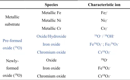
Species Characteristic ion
Metallic substrate Metallic Fe Fe2 -Metallic Ni Ni2 -Metallic Cr Cr2 -Pre-formed oxide (16O) Oxide/Hydroxide 16O- / 16OH -Iron oxide Fe16O2- ; Fe216O3 -Chromium oxide Cr16O2 - Newly-formed oxide (18O) Oxide 18O -Iron oxide Fe18O2 -Chromium oxide Cr18O3
ToF-SIMS depth profiles were recorded to determine the composition and layer structure of the passive film. The characteristic ions were selected as shown in Table 1. The pre-formed iron oxide is associated to two different characteristic ions (Fe16O
2- with high intensity and Fe216O3- with low intensity). Despite the high mass resolution that allows us to locate the maximum intensity of each single peak, the characteristic ion of newly formed chromium oxide (Cr18O
2-) slightly overlaps with the tail of characteristic ion of the pre-formed iron oxide (Fe16O
2-). To avoid this problem, Cr18O3- and Fe216O3- ions (that do not suffer for overlapping) are used to characterize the oxide composition during re-oxidation in 18O
2 of the oxide pre-formed in H2SO4. It should be noted that the selected ions do not reflect the real stoichiometry of the species constituting the sample, but are the appropriate markers of the studied species. Since ToF-SIMS is a non-quantitative technique (due to strong matrix effect on secondary ion emission), the intensities of the plotted ions in the depth profiles cannot be compared directly and do not reflect the concentrations of the associated species in the substrate. However, variations of a single signal reflect mainly in-depth variations of the concentration. The depth profiles are plotted versus sputtering time. The sputtering rate has been calculated knowing for passivated 304L SS (in H2SO4 at 0.4V/SCE) (i) the total oxide layer thickness measured from XPS (results not shown here), and (ii) the position of the metal/oxide interface on the ToF-SIMS depth profile. Assuming a constant sputtering rate (0.015 nm/s) in the oxide, independent of the oxide layer composition, the sputtering time directly translates into oxide thickness.
Immediately after the specimen was introduced into the ToF-SIMS analysis chamber, and prior to the application of heat treatment, the passive film was analysed at both primary beam currents. Due to the destructive nature of sputtering, each depth profile was collected at a different, unperturbed area of the sample surface. The specimen was then heated up to a temperature of 300 ± 1 °C (an IONTOF heating stage was used). Modifications of the passive oxide film due to the increase of temperature were studied [34]. A precision leak valve was then used to introduce a constant and low 18O
2 pressure into the analysis chamber, such that the partial pressure P(18O2) was constant at 1×10⁻⁵ mbar. After the designated oxidation time had elapsed, the leak valve was closed while the sample temperature was maintained at 300 ± 1 °C, and the chamber immediately pumped down to the base pressure (10−9 mbar), in order to record the ToF-SIMS depth profiles. A scheme of the in situ re-oxidation experiment is shown in Fig.1.
Results and discussion
Passive film formation and analysis
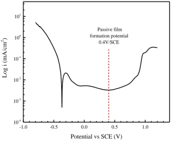
-1.0 -0.5 0.0 0.5 1.0 10-4 10-3 10-2 10-1 100 101 Log i (mA/cm 2 ) Potential vs SCE (V) Passive film formation potential 0.4V/SCE
Fig.2 Polarization curve of the 304L SS in 0.05M H2SO4 with a scan rate of 1mV/s
Fig. 2 shows the polarization curve obtained on 304L SS in 0.05M H2SO4 aqueous solution with a scan rate of 1mV/s. Within the passive range (-0.1~0.8V/SCE), the lowest current is obtained at 0.4V/SCE. Anodic polarization at this potential in sulfuric acid causes the Cr oxide enrichment and Fe oxide dissolution, which leads to the formation of passive film with high corrosion resistance [7, 8]. In this study, a static potential polarization at 0.4V/SCE was applied to the 304L SS for 1h to form a protective passive film on the stainless steel surface.
Fig.3 ToF-SIMS depth profiles for passive film formed on 304L SS surface in 0.05M H2SO4(aq) at 0.4V/SCE for 1h at (a) room
maximum intensity of Ni2- in the depth profile)
Fig.3(a) shows the ToF-SIMS negative ion depth profiles for the passive film formed on 304L SS at room temperature. The position of the metal/oxide interface on the ToF-SIMS depth profiles is crucial to extract the total thickness of the oxide layer. As previously reported, the Ni2- species is a good marker of the metal/oxide interface (maximum intensity of Ni2- signal) [27, 35]. Thus, the passive film thickness is estimated to be 2 nm when the 304L SS sample is passivated 1h at room temperature in 0.05M H2SO4 at 0.4V/SCE.
Focusing on the layered structure of the passive film formed at room temperature (Fig.3(a)), it appears that the Fe16O
2- signal peaks in the outer part of the passive film and decreases slowly through the oxide film region, indicating that oxidized iron is mainly located in the outer part of the film with low content in the inner part of the film. On the contrary, the Cr16O
2- depth profile shows its maximum in the inner part of the film. Thus, chromium oxide is mainly located in the inner oxide layer. This duplex structure for the passive oxide film on 304L SS surface at room temperature is consistent with previous studies [7, 8].
In order to investigate at higher temperature, the transport of species in the passive film formed on 304L SS, we first studied the thermal stability of this electrochemically formed passive film when heated up to 300°C, so that the re-oxidation experiments could be carried out under 18O
2 at this temperature. At lower temperatures, the ion-transport process would be very slow, and it would take a long time to observe the transport of species in the passive film. At temperatures higher than 300°C, the composition and structure of the pre-formed passive film would be changed too rapidly. Based on the previous study of the thermal stability of the passive film in ultra-high vacuum by ToF-SIMS, it is known that 300°C is a suitable temperature, ensuring sufficiently fast kinetics of the ion transport and stability of pre-formed passive film [34].
Fig.3(b) shows the characteristic ToF-SIMS depth profile for the electrochemically formed passive film in H2SO4 after increasing the temperature to 300°C under UHV. The passive film thickness is estimated to be 1.9 nm. The structure of the passive film remains duplex as indicated by the Fe16O2- signal that reaches its maximum intensity in the outer part of the oxide scale whereas the Cr16O
2- signal has its maximum in the inner part of the oxide. However some changes are observed when increasing the temperature from room temperature to 300°C: (i) the lower intensity of the Fe16O
2- in the outer oxide, which is correlated with (ii) the slightly higher intensity of the Cr16O
2- signal, as well as its distribution throughout the oxide film (signal spreading in the oxide). These observations have already been done and explained in a previous work on 316 SS when studying the thermal stability of the native oxide film [34]. This is assigned to (i) the reduction of the Fe hydroxide species that happens above 100°C and (ii) the formation of chromium oxide at the expense of iron oxide when the temperature is above 250°C. The decrease of the passive oxide film thickness is
assigned to the dehydroxylation and dehydration. Nevertheless, the structure of the oxide is still a bilayer and the main species in the film are still iron and chromium oxides.
Investigation of ion transport process in oxide layers
Fig.4 ToF-SIMS profiles obtained on pre-passivated 304L SS after re-oxidation at 300℃ for (a) 1 min, (b) 5 min, (c) 15min, (d) 1h and (e) 2
h (the position of the oxide/metal interface is determined by the maximum intensity of Ni2- in the depth profile)
Fig.4 shows the ToF-SIMS depth profiles obtained on the pre-passivated 304L SS after exposure to low pressure isotopic 18O2 gas (10-5 mbar) at 300°C for different times. After 1 min of re-oxidation (Fig.4(a)),
the metal/oxide interface, still defined by the maximum intensity of the Ni2- signal, is located at 137.5s of sputtering time, which corresponds to 2.1 nm. By looking at both the 16O- and 18O- signals on the depth profile, one observes that the new 18O- signal is located at the prior oxide surface. A closer examination of Cr16O2-, Fe216O3-, Cr18O3- and Fe18O2-, characteristic signals of the Cr oxide and Fe oxide formed with 16O and 18O, respectively, confirms the location of the newly formed oxides (during re-oxidation in 18O gas) at the surface of the prior oxide film, the pre-formed 16O species remaining deeper into the oxide scale.
With further exposure to 18O2 for 5 min, the oxide film becomes thicker, from 2.1 nm to 3.1 nm (oxide/metal interface recorded for 205s of sputtering). The intensities of Cr18O
3- and Fe18O2- profiles are found to significantly increase, and their location at the external surface indicates that the oxide film thickens at the oxide/gas interface due to the formation of both chromium and iron oxides. The re-oxidation process clearly reveals that, for short re-oxidation time, the oxide growth is mainly governed by the transport of Cr and Fe cation species through the pre-formed oxide.
For a re-oxidation time of 15 min, the Ni2- signal indicates that the metal/oxide interface is reached after 360s of sputtering, corresponding to a total oxide layer thickness of 5.4 nm. The oxide film is clearly growing. Looking at the depth profile of 18O- signal, its maximum intensity remains located at the outer surface, and its intensity slowly decreases through the pre-formed passive oxide layer. This shows that the newly oxides (18O) is still formed at the oxide/gas interface due to cation diffusion through the oxide, but that 18O species also diffuse towards the metal/oxide interface following an inward isotopic exchange. Although the main growth mechanism is still by outward cation diffusion, there is clearly an 18O- concentration gradient in the inner oxide, assigned to O2- inward diffusion and isotopic exchange. Isotopic exchange in such experiments has already been described by Poulain et al. [23] for oxidation of pure Cr sample and Voyshnis et al. [27] for oxidation of nickel base alloy.
For 1h of re-oxidation, the oxide film thickness is 6.5 nm (corresponding to 435s of sputtering), while for a 2h re-oxidation time, the thickness of the oxide film remains nearly constant (6.8 nm, ~ 450s of sputtering time). By looking at the depth profiles of the 16O- and 18O- signals, one observes that the 18O- signal still has a wide peak in the external surface and the 16O- signal peak in the inner region for 1h re-oxidation, whereas for 2h re-oxidation, the 16O- and 18O- depth profiles overlap in the oxide film region, which is assigned to the 16O/18O isotopic exchange. Thus, after 1h of re-oxidation, the oxide film reaches a quasi-stationary state, and the main mechanism dominating the depth profile is the 16O/18O isotopic exchange.
Fig. 5 Scheme of the evolution of the passive oxide layer exposed to 18O
2 at 300°C
A scheme of the evolution of the oxide layer exposed to 18O
2 at 300°C based on the ToF-SIMS depth profiling data is shown in Fig.5. As demonstrated before, the main mechanism governing the growth of the oxide for short re-oxidation of the pre-passivated surface is the outward cation transport, meaning that iron and chromium cations diffuse from the metallic substrate to the oxide/gas interface where they react with oxygen (18O
2). Thus, the newly formed chromium and iron oxides are located at the outer oxide/gas interface. The higher 18O concentration at the external surface leads to an oxygen isotopic exchange with 16O. After 15 min of re-oxidation with low 18O2 pressure, cation diffusion is still governing the growth of the oxide. Isotopic exchange is also evidenced as shown by the 18O- profile into the pre-formed 16O oxide. At longer re-oxidation time (more than 1h), the oxide film reaches a quasi-stationary thickness (the growth rate becomes very low) and the isotopic exchange becomes the main mechanism leading to increased amount of 18O in the inner part of the oxide layer. Some volatilization of chromium oxide evidenced by Poulain et al. [23] for oxidation of pure Cr at the same temperature cannot be excluded.
1 2 3 4 5 6 7 0 20 40 60 80 100 120 304 SS Fit Re oxidation ti me in 18 O 2 (mi n) Oxide thickness (nm) Kp= 0.018 nm2.s-1 Kv= 0.0037 nm.s-1
The thickness of the oxide layer, measured by ToF-SIMS at different oxidation times on pre-passivated 304L SS during re-oxidation at 300°C is reported in Fig.6. The kinetics of re-oxidation is well fitted by a logarithmic-type law, which, according to previous work [23, 35], can be explained by the competition between parabolic growth and oxide layer volatilization. From Fig.6, two regions can be observed: a first region from 0 min to 30 min of fast oxide layer growth (with chromium and iron oxides appearing at the outer surface), as previously. Then, for longer re-oxidation, the oxide film growth becomes slower and a quasi-stationary thickness is observed (which is explained by a growth rate equal to the volatilization rate).
The oxidation kinetics can then be fitted by Eq. (1), as shown in a previous paper [23]:
𝑡 =𝑘𝑝 𝑘𝑣2[− 𝑘𝑣 𝑘𝑝(𝑥 − 𝑥0) − ln (1 − 𝑘𝑣 𝑘𝑝(𝑥 − 𝑥0))] [1]
where kp is the parabolic constant, kv is the constant of volatilization, and x0 is the thickness of pre-passivated oxide film.
Both the fitting curve and the results calculated for the parabolic and volatilization constants are shown in Fig.6. The values of kp derived from the fit is 1.8×10⁻² nm2.s-1 and kv is 3.7×10⁻³ nm.s-1. These values are close to that calculated for pure Cr at 300°C (kp = 1.9×10⁻² nm2.s-1 and kv = 1.8×10⁻³ nm.s-1) [23], but kp is higher than the value obtained on polycrystalline Ni alloy (kp = 5.9×10⁻³ nm2.s-1) [35]. This difference is assigned to the different structures of the oxide film.
On the basis of a simplified relationship between diffusion coefficient and parabolic rate constant 𝑘𝑝 = 2𝐷𝑐
[36]
, one would conclude that the diffusion coefficient for outward cation diffusion (Dc) in the oxide film is 9×10-17 cm2.s-1.Although, as discussed above, the main ion transport mechanism during the re-oxidation process of passive film formed on 304L SS is outward cation diffusion, a mixed mechanism including both cation and anion diffusion cannot be excluded, according to the depth profile of 18O- in the oxide layer. Thus, it is interesting to assess the oxygen diffusion coefficient through the oxide film and compare it to the cation diffusion coefficient. The semi-infinite integration of Fick’s second diffusion law for one-dimensional diffusion, modified to include isotopic exchange can be tested on the 18O- depth profiles, as already done by Voyshnis
et al. [27]. Possible chemical reactivation of Cs implanted during the acquisition of the depth profile and chemical activity of the diffusing species are not considered in the model. With this model, the oxygen diffusion coefficient [Do, in cm2.s-1] and isotopic exchange coefficient [k, in at.cm-2.s-1] can be obtained. The differential equation to be solved is the following:
𝜕𝐶 𝜕𝑡 = − 𝐷
𝜕2𝐶
where the first term is for the diffusion and the second one for isotopic exchange, C is the concentration of 18O in the oxide film at depth x and after oxidation time t.
This differential equation can be analytically solved:
𝐶(𝑥,𝑡)−𝐶𝑚𝑎𝑥 𝐶0−𝐶𝑚𝑎𝑥 = 1 2{𝑒𝑥𝑝 − (√ 𝑘 𝐷𝑥)𝑒𝑟𝑓𝑐 ( 𝑥 2√𝐷𝑡− √𝑘𝑡) + 𝑒𝑥𝑝(√ 𝑘 𝐷𝑥)𝑒𝑟𝑓𝑐 ( 𝑥 2√𝐷𝑡+ √𝑘𝑡)} [3]
using the following boundary conditions:
{
𝐶(𝑥, 0) = 0, 𝑥 > 0 𝐶(0, 𝑡) = 𝐶0, 𝑡 > 0
𝐶(𝑥 = 𝑀 𝑂⁄ , 𝑡) = 𝐶𝑚𝑎𝑥, 𝑡 > 0
[4]
where Cmax is the initial 18O concentration at the metal/oxide interface, C0 the initial 18O concentration at the outer interface (x = 0).
More details on this model are provided in ref [23, 27].
Fig.7 Normalized 18O− ion ToF-SIMS depth profiles (black points) and fit of the experimental data with the theoretical model (red solid
line), for the 304L SS re-oxidized in 18O
2 in situ in the ToF-SIMS chamber at 300 °C during (a)1 min and (b) 5 min.
Eq.3 takes into account both the solid state diffusion and the isotopic exchange, and is used to fit the experimental data and determine the oxygen diffusion coefficient (Do) and the isotopic exchange coefficient of the O species. The results shown in Fig.7 are the best fits of the experimental data of normalized depth profile of the 18O- signals (black points) with the theoretical model (red curve) for the 304L SS re-oxidation in 18O
2 for 1 min (Fig.6(a)) and 5 min (Fig.6(b)). It is observed that the model fits well the experimental data. The oxygen diffusion coefficients (Do) determined in this way are 2 × 10-17 cm2.s-1 (Fig.7(a)) and 1.6 × 10 -17 cm2.s-1 (Fig.7(b)), and the isotopic exchange coefficients (k) are 3.7 × 10⁻¹⁰ at.cm-2.s-1 (Fig.7(a)) and 1 × 10-11 at.cm-2.s-1 (Fig.7(b)). For longer re-oxidation times, the experimental data cannot be fitted by this model, because of the significant modifications of the structure and composition of the oxide after 15 min of re-oxidation, as shown in Fig.4(c).
It appears that the cation diffusion coefficient (9×10-17 cm2.s-1) deduced from the thickness measurement is about 5 times higher than the oxygen diffusion coefficient (1.6 ~ 2× 10-17 cm2.s-1). Thus, outward cation diffusion plays the main role in the oxide growth on 304L SS. This is in agreement with observations done during the oxidation of Fe-15%Cr alloy [32], at 750°C, where DFe (3.6×10⁻¹⁶ cm2.s-1) and DCr (2×10-16 cm2.s -1) are one order of magnitude higher than D
O (3.4×10⁻¹⁷ cm2.s-1).
Conclusion
The nature, composition and structure of the passive film formed on 304L SS in acid solution and after heating in vacuum at 300°C has been investigated by ToF-SIMS. The passive oxide film formed electrochemically in 0.05 M H2SO4 solution at 0.4V/SCE for 1h has a duplex structure, comprising an iron rich outer layer and a chromium rich inner layer. The duplex structure of the passive film is thermally stable at 300°C in vacuum, with however partial dehydroxylation and iron oxide reduction.
Insight into the ion transport process in the pre-formed passive film on 304L SS has been obtained by ToF-SIMS. The pre-formed passive film was exposed to 18O
2 at 300°C. The enrichment of 18O species at the oxide/gas interface, provided direct evidence of outward cation diffusion. After 1h re-oxidation, the oxide film reaches a quasi-stationary thickness, assigned to the limiting transport of cation from the substrate and the chromium oxide evaporation. Then the 16O /18O isotopic exchange becomes the main mechanism governing the O depth profile. Based on a model built up to describe the re-oxidation kinetics obtained from the measurements, the parabolic and volatilization constants for the oxide growth are determined. They are 0.018 nm2.s-1 and 0.0037 nm.s-1, respectively. According to the parabolic constant, the diffusion coefficient of cations is found to be Dc=9×10-17 cm2.s-1, which is several times higher than the oxygen diffusion coefficient (Do=1.6 ~ 2× 10-17 cm2.s-1) determined from the 18O depth profile. This work evidences that the growth of the passive oxide film is governed by outward cation diffusion.
Declaration of Competing Interest
The authors declare that they have no known competing financial interests or personal relationships that could have appeared to influence the work reported in this paper.
Author Statement
Luntao WANG: Investigation, Data acquisition, Writing - original draft
Svetlana VOYSHNIS: Methodology
Philippe MARCUS: Conceptualization, Writing - review & editing, Supervision. All authors contributed to the interpretation of the results and to the writing of the paper.
Data availability statement
The datasets generated for this study are available on request to the corresponding author.
Acknowledgements
This project has received funding from the European Research Council (ERC) under the European Union’s Horizon 2020 research and innovation program (ERC Advanced grant CIMNAS: Corrosion Initiation Mechanisms at the Atomic and Nanometric Scales Advanced, grant no. 741123). Région île-de-France is acknowledged for partial funding of the ToF-SIMS equipment. China Scholarship Council (CSC) is acknowledged for the scholarship to the first author (No. 201706460018).
References
[1] V. Maurice, P. Marcus, Current developments of nanoscale insight into corrosion protection by passive oxide films, Curr Opin Solid State Mater Sci, 22 (2018) 156-167.
[2] T. Massoud, V. Maurice, L.H. Klein, P. Marcus, Nanoscale morphology and atomic structure of passive films on stainless steel, J. Electrochem. Soc. 160 (2013) C232-C238.
[3] V. Maurice, P. Marcus, Passive films at the nanoscale, Electrochim. Acta 84 (2012) 129-138. [4] J.R. Davis, Stainless steels, ASM international, 1994.
[5] S.E. Ziemniak, M. Hanson, Corrosion behavior of 304 stainless steel in high temperature, hydrogenated water, Corros. Sci. 44 (2002) 2209-2230.
[6] B. Stellwag, The mechanism of oxide film formation on austenitic stainless steels in high temperature water, Corros. Sci. 40 (1998) 337-370.
[7] V. Maurice, H. Peng, L.H. Klein, A. Seyeux, S. Zanna, P. Marcus, Effects of molybdenum on the composition and nanoscale morphology of passivated austenitic stainless steel surfaces, Faraday Discuss. 180 (2015) 151-170. [8] Z. Wang, F. Di-Franco, A. Seyeux, S. Zanna, V. Maurice, P. Marcus, Passivation-induced physicochemical alterations of the native surface oxide film on 316L austenitic stainless steel, J. Electrochem. Soc. 166 (2019) C3376-C3388.
[9] E. Gardin, S. Zanna, A. Seyeux, A. Allion-Maurer, P. Marcus, XPS and ToF-SIMS characterization of the surface oxides on lean duplex stainless steel – Global and local approaches, Corros. Sci. 155 (2019) 121-133. [10] E. Gardin, S. Zanna, A. Seyeux, A. Allion-Maurer, P. Marcus, Comparative study of the surface oxide films on lean duplex and corresponding single phase stainless steels by XPS and ToF-SIMS, Corros. Sci. 143 (2018) 403-413.
[11] Z. Wang, E.-M. Paschalidou, A. Seyeux, S. Zanna, V. Maurice, P. Marcus, Mechanisms of Cr and Mo Enrichments in the Passive Oxide Film on 316L Austenitic Stainless Steel, Front. Mater. 6 (2019) 232.
stainless steel by XPS and ToF-SIMS, J. Vac. Sci. Technol. A 33 (2015) 05E122.
[13] P. Stefanov, D. Stoychev, M. Stoycheva, T. Marinova, XPS and SEM studies of chromium oxide films chemically formed on stainless steel 316 L, Mater. Chem. Phys. 65 (2000) 212-215.
[14] X. Cheng, Z. Feng, C. Li, C. Dong, X. Li, Investigation of oxide film formation on 316L stainless steel in high-temperature aqueous environments, Electrochim. Acta 56 (2011) 5860-5865.
[15] A. Seyeux, V. Maurice, P. Marcus, Oxide film growth kinetics on metals and alloys I. Physical model, J. Electrochem. Soc. 160 (2013) C189-C196.
[16] D. Barnes, J. Calvert, K. Hay, D. Lees, The role of oxygen transport in oxidation of Fe-Cr alloys, Philos Mag. 28 (1973) 1303-1318.
[17] K. Leistner, C. Toulemonde, B. Diawara, A. Seyeux, P. Marcus, Oxide film growth kinetics on metals and alloys II. Numerical simulation of transient behavior, J. Electrochem. Soc. 160 (2013) C197-C205.
[18] J.D. Henderson, A. Seyeux, S. Zanna, M.C. Biesinger, D.W. Shoesmith, J.J. Noël, P. Marcus, Investigating the transport mechanisms governing the oxidation of Hastelloy BC-1 by in situ ToF-SIMS, Corros. Sci. (2019) 108138.
[19] M.J. Graham, J. Eldrige, D. Mitchell, R. Hussey, Anion transport in growing Cr2O3 scales, Mater. Sci. Forum
1989, pp. 207-242.
[20] C. Wagner, Theoretical analysis of the diffusion processes determining the oxidation rate of alloys, J. Electrochem. Soc. 99 (1952) 369-380.
[21] S. Brenner, Oxidation of Iron‐Molybdenum and Nickel‐Molybdenum Alloys, J. Electrochem. Soc. 102 (1955) 7-15.
[22] J.A. Bardwell, B. MacDougall, M. Graham, Use of 18O/SIMS and electrochemical techniques to study the reduction and breakdown of passive oxide films on iron, J. Electrochem. Soc. 135 (1988) 413-418.
[23] C. Poulain, A. Seyeux, S. Voyshnis, P. Marcus, Volatilization and Transport Mechanisms During Cr Oxidation at 300 °C Studied In Situ by ToF-SIMS, Oxid. Met. 88 (2017) 423-433.
[24] P. Kofstad, K. Lillerud, On high temperature oxidation of chromium II. Properties of and the oxidation mechanism of chromium, J. Electrochem. Soc. 127 (1980) 2410-2419.
[25] P. Kofstad, Nonstoichiometry, diffusion, and electrical conductivity in binary metal oxides, Wiley (1972). [26] G. Bakradze, L. Jeurgens, T. Acartürk, U. Starke, E. Mittemeijer, Atomic transport mechanisms in thin oxide films grown on zirconium by thermal oxidation, as-derived from 18O-tracer experiments, Acta Mater. 59 (2011) 7498-7507.
[27] S. Voyshnis, A. Seyeux, S. Zanna, B. Martin-Cabanas, T. Couvant, P. Marcus, Oxide layer growth on nickel-base alloy surfaces in high temperature water and in O2 studied by ToF-SIMS with isotopic tracers, Corros. Sci. 145 (2018) 212-219.
[28] R. Lobnig, H. Schmidt, K. Hennesen, H. Grabke, Diffusion of cations in chromia layers grown on iron-base alloys, Oxid. Met. 37 (1992) 81-93.
[29] A.C.S. Sabioni, A.M. Huntz, J. Philibert, B. Lesage, C. Monty, Relation between the oxidation growth rate of chromia scales and self-diffusion in Cr2O3, J. Mater. Sci. 27 (1992) 4782-4790.
[30] T. Horita, K. Yamaji, Y. Xiong, H. Kishimoto, N. Sakai, H. Yokokawa, Oxide scale formation of Fe–Cr alloys and oxygen diffusion in the scale, Solid State Ion. 175 (2004) 157-163.
[31] A.C.S. Sabioni, E.A. Malheiros, V. Ji, F. Jomard, W.A. de Almeida Macedo, P.L. Gastelois, Ion Diffusion Study in the Oxide Layers Due to Oxidation of AISI 439 Ferritic Stainless Steel, Oxid. Met. 81 (2014) 407-419. [32] A.C.S. Sabioni, J.N.V. Souza, V. Ji, F. Jomard, V.B. Trindade, J.F. Carneiro, Study of ion diffusion in oxidation films grown on a model Fe–15%Cr alloy, Solid State Ion. 276 (2015) 1-8.
Role of Chromium and Oxygen Ion Diffusion on the Growth Mechanism of Oxidation Films of the AISI 304 Austenitic Stainless Steel, Oxid. Met. 78 (2012) 211-220.
[34] L. Wang, A. Seyeux, P. Marcus, Thermal stability of the passive film formed on 316L stainless steel surface studied by ToF-SIMS, Corros. Sci. (2019) 108395.
[35] X. Wu, S. Voyshnis, A. Seyeux, Y. Chumlyakov, P. Marcus, ToF-SIMS study of oxide films thermally grown on nickel-base alloys, Corros. Sci. 141 (2018) 175-181.
[36] National Research Council . Committee on Coatings, High-temperature oxidation-resistant coatings: coatings for protection from oxidation of superalloys, refractory metals, and graphite, National Academies, 1970.