Forelimb and hindlimb adjustments to treadmill speed during quadrupedal locomotion after a lateral spinal hemisection in adult cats
Par Adam Doelman Programmes de Physiologie
Mémoire présenté à la Faculté de médecine et des sciences de la santé en vue de l’obtention du grade de maitre des sciences (M. Sc.)
en Physiologie
Sherbrooke, Québec, Canada July, 2019
Membres du jury d’évaluation Alain Frigon, Programme de physiologie Dimitri Ryczko, Programme de physiologie
Nathaly Gaudreault, École de réadaptation
thesis:
Alain Frigon, thank you. As my first supervisor in medical research, I was incredibly fortunate to have you as a mentor. You taught me the meaning of true science and always pushed me to present my findings in the most concise and accurate form. Your guidance has been greatly appreciated.
Johnathan Harnie, merci mon homme! Your pursuit of excellence in life and in academics is remarkably infectious and I thank you for being there to push me and to answer all of the questions I’ve thrown your way. You are a terrific scientist and I look forward to seeing what’s in store for your future.
Marie-France Hurteau & Étienne Desrochers, thank you for welcoming me to the lab and providing all of the necessary tools to succeed in the Frigon lab and in Sherbrooke, QC.
I would like to thank all of the animals that were used in the present study along with amazing animal care staff at the Université de Sherbrooke’s Animalerie. It was a pleasure interacting with you on a regular basis. Without your contributions, this project would not be possible.
Finally, I would like to extend a sincere thank you to my friends and family for supporting me along this long and arduous journey. Your relentless positivity means the world to me.
la locomotion quadrupède après une hémisection de la moelle épinière chez le chat adulte
Par Adam Doelman Programmes de Physiologie
Mémoire présenté à la Faculté de médecine et des sciences de la santé en vue de l’obtention du diplôme de maitre ès sciences (M.Sc) en Physiologie, Faculté de médecine et des sciences de la santé, Université de Sherbrooke, Sherbrooke, Québec, Canada, J1H 5N4
La capacité à atteindre et à maintenir des vitesses de marche modérées est d'une importance capitale pour diverses tâches locomotrices chez les animaux comme capturer une proie ou échapper à des prédateurs. Chez l'humain, cette capacité est essentielle au quotidien pour évoluer efficacement dans notre environnement comme par exemple accélérer pour traverser la rue en toute sécurité. Suite à une lésion incomplète de la moelle épinière (LIME), un des déficits engendrés est l’incapacité à atteindre et à maintenir des vitesses modérées. Le but de la présente étude est de décrire les ajustements à différentes vitesses (0.4-0.8 m/s) avant et après une LIME dans le modèle du chat. Des analyses expérimentales ont été effectuées avant, une semaine (H1) et huit semaines (H2) après une hémisection latérale de la moelle épinière au niveau de la 5-6ème vertèbre thoracique chez 8 chats adultes. Nous avons constaté que 4 chats étaient incapables de réaliser 10 à 15 pas de manière constante sur le tapis roulant aux vitesses les plus rapides (0.9 et 1.0 m/s) après l'hémisection à H1 et H2, respectivement. La modulation de la durée du cycle de marche, la phase d’appui et de balancement en fonction de la vitesse a été maintenue chez tous les chats. Cependant, une diminution significative de la durée du cycle des membres antérieurs et une augmentation de la durée du cycle des membres postérieurs a pu être observée. Cette dissociation du rythme antérieur-postérieur fait apparaître une coordination 2-1 mais qui ne semble toutefois pas être influencée par la vitesse de marche. La modulation de la longueur du pas et de la foulée a été maintenue après la LIME. Par ailleurs, à mesure que la vitesse du tapis augmente, le contact de la patte du membre postérieur est resté nettement plus caudal par rapport à la hanche à H1 et H2. Les intervalles de phase ont été relativement peu affectés par la vitesse du tapis roulant ou l'hémisection latérale. Bien que la modulation de la durée des périodes de support en fonction de la vitesse ait été conservée après la LIME, la durée des périodes de double support a augmenté tandis que les périodes de triple et quadruple support ont diminué avec l'augmentation de la vitesse. Nous avons noté une diminution significative de la durée de la période de triple support pendant laquelle deux membres postérieurs et un membre antérieur sont en contact avec le tapis roulant, ainsi qu'une augmentation significative de la fréquence et de la durée de quatre supports. De plus, avec l’augmentation de la vitesse de marche, nous avons observé une augmentation de l'activité électromyographique dans tous les muscles testés, à l'exception du vaste latéral ipsilatérale à la lésion (RVL; extenseur du genou). Nous avons également noté une diminution de la durée d'activation du RVL ainsi qu'une augmentation de la durée d’activation du muscle sartorius ipsilatéral à la lésion (RSRT; fléchisseur de la hanche) à toutes les vitesses après l'hémisection. Nous avons également
remarqué que l'activation du RSRT se poursuivait pendant la phase d'appui, probablement pour stabiliser le genou. Dans leur ensemble, les résultats actuels démontrent que les chats peuvent apporter les ajustements nécessaires à leur patron locomoteur sur une plage de vitesses modérée suivant une lésion incomplète de la moelle épinière. Cependant, 4 des 8 chats n'ont pas été capables d'atteindre des vitesses plus rapides, ce qui peut être attribué aux divers changements survenus après l'hémisection. Des études futures pourraient chercher à déterminer quels ajustements limitent cette capacité à atteindre des vitesses plus rapides afin d'optimiser la récupération et une marche fonctionnelle après la blessure.
SUMMARY
Forelimb and hindlimb adjustments to treadmill speed during quadrupedal locomotion after a lateral spinal hemisection in adult cats
By Adam Doelman Physiology Program
Thesis presented at the Faculty of medicine and health sciences for the obtention of Master of science degree diploma (M.Sc.) in Physiology, Faculty of medicine and health sciences,
Université de Sherbrooke, Sherbrooke, Québec, Canada, J1H 5N4
The ability to attain and sustain moderately fast walking speeds is critically important for various locomotor tasks in animals, including humans. In animals, it is vital for survival, to capture prey or escape from predators and to find a mate. In humans, modulating walking speed is important for several activities, including sports, exercise and effective community ambulation, such as safely crossing a street. Following spinal cord injury (SCI), a common deficit includes an inability to attain and sustain moderately fast speeds. The goal of the present study was to describe adjustments to different speeds (0.4m/s-0.8m/s) before and after an incomplete SCI in the cat model. Experimental analyses were performed before, one week (H1) and eight weeks (H2) after a lateral spinal hemisection between the 5-6th thoracic vertebrae in 8 cats. We found that 4 cats were unable to perform 10-15 steps consistently on the treadmill at the fastest speeds (0.9m/s-1.0m/s) following hemisection (H1 and H2). Speed-dependent modulation of cycle, stance and swing durations were maintained in all cats, albeit with a significant decrease in forelimb cycle duration and increase in hindlimb cycle duration. The dissociation of fore-hindlimb rhythms lead to the appearance of 2-1 coordination which was not influenced by stepping speed. Step and stride length modulation was maintained after SCI, however, as speeds increased, hindlimb paw contact remained significantly more caudal relative to the hip at H1 and H2. Interlimb phase intervals were relatively unaffected by walking speed or lateral hemisection. Speed-dependent modulation of support period durations were conserved following SCI such that double support durations increased, while triple and quadruple support periods decreased with increasing speed. We did note a significant decrease in triple support period durations where two hindlimbs and one forelimb are in contact with the treadmill, along with a significant increase in quadruple support frequency and duration. As speeds increased, electromyographic (EMG) activity increased in all muscles tested, with the exception of the ipsilesional knee extensor, right vastus lateralis (RVL). We also noted a decreased RVL activation duration along with increased ipsilesional Sartorius (RSRT, hip flexor/knee extensor) burst duration across all speeds after hemisection. RSRT showed significant activation during the stance phase at both timepoints following hemisection, presumably to stabilize the knee post-SCI. Taken together, the present results demonstrate that felines can make the necessary adjustments to their locomotor pattern across a moderate range of speeds following hemisection at mid-thoracic levels. However, many of these cats could not attain the fastest speeds, which may be attributed to the various changes that occurred following hemisection. Future studies may seek to explore which adjustments limit SCI patients and quadrupeds most from attaining faster speeds to optimize recovery and ambulation after injury.
Résumé ... iv
Summary ... vi
Table of Contents ... viii
Figures List ... x
List of Tables ... xi
List of Abbreviations ... xii
Introduction ... 1
1. Speed modulation during bipedal and quadrupedal locomotion ... 2
1.1. Temporal adjustments to changes in speed ... 3
1.1.1. Cycle and phase durations ... 3
1.1.2. Interlimb coordination as a function of speed ... 5
1.2. Spatial adjustments to changes in speed ... Error! Bookmark not defined. 1.3. Electromyographic adjustments to changes in speed ... 9
2. Neural control of locomotion ... 11
2.1. Spinal mechanisms and speed-dependent adjustments Error! Bookmark not defined. 2.1.1. Commissural interneurons ... 14
2.1.2. Propriospinal interneurons ... 15
2.2. Sensory feedback and speed-dependent adjustments ... 16
2.3. Supraspinal mechanisms and speed-dependent adjustments ... 18
3. Spinal cord injury ... 19
3.1. Complete spinal cord injury ... 19
3.2. Incomplete spinal cord injury ... 20
3.3. Lateral spinal hemisection ... 20
4. Adjustment to changes in speed during locomotion following SCI ... 24
4.1. Adjustments to changes in speed during human locomotion following SCI ... 25
4.2. Adjustments to changes in speed during mammalian locomotion following SCI ... 26
5. Objective ... 27
Materials and Methods ... 29
Animals and ethical approval ... 29
Surgical procedures ... 29
Electrode implantation ... 29
Surgical hemisection ... 30
Data acquisition and analysis ... 31
Histological procedures ... 33
Statistical analysis ... 33
Results ... 35
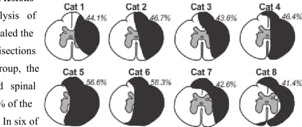
Extent of the spinal lesions ... 35
Adjustments to speed during treadmill locomotion after incomplete spinal cord injury ... 36
Spatial adjustments to speed before and after incomplete spinal cord injury ... 37
Temporal adjustments in the locomotor pattern at different speeds before and after hemisection ... Error! Bookmark not defined. Cycle-stance-swing durations ... Error! Bookmark not defined. Support periods ... 44
Interlimb phasing ... 47
Modulation of forelimb and hindlimb muscle activity at different speeds before and after spinal cord injury ... 50
Discussion ... 52
Speed-dependent adjustments in the locomotor pattern before and after spinal hemisection ... 52
Cycle and phase durations ... 52
Cycle and phase variations ... 53
Spatial parameters ... 54
Interlimb coordination ... 55
Support periods ... 57
Phase intervals ... 58
Changes in fore- and hindlimb EMG activity following spinal hemisection ... 59
Limitations and perspectives ... 60
Bibliography ... 63 Annexes ... Error! Bookmark not defined.
Figure 1. Descent of Inclined Plane - Étienne-Jules Marey (1882). ... 1
Figure 2. Examples of phase-dominance patterns. ... 3
Figure 3. Temporal analysis. ... 6
Figure 4. Spatial analysis. ... 8
Figure 5. Electromyographic analysis. ... 10
Figure 6. Neural control of locomotion. ... Error! Bookmark not defined. Figure 7. Neural control of locomotion following mid-thoracic lateral hemisection. ... 22
Figure 8. Experimental methods. ... 30
Figure 9. Lesion extent. ... 35
Figure 10. Modulation of the locomotor pattern before and after hemisection in a single cat. ... 36
Figure 11. Qualitative representations of hindlimb movements at two speeds before and one week after hemisection in a single cat. ... 37
Figure 12. Changes in limb trajectory before and after hemisection at two speeds in all cats. ... 38
Figure 13. Temporal variables before and after hemisection at various speeds in all limbs. ... 41
Figure 14. Changes in step cycle structure before and after hemisection. ... 43
Figure 15. Effect of treadmill speed on the 8 support periods at two speeds before and after hemisection. ... 45
Figure 16. Changes in interlimb phase durations at two speeds before and after hemisection. ... 47
Figure 17. Modulation of fore- and hindlimb electromyographic (EMG) burst duration and amplitude as a function of speed in various limb muscles before and after hemisection. ... 50
Figure 18. Change in EMG of hip flexor (SRT) as a function of speed before and after hemisection. ... 51
Table 1. Number of cycles with a 1-1, 2-1 and 3-1 coordination before and at two timepoints after hemisection. ... 40
BR CPG EMG FL-HL GTO LF LGS LH RH RF SCI SRT SOL SP TA TRI VL Biceps brachii
Central pattern generator / Générateur de patron central Electromyography / Électromyographie
Forelimb-Hindlimb / Antéro-postérieur
Golgi tendon organ / Organe tendineux de Golgi Left forelimb / Patte antérieur gauche
Lateral gastrocnemius-soleus / Gastrocnemius-soleus lateral Left hindlimb / Patte postérieur gauche
Right hindlimb / Patte postérieur droite Right forelimb / Patte antérieur gauche
Spinal cord injury / Lésion de la moelle épinière Sartorius
Soleus
Superficial peroneal / Péronier superficiel Tibialis anterior / Tibial antérieur
Triceps brachii
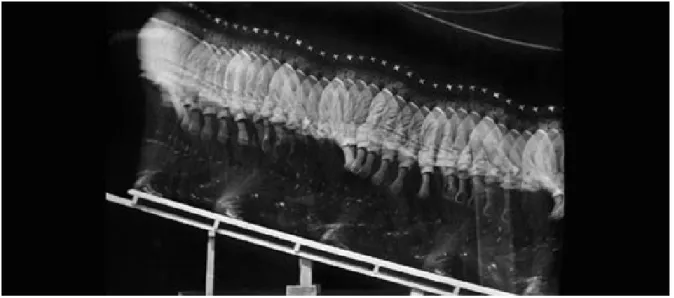
years, even appearing on the walls of caves inhabited by Upper Paleolithic humans (Windels & Laming-Emperaire, 1949). Aristotle (384-322 BCE), a prominent Greek philosopher and scientist made one of the first written contributions to the analysis of human locomotion by stating “If a man were to walk on the ground alongside a wall with a reed dipped in ink attached to his head the line traced by the reed would not be straight but zig-zag, because it goes lower when he bends and higher when he stands upright and raises himself.”(Aristotle 1968; Baker 2007). It was not until the late 19 th century that the analysis of gait kinematics and the measurement of limb movements during locomotion at different speeds appeared. German brothers, Wilhelm and Eduard Weber (1836) were credited with the first scientific analysis of human gait by measuring kinematics from successive images (Weber & Weber, 1991). This method later evolved with the invention of chronographic photography adopted by Edward Muybridge in California who captured serial images of quadrupedal (equine) locomotion using multiple cameras arranged at specific intervals along a track to record trot and galloping gaits. Inspired by this work, French physiologist Jules Etienne Marey (1885)
Figure 1. Descent of Inclined Plane - Étienne-Jules Marey (1882). This photograph is one of the first cinematic recordings of human locomotion using successive serial images. In the photograph, we can see the transition from stance to swing phase along with the movement of the head, arms and legs from the right side of the body as a man descends an inclined plane.
used a shutter system which enabled multiple photographs to be taken with the same camera (Fig. 1) (Baker, 2007).
These pioneer studies enabled scientists to significantly improve their analyses of human and animal locomotion. Gait analysis then further evolved with the development of more sophisticated methods of recording limb muscle activity, known as electromyography (EMG) (Sutherland, 2001). As muscles are known to be the engines that produce movement, it was essential to develop a technique to quantify the electrical activity produced by limb muscles in real time during locomotion. EMG recordings allowed for the measurement of limb muscle activation timing and amplitude. Together, these methods permit a rigorous gait analysis for quantifying adjustments that occur to adapt to changing environments. One such adjustment is the ability to modulate speed, which is important for goal-oriented behaviors. Evolutionarily, the development of faster gaits would have been essential for chasing prey and evading predators (Bramble & Lieberman, 2004). Today, humans adjust their walking speed for various activities, including sports, exercise and effective community ambulation, such as safely crossing a street. In various movement disorders, including spinal cord injury (SCI), individuals are often left with minimal ambulatory capabilities, that includes a significant reduction in walking speed (Dietz & Harkema, 2004; Pepin et al., 2003; Waters et al. 1989). However, despite its obvious importance, few studies have performed gait analysis on human or quadrupedal locomotion following SCI at different speeds.
1. Speed modulation during bipedal and quadrupedal locomotion
To increase speed during locomotion, bipedal and quadrupedal animals make one or both of the following adjustments to their locomotor pattern: increase step cycle frequency and/or increase the distance covered by each stride. To increase cadence or stride length, more muscular force is required to propel the limbs farther and faster during each cycle. At the same time, the limbs must be properly coordinated to maintain a stable and efficient gait. Another method used to increase speed during locomotion is through gait transitions. In four-limbed terrestrial animals, there are a number of different gaits that can be utilised to locomote most efficiently at a given speed. The four primary quadrupedal gaits are (from slowest to fastest) walk, trot, gallop and bound. In the present thesis we will define gaits based on their duty factor, or phase relationship, between ipsilateral (fore-hind) and homologous (left-right) limbs.
1.1.Temporal adjustments to changes in speed
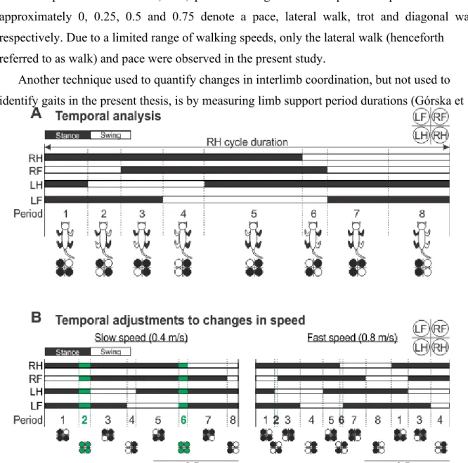
Before describing adjustments to speed, it is important to define a locomotor cycle. We define the locomotor cycle as successive contacts of a given limb. The period during which the limb is in contact with the support surface is referred to as the stance phase, whereas the non-contact phase is termed swing. At comfortable walking speeds, the limb is in stance for approximately 60-70 percent of the step cycle. During bipedal locomotion, when one limb is in the swing phase, the other limb is in single support. At slow to moderate speeds, periods of double support are observed more frequently as both limbs are in contact with the support surface. The absence of double support differentiates running from walking. The support pattern is more complex in quadrupeds, with periods of double, triple and quadruple support during each step cycle. We can quantify cycle and phase durations, as well as support periods, with increasing speed to determine changes in the temporal structure of the step cycle.
1.1.1. Cycle and phase durations
As mentioned previously, increasing speed requires an increase in step frequency, or a decrease in cycle duration, and/or an increase in stride length. Arshavsky et al. (1965) observed in dogs that increasing treadmill speed from 3 km/h to 8 km/h resulted in a significant decrease in cycle duration, which was primarily due to a reduction in stance
Figure 2. Theoretical representation of phase-dominance patterns. Phase-dominance is used to describe the phase (stance or swing) showing a larger variation as a function of cycle duration. There are three forms of phase dominance (left to right in figure): Stance-dominant, which occurs when the stance phase duration varies more with cycle duration than swing phase. Swing-dominant, which occurs when swing phase duration varies more with cycle duration than stance phase and Co-varying pattern, which occurs when stance and swing phases vary approximately equally with cycle duration.
duration, whereas swing duration remained approximately constant across all speeds (Arshavsky et al. 1965). Similar findings were then confirmed in cats and humans [reviewed in (Gossard et al., 2011; Frigon, 2012)]. The variation of stance duration with cycle duration can be seen graphically by plotting stance and swing durations as a function of cycle duration (Fig. 2). A linear regression analysis can then be performed to determine the relationship between cycle and phase durations. During terrestrial locomotion in cats, dogs, mice, rats and insects (stick bug), the slope of the linear regression between stance and cycle duration is significantly steeper than the slope of the relationship between swing and cycle duration (such as the leftmost graph seen in Figure 2) (Goslow et al., 1973; Wetzel & Stuart, 1976; Grillner et al., 1979; Williams, 1981; Jacobson & Hollyday, 1982; Halbertsma, 1983;Grillner & Zangger, 1984; Nilsson et al., 1985; Vilensky, 1987; Büschges et al., 2007; Musselman & Yang, 2007; Yang et al., 2005; Gruhn et al., 2009; Frigon et al., 2013). In other words, cycle duration is ‘dominated’ by the stance phase (Yakovenko et al., 2005; Frigon & Gossard, 2009, 2010; Frigon, 2012; Gossard et al., 2011;). Cycle duration may also be “swing-dominant” when the swing phase varies more with cycle duration compared to the stance phase, or “co-varying” when both stance and swing phases vary approximately equally with cycle duration. Generally, extensor muscles are activated during the stance phase, whereas flexor muscles are activated during the swing phase. Therefore, the terms stance-dominant and extensor-dominant are interchangeable. Similarly, swing-dominant and flexor-dominant sequences are considered the same. It was reported that phase dominance can be influenced by different factors, such as the stimulation of supraspinal centers and sensory feedback (Frigon & Gossard, 2009, 2010; Juvin et al., 2007; Kriellaars et al., 1994; Musselman &
Yang, 2007; Pearson & Rossignol, 1991). For instance, during fictive locomotion in curarized postmamillary-precollicular decerebrate cats, Yakovenko et al. (2005) showed that stimulating the mesencephalic locomotor region (MLR), a region known to initiate locomotion in a wide variety of vertebrate species from lampreys to humans (Shik et al. 1966; Le Ray et al., 2011; Ryczko & Dubuc, 2013), produced flexion-dominant locomotion (Yakovenko et al., 2005). In contrast, Frigon and Gossard (2009) showed that the pattern was extension-dominated during spontaneous fictive locomotion in pre-mamillary/pre-collicular decerebrate cats. Moreover, group I sensory feedback from ankle extensors can strengthen extensor-dominance and even change flexion-dominated patterns to extension-dominated ones (Frigon & Gossard, 2009).
1.1.2. Interlimb coordination as a function of speed
To maintain dynamic stability during locomotion, the limbs must be precisely coordinated. During human walking at comfortable speeds, the arms swing in opposition to the legs, such that the right leg and left arm move synchronously, as well as the left leg and right arm. This coordination reduces energy expenditure by balancing the angular momentum generated from the legs and hips (Elftman, 1939; Pontzer et al., 2009; Hinrichs, 2011). As locomotor speeds increase, the frequency and amplitude of arm and leg swings increase, however the coordination between limbs remains the same. It is only at very slow walking speeds that arm-leg coordination changes, such that arm swing frequency can be up to two times faster than leg frequency, generating a 2:1 arm-leg coordination (Craik et al., 1976; Van Emmerik & Wagenaar, 1994, 1996, 2000).
During quadrupedal locomotion, interlimb coordination must be flexible enough to allow for changes in gait. Historically, gaits have been identified by their “footfall” or support pattern (Hildebrand, 1965, 1976) and determined to be either symmetrical, in which the second half of the gait cycle is identical to the first half, or asymmetrical, when the second half is different relative to the first half. Based on this definition, walk, trot and bound are symmetrical gaits, whereas the trot is asymmetrical. Furthermore, gaits were more specifically identified, using a “gait formula” by comparing the percentage of the gait cycle that each hindfoot is on the ground to the percentage of the gait cycle that the forefoot follows the hindfoot on the same side (Fig. 1, Hildebrand, 1965). This method of gait identification is limited however, as it operates under the assumption that the forelimb and hindlimb step cycles are equal in duration.
In the present study, we will be identifying gaits based on their duty factor, or phase relationship, measured as the interval of time between successive limb contacts as a function of cycle duration (English, 1979; English & Lennard, 1982; Orsal et al., 1990). For example, a phase relationship of 0.5 between homologous limbs would indicate perfect left-right alternation. This method of gait identification is ideal for the present study due to the nature of the experimental protocol and how it may influence the cycle duration of each limb differently. Lemieux et al. (2016) has elaborated on this concept to develop a clear method of identifying quadrupedal gaits based on their duty factor (Fig 1. Lemieux et al., 2016). If homologous limbs maintain a phase relationship of approximately 0.5, the gait is said to be
anti-phase. Anti-phase gaits are then broken down based on their ipsilateral phase relationships into lateral walk, trot, pace and diagonal walk. Ipsilateral phase values of approximately 0, 0.25, 0.5 and 0.75 denote a pace, lateral walk, trot and diagonal walk, respectively. Due to a limited range of walking speeds, only the lateral walk (henceforth
referred to as walk) and pace were observed in the present study.
Another technique used to quantify changes in interlimb coordination, but not used to identify gaits in the present thesis, is by measuring limb support period durations (Górska et
Figure 3. Temporal analysis. (A) Cycle duration is defined by the sum of the stance (black bars) and swing (white bars) duration for each limb (RH, RF, LH, LF). The step cycle is define from RH contact to subsequent RH contact. Support periods are divided into 8 epochs based on the pattern of limb support. Limbs contacting the surface during each period are shown using a footfall diagram which consists of four circles, each representing one of four limbs (black circle = contact) and using a cat schematic (black limb = contact) (B) As step speed increases from slow (left panel) to fast (right panel) walking, stance duration decreases, whereas swing duration remains invariant. Generally at slow speeds, RF contact precedes LH liftoff at SP2 and LF contact precedes RH liftoff at SP6 resulting in a period of quadrupedal limb support (indicated using green bars/circles). At faster speeds, LH liftoff precedes RF contact at SP2 and RH liftoff precedes LF contact at SP6, resulting in periods of diagonal double limb support. RH, right hindlimb; RF, right forelimb; LH, left hindlimb; LF, left forelimb.
al., 1996; Barrière et al., 2010; Frigon et al., 2014; Escalona et al., 2017). The measurement of limb support periods allows for the characterization limb support patterns and the time spent in each type of support. During bipedal upright locomotion, only one and two-limb support is possible. As locomotor speed increases and a greater percentage of the step cycle becomes occupied by the swing phase, bipeds decrease the amount of time spent in double-support and increase the duration of single-limb double-support (Nilsson et al., 1985). However, during quadrupedal walking and pacing, eight different support periods are generally observed during each step cycle, which include double, triple and quadruple-limb support (see Figure 3A). Double support periods include diagonal (support periods 2 and 6) and homolateral (support periods 4 and 8) support. Triple support periods include posterior (both hindpaws and one forepaw, support periods 1 and 5) and anterior tripod (both forepaws and one hindpaw, support periods 3 and 7) support. As locomotor speed increases during walking and pacing gaits, quadrupedal animals spend a greater percentage of the step cycle in double support, whereas the percentage of triple and quadrupedal support periods decrease proportionally (Frigon et al. 2014). Indeed, diagonal double support periods replace quadruple support as locomotor speed increases (see Figure 3B). For instance, Frigon et al. (2014) reported that at slower speeds, support period 2 began with the contact of the right forelimb (RF) and ended with the liftoff of the left hindlimb (LH), creating a period of quadruple support. At faster speeds however, support period 2 began with LH liftoff and
ended with RF contact, thus creating a diagonal double support period.
1.2.Spatial adjustments to changes in speed
Another method used to analyze limb displacement during locomotion is through spatial analysis. In the present study, we analysed three spatial parameters: step length, stride length and maximum swing height (Figure 4A). Stride lengths were measured as the distance between stance onset and offset of the right hindlimb added to the distance traveled by the treadmill during the swing phase, which was calculated by multiplying swing duration by treadmill speed (Thibaudier & Frigon, 2014; Dambreville et al., 2015; Harnie et al., 2018).
Step length is the distance between the leading and trailing limbs at stance onset of the leading limb (Hoogkamer et al., 2013). Maximum swing height is defined as the vertical distance (cm) between the hip and ankle markers at the highest point achieved during the
swing phase. As mentioned previously, increases in stepping speed are accomplished by increasing cadence and/or stride length.
In humans and quadrupedal animals, step and stride lengths increase from slow to fast speeds (Grillner et al., 1979; Afelt et al., 1983; Nilsson et al., 1985; Blaszczyk & Dobrzecka, 1989; Thibaudier & Frigon, 2014). Halbertsma (1983) noted that increases in step and stride lengths were mediated by a more caudal position of the hindpaw relative to the hip at liftoff
Figure 4. Spatial analysis. In the present thesis, we analyzed several adjustments in limb trajectory that take place when stepping at different speeds on a treadmill. (A) Distance of the paw relative to the hip at stance offset (a), distance at stance onset (b), maximum paw height attained during swing (h), step length and stride length. (B) As stepping speed increases, RH stance offset occurs at a more caudal position relative to the hip, swing height increases slightly and stance onset position is invariant. Step and stride lengths increase proportionally with speed. RH, Right hindlimb.
whereas paw placement at contact remained relatively invariant in intact cats walking on a treadmill (see Figure 4B). Paw placement remains relatively invariant with increasing speed because it is critical in establishing a solid base of support (Hof et al., 2005; Farrell et al., 2014; Klishko et al., 2014).
1.3.Electromyographic adjustments to changes in speed
Animals move by contracting muscles, which pull or push on the skeleton. In limbed animals, the central nervous system (CNS) augments the neural drive to limb muscles to increase speed, which can be measured by recording the electrical activity of muscle (EMG, electromyography). EMG is defined as the summation of motor unit action potentials at a particular moment and at a precise location in a contracting skeletal muscle (Herzog, 1998). It can be recorded with intramuscular or surface electrodes. In animal models, chronically implanted intramuscular EMG electrodes provide an effective method of recording muscle activity, as electrodes remain in the same position during movement and do not need to be repositioned at the beginning of each experiment, reducing electrode placement variability. Within a locomotor cycle, a given muscle can have one or two periods, or bursts, of activity (Engberg & Lundberg, 1969). As mentioned previously, extensor muscles are activated during the stance phase, whereas flexor muscles are activated during the swing phase. For example, in Figure 5A, we show the activation pattern of the anterior sartorius (hip flexor/knee extensor) and vastus lateralis (knee extensor). The right vastus lateralis (RVL) is activated during the right hindlimb (RH) stance phase. In contrast, the right anterior sartorius (RSRT), which is primarily a hip flexor, is activated during the RH swing phase. However, as the sartorius is a biarticular muscle, spanning two joints from the patellar tendon to the iliac crest, it can also display some activity during stance to stabilize the knee during extension (Engberg & Lundberg, 1969; Rasmussen et al., 1978; Hoffer et al., 1987).
We can quantify EMG bursts by measuring their burst duration, amplitude and timing (Figure 5B). With increasing speed, extensor burst duration is reduced while flexor burst duration remains relatively unchanged (Fig 5C), mirroring changes in stance and swing phases, respectively [reviewed in (Gossard et al., 2011)]. Increases in stepping speed require limb muscles to produce more force to increase limb propulsion and support. The CNS can increase muscle force by recruiting additional motor units or by increasing the firing rate of
Figure 5. Electromyographic analysis. (A) Example of EMG recordings from one cat (Cat 2) for a right hindlimb hip flexor (RSRT) and knee extensor (RVL) over the course of ~5 right hindlimb (RH) step cycles. (B) EMG analysis was based on measuring burst duration, which is the time (ms) between the onset and offset of each muscle burst, and burst amplitude, which is measured by integrating the full-wave rectified EMG burst from onset to offset and dividing it by its burst duration. (C) As step speed increases from 0.4m/s (left column) to 0.8m/s (right column), burst durations decrease, particularly in extensor muscles, and burst amplitudes generally increase. R, right; L, left; VL, Vastus lateralis; SRT, Sartorius; TRI, Triceps brachii (elbow extensor); BR, Biceps brachii (elbow flexor); RH, Right hindlimb; RF, Right forelimb; LH, Left hindlimb; LF, Left forelimb.
active motor units. As motor unit recruitment and discharge rate increases, so does EMG activity. In agreement, studies have shown an increase in EMG burst amplitude in leg muscles with increasing speed in several mammals, including rats (Hutchison et al., 1989a,b; Roy et al., 1991), cats (Pierotti et al., 1989; Frigon et al., 2015; Hurteau et al., 2017) and humans (Nilsson et al., 1985; Ivanenko et al., 2002; Den Otter et al., 2004; Cappellini, 2006; Liu et al., 2008). Furthermore, various limb muscles such as VL and rectus femoris (RF, knee extensor/hip flexor) are essentially silent at low speeds (<4km/h) in humans, and exhibit non-linear increases in EMG burst activation as locomotor speeds increase (Nilsson et al., 1985; Pepin et al., 2003a; den Otter et al., 2004; Ivanenko et al., 2002, 2009). Other muscles in
more distal regions of the leg, such as the ankle flexor (Tibialis anterior, TA) and ankle extensors (Soleus, SOL; Lateral gastrocnemius, LG) maintain a more linear increase in amplitude as speeds increase (Pepin et al., 2003a; Liu et al., 2008; Ivanenko et al., 2009).
2. Neural control of locomotion
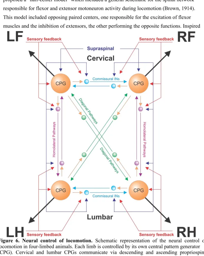
An increase in EMG activity reflects changes in motoneuronal output, which is controlled directly or indirectly by different levels of the CNS that interact dynamically [reviewed in (Rossignol, 2006)]. These include supraspinal structures, spinal circuits and sensory feedback from the periphery. Figure 6 provides an overview of the neural control of locomotion.
2.1. Spinal mechanisms and speed-dependent adjustments
In the early 20th century, Sherrington (1910) showed that hindlimb locomotion could be generated after complete spinal transection (spinalization) in cats and dogs (Sherrington, 1910). Sherrington then proposed that the locomotor rhythm was generated by proprioceptive reflexes, such that sensory receptors in muscles are “themselves the seat of the intrinsic stimuli for reflex stepping” (Sherrington, 1910). However, one year later, Thomas Graham-Brown reported that rhythmic alternating contractions between antagonist muscles (ankle flexor and extensor) could be elicited in spinalized cats after sectioning the dorsal roots caudal to the spinal transection, effectively eliminating sensory feedback (Brown, 1911). Brown proposed that the rhythmic sequence of locomotion is determined by an intrinsic spinal mechanism, which is not essentially controlled by peripheral feedback. In 1914, he
proposed a “half-center model” which included a general schematic for the spinal network responsible for flexor and extensor motoneuron activity during locomotion (Brown, 1914). This model included opposing paired centers, one responsible for the excitation of flexor muscles and the inhibition of extensors, the other performing the opposite functions. Inspired
Figure 6. Neural control of locomotion. Schematic representation of the neural control of locomotion in four-limbed animals. Each limb is controlled by its own central pattern generator (CPG). Cervical and lumbar CPGs communicate via descending and ascending propriospinal pathways with homolateral (purple) and diagonal (green) projections. Left-right coordination is mediated by commissural interneurons (light blue). Sensory feedback from the limbs and supraspinal inputs access the spinal CPGs directly and via commissural and propriospinal pathways. LF, left forelimb; LH, left hindlimb; RF, right forelimb; RH, right hindlimb. Adapted from (Frigon, 2017).
by Brown, work by Anders Lundberg and colleagues further supported the half-center model with the administration of L-DOPA, a dopaminergic and noradrenergic precursor, in unanaesthetized spinal cats that revealed a spinal network of interneurons (INs) with mutual reciprocal inhibition between flexors and extensors (Enberg & Lundberg, 1962; Enberg, 1964; Jankowska et al. 1965). Sten Grillner then showed that the application of L-DOPA to acute spinal, decerebrate cats could elicit coordinated locomotion on a treadmill (Grillner, 1969; Forssberg & Grillner, 1973; Grillner & Zangger, 1979). Studies performed in the cat model reveled important discoveries pertaining to locomotor initiation, central pattern generation and complex reflexes (Grillner 1981; Grillner 1975; PSG Stein, 1987; Arshavsky et al., 1983). Scientists then explored more primitive models, including the lamprey, to better understand the complex neural control networks governing locomotor rhythms. Studies performed in the lamprey model during fictive preparations furthered our understanding of locomotor initiation via the stimulation of specific brainstem regions (McClellan & Grillner, 1984), sensory stimuli (McClellan, 1984) or the pharmacological excitation of specific receptors in the spinal cord (Grillner et al. 1981). Key insights were also discovered pertaining to motoneuron morphology (Teravainen & Rovainen 1971), excitation (Buchanan & Cohen, 1982; Grillner & Wallén, 1985), and central pattern generator distribution (Cohen & Wallén, 1980; Wallén & Williams 1984). These findings contributed to the Unit Burst Generator theory which hypothesized that CPG circuits or limb networks could be broken down to smaller subunits referred to as unit burst generators and coupled to produce more complex motor movements (Grillner, 1981). Since that time, advances in our understanding of spinal locomotor networks have resulted in increasingly complicated models [For instance see (McCrea & Rybak, 2008; Frigon, 2017)]. In summary, it is important to note that today we define the locomotor central pattern generator (CPG) as a network of neurons within the spinal cord capable of autonomously generating a rhythmic pattern of activity [reviewed in (Dietz, 2003; McCrea & Rybak, 2008; Kiehn et al., 2010; Frigon, 2012). It is now thought that each limb is controlled by its own CPG, which are coordinated by propriospinal pathways that receive inputs from supraspinal pathways and peripheral receptors [reviewed in (Frigon, 2017)]. In cats, forelimb CPGs are thought to reside at cervico-thoracic levels (C4-T1) (Yamaguchi, 2004), whereas hindlimb CPGs can be found more caudally, at lumbar levels of the spinal cord (L1-L4) (Langlet et al., 2005; Delivet-Mongrain et al., 2008; Marcoux & Rossignol, 2000). It must also be noted that, although specific regions of the
spinal cord are critical for fore- and hindlimb rhythms, studies have shown that the rhythmogenic capacities are distributed throughout the spinal cord (Kjaerulff & Kiehn, 1996; Cowley & Schmidt, 1997; Delivet-Mongrain et al., 2008; Hagglund et al., 2013) which were initially shown in the lamprey model (Cohen & Walén, 1980; Walén & Williams, 1984).
2.1.1. Commissural interneurons
Locomotor behaviors, such as walking and running, in vertebrates require intricate control of left-right coordination in order to execute efficient rhythmic movement of the limbs. Commissural interneurons (CINs) are thought to be principally responsible for left-right alternation during locomotion. Such an interaction is crucial during quadrupedal locomotion as these animals must frequently adjust their gait to adapt to changes in speed, requiring a shift from out-of-phase gaits at low speeds to in-phase (synchronous) gaits at high speed (English, 1979). Commissural interneurons involved in rhythmic left-right alternation include, among others, V0 and V3 interneurons, which are ipsilaterally projecting CINs and represent an important class of CINs in the ventral spinal cord [reviewed in (Kiehn, 2016)]. V0 neurons have been reported to be crucial in the execution of left-right alternation at various speeds during quadrupedal locomotion due to their monosynaptic contact to contralateral motoneurons (Lanuza et al., 2004; Crone et al., 2009; Talpalar et al., 2013). Talapar et al. (2013) reported that the genetic ablation of these neurons results in complete abolishment of left-right alternation and the production of a bound gait at all speeds tested in the mouse model. Furthermore, subpopulations of V0 neurons have been identified as excitatory CINs in the ventral spinal cord, V0V, and inhibitory CINs in the dorsal spinal cord, V0D. The same group also examined the speed-dependent adjustments to left-right alternation following the genetic ablation of V0V, V0D or both subpopulations of V0 neurons. They reported that selective ablation of inhibitory V0D neurons lead to mice dying shortly before birth. The group then chose to examine locomotor-like activity in the hindlimbs via ventral root recordings during drug-induced fictive locomotion in the isolated spinal cord extracted from embryonic mice. They found that selective ablation of V 0D neurons lead to a lack of left-right walking alternation at low frequencies of locomotor activity (<0.20Hz), whereas clear left-right walking alternation in L2 flexor roots was observed at high frequencies (>0.45Hz). Alternatively, in vivo examination of locomotion was possible in V 0V -ablated mice. In the hindlimbs, they found that an alternating walking pattern was produced at low locomotor
frequencies (2-4Hz) and a synchronous hoping gait at high frequencies (4-10Hz). Interestingly, the forelimbs maintained consistent left-right walking alternation at all locomotor frequencies (Talpalar et al., 2013). Taken together, the group highlighted the importance of V0V neurons for hindlimb alternation at high locomotor frequencies but not low frequencies. In contrast, in the forelimbs, V0V neurons are dispensable for rhythmic walking alternation at all speeds, thus hypothesizing that V 0D neurons are the major alternation neurons governing the forelimb locomotor network. In summation, the study highlights the role of V0 neurons during locomotion and their importance in appropriate speed-dependent adjustments to left-right coordination. Another form of excitatory CIN include the V3 interneurons, which project directly to contralateral motoneurons and interneurons and are rhythmically active during locomotion (Borowska et al., 2013). When V3 neurotransmission is selectively blocked by the expression of tetanus toxin light chain subunit in mice, the regularity of the locomotor rhythm was disrupted based on extracellular recordings of ventral roots using an electroneurogram, without disturbing left-right alternation during pharmacologically induced locomotor activity in mice (Zhang et al., 2008).
2.1.2. Propriospinal interneurons
Propriospinal neurons interconnect spinal segments with ascending and descending axons which can project ipsilaterally or diagonally [reviewed in (Frigon, 2017)]. These propriospinal projections can either be short (1-6 spinal segments) or long (>6 spinal segments) and are thought to be responsible for fore-hindlimb coordination (Skinner et al., 1979; Saywell et al., 2010; Flynn et al., 2011). Cowley et al. (2010) showed that following staggered contralateral thoracic spinal lesions in which half of the spinal cord was sectioned laterally at different locations on opposite sides, brainstem stimulation could still elicit locomotor activity in the hindlimbs. This finding demonstrated that despite eliminating all long direct bulbospinal transmission, locomotion could still be elicited via diagonally projecting propriospinal connections during in vitro preparations in neonatal rats (Cowley et al., 2010). Taken together, the spinal control of interlimb coordination and locomotor speed generation is a complex process that requires the involvement of various spinal neurons. Today, behavioral analysis following experimental genetic or surgical ablation of these neurons in various animal models remains one of the most prominent ways to characterize their function during stepping.
2.2. Sensory feedback and speed-dependent adjustments
Although the basic locomotor rhythm can be produced by CPGs alone in the absence of sensory information, afferent signals from muscles, joints and skin are necessary to allow for adjustments to the environment. Sensory feedback from muscle afferents is generated by various peripheral receptors, such as stretch-sensitive muscle spindles (group Ia and II afferent) and tension-sensitive Golgi tendon organs (GTOs, group Ib afferents) [reviewed in (Rossignol et al. 2006)]. Although flexion-extension alternation has been shown to occur during many preparations in the absence of sensory feedback, the regulation of phase transitions has been shown to be regulated by stretch sensitive receptors during locomotion. For instance, afferent stimulation of group I afferents in many extensor muscles, such as the lateral/medial gastrocnemius (LG/MG) and soleus (SOL), prolongs the stance phase and delays swing phase onset in walking decerebrate cats (Conway & Hultborn, 1987; Guertin et al., 1995; Hiebert & Pearson, 1999). Furthermore, to facilitate flexor burst activation and initiate the swing phase, caudally extending the hip promoted the activation of various flexor muscles responsible for swing generation during fictive locomotion in decerebrate cats (Kriellaars et al., 1994) and during walking in chronic spinal cats (Grillner & Rossignol 1978). In contrast, loading ankle extensors can inhibit or prevent flexor burst initiation during fictive preparations in decerebrate anesthetized spinal cats [reviewed in (Pearson, 2004)].
Feedback from proprioceptors can influence the activation of extensor muscles such that increased limb loading results in greater EMG amplitude. For instance, studies examining human subjects who walk while partially suspended by a harness show a considerable reduction in extensor burst amplitude during walking (Harkema et al., 1997; Dietz & Colombo, 1998; Dietz & Duysens, 2000). Similar results have been shown in cats using “foot-in-the-hole” experiments to temporarily reduce sensory feedback (Donelan & Pearson, 2004). The modulation of EMG amplitude as a function of limb loading was similar in patients with normal, partial or no supraspinal input (Harkema et al., 1997), indicating that it does not require supraspinal input. As humans and other animals increase their stepping speed, more significant load bearing on limb muscles results in the increased activation of afferent neurons, leading to increased EMG activation in extensor muscles (Donelan & Pearson, 2004; Pearson, 2004). Mayer et al. (2018) reported that speed attainment and extensor burst amplitude modulation is severely limited in mice with genetic ablation of
muscle spindle afferents, further indicating that afferent feedback is responsible for mediating proper EMG activation in various extensor muscles during quadrupedal locomotion (Mayer et al., 2018).
Cutaneous afferent feedback is generated as a result of the activation of sensory receptors in the skin, which are capable of informing the spinal circuitry of limb placement in order to adjust or maintain movements (Bouyer & Rossignol, 2003a) as well as provide stability following perturbations (Bolton & Misiaszek, 2009). A common example of the role of cutaneous feedback is the “stumbling corrective response”, whereby cutaneous inputs signal contact with an obstacle and alter limb trajectory and stabilize the contralesional limb, as shown in cats (Forssberg, 1977; Prochazka et al., 1978) and humans (Schillings et al., 2000, 2005; Haridas & Zehr, 2006) . The modulation of cutaneous feedback is task- and phase-dependent. Phase-dependent modulation of cutaneous feedback involves differences in reflex responses during different phases of the step cycle. For instance, stimulation of the hindpaw or the superficial peroneal (SP) nerve, in decerebrate cats during the swing phase elicits a response similar to the stumbling corrective response (Miller et al., 1977; Drew & Rossignol, 1985, 1987; Fuwa et al., 1991). However, the same stimulation during the stance phase prevents limb flexion to maintain stability, or a “stumbling preventive reaction” (Quevedo et al., 2005). Cutaneous afferent feedback is also sensitive to the task during which it is being evoked. For instance, the amplitude of cutaneous reflexes was significantly greater during running than standing when stimulating the sural nerve at the ankle in healthy human subjects (Duysens et al., 1991). Moreover, Hurteau et al. (2017) reported that cutaneous reflexes in spinalized cats were modulated non-linearly with increasing speed, such that the largest responses were evoked at intermediate speeds (0.4 m/s and 0.6m/s), whereas smaller responses were observed at slower (0.2m/s) and faster (0.8m/s) speeds. Overall, studies examining task-dependent modulation of cutaneous feedback have revealed that phase-dependent modulation is conserved with increased stepping speeds in humans (Duysens et al., 1991; Hauglustaine et al., 2001; Baken et al. 2005) and spinal cats (Hurteau et al., 2017). Although cutaneous receptors in the paw are important sources of input for speed-dependent modulation of limb movements, most studies suggest that afferent feedback from muscles and joints are the primary contributors [reviewed in (Rossignol, 2006)].
2.3. Supraspinal mechanisms and speed-dependent adjustments
Despite locomotion being a largely unconscious act, there are various regions of the brain that regulate locomotion. Russian scientists Mark Shik, Fidor Severin and Grigori Orlovsky were credited with the discovery of a region within the mesencephalon known as the Mesencephalic locomotor region (MLR) that, when stimulated, could initiate locomotion in cats. Furthermore, the group found that stimulation could not only initiate locomotion but also modulate its speed and induce gait transitions. Weak stimulation intensity induced a walking gait that gradually increased in speed as simulation intensity increased, transitioning to trot and gallop (Shik et al. 1966). Since its original discovery in 1966, the MLR has been confirmed in other species, including lamprey (Sirota et al., 2000), birds (Sholomenko et al. 1991) and monkeys (Karachi et al., 2010; Goetz et al., 2016;). A number of rostral brain regions have been shown to send projections to the MLR including the motor cortex, basal ganglia, periaqueductal grey matter, lateral hypothalamus and substantia nigra pars compacta (SNc) [reviewed in (Ryczko & Dubuc, 2013)]. For instance, the SNc, and its homologue in the lamprey, the posterior tuberculum (PT), projects descending dopaminergic (Ryczko et al., 2013; Pérez-Fernández et al., 2014) and glutamatergic (Ryczko et al., 2017) neurons to the MLR. Signals derived from the SNc have been shown to activate the MLR and induce locomotor behavior (Derjean et al., 2010; Gariepy et al., 2012a; Ryczko et al., 2013). Furthermore, Ryczko et al. (2017) showed that incremental stimulation of the SNc increased the activation of MLR cells, which increased swimming frequency in lampreys.
The MLR does not project directly to the spinal cord but instead activates neurons in the reticular formation that then activate locomotor networks in the spinal cord (Garcia-Rill & Skinner, 1987a,b; Steeves & Jordan, 1984). Investigation of the downstream effects of MLR stimulation have been most extensively performed in the lamprey [reviewed in (Ryczko & Dubuc, 2013)]. These studies have shown that, in addition to increasing locomotor frequency (Brocard & Dubuc, 2003; Brocard et al., 2010), MLR stimulation activates reticulospinal cells which receive monosynaptic inputs from the MLR (Brocard & Dubuc, 2003) which can then receive additional excitation via a parallel, or “hyperdrive” (Ryczko & Dubuc, 2013), pathway in which the MLR recruits a muscarinoceptive group of cells which function to increase the excitation of reticulospinal cells (Smetana et al. 2010), increase the activity of neurons in respiratory centers to compensate for the increased metabolic cost of faster movement (Gariépy et al. 2012b) and also decrease sensory inputs to reticulospinal neurons,
via the depression of trigeminal sensory transmission (Le Ray et al., 2010). Furthermore, using fMRI studies examining humans visualizing walking, the two structures of the MLR, the Pedunculopontine nucleus (PPN) and Cuneiform nucleus (CuN) are activated (Jankowska, 1992; Duysens et al., 2000). When subjects imagined their walking speed increasing, a proportional increase in PPN and CuN activation was observed (Jahn et al., 2008). Alternatively, significant lesions compromising cholinergic neurons in the PPN have resulted in significant reductions in stride length and stepping speed in patients with Parkinson’s disease (Karachi et al., 2010).
Taken together, there exists a complex interconnected network of supraspinal input, sensory afferent feedback and spinal mechanisms that are responsible for controlling the execution of locomotion across a wide range of speeds in all animals.
3. Spinal cord injury
Spinal cord injury (SCI) is a devastating movement and sensory disorder with significant physical, psychological and financial impact on the lives of patients and their respective families. In Canada, the incidence of SCI in the population varies between 25-55 incidences per million depending on the province and occurs most frequently in males (Singh et al., 2014). Furthermore, the economic burden of an SCI patient ranges from about $1.5 million for incomplete paraplegia up to $3.0 million (CAD) for complete tetraplegia over the course of their lifetime (Krueger et al., 2013). Spinal cord injuries can be broadly defined as complete and incomplete. Complete spinal cord injuries involve the complete abolishment of motor and sensory functions below the spinal lesion, whereas with incomplete spinal cord injuries (iSCI), some sensory and/or motor functions are preserved below the lesion.
3.1.Complete spinal cord injury
Following complete spinal cord injury, humans cannot produce any voluntary movement nor receive sensory feedback caudal to the level of the lesion. Other mammals, such as mice, rats, cats and dogs, however, are generally able to recover some form of hindlimb locomotion, although involuntary, following a few weeks of locomotor training with substantial assistance provided for hindlimb support and with no coordination between the fore- and hindlimbs (Forssberg, 1980a,b; Rossignol et al., 2008). The EMG activity of
hindlimb muscles following complete spinal transection remains relatively similar to intact recordings in adult cats (Belanger et al., 1996; Desrochers et al., 2018). Spinalized adult cats can adjust to changes in speed and even to different speeds for the left and right hindlimbs during hindlimb treadmill stepping while the forelimbs are supported on a stationary, fixed platform (Dambreville et al., 2015; Frigon et al., 2013; Frigon, 2017; Harnie et al., 2018).
3.2. Incomplete spinal cord injury
Experimentally, incomplete SCIs can be performed using different approaches, for instance by compression, contusion or surgical sections. Compression or contusion studies more closely reflect clinical injuries that are predominantly induced via blunt force trauma, such as car or sports accidents. Contusion injuries caused by weight drop or compression clips produce a more diffuse area of damaged spinal tissue (Choo et al., 2007). These models have helped guide our understanding of the primary and secondary damage following SCI (Young, 2002). Not surprisingly, it has been reported that the extent of spinal cord white matter damage is positively correlated with locomotor deficits following contusive injury in animal models (Cao et al., 2005; Poon et al., 2007). Contusion level is also important in determining the extent of locomotor deficits as lesions closer to the theorized lumbar CPG region [L1-L2 in rats (Magnuson et al., 1999; Hadi et al., 2000)], produced more substantial loss of locomotor function compared to more caudal lesions (Magnuson et al., 2005). Although less reflective of the more common forms of SCI observed in the clinical setting, surgical sections of the spinal cord allow researchers to investigate the behavioral outcomes following lesions to specific regions of the spinal cord.
3.3. Lateral spinal hemisection
In the present study, we will be characterizing speed-dependent adjustments following section of the lateral half of the spinal cord, termed a lateral spinal hemisection. The clinical equivalent is the Brown-Séquard syndrome (BSS), which is caused most frequently by penetrating trauma, such as gun or stab wounds (Peacock et al. 1977). This condition was originally described in 1850 by the French physiologist Charles-Édouard Brown-Séquard and was characterized as weakness or paralysis on one side of the body (hemiplegia) and loss of sensation (hemianesthesia) on the other (Brown-Séquard, 1850). The behavioural and pathological outcomes of patients suffering from BSS have been described in a number of
studies since it was initially characterized (Bosch et al., 1979; Cabezudo et al., 1980; Gentleman & Harrington, 1984; Little & Halar, 1985; Miranda et al., 2007). Generally, patients are able to recover both voluntary movements and functional ambulation following a period of complete paresis on the side of the lesion (Little & Halar, 1985). In humans, motor recovery occurs over the course of one to six months (Nathan & Smith, 1973). Initial neuromuscular deficits include a generalized weakness of hip flexor, knee extensor and ankle dorsiflexor muscles (Bosch et al., 1979; Little & Halar, 1985).
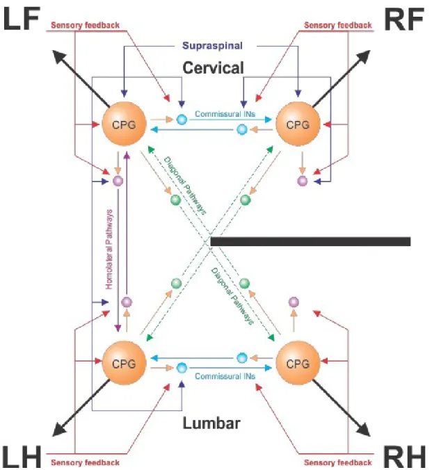
Lateral spinal hemisections have been used in various preclinical models, including mice (Courtine et al., 2008; Goldshmit et al., 2008), rats (Little et al., 1988; Kunkel-Bagden & Bregman, 1990; Webb & Muir, 2004), monkeys (Mettler, 1943; Babu & Namasivayam, 2000; Babu et al., 2008) and cats (Jane et al., 1964; Murray & Goldberger, 1974; Hultborn, 1983; Bregman & Goldberger, 1983; Kato et al., 1984, 1985; Alstermark et al., 1987; Helgren & Goldberger, 1993; Jiang & Drew, 1996; Kuhtz-Buschbeck, et al., 1996; Brustein & Rossignol, 1999; Rossignol et al., 1999; Tester & Howland, 2008; Barrière et al., 2008, 2010; Jefferson et al., 2010; Doperalski et al., 2011; Jefferson et al., 2011; Martinez et al., 2011, 2012, 2013; Thibaudier et al., 2017; Escalona et al. 2018). When performed at thoracic levels, spinal hemisections not only disrupt descending supraspinal inputs (such as reticulospinal neurons controlled by the MLR), but also descending and ascending propriospinal pathways and ascending somatosensory pathways [reviewed in (Frigon, 2017)]. Figure 7 shows a schematic representation of the effects of a lateral spinal hemisection on various neural pathways controlling locomotion.
Following hemisection, adult cats generally recover stable treadmill quadrupedal locomotion within the first 1-2 weeks, although assistance for weight bearing and/or balance may be necessary, depending on lesion extent (Helgren & Goldberger, 1993; Barriere et al., 2008). Immediately after lateral hemisection, studies have shown flaccid paresis in the ipsilesional hindlimb, which starts to improve after about 3 days [reviewed in (Rossignol et al., 2009; Rossignol & Frigon, 2011). Maximum recovery is achieved three to eight weeks after lateral hemisection (Jane et al., 1964). After lateral hemisection, studies have shown an increase in hindlimb cycle duration relative to the intact state (Helgren & Goldberger, 1993;
Kuhtz-Buschbeck et al., 1996; Barrière et al., 2010; Martinez et al., 2011, 2012, 2013). Furthermore, stance duration of the ipsilesional hindlimb is reduced whereas swing phase duration is increased (Brustein & Rossignol, 1999; Martinez et al., 2012). The contralesional hindlimb shows a reverse pattern with increased stance duration and reduced swing duration,
Figure 7. Neural control of locomotion following mid-thoracic lateral hemisection. Schematic representation of the effects of a lateral hemisection (black bar) on the neural
control of locomotion depicted in Figure 6. Dashed lines for diagonal pathways indicate weakened or damaged projections following hemisection. CPG, Central pattern generator; LF, Left forelimb; LH, Left hindlimb; RF, Right forelimb; RH, Right hindlimb.
presumably as a compensatory mechanism to support the ipsilesional hindlimb (Martinez et al., 2012). This effect has also been reported in hemisected mice (Courtine et al., 2008).
Furthermore, lateral hemisections alter the coordination between fore- and hindlimb rhythms. Several studies have reported a dissociation between fore- and hindlimb rhythms, with the forelimbs taking ‘unpaired steps’ (Kato et al., 1984; Górska et al., 1996, 2013; Jiang & Drew, 1996). This produces 2:1 coordination patterns between the fore- and hindlimbs (Jiang & Drew, 1996; Thibaudier & Frigon, 2014; Thibaudier et al., 2017). The disruption of arm-leg rhythms has also been observed in human SCI patients (Tester et al., 2011, 2012). Although it is presently unclear why cats develop 2-1 coordination following incomplete SCI, Thibaudier et al. (2017) proposed that 2-1 coordination could be used as a compensatory strategy to increase stability by increasing the surface area of their support polygon during locomotion. Briefly, Cartmill et al. (2002) reported that the support polygon is created by joining the different points of limb contact with the walking surface. Thibaudier et al. (2017) proposed that when the forelimbs step faster than the hindlimbs, a more caudal extension of the forelimb brings the fore- and hindlimb closer together, thus decreasing the support polygon. Smaller forelimb steps observed with 2-1 FL-HL coordination offsets this interference between homolateral limbs, thus increasing the base of support.
The extent to which forelimb and hindlimb rhythms become impaired following injury appears to depend on lesion extent. Cats with smaller spinal lesions generally preserve a 1-1 FL-HL coordination during locomotion, whereas larger lesions result in the appearance of 2-1 coordination (Brustein & Rossignol, 2-1999; Górska et al., 2-1993, 2-1996, 202-13). Support periods have also been shown to change following hemisection, with reductions in the amount of time spent in diagonal support and increases in the duration of quadrupedal support (Escalona et al. 2017). However, it should be noted that these temporal changes were investigated at a single treadmill or overground speed only.
Spatial analyses of animals stepping overground or on a treadmill following a lateral spinal hemisection has generally consisted of step and stride length measurements and the placement of the paw at contact and liftoff. In mice, Courtine et al. (2008) showed that, following a low thoracic hemisection, the placement of the ipsilesional hindpaw was significantly more caudal relative to the hip. Furthermore, hindpaw placement relative to the hip at stance offset was also more caudal [see Fig.1E (Courtine et al. 2008). In cats, Martinez et al. (2011) reported that ipsilesional hindlimb paw placement at contact was significantly
more caudal relative to the hip following lateral thoracic hemisection (see Fig. 4C). They also reported a significantly shorter stride length on the ipsilesional side at all timepoints after hemisection [see Fig. 4B (Martinez et al. 2011)]. Barrière et al. (2010) showed that two hemisected cats significantly decreased their stride length following hemisection for approximately twenty days before returning to intact values with one cat (GB6), increasing their stride length thirty to sixty days after injury [see Fig. 7A,B (Barrière et al. 2010)]. Decreased step and stride lengths have also been reported in human SCI subjects (Leroux et al. 2002).
The EMG of various ipsilesional hindlimb muscles is reduced after hemisection (Courtine et al. 2005, 2008). For example, although locomotion recovered 10-14 days following hemisection at L1-L2 in rats, EMG activity in TA and VL were reduced (Courtine et al. 2008). Decreased knee extensor (VL) EMG activity was also observed in monkeys (Macaca mulatta) following spinal hemisection (Courtine et al. 2005). EMG burst durations tend to mirror changes in stance and swing durations. For example, Martinez et al. (2011) reported that the EMG burst durations of the ipsilesional Sartorius and semitendinosus muscles increased with swing. Similarly, reduced ipsilesional hindlimb stance duration was reflected by shorter burst durations in the VL of the same limb [see Fig. 5A-D (Martinez et al. 2011)].
Other notable deficits observed following lateral hemisection include a pronounced ipsilesional hindpaw drag at the initiation of swing (Barrière et al. 2010), likely caused by the improper timing of hip and knee flexor muscle activations (Martinez et al. 2011). The ability to avoid objects during swing is also impaired following a lateral spinal hemisection in cats (Doperalski et al. 2011), which requires corticospinal inputs (Bretzner & Drew, 2005; Drew et al., 2002). Skilled locomotor tasks, such as ladder or grid walking, are also impaired in cats (Escalona et al. 2017; Helgren & Goldberger, 1993), rats (Gulino et al., 2007) and monkeys (Babu et al., 2000).
4. Adjustment to changes in speed during locomotion following SCI
People with SCI often state that recovery of walking is one of their primary rehabilitation goals (Ditunno et al., 2008). In general, the recovery of walking only occurs in people with SCI that retain good residual function, classified as ASIA C or D (Vazquez et al. 2008). In 2009, the likelihood a patient classified as ASIAD recovered the ability to walk was over
80% (Scivoletto et al., 2008). However, despite recovering the ability to rhythmically control stepping movements, these patients have been shown to develop a number of alterations to their locomotor pattern, including abnormal muscle activity (Fung & Barbeau, 1989; Crozier et al. 1991; Pepin et al., 2003), changes in hip, knee and ankle trajectory (Dietz et al., 1981) and hyperactive reflexes (Yang et al., 1991; Fung & Barbeau, 1994).
4.1. Adjustments to changes in speed during human locomotion following SCI One of the most common deficits observed in people with SCI is the inability to attain and sustain moderate to fast walking speeds. Healthy humans walk at a speed of roughly 1.4 m/s (Winter, 1992), whereas walking speed in people with SCI varies from 0.2 in the more severely injured to 0.6 m/s in the less severely injured (Waters et al. 1989). Higher-functioning SCI patients have been reported to walk at speeds of up to 0.8 m/s (Krawetz & Nance, 1996; Melis et al., 1999). Decreased walking speed has been proposed as a strategy to decrease fall-risk in elderly and neurologically impaired people (Dingwell & Marin, 2006; England & Granata, 2007; Kang & Dingwell, 2008; Krasovsky et al., 2012). However, whether those are compensatory strategies or merely after-effects that result in greater instability remains to be determined (Espy et al., 2010).
Pepin et al. (2003a) reported that SCI patients (ASIA D) were able to adjust their locomotor pattern from 0.3 m/s to 0.8 m/s by reducing cycle duration and increasing stride length, similar to healthy volunteers. However, they noted that only two SCI patients could reduce their cycle duration to match the treadmill speed like healthy subjects. Instead, subjects with SCI favored increasing stride length to achieve faster walking speeds. In the same study, they found that, compared to healthy subjects, SCI patients presented greater hip excursion. Moreover, they reported that the knee was more flexed at contact, which was maintained throughout the stance phase (see Fig. 2C). Ankle extensor activation was also significantly reduced as a function of speed in SCI patients compared to intact humans [see Fig. 5 (Pepin et al. 2003a]. Weakened EMG activation of leg extensor muscles has been proposed as a key factor inhibiting SCI patients from attaining faster walking speeds.