UNIVERSITÉ DE LILLE
FACULTE DE MÉDECINE HENRI WAREMBOURG
Année :2020T H È S E P O U R L E D I P L O M E D ' É T A T D E D O C T E U R E N M É D E C I N E
Les taux sériques d’Hormone Anti Müllerienne sont-ils normaux chez les
patientes présentant une Anovulation Hypothalamique Fonctionnelle ?
Présentée et soutenue publiquement le 23 Juin 2020 à 14h
au Pôle Recherche
par Sarah MAKOLLÉ
_______________
JURY
Président :
Madame le Professeur Sophie CATTEAU-JONARD
Assesseurs :
Monsieur le Docteur Geoffroy ROBIN
Madame le Docteur Camille ROBIN
Directeur de thèse :
Monsieur le Professeur Didier DEWAILLY
Avertissement
La Faculté n’entend donner aucune approbation aux opinions
émises dans les thèses : celles-ci sont propres à leurs auteurs
LISTE DES ABRÉVIATIONS
AHF Aménorrhée Hypothalamique Fonctionnelle AMH Anti Müllerian Hormone
BMI Body Mass Index
CFA Compte Folliculaire Antral
E2 Estradiol
FNPO Follicle number per ovary FSH Follicle Stimulating Hormone GCs Granulosa cells
GnRH Gonadotrophin Releasing Hormone HGHG Hypogonadisme Hypogonadotrope LH Luteinizing Hormone
OPK Ovaires polykystiques
PCOM Polycystic Ovarian Morphology
TABLE DES MATIÈRES
INTRODUCTION ... 1
ARTICLE ... 4
INTRODUCTION : ... 4
SUBJECTS AND METHODS: ... 6
RESULTS: ... 8 DISCUSSION : ... 9 TABLE 1 : ... 13 TABLE 2 : ... 14 TABLE 3 : ... 15 TABLE 4: ... 16 FIGURE 1 : ... 17 FIGURE 2 : ... 18 FIGURE 3 : ... 19 DISCUSSION ... 20 CONCLUSION ... 24 RÉFÉRENCES ... 25
INTRODUCTION
Les insuffisances gonadotropes sont caractérisées par une sécrétion insuffisante des gonadotrophines hypophysaires : la LH (Luteinizing hormone) et la FSH (Follicle stimulating hormone) conduisant à un état d’hypoestrogénie chez la femme. Il en existe plusieurs types que l’on peut classer en 2 grandes catégories : les hypogonadismes hypogonadotropes congénitaux (HGHG) et les insuffisances gonadotropes acquises.
Les HGHG sont le plus souvent de cause génétique d’origine familiale (1). Ils peuvent être isolés avec olfaction normale (mutation du récepteur à la GnRH, KISS1/KISS1R, TAC3), rentrer dans le cadre de syndrome complexe (Charge, Prader- Willi) ou être un syndrome de Kallmann avec hyposmie-anosmie.
Les insuffisances gonadotropes acquises peuvent être secondaires à une atteinte de la région hypothalamo-hypophysaire (iatrogène, traumatique, tumorale), une hyperprolactinémie. La principale cause est l’aménorrhée hypothalamique fonctionnelle (AHF) responsable d’environ 15 à 30% des insuffisances gonadotropes acquises (2). Il s’agit d’un diagnostic d’élimination après avoir exclu toutes les autres causes organiques et acquises.
La physiopathologie de l’AHF est complexe faisant intervenir de nombreux acteurs. La perturbation de l’axe hypothalamo-hypophysaire est due à un déficit énergétique en lien avec une perte de poids importante et/ou une activité physique excessive et/ou un stress psychologique intense (2–4). Cela entraine une inhibition de la sécrétion pulsatile des neurones à GnRH conduisant à une baisse de la sécrétion de LH et de FSH.
De nombreuses études ont montré le rôle majeur de certains médiateurs dans la physiopathologie de l’AHF (4,5). C’est le cas de la leptine, hormone sécrétée par les adipocytes et régulant la masse grasse en ayant un effet direct sur l’hypothalamus. Des travaux ont retrouvé une leptinémie basse chez les femmes en AHF (6–8) et l’étude de Welt et al (9) montre que l’administration exogène de leptine recombinante humaine permet de restaurer une ovulation chez ces mêmes femmes. Le système Kisspeptine/KISS1R est également impliqué ; en effet les kisspeptines stimulent la sécrétion de GnRH en se liant à leur récepteur KISS1R directement exprimé à la
surface des neurones à GnRH (10). Caronia et al (11) ont également mis en évidence une prédisposition génétique chez certaines femmes en AHF. En effet elles ont une prévalence plus élevée de certaines mutations à l’état hétérozygote simple dans des gènes impliqués dans les HGHG (FGFR1, PROKR2, récepteur à la GnRH, KAL1). L’hormone anti müllerienne (AMH) est une glycoprotéine appartenant à la famille des TGFß. Dans les années 1950, ce sont les travaux du Pr Alfred Jost qui ont permis de découvrir son rôle principal dans la différenciation sexuelle mâle. Nous savons désormais que chez la femme, l’AMH est sécrétée par les cellules de la granulosa des follicules primaires et secondaires (pré-antraux et petits antraux) et qu’elle intervient dans la folliculogénèse en régulant le recrutement folliculaire initial et cyclique (12,13). Plus récemment des travaux d’une équipe lilloise ont montré que l’AMH aurait un rôle sur l’axe gonadotrope en interagissant directement avec les neurones à GnRH (14). Ils ont mis en évidence qu’une partie des neurones à GnRH exprimaient le récepteur à l’AMH (AMHRII) et que l’administration intracérébroventriculaire d’AMH chez des souris femelles augmentait la sécrétion et la pulsatilité de la LH suggérant donc un effet direct de l’AMH sur les neurones à GnRH.
Dans la plupart des situations avec LH basse, soit physiologique (grossesse (13)), soit pharmacologique (contraception oestroprogestative (15,16)), soit pathologique (HGHG) (17), les taux d’AMH sont bas. Toutefois, ce déficit en AMH n’est pas l’explication primitive mais est au contraire la conséquence du défaut de la folliculogénèse secondaire à la carence en LH et FSH. La seule exception est celle des HGHG secondaires à une mutation dans les gènes de l’AMH ou de l’AMHRII (3% des HGHG) (18) où le déficit en AMH serait a priori la cause de l’hypogonadisme hypogonadotrope.
De façon paradoxale, certaines publications rapportent des taux d’AMH normaux voire élevés chez les patientes en AHF (19–24) alors que la folliculogénèse est supposée être défectueuse comme en témoignent l’aménorrhée et l’hypoestrogénie que présentent ces femmes. Toutefois, les séries publiées incluaient des femmes en AHF dont une grande proportion présentait un nombre important de petits follicules antraux et donc un taux d’AMH augmenté puisque positivement corrélé à ces follicules. Cela suggère l’existence d’une morphologie ovarienne d’ovaires polykystiques (Polycystic
Ovarian Morphology, PCOM). Le statut PCOM est fréquemment retrouvé en population générale ainsi que chez les femmes en AHF. En population générale, la prévalence varie de 7 à 24% (25–30). Cette prévalence a même été surestimée dans des publications plus récentes (jusqu’à 80%) (31,32) car les auteurs avaient utilisé le seuil de 12 follicules/ovaire alors qu’il est bien établi maintenant que ce seuil est trop bas avec les échographes de nouvelles générations (33). Chez les AHF, cette prévalence varie de 30 à 50% (19,34–36). L’inclusion de patientes avec PCOM est donc suspecte d’avoir biaisé les résultats. Dans d’autres études, les critères échographiques pour définir le PCOM n’étaient pas précisés et/ou seuls la surface ou le volume ovarien était utilisés alors qu’ils sont moins sensibles que le comptage des follicules antraux (CFA) (37).
L’objectif de cette étude était donc de refaire le point sur les taux plasmatiques d’AMH chez les patientes présentant une LH basse liée à une AHF en utilisant des critères plus stricts pour dépister un éventuel PCOM chez ces femmes.
ARTICLE
Revisiting the serum level of Anti Müllerian Hormone in patients with Functional Hypothalamic Anovulation.
Introduction :
Gonadotropic insufficiencies are characterized by insufficient secretion of pituitary gonadotropins, LH (Luteinizing hormone) and FSH (Follicle stimulating hormone), leading to a state of hypoestrogenism in women. There are several types that can be classified into 2 main categories: congenital hypogonadotropic hypogonadism (HGHG) and acquired gonadotropic insufficiencies.
HGHG are more often due to a genetic cause of family origin (1). They can be isolated with normal olfaction (GnRH receptor mutation. KISS1/KISS1R. TAC3), be part of a complex syndrome (Charge. Prader- Willi) or be a Kallmann’s syndrome with hyposmia-anosmia. Acquired gonadotropic insufficiencies may be secondary to damage to the hypothalamic-pituitary region (iatrogenic. traumatic. tumor) or hyperprolactinemia. The main cause is functional hypothalamic amenorrhea (FHA) responsible for about 15 to 30% of acquired gonadotropic insufficiencies (2). It is a diagnosis of elimination after excluding all other organic and acquired causes.
The pathophysiology of FHA is complex involving many actors. The disruption of the hypothalamic-pituitary axis is due to an energy deficit related to significant weight loss and/or excessive physical activity and/or intense psychological stress (2–4). This induces an inhibition of pulsatile secretion of GnRH neurons leading to a decrease in LH and FSH secretion. Many studies have shown the major role of some mediators in the pathophysiology of FHA (4,5). This is the case of leptin, a hormone secreted by adipocytes and regulating fat mass by having a direct effect on the hypothalamus. Studies have found low leptinemia in women with FHA (6–8) and Welt’s et al study (9) shows that exogenous injection of recombinant human leptin can restore ovulation in these same women. The Kisspeptine/KISS1R system is also involved as kisspeptins stimulate the secretion of GnRH by binding to their KISS1R receptor directly expressed on the surface of GnRH neurons (10). Caronia et al (11) also shown a genetic predisposition in some FHA women. Indeed, they have a higher prevalence of some
mutations in the simple heterozygous state in genes involved in HGHG (FGFR1. PROKR2. GnRH receptors. KAL1).
Anti Müllerian Hormone (AMH) is a glycoprotein belonging to the family of TGFß. Initially, it was considered as a testis hormone involved in male sexual differentiation. We know now that in women, AMH is secreted by the granulosa cells (GCs) of primary and secondary follicles (pre-antral and small antral) and that it is involved in the regulation of follicle recruitment and growth (12,13). More recently, it has been shown that AMH has also an effect on the gonadotropic axis by interacting directly with GnRH neurons (14). This study demonstrated that some GnRH neurons express the AMH receptor (AMHRII) and that intracerebroventricular administration of AMH in female mice increased LH secretion and pulsatility, suggesting a direct effect of AMH on GnRH neurons.
In most situations with low LH, either physiological (pregnancy (13)), pharmacological (oral contraceptive pill (15,16)), or pathological (HGHG) (17), AMH levels are low. However, this AMH deficiency is not the primary explanation but is on the contrary the consequence of a defect in folliculogenesis secondary to LH and FSH insufficiency. The only exception is the HGHG secondary to a mutation in the AMH or AMHRII genes (3% of HGHGs) (18) where AMH deficiency is thought to be the cause of hypogonadotropic hypogonadism.
Paradoxically, some publications report normal or even high serum level of AMH in FHA patients (19–24) when folliculogenesis is thought to be defective, as evidenced by the amenorrhea and hypoestrogenism in these women. However, most of the published series included a significant number of FHA women having an excess number of small antral follicles and therefore an increased AMH level, since it is positively correlated with these follicles. This paradoxical finding suggests the existence of an incidentally associated polycystic ovarian morphology (PCOM). PCOM is frequently found in the general population, with a prevalence ranging from 7% to 24% (25–30). This prevalence has even been overestimated in more recent publications (up to 80%) (31,32) because the authors had used the threshold of 12 follicles/ovary whereas it is now well established that this threshold is too low with new generation ultrasound scanners (33). Intriguingly, in FHA, this prevalence is even higher, ranging from 30 to 50% (19,34–36). As concerns the serum AMH level, the
inclusion of patients with PCOM in some studies may have biased the results. In other studies, the ultrasound criteria for defining PCOM were not specified and/or only the ovarian surface area or volume was used, both being less sensitive than the follicle count (37).
The aim of this study was therefore to re-examine the serum AMH levels in patients with low LH related to FHA using strict criteria to screen a PCOM in these women.
Subjects and Methods: Population:
This is a retrospective observational study carried out at the University Hospital of Lille. Patients in the study had a FHA and were compared to a control group matched on age and body mass index (BMI). Patients and control data were obtained retrospectively from a database in our department, including clinical, hormonal, and ultrasound parameters collected between 2006 to 2015. The Institutional Review Board of the University Hospital of Lille approved this study. All patients gave their informed consent before inclusion in this study.
Patients with FHA:
Data from 45 patients with FHA were used for this study. Diagnosis of FHA was based on a combination of the following criteria; patients had a secondary amenorrhea for more than 6 months (after excluding pregnancy) in a context of recent weight loss and/or insufficient caloric intake and/or intense physical activity and/or notion of recent psychological stress. Biologically, they had hypogonadotropic hypogonadism, i.e., low serum LH (<2 IU/L) with low estradiol (E2) and normal or low FSH. The progestin test (Duphaston 10 mg/d for 10 days) was negative reflecting hypoestrogenicity. Hyperprolactinemia (defined as plasma levels greater than 20 ng/mL on two occasions) had to be excluded. For a diagnosis of elimination, a pituitary MRI was performed to rule out an organ cause. Isolated HGHG and Kallmann's syndrome were excluded on the basis of normal smell, history of normal puberty, and secondary
amenorrhea in all FHA patients. Premature ovarian failure and a history of drug abuse or hormonal treatment during the 3 months before the study were exclusion criteria.
Controls:
The control group consisted of 37 women, matched to patients on age and BMI, who had regular cycles, between 25 and 35 days. The exclusion criteria were LH<2 IU/L; polycystic ovary syndrome (PCOS) status according to the Rotterdam criteria (38); suspicion of low ovarian reserve (FSH>12 IU/L) or hyperprolactinemia. The existence of a PCOM was also an exclusion criterion, even if it was asymptomatic. In this study we defined it with strict criteria, namely: an antral follicle number per ovary (FNPO) ≥12 or ≥19 depending on the date on which the assessment was carried out and the type of ultrasound device used (see below) and/or an ovarian volume ≥ 10 cm3 and/or an ovarian area ≥ 5.5 cm2. In addition, as demonstrated in our previous study (39), an AMH level ≥ 35 pmol/L could substitute for an excess FNPO.
Investigations:
The hormonal work-up was performed between the second and fifth day of the cycle in the controls, and at any time in patients with FHA. LH, FSH, E2, 17-Hydroxy-Progesterone (17-OHP), Dehydroepiandrosterone Sulfate (SDHEA), Androstenedione (A), Total Testosterone (T), Prolactin were measured by immunoassay as described previously (39). The limit of quantification (LoQ) was 0.1 U/L for LH and 0.2 U/mL for FSH (automated assays, Abott Architect). Plasma AMH levels were estimated by ELISA using a 2nd generation Immunotech kit (provided by Beckman Coulter Immunotech) (39).
The ultrasound was performed the same day by endovaginal route with a Logic 400 General Electric Milwaukee from 2006 to 2008 and a General Electric Voluson E8 from 2008. According to a pre-established protocol, a morphological study of the uterus and ovaries was conducted, as well as an assessment of the FNPO (all follicles < 10 mm in diameter were counted by a slow scan across the ovary) (39). The FNPO threshold
for defining follicular excess was ≥12 follicles per ovary before 2008 and ≥ 19 follicles after 2008. Patients in whom vaginal ultrasound was not possible (virgin or refusal) were excluded as well as those with no follicle visualized on the ovaries and/or with an ovarian area below the lower limit (i.e. 2.5 cm2) and/or if there was already a follicle >9 mm with a serum E2 level >80 pg/ml.
Statistical analysis:
Given their non-Gaussian distribution, continuous variables were expressed as medians, with the 5th and 95th percentiles. The non-parametric Mann-Whitney test was used to compare the continuous variables and the Chi-2 test was used to compare categorical variables between two groups. Univariate correlations between some variables were sought by Spearman's non-parametric test. The IBM Statistical Package for Social Science software (SPSS 25.0) was used for all statistical tests. The results were considered statistically significant for a p-value below the alpha risk of 0.05.
Results:
A total of 45 FHA patients and 37 controls were included in the study. Table 1 summarizes the clinical and biological data of the controls and FHA patients. As expected, there were no significant differences in age and BMI since controls were matched to patients on these items.
Conversely, the ranks of serum E2, LH and FSH levels were significantly lower in FHA patients compared to controls (Table 1), as well as the ranks of serum A levels. No significant difference was observed for the ranks of T, 17OHP and Sex Hormone Binding Globulin (SHBG) (Table 1).
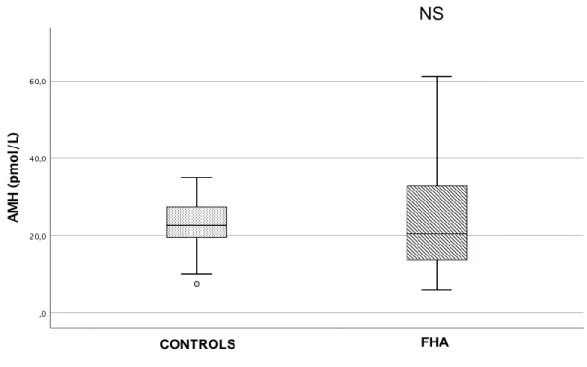
Finally, no significant difference was found in the rankings of AMH levels between the two groups (Table 1 and Figure 1A).
Applying strict criteria for defining PCOM status (see Material and Methods), we found an aspect of PCOM in 21 (46.7%) of the 45 FHA patients. After excluding the PCOM+ patients, we found significantly lower ranks for AMH levels (p<0.002) in FHA patients
compared with controls in which PCOM had been excluded (see Materials and Methods) (Table 2, Figure 1B). As in the whole FHA group, the ranks of LH and A levels were also lower (p<0.0001 and <0.01, respectively) and there was still no significant difference for T. The lower ranks of FSH levels were close to significance (p=0.08) (Table 2).
In the 21 PCOM+ FHA patients, AMH levels ranked significantly higher than in controls (33.2 (11.3-59) vs 22.6 (10.1-34.6), p<0.02) and in PCOM- FHA patients (Table 3). However, their ranks of LH, FSH, and A levels were still lower than in the controls (p<0.0001, <0.002 and <0.05, respectively). Interestingly, they had significantly higher BMI and lower SHBG ranks than in the PCOM- FHA group (Table 3). There was a trend to lower LH and FSH ranks than in the PCOM- FHA group, close to significance (Table 3).
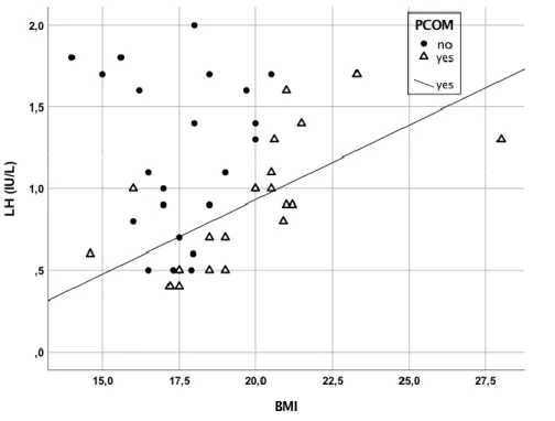
Statistical analysis using the Spearman’s test showed that AMH levels were negatively correlated with age in FHA PCOM- patients exclusively (r=-0.414, p<0.04), but were not correlated with anything else, particularly not LH or FSH levels in the 2 subgroups. In contrast, LH levels were significantly and strongly positively correlated with BMI in FHA PCOM+ patients exclusively (r=0.743), p<0.0001) (Figure 2).
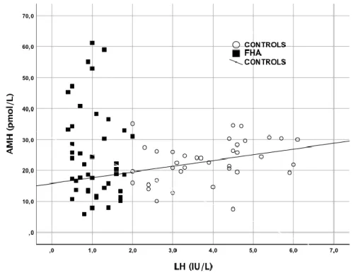
In controls, AMH levels were significantly positively correlated with those of LH (r=0.366, p<0.002) (Figure 3) and with a trend close to significance with T, A and 17OHP levels (p=0.09, 0.09 and 0.07, respectively).
Discussion :
Our study confirms that in a general FHA population, AMH levels are not decreased, despite the low LH and FSH levels, in line with previous publications (19–24) (Table 4). In some studies (21,22,24), AMH levels were even significantly increased compared to a control population. However, it is important to note the heterogeneity of these studies in terms of inclusion criteria, patient phenotypes and serum AMH assay techniques (Table 4).
In the 4 studies where data were available (19,21,23,24), FSH levels were lower than controls, as in this study. Based on our current knowledge of GCs physiology (40), the absence of a decrease in AMH levels in face of decreased FSH levels is paradoxical. Indeed, knowing the stimulating effect of FSH on AMH production (40), its decreased level should be accompanied by a lower AMH level as reported in other situations of
gonadotropic insufficiency (13,15–17). In addition, deficiency in circulating LH leads to a lower production of androgens by theca cells, and therefore in theory, a decrease in their stimulating effect on the richness of FSH receptors (FSHr) in the GCs (40). This would result in reduced FSH activity on these cells, as previously suggested (21), amplifying the negative effect of the relative deficiency of circulating FSH on AMH production. The latter effect may not have been present in the 2 studies that did not exclude FHA patients with basal LH >2 IU/L (21,23) (Table 4). However, AMH levels were not decreased ((19), present study), or even increased (24), in studies that included only FHA patients with basal LH<2 IU/L (Table 4).
Therefore, the explanation for the paradox of a normal or even high AMH in FHA patients is perhaps to be sought elsewhere. In addition to their effects on GCs, FSH and LH deficiency should in theory lead to a decrease in the pool of growing follicles and therefore to a decrease in ovarian AMH production. It is possible to apprehend this phenomenon directly by ultrasound because the pool of 2 to 9mm follicles is supposed to be a reflection of the entire pool of growing follicles. In this study, due to the change of ultrasound scanner in 2008, we were not able to compare the average FNPO of our patients with that of the controls and to look for a possible correlation of FNPO with FSH levels. In all studies that reported it (20–22,24), the FNPO was similar between the FHA and control groups. This is consistent with the normality of AMH levels reported in one study (20), but inconsistent with high AMH levels in the other 3 studies (21,22,24) (Table 4). To explain this singularity, the authors speculate on an increase in the number of small follicles <2mm, not visible on ultrasound (21,24), and/or on a deregulation of the GCs (21).
The latter two phenomena are pathognomonic of PCOS (40), but remain completely speculative for FHA. This raises the hypothesis of a bias related to the presence of PCOM in some of the patients included in the various studies, including our present study. Our results confirm this hypothesis because after exclusion of FHA patients with PCOM, AMH levels became significantly lower than those of controls, in which PCOM was an exclusion criterion. No other studies have reported results after exclusion of FHA patients with PCOM except one of our previous studies (21). However, in this relatively old study, indicating higher AMH levels in FHA patients compared to controls, the PCOM definition did not include the serum AMH level, as in the present study. In view of rather high AMH levels in some controls and patients in this study (21), it can be speculated that the exclusion of patients with PCOM was not complete. In another
study (22), it is not clear whether this exclusion criterion was applied in FHA patients (Table 4). On the other hand, Aleymyar et al (24) deliberately omitted this criterion and found a PCOM in 31.4% of their FHA patients against 24.1% of their controls. Although the difference was not significant, this high prevalence of PCOM among FHA patients may have biased their results. In another of our studies (19), cluster analysis identified a subgroup of FHA patients with PCOM, representing 48.3% of the total FHA population. Our results confirm these data with 46.7% of FHA patients with PCOM. The prevalence of PCOM in FHA patients seems to be higher than in the general population, which ranges from 7% to 24% (25–30). On the other hand, this result is in disagreement with the study by Aleymyar et al (24) which did not find a significantly higher frequency of PCOM in FHA compared to controls, but a trend can nevertheless be observed (31.4% vs 24.1%). This singularity has never been clearly explained. Our study does not shed light on this mystery, but the strong positive correlation between BMI and LH, not found in FHA PCOM- patients, opens up new avenues. The higher BMI in FHA patients with PCOM, along with lower SHBG levels, could suggest that these women had initially the metabolic component of PCOS, before losing weight. Would the effect of weight loss be more directly involved in the inhibition of GnRH neurons than in FHA patients without PCOM? This particular sensitivity could then explain why women with PCOM are over-represented in a population of FHA patients. More data are needed to elucidate this issue.
That FSH deficiency does not prevent some patients with FHA from having a PCOM and therefore a high AMH level contradicts our “triangle theory” (40). Indeed, based on this theory, the follicular and AMH excess in FHA PCOM+ patients should be explained by an excess of intra-ovarian androgens, promoting the expression of FSH receptors on GCs. The more active FSH would stimulate follicle growth and AMH production. On the contrary, our results indicate circulating levels of A identical to PCOM- patients and a trend close to significance at lower circulating FSH levels. To explain this paradox, it is necessary to speculate on differences within the GCs themselves, not perceptible by plasma assays, concerning for example the richness and/or affinity of androgen and/or FSH receptors. These differences could be the consequence of epigenetic reprogramming in utero, which is currently a popular hypothesis for PCOS (41,42). Therefore, in a woman with PCOM, the decrease in gonadotropins due to FHA would
not affect follicular excess and AMH production but would result in hypoestrogenic amenorrhea as in FHA patients without PCOM.
This study did not find a correlation between AMH and LH in FHA patients. On the other hand, this correlation was present in the controls, which is in agreement with our previous publication (14). The lack of correlation between AMH and LH in FHA patients may be explained by the fact that the defects in neural systems involved in the pathophysiology of FHA, such as KISS/KISS1R or Neurokinin (10), likely occur upstream the effect of AMH on GnRH neurons. More pragmatically, the lack of correlation could also be explained by the inaccuracy of the circulating LH assay at low levels, although it was above LoQ in all of our FHA patients.
In conclusion, our study confirms that AMH levels are paradoxically not decreased in a general FHA population but that finally, after excluding with strict criteria the FHA patients with a PCOM, these levels become significantly lower than in controls. This is in line with our current knowledge of GCs physiology. For clinical practice, it seems important not to diagnose too quickly a low ovarian reserve in FHA patients on the basis of a decreased AMH level alone. On the other hand, a high level of AMH in the context of a cycle disorder and a PCOM should not lead to a misdiagnosis of PCOS if the basal LH is low. This means that the clinical analysis and the FSH and LH assays remain essential in the assessment of a cycle disorder to avoid these two pitfalls.
Table 1 : Clinical and hormonal data for patients with Functional Hypothalamic Amenorrhea (FHA) and controls
CONTROLS (n=37) FHA (n=45) p
Median Percentile 05 Percentile 95 Median Percentile 05 Percentile 95
AGE (years) 29.0 21.0 36.0 29.0 16.0 36.0 NS BMI* (kg/m2) 18.5 17.5 19.8 18.5 15.0 21.5 NS T* (ng/ml) 0.17 0.06 0.47 0.20 0.05 0.36 NS A* (ng/ml) 1.44 0.67 2.59 1.00 0.48 1.94 <0.005 DHEAS*(µmol/l) 4.40 1.40 8.60 3.50 2.10 6.80 NS 17OHP* (ng/ml) 0.45 0.24 0.86 0.35 0.14 0.82 NS E2 (pg/ml) 39.0 20.0 72.0 17.0 12.0 36.0 <0.0001 AMH* (pmol/l) 22.6 10.1 34.6 20.4 8.0 55.1 NS LH* (UI/l) 3.9 2.0 6.0 1.0 0.5 1.8 <0.0001 FSH* (UI/l) 5.7 4.2 8.4 4.9 2.6 8.6 <0.004 SHBG* (nmol/l) 57.1 33.6 97.0 58.0 33.1 105.0 NS
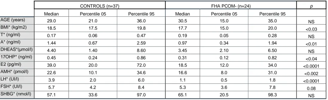
Table 2 : Clinical and hormonal data in FHA patients without PCOM (PCOM-) and in controls
CONTROLS (n=37) FHA PCOM- (n=24) p
Median Percentile 05 Percentile 95 Median Percentile 05 Percentile 95
AGE (years) 29.0 21.0 36.0 30.5 15.0 35.0 NS BMI* (kg/m2) 18.5 17.5 19.8 17.7 15.0 20.0 <0.03 T* (ng/ml) 0.17 0.06 0.47 0.19 0.05 0.28 NS A* (ng/ml) 1.44 0.67 2.59 0.97 0.34 1.94 <0.01 DHEAS*(µmol/l) 4.40 1.40 8.60 3.45 2.10 6.50 NS 17OHP* (ng/ml) 0.45 0.24 0.86 0.31 0.12 0.82 <0.04 E2 (pg/ml) 39.0 20.0 72.0 18.5 12.0 34.0 <0.0001 AMH* (pmol/l) 22.6 10.1 34.6 16.6 8.0 31.0 <0.002 LH* (UI/l) 3.9 2.0 6.0 1.1 0.5 1.8 <0.0001 FSH* (UI/l) 5.7 4.2 8.4 5.3 3.6 7.8 0.08 SHBG* (nmol/l) 57.1 33.6 97.0 65.1 20.5 98.3 NS
Table 3 : Comparison between FHA patients with (PCOM +) and without PCOM (PCOM -).
FHA PCOM- (n=24) FHA PCOM+ (n=21) p
Median Percentile 05 Percentile 95 Median Percentile 05 Percentile 95
AGE (years) 30.5 15.0 35.0 28.0 17.0 36.0 NS BMI* (kg/m2) 17.7 15.0 20.0 20.0 16.0 23.3 <0.01 T* (ng/ml) 0.19 0.05 0.28 0.21 0.10 0.37 NS A* (ng/ml) 0.97 0.34 1.94 1.00 0.64 1.77 NS DHEAS*(µmol/l) 3.45 2.10 6.50 3.70 2.20 6.80 NS 17OHP* (ng/ml) 0.31 0.12 0.82 0.37 0.14 0.96 NS E2 (pg/ml) 18.5 12.0 34.0 17.0 12.0 36.0 NS AMH* (pmol/l) 16.6 8.0 31.0 33.2 11.3 59.0 <0.0001 LH* (UI/l) 1.1 0.5 1.8 0.9 0.4 1.7 0.07 FSH* (UI/l) 5.3 3.6 7.8 4.1 2.2 9.0 0.06 SHBG* (nmol/l) 65.1 20.5 98.3 48.6 33.8 105.0 0.04
Figure 1 : « Box-plot » showing AMH levels in controls and FHA patients
A : before exclusion of FHA patients with PCOM; B : after exclusion of FHA patients with PCOM
A
NSB
p<0.002
Figure 2 : LH-BMI Correlation in FHA patients
The positive BMI-LH correlation is significant in PCOM+ patients (solid line) but no in PCOM – patients.
Figure 3 : AMH-LH correlation in Controls and FHAs
The positive AMH-LH correlation is significant in controls (solid line) but not in FHA patients.
DISCUSSION
Notre étude confirme que dans une population d’AHF « tout venant », les taux d’AMH ne sont pas diminués, malgré les taux bas de LH et FSH, en accord avec les publications antérieures (19–24) (Table 4). Dans certaines études (21,22,24), les taux d’AMH étaient même significativement augmentés par rapport à une population contrôle. Il faut souligner toutefois l’hétérogénéité de ces études quant aux critères d’inclusion, des phénotypes des patientes et des techniques de dosage de l’AMH sérique (Table 4).
Dans les 4 études où les données étaient disponibles (19,21,23,24), les taux de FSH étaient inférieurs à ceux des contrôles, comme dans la présente étude. Sur la base de nos connaissances actuelles sur la physiologie des cellules de la granulosa (GCs) (40), l’absence de diminution des taux d’AMH en regard d’une diminution de la FSH est paradoxale. En effet, compte tenu de l’effet stimulant de la FSH sur la production d’AMH (40), la diminution de la FSH devrait s’accompagner d’un taux abaissé d’AMH comme cela est reporté dans d’autres situations d’insuffisance gonadotrope (13,15– 17). En outre, la carence en LH circulante entraine une moindre production d’androgènes par la thèque, et donc en théorie, une diminution de leur effet stimulant sur la richesse en récepteurs de la FSH (FSHr) au niveau des GCs (40). Il en résulte une moindre activité de la FSH sur ces cellules, comme nous l’avions suggéré antérieurement (21), amplifiant l’effet négatif de la carence relative en FSH circulante sur la production d’AMH. Ce dernier effet n’a peut-être pas joué dans les 2 études n’ayant pas exclus les patientes AHF avec LH basale >2 IU/L (21,23) (Table 4). Il n’en reste pas moins que les taux d’AMH ne sont pas abaissés ((19), présente étude), voire même élevés (24), dans les études n’ayant inclus que des patientes AHF avec taux basal de LH<2 IU/L (Table 4).
Dès lors, l’explication du paradoxe d’une AMH normale, voire élevée, chez les patientes AHF est peut-être à rechercher ailleurs. Outre leurs effets au niveau des GCs, la carence en FSH et en LH devrait en théorie entrainer une diminution du pool de follicules en croissance et donc une baisse de production ovarienne d’AMH. Il est possible d’appréhender directement ce phénomène par l’échographie car le pool des follicules de 2 à 9 mm est supposé être un reflet de l’ensemble du pool de follicules en croissance. Dans la présente étude, du fait du changement d’échographe en 2008,
nous n’avons pas été en mesure de comparer le FNPO moyen de nos patientes avec celui des contrôles et de rechercher une éventuelle corrélation du FNPO avec les taux de FSH. Dans toutes les études l’ayant rapporté (20–22,24), le FNPO était similaire entre les groupes AHF et contrôles. Cela est en accord avec la normalité des taux d’AMH rapporté dans 1 étude (20) mais discordant avec un taux d’AMH élevé dans les 3 autres études (21,22,24) (Table 4). Pour expliquer cette singularité, les auteurs spéculent sur une augmentation du nombre des petits follicules <2 mm, non visibles à l’échographie (21,24), et/ou sur une dysrégulation des GCs (21).
Ces deux derniers phénomènes sont bien démontrés chez les patientes ayant un SOPK (40), mais restent totalement spéculatifs en ce qui concerne l’AHF. Dès lors se pose l’hypothèse d’un biais lié à la présence d’un PCOM chez certaines des patientes incluses dans les différentes études, ce qui a motivé la présente étude. De fait, nos résultats confirment cette hypothèse car après exclusion des patientes AHF avec PCOM, les taux d’AMH deviennent significativement inférieurs à ceux des contrôles, chez lesquels le PCOM était un critère d’exclusion. Aucune autre étude n’a rapporté de résultats après exclusion des patientes AHF avec PCOM excepté une de nos études antérieures (21). Toutefois dans cette étude relativement ancienne, indiquant des taux plus élevés d’AMH chez les patientes AHF par rapport aux contrôles, la définition du PCOM n’avait pas inclus le taux d’AMH >35 pmol/l, comme dans la présente étude. Au vu des taux d’AMH supérieurs à ce seuil chez environ la moitié des contrôles et des patientes, il semble que l’exclusion des patientes avec PCOM n’ait pas été complète. Dans une autre étude (22), il n’est pas précisé si ce critère d’exclusion a été appliqué chez les patientes AHF (Table 4). En revanche, Aleymyar et al (24) ont délibérément omis ce critère et ont retrouvé un PCOM chez 31.4% de leurs patientes AHF contre 24.1% de leurs contrôles. Bien que la différence ne soit pas significative, cette prévalence importante du PCOM chez les AHF peut avoir biaisé leurs résultats. Dans une autre de nos études (19), l’analyse par clusters a permis d’identifier un sous-groupe de patientes AHF avec PCOM, représentant 48.3% de la population AHF totale. Nos résultats confirment ces données avec 46.7% de patientes AHF ayant un PCOM.
La prévalence du PCOM chez les patientes AHF semble supérieure à celle d’une population générale qui se situe entre 7 et 24% (25–30). En revanche ce résultat est en désaccord avec l’étude de Aleymyar et al (24) qui ne retrouve pas de fréquence significativement plus élevée du PCOM chez les AHF par rapport aux contrôles, mais
on remarque néanmoins une tendance (31.4% vs 24.1%). Cette singularité n’a jamais été clairement expliquée. Notre étude ne permet pas d’élucider ce mystère mais la forte corrélation positive entre BMI et LH, non retrouvée chez les patientes AHF PCOM-, ouvre des voies. Le BMI plus élevé chez les patientes avec PCOM pourrait laisser penser que leur BMI initial était également plus élevé, connaissant la forte association entre BMI excessif et SOPK. L’effet de la perte de poids serait-il plus directement impliqué dans l’inhibition des neurones à GnRH que chez les patientes AHF sans PCOM ? Cette sensibilité particulière pourrait alors expliquer que les femmes avec PCOM soient surreprésentées au sein d’une population de patientes avec AHF.
Toutefois, le déficit en FSH n’empêche pas certaines patientes avec AHF d’avoir un PCOM et donc un taux d’AMH élevé. Cela contredit notre théorie des triangles (40). En effet, en se basant sur cette théorie, l’excès folliculaire et celui d’AMH chez les patientes AHF PCOM+ devraient s’expliquer par un excès d’androgènes intra-ovariens, favorisant l’expression des récepteurs de la FSH sur les cellules de la granulosa. La FSH, plus active, stimulerait la croissance folliculaire et la production d’AMH. Nos résultats indiquent au contraire des taux circulants d’androsténédione identiques aux patientes PCOM- et une tendance proche de la significativité à des taux circulants de FSH inférieurs. Pour expliquer ce paradoxe, force est de spéculer sur des différences au sein même des GCs, non perceptibles par les dosages plasmatiques, concernant par exemple la richesse et/ou l’affinité des récepteurs des androgènes et/ou ceux de la FSH. Ces différences pourraient être la conséquence d’une reprogrammation épigénétique in utero, hypothèse actuellement très en vogue pour le SOPK (41,42). En conséquence, chez une femme ayant un PCOM, la baisse des gonadotrophines n’affecterait pas l’excès folliculaire et la production d’AMH mais entrainerait une aménorrhée hypoestrogénique comme chez les AHF sans PCOM. Cette étude n’a pas permis de retrouver de corrélation entre l’AMH et la LH chez les patientes AHF. En revanche, cette corrélation est présente chez les contrôles ce qui est en accord avec une publication antérieure qui suggérait un effet direct et stimulant de l’AMH sur les neurones à GnRH et donc sur la sécrétion de LH (14). L’absence de corrélation AMH-LH chez les patientes AHF peut s’expliquer par le fait qu’il existe d’autres mécanismes impliqués dans la physiopathologie de l’AHF qui interviennent bien en amont de l’effet de l’AMH sur les neurones à GnRH et qui peuvent ainsi
masquer l’effet de l’AMH sur la LH en mettant déjà au repos les neurones à GnRH (comme le système neuronal KISS/KISS1R, Neurokinine B) (10). De façon plus pragmatique, l’absence de corrélation pourrait aussi s’expliquer par l’imprécision du dosage de la LH circulante quand son taux est bas, bien qu’il était au-dessus de la LoQ chez toutes nos patientes avec AHF.
CONCLUSION
Notre étude montre des taux d’AMH qui ne sont pas diminués dans une population d’AHF « tout venant » mais qui finalement, après avoir exclu avec des critères stricts les AHF ayant un PCOM, deviennent significativement bas. Pour la pratique clinique, il semble donc important de ne pas diagnostiquer trop rapidement une baisse de réserve ovarienne chez les AHF en se basant uniquement sur une AMH diminuée. A contrario, un taux élevé d’AMH dans un contexte de trouble du cycle et d’un PCOM ne doit pas faire porter abusivement un diagnostic de SOPK si la LH basale est basse. C’est dire que l’analyse clinique et les dosages de la FSH et de la LH restent indispensables dans le bilan d’un trouble du cycle pour éviter ces 2 écueils.
RÉFÉRENCES
1. Bry-Gauillard H, Trabado S, Bouligand J, Sarfati J, Francou B, Salenave S, et al. Congenital hypogonadotropic hypogonadism in females: Clinical spectrum, evaluation and genetics. Ann Endocrinol. 1 mai 2010;71(3):158‑62.
2. Couzinet B, Young J, Brailly S, Bouc YL, Chanson P, Schaison G. Functional hypothalamic amenorrhoea: a partial and reversible gonadotrophin deficiency of nutritional origin. Clin Endocrinol (Oxf). 1999;50(2):229‑35.
3. Fourman LT, Fazeli PK. Neuroendocrine Causes of Amenorrhea—An Update. J Clin Endocrinol Metab. 1 mars 2015;100(3):812‑24.
4. Meczekalski B, Katulski K, Czyzyk A, Podfigurna-Stopa A, Maciejewska-Jeske M. Functional hypothalamic amenorrhea and its influence on women’s health. J Endocrinol Invest. 1 nov 2014;37(11):1049‑56.
5. Brioude F, Bouvattier C-E, Lombès M. Hypogonadisme hypogonadotrope : notions récentes sur la régulation de l’axe hypothalamo-hypophyso-gonadique. Ann Endocrinol. sept 2010;71:S33‑41.
6. Miller KK, Parulekar MS, Schoenfeld E, Anderson E, Hubbard J, Klibanski A, et al. Decreased Leptin Levels in Normal Weight Women with Hypothalamic Amenorrhea: The Effects of Body Composition and Nutritional Intake. J Clin Endocrinol Metab. 1 juill 1998;83(7):2309‑12.
7. Andrico S, Gambera A, Specchia C, Pellegrini C, Falsetti L, Sartori E. Leptin in functional hypothalamic amenorrhoea. Hum Reprod. 1 août 2002;17(8):2043‑8. 8. Warren MP, Voussoughian F, Geer EB, Hyle EP, Adberg CL, Ramos RH. Functional Hypothalamic Amenorrhea: Hypoleptinemia and Disordered Eating. J Clin Endocrinol Metab. 1 mars 1999;84(3):873‑7.
9. Welt CK, Chan JL, Bullen J, Murphy R, Smith P, DePaoli AM, et al. Recombinant Human Leptin in Women with Hypothalamic Amenorrhea. N Engl J Med. 2 sept 2004;351(10):987‑97.
10. Maione L, Christin-Maître S, Chanson P, Young J. Contrôle de l’axe gonadotrope : nouveaux aspects physiologiques et thérapeutiques: Control of the gonadotrope axis: new physiologic and therapeutic aspects. Ann Endocrinol. 1 oct 2017;78:S31‑40.
11. Caronia LM, Martin C, Welt CK, Sykiotis GP, Quinton R, Thambundit A, et al. A Genetic Basis for Functional Hypothalamic Amenorrhea. N Engl J Med. 20 janv 2011;364(3):215‑25.
12. Durlinger ALL, Gruijters MJG, Kramer P, Karels B, Kumar TR, Matzuk MM, et al. Anti-Müllerian Hormone Attenuates the Effects of FSH on Follicle Development in the Mouse Ovary. Endocrinology. 1 nov 2001;142(11):4891‑9.
13. Seroka-Vanhove A, Sonigo C, Roche C, Grynberg M. Quoi de neuf en 2014 sur l’hormone anti-müllérienne ? J Gynécologie Obstétrique Biol Reprod. oct 2014;43(8):559‑71.
14. Cimino I, Casoni F, Liu X, Messina A, Parkash J, Jamin SP, et al. Novel role for anti-Müllerian hormone in the regulation of GnRH neuron excitability and hormone secretion. Nat Commun. 12 janv 2016;7:10055.
15. Kallio S, Puurunen J, Ruokonen A, Vaskivuo T, Piltonen T, Tapanainen JS. Antimüllerian hormone levels decrease in women using combined contraception independently of administration route. Fertil Steril. 1 avr 2013;99(5):1305‑10.
16. Bentzen JG, Forman JL, Pinborg A, Lidegaard Ø, Larsen EC, Friis-Hansen L, et al. Ovarian reserve parameters: a comparison between users and non-users of hormonal contraception. Reprod Biomed Online. 1 déc 2012;25(6):612‑9.
17. Bry-Gauillard H, Larrat-Ledoux F, Levaillant J-M, Massin N, Maione L, Beau I, et al. Anti-Müllerian Hormone and Ovarian Morphology in Women With Isolated Hypogonadotropic Hypogonadism/Kallmann Syndrome: Effects of Recombinant Human FSH. J Clin Endocrinol Metab. 1 avr 2017;102(4):1102‑11.
18. Malone SA, Papadakis GE, Messina A, Mimouni NEH, Trova S, Imbernon M, et al. Defective AMH signaling disrupts GnRH neuron development and function and contributes to hypogonadotropic hypogonadism. Elmquist JK, Dulac C, Elias CF, McNutt M, éditeurs. eLife. 10 juill 2019;8:e47198.
19. Robin G, Gallo C, Catteau-Jonard S, Lefebvre-Maunoury C, Pigny P, Duhamel A, et al. Polycystic Ovary-Like Abnormalities (PCO-L) in Women with Functional Hypothalamic Amenorrhea. J Clin Endocrinol Metab. 1 nov 2012;97(11):4236‑43. 20. La Marca A, Pati M, Orvieto R, Stabile G, Carducci Artenisio A, Volpe A. Serum anti-müllerian hormone levels in women with secondary amenorrhea. Fertil Steril. mai 2006;85(5):1547‑9.
21. Jonard S, Pigny P, Jacquesson L, Demerle-Roux C, Robert Y, Dewailly D. The ovarian markers of the FSH insufficiency in functional hypothalamic amenorrhoea. Hum Reprod. 1 janv 2005;20(1):101‑7.
22. Lie Fong S, Schipper I, Valkenburg O, de Jong FH, Visser JA, Laven JSE. The role of anti-Müllerian hormone in the classification of anovulatory infertility. Eur J Obstet Gynecol Reprod Biol. 1 mars 2015;186:75‑9.
23. Luisi S, Ciani V, Podfigurna-Stopa A, Lazzeri L, De Pascalis F, Meczekalski B, et al. Serum anti-Müllerian hormone, inhibin B, and total inhibin levels in women with hypothalamic amenorrhea and anorexia nervosa. Gynecol Endocrinol Off J Int Soc Gynecol Endocrinol. janv 2012;28(1):34‑8.
24. Alemyar A, van der Kooi A-LLF, Laven JSE. Anti-Müllerian hormone and ovarian morphology in women with Hypothalamic Hypogonadism. J Clin Endocrinol Metab. 14 mars 2020;
25. Clayton RN, Ogden V, Hodgkinson J, Worswick L, Rodin DA, Dyer S, et al. How common are polycystic ovaries in normal women and what is their significance for the fertility of the population? Clin Endocrinol (Oxf). août 1992;37(2):127‑34.
26. Polson DW, Adams J, Wadsworth J, Franks S. Polycystic ovaries--a common finding in normal women. Lancet Lond Engl. 16 avr 1988;1(8590):870‑2.
27. Farquhar CM, Birdsall M, Manning P, Mitchell JM, France JT. The Prevalence of Polycystic Ovaries on Ultrasound Scanning in a Population of Randomly Selected Women. Aust N Z J Obstet Gynaecol. 1994;34(1):67‑72.
28. Abdel Gadir A, Khatim MS, Mowafi RS, Alnaser HM, Muharib NS, Shaw RW. Implications of ultrasonically diagnosed polycystic ovaries. I. Correlations with basal hormonal profiles. Hum Reprod Oxf Engl. avr 1992;7(4):453‑7.
29. Michelmore KF, Balen AH, Dunger DB, Vessey MP. Polycystic ovaries and associated clinical and biochemical features in young women. Clin Endocrinol (Oxf). 1999;51(6):779‑86.
30. Koivunen R, Laatikainen T, Tomás C, Huhtaniemi I, Tapanainen J, Martikainen H. The prevalence of polycystic ovaries in healthy women. Acta Obstet Gynecol Scand. 1999;78(2):137‑41.
31. Johnstone EB, Rosen MP, Neril R, Trevithick D, Sternfeld B, Murphy R, et al. The Polycystic Ovary Post-Rotterdam: A Common, Age-Dependent Finding in Ovulatory Women without Metabolic Significance. J Clin Endocrinol Metab. 1 nov 2010;95(11):4965‑72.
32. Duijkers IJM, Klipping C. Polycystic ovaries, as defined by the 2003 Rotterdam consensus criteria, are found to be very common in young healthy women. Gynecol Endocrinol Off J Int Soc Gynecol Endocrinol. mars 2010;26(3):152‑60.
33. Dewailly D, Lujan ME, Carmina E, Cedars MI, Laven J, Norman RJ, et al. Definition and significance of polycystic ovarian morphology: a task force report from the Androgen Excess and Polycystic Ovary Syndrome Society. Hum Reprod Update. 1 mai 2014;20(3):334‑52.
34. Futterweit W, Yeh HC, Mechanick JI. Ultrasonographic study of ovaries of 19 women with weight loss-related hypothalamic oligo-amenorrhea. Biomed Pharmacother Biomedecine Pharmacother. 1988;42(4):279‑83.
35. Sum M, Warren MP. Hypothalamic amenorrhea in young women with underlying polycystic ovary syndrome. Fertil Steril. 1 déc 2009;92(6):2106‑8.
36. Schachter M, Balen AH, Patel A, Jacobs HS. Hypogonadotropic patients with ultrasonographically detected polycystic ovaries: endocrine response to pulsatile gonadotropin-releasing hormone. Gynecol Endocrinol Off J Int Soc Gynecol Endocrinol. oct 1996;10(5):327‑35.
37. Jonard S, Robert Y, Dewailly D. Revisiting the ovarian volume as a diagnostic criterion for polycystic ovaries. Hum Reprod. 1 oct 2005;20(10):2893‑8.
38. Rotterdam ESHRE/ASRM-Sponsored PCOS consensus workshop group. Revised 2003 consensus on diagnostic criteria and long-term health risks related to polycystic ovary syndrome (PCOS). Hum Reprod Oxf Engl. janv 2004;19(1):41‑7. 39. Dewailly D, Gronier H, Poncelet E, Robin G, Leroy M, Pigny P, et al. Diagnosis of polycystic ovary syndrome (PCOS): revisiting the threshold values of follicle count on ultrasound and of the serum AMH level for the definition of polycystic ovaries. Hum Reprod. 1 nov 2011;26(11):3123‑9.
40. Dewailly D, Robin G, Peigne M, Decanter C, Pigny P, Catteau-Jonard S. Interactions between androgens, FSH, anti-Müllerian hormone and estradiol during folliculogenesis in the human normal and polycystic ovary. Hum Reprod Update. 2016;22(6):709‑24.
41. Tata B, Mimouni NEH, Barbotin A-L, Malone SA, Loyens A, Pigny P, et al. Elevated prenatal anti-Müllerian hormone reprograms the fetus and induces polycystic ovary syndrome in adulthood. Nat Med. juin 2018;24(6):834‑46.
42. Abbott DH, Barnett DK, Bruns CM, Dumesic DA. Androgen excess fetal programming of female reproduction: a developmental aetiology for polycystic ovary syndrome? Hum Reprod Update. 1 juill 2005;11(4):357‑74.
AUTEUR : Sarah Makollé Date de soutenance : 23 juin 2020
Titre de la thèse : Les taux sériques d’Hormone Anti Müllerienne sont-ils normaux chez les patientes présentant une Anovulation Hypothalamique Fonctionnelle ?
Thèse - Médecine - Lille 2020
Cadre de classement : Médecine de la reproduction DES + spécialité : Gynécologie médicale
Mots-clés : Aménorrhée Hypothalamique Fonctionnelle, Hormone anti müllérienne, ovaires polykystiques
Résumé :
Les taux sériques d’Hormone Anti Müllérienne sont-ils normaux chez les patientes présentant une Anovulation Hypothalamique Fonctionnelle ?
Contexte : L’aménorrhée hypothalamique fonctionnelle est l’une des principales causes d’insuffisance
gonadotrope acquise responsable d’une sécrétion insuffisante des gonadotrophines hypophysaires : la LH et la FSH. Dans la plupart des situations avec LH basse (physiologique, pharmacologique ou pathologique) les taux d’AMH sont bas. Cependant, de façon paradoxale, de nombreuses publications
ont rapporté des taux d’AMH normaux voire augmentés chez les patientes en AHF (souvent lié à
l’existence d’un statut PCOM chez ces patientes.) L’objectif de cette étude était de refaire le point sur
les taux plasmatiques d’AMH chez les patientes présentant une LH basse liée à une AHF en utilisant des critères plus stricts pour dépister un éventuel PCOM chez ces femmes.
Méthode : Étude observationnelle rétrospective menée dans le service de Gynécologie endocrinienne
du Centre Hospitalier Universitaire de Lille. Les données concernant la population de l’étude ont été collectées entre 2006 et 2015 à partir d’une base de données regroupant des informations clinico-biologiques et échographiques. Au total, 45 patientes en AHF ont été comparées à 37 contrôles appariés sur l’âge et le BMI.
Résultats : On ne retrouvait pas de différence significative pour les rangs des taux d’AMH entre les
AHF et les contrôles. En utilisant des critères plus stricts pour définir le statut PCOM, on constatait que 46,7% des AHF étaient PCOM+. Après avoir exclu a posteriori ces AHF PCOM+, on retrouvait des taux significativement plus bas d’AMH chez les AHF par rapport aux contrôles. Au sein des AHF, les PCOM+ avaient des rangs de taux d’AMH et de BMI significativement supérieurs au groupe PCOM -, cependant chez les PCOM+ les rangs des taux de LH, FSH, A restaient toujours abaissés par rapport aux contrôles. La corrélation positive AMH-LH était présente chez les contrôles mais non retrouvée chez les AHF. Cependant chez les AHF PCOM+ il existait une forte corrélation positive entre le BMI et la LH.
Conclusion : Notre étude confirme que dans une population d’AHF « tout venant », les taux d’AMH ne
sont pas diminués, malgré les taux bas de LH et FSH. Cependant, si on exclue les patientes AHF avec un statut PCOM défini de façon plus strict, les taux deviennent significativement plus bas, comme dans
les autres situations d’insuffisance gonadotrope. Il est donc important de ne pas diagnostiquer trop
rapidement une baisse de réserve ovarienne chez les AHF en se basant uniquement sur une AMH diminuée et a contrario, ne pas poser un diagnostic de SOPK sur des troubles du cycles et un PCOM
Composition du Jury :
Président : Madame le Professeur Sophie Catteau-Jonard Assesseurs : Monsieur le Docteur Geoffroy Robin
Madame le Docteur Camille Robin Directeur de thèse : Monsieur le Professeur Didier Dewailly