LS-7-P-2906 Microscopy Techniques Unveil the
Mechanism of Action of Berenil, a DNA Binding
Drug, on Trypanosoma cruzi
Zuma A. A.1, Cavalcanti D. P.2, Thiry M.3, de Souza W.1,2, Motta M. C.1
1
Laboratório de Ultraestrutura Celular Hertha Meyer, Instituto de Biofísica Carlos Chagas Filho, Universidade Federal do Rio de Janeiro (UFRJ), Rio de Janeiro, Brazil , 2Instituto Nacional de Metrologia, Qualidade e Tecnologia (INMETRO), Rio de Janeiro, Brazil, 3Department of life Sciences, GIGA-Neurosciences, Unit of Cell and Tissue Biology, University of Liege, Belgique
alinezuma@gmail.com
The protozoa Trypanosoma cruzi, the aetiological agent of Chagas disease, belongs to the Trypanosomatidae family that presents as a main characteristic a single mitochondrion with an enlarged portion termed
kinetoplast. This structure contains the mitochondrial DNA (kDNA) that is composed of interlocked maxi and minicircles that are released from this network for replication. Since the kDNA arrangement is unique in nature and represents a hallmark of kinetoplastids, it constitutes a valuable target in chemotherapeutic and cell biology studies. In this work, we analyzed the effects of berenil, a minor-groove binding agent that targets preferentially the kDNA, on the proliferation and ultrastructure of T. cruzi, using different microscopy
approaches. For this purpose, cells were cultivated in medium containing different drug concentrations (2, 10, 20 and 50 µM) and samples were collected after each 24 hours for counting on Neubauer’s chamber and for analysis by optical and electron microscopy. The presence of dyskinetoplastic cells, which lost partially or totally the kDNA, was revealed after DAPI staining and cell viability was verified using MTS/PMS method based on mitochondrial viability. Our results showed that berenil promoted a slight effect on parasite growth and its viability was not affected. However, this compound caused significant changes at ultrastructural level as revealed by transmission electron microscopy, when comparing control and treated cells, such as
mitochondrial swelling, including loss of matrix, and strong changes on kDNA arrangement. Furthermore, membrane profiles were observed in the middle of the kinetoplast network, as well as an electron-lucid area close to the kDNA. In order to investigate if such areas corresponded to uncatenated minicircles, we used the TdT technique that specifically recognizes DNA, however no labeling was detected in this kinetoplast region. Using atomic force microscopy, we observed that the isolated kDNA presented a more compact arrangement after berenil treatment, when compared to control cells. Taking our results together we can assume that berenil impeaches the minicircle decatenation of the network, thus impairing DNA replication and
culminating in the appearance of dyskinetoplastic cells. Since berenil affected directly the kDNA topology, our data reinforce the idea that the kinetoplast represents a potential target for chemotherapy against
trypanosomatids.
Fig. 1: Figure 1: Non-treated Trypanosoma cruzi, showing the bar shape kinetoplast, the nucleus and the mitochondrion. n = nucleus, ht = heterochromatin, m = mitochondrion, k = kinetoplast, f = flagellum, gc = Golgi complex, cy = cytostome.
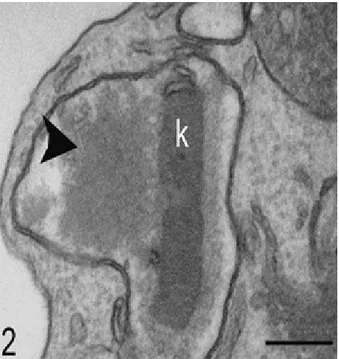
Fig. 2: Figure 2: Transmission electron microscopy of parasite treated with 20 µM berenil for 48 hours. Note the electron-lucid area close to the kDNA network (arrowhead).
Fig. 3: Figure 3: TdT technique applied on parasite treated with 20 µM berenil for 48 hours. Gold particles were observed in the compact kDNA, but not at the kinetoplast electron-lucid area (arrowhead).
Fig. 4: Figure 4: Atomic force microscopy of the kDNA after treatment with 50 µM berenil for 72 hours. Note that the network periphery is more compact (white arrow).