Université de Montréal
Sleep and circadian rhythms in the acute phase of
moderate to severe traumatic brain injury
par Catherine Duclos
Département de psychiatrie Faculté de médecine
Thèse présentée
en vue de l’obtention du grade de doctorat en sciences biomédicales
option sciences psychiatriques
Novembre, 2017
Résumé
Les traumatismes craniocérébraux (TCC) sont la principale cause d’invalidité chez les jeunes adultes, engendrant d’importantes séquelles cognitives, physiologiques et comportementales. Les perturbations du cycle veille-sommeil sont parmi les symptômes les plus persistants à la suite d’un TCC et pourraient nuire à la récupération. En effet, le sommeil est nécessaire à l’apprentissage, la plasticité cérébrale et la génération de nouveaux neurones dans le cerveau adulte. Les observations cliniques suggèrent que ces perturbations apparaissent dès les premières semaines suivant le TCC et pourraient suggérer une altération de l’horloge circadienne. Cependant, aucune étude n’a encore documenté comment les perturbations du cycle veille-sommeil émergent et évoluent dans la phase aiguë du TCC, ni leur association à la récupération fonctionnelle et cognitive à court-terme.
Conséquemment, cette thèse vise à caractériser le sommeil et les rythmes circadiens des patients hospitalisés avec un TCC modéré ou sévère et déterminer si les perturbations du cycle veille-sommeil sont causées par un dérèglement de l’horloge circadienne. Pour ce faire, nous avons utilisé des mesures objectives et quantitatives de sommeil et des rythmes circadiens, incluant l’actigraphie, la polysomnographie (PSG) et la mélatonine, dès la phase d’éveil aux soins intensif. Afin de comprendre le rôle du TCC dans ces perturbations, nous avons comparé les patients TCC à des patients hospitalisés avec blessures orthopédiques graves, sans TCC. Ce protocole a mené à cinq articles de recherche.
En premier lieu, nous démontrons que le cycle veille-sommeil des patients TCC est sévèrement perturbé, mais s’améliore chez 50% d’entre eux au cours de leur séjour hospitalier. Les patients avec une amélioration de la consolidation du cycle veille-sommeil ont un meilleur fonctionnement cognitif et fonctionnel au congé de l’hôpital. Ensuite, dans une étude de cas, nous démontrons qu’un patient TCC peut avoir un cycle veille-sommeil complètement différent dans un même environnement, selon son stade de récupération. Notre troisième article confirme que la consolidation du cycle veille-sommeil évolue en synchronie avec la récupération de la conscience et des fonctions cognitives dans la phase aiguë du TCC.
Notre quatrième article compare le sommeil des patients TCC à celui des blessés orthopédiques graves, sans TCC, en utilisant un système de PSG ambulatoire au chevet. Nous démontrons que, contrairement à notre hypothèse, le sommeil des patients TCC comprend tous les éléments et stades d’un sommeil normal. Cependant, ces patients s’endorment plus tôt et ont un sommeil de plus longue durée, mais plus fragmenté, que les patients sans TCC. Dans les deux groupes, le sommeil est de mauvaise qualité, reflétant probablement l’effet de facteurs non-spécifiques associés avec les blessures physiques et l’environnement hospitalier. Conséquemment, la PSG en phase aiguë permet difficilement de distinguer les patients TCC des patients sans TCC.
Notre dernier article confirme que les patients avec TCC ont une consolidation du cycle veille-sommeil et une qualité de sommeil nocturne inférieures à celles des patients sans TCC, ce qui confirme le rôle du TCC dans les perturbations du cycle veille-sommeil. Cependant, malgré ces perturbations plus sévères, les patients TCC ont un rythme normal de la mélatonine et celui-ci n’est pas associé aux perturbations observées. Cet article suggère que des mécanismes neuronaux autres que l’horloge circadienne seraient responsables des perturbations du cycle veille-sommeil à la suite d’un TCC.
Cette thèse est la première à évaluer le sommeil et le fonctionnement de l’horloge circadienne de patients hospitalisés avec un TCC modéré ou sévère ayant atteint la stabilité médicale. En isolant le rôle du TCC de celui du traumatisme physique et du milieu hospitalier, ces études contribuent à comprendre les caractéristiques, les conséquences et la pathophysiologie des perturbations du cycle veille-sommeil à la suite d’un TCC, ouvrant la voie à de possibles interventions visant à améliorer le sommeil et optimiser la récupération. Mots-clés : traumatisme craniocérébral, sommeil, rythmes circadiens, mélatonine, actigraphie, polysomnographie, soins aigus, récupération, conscience, cognition
Abstract
Traumatic brain injuries (TBI) are the leading cause of disability among young adults, causing debilitating cognitive, psychological and behavioural impairments. Sleep-wake disturbances (SWD) are among the most persistent sequelae following TBI, and could impede recovery. Indeed, sleep is essential to learning, plasticity and neurogenesis. Clinical observations suggest that these disturbances arise in the first weeks following injury, and could suggest a circadian disturbance. However, no study has yet documented how SWD arise and evolve in the acute phase of TBI, or how they are associated to short-term cognitive and functional recovery.
Consequently, this thesis aims to characterize the sleep and circadian rhythms of patients hospitalized with moderate or severe TBI, and determine whether SWD are caused by a deregulation of the circadian clock. To achieve this goal, we used objective and quantitative measures of sleep and circadian rhythms including actigraphy, polysomnography (PSG), and melatonin, beginning in the awakening stage in the Intensive Care Unit. In order to understand the specific role of TBI on SWD, we compared TBI patients to other hospitalized trauma patients, without TBI. Our comprehensive study protocol led to five research articles.
First, we show that the sleep-wake cycle of TBI patients is severely disturbed, but improves for 50% of patients during their hospital stay. Patients whose sleep-wake cycle consolidation improves have better cognitive and functional outcome at hospital discharge. Then, in a single case study, we demonstrate how a patient can have drastically different sleep-wake patterns in the same environment, according to recovery stage. In our third research article, we show that the consolidation of sleep and wake states evolves synchronously with the recovery of consciousness and cognition in the acute phase of TBI.
Our fourth article compares the sleep of TBI patients to that of non-TBI trauma patients using ambulatory PSG at bedside. Contrary to our hypothesis, TBI patients have normal sleep elements and normal proportions of each sleep stages. However, they have
non-TBI patients. In both groups, sleep quality is poor, which most likely reflects non-specific factors associated with the physical trauma and hospital environment. Therefore, PSG reveals little information able to distinguish TBI patients from other non-TBI trauma patients at this stage post-injury.
Our final article shows that TBI patients have poorer sleep-wake cycle consolidation and nighttime sleep quality than non-TBI patients, confirming the role of the TBI in altering sleep and wake states. However, despite having more severe SWD, TBI patients have a normal melatonin rhythm, and this rhythm is not associated with the observed SWD. This article suggests that neural mechanisms other than the circadian clock may be responsible for post-TBI SWD.
This thesis is the first to investigate the sleep and circadian clock of hospitalized moderate to severe TBI patients who are medically stable. By isolating the role of the injured brain from that of overall trauma and the hospital setting, these studies contribute to understanding the characteristics, consequences and pathophysiology of post-TBI SWD, unlocking the possibility to design interventions aiming to improve sleep and optimize recovery.
Keywords: traumatic brain injury, sleep, circadian rhythms, melatonin, actigraphy, polysomnography, acute care, recovery, consciousness, cognition
Table of Contents
Résumé ... i
Abstract ... iii
List of tables ... viii
List of figures ... x
List of acronyms ... xii
List of abbreviations ... xv
Acknowledgements ... xvii
Chapter I: Introduction and theoretical background ... 1
1. General introduction ... 2
2. Traumatic Brain Injury ... 3
2.1. Characteristics and diagnostic criteria ... 3
2.2. Pathophysiology ... 4
2.3. Functional and cognitive consequences of moderate to severe TBI ... 5
2.4. The acute phase of TBI ... 6
3. Sleep ... 7
3.1. Normal sleep ... 7
3.2. Sleep in acute care in non-TBI patients ... 9
3.3. Sleep in the acute and chronic stages of TBI ... 9
4. Circadian rhythms ... 11
4.1. Processes of sleep regulation ... 11
4.2. The circadian system ... 11
4.3. Circadian rhythms after TBI ... 13
5. Review articles ... 14
Article 1: The impact of poor sleep on cognition and activities of daily living after traumatic brain injury: a review ... 15
6. Current gaps in the literature ... 74
7. Objectives and hypotheses ... 75
7.1. Articles 3 and 4 ... 75
7.2. Article 5 ... 76
7.3. Article 6 ... 76
7.4. Article 7 ... 76
Chapter II: Methodology and Results ... 78
1. Overview of research protocol ... 79
Article 3: Rest-activity cycle disturbances in the acute phase of moderate to severe traumatic brain injury ... 83
Article 4: Evolution of severe sleep-wake cycle disturbances following traumatic brain injury: A case study in both acute and subacute phases post-injury ... 115
Article 5: Parallel recovery of consciousness and sleep in acute traumatic brain injury 141 Article 6: Sleep in the acute phase of severe traumatic brain injury: A snapshot of polysomnography ... 161
Article 7: Sleep-wake cycle deregulation but normal circadian clock signal in acute traumatic brain injury ... 182
Chapter III: General Discussion ... 208
1. Overall study conclusions ... 209
2. Brain mechansims that could explain acute sleep-wake disturbances ... 211
2.1. Disruption of sleep- and wake-regulatory pathways ... 211
2.2. Impairment of neurotransmitters involved in sleep-wake regulation ... 213
2.3. Defective switching mechanism between sleep and wake ... 215
2.4. Disturbance in the homeostatic process of sleep regulation ... 216
2.5. Inflammation ... 218
3. Potential confounding variables ... 219
3.1. Sedative and analgesic medication ... 219
3.2. Delirium in ICU ... 221
3.3. Pain ... 223
5. Strengths of the studies presented in this thesis ... 227
5.1. Research protocol and patients ... 227
5.2. Development of novel measures to assess sleep-wake rhythm ... 228
6. Limits ... 229
6.1. Actigraphy as a proxy measure of the sleep-wake cycle ... 229
6.2. Weaknesses of the DAR as a measure of sleep-wake cycle consolidation ... 230
6.3 Limits imposed by polysomnography ... 231
6.4. Using the RLA for the assesment of consciousness ... 232
6.5. Hospital environment ... 233
7. Future research priorities ... 233
7.1. Sleep disturbance, wake disturbance, or faulty switching mechanism? ... 233
7.2. Can we improve nighttime sleep quality and optimize recovery? ... 235
8. Conclusion ... 235
List of tables
Chapter I: IntroductionArticle 2: Sleep and wake disturbances following traumatic brain injury
Table 1. Screening and interventions for post-TBI insomnia and hypersomnia (list not exhaustive)………61-62 Chapter II: Methodology and results
Article 3: Rest-activity cycle disturbances in the acute phase of moderate to severe traumatic brain injury
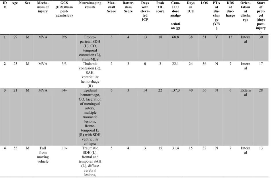
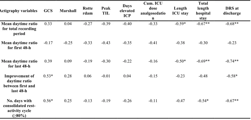
Table 1. Demographic and clinical characteristics of patients ………88-90 Table 2. Pearson correlation coefficients between rest-activity cycle and clinical
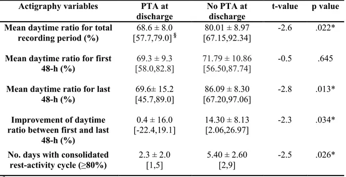
variables……….97 Table 3. Association between PTA upon discharge and variables of actigraphy
compared between sub-groups of patients with (n=6) and without (n=10) PTA at discharge………...99 Supplementary Table A1. Total fraction of time moving (%) as scored by the
Actiware program with a threshold of ≥10 activity counts per minute…………...….114 Article 4: Evolution of severe sleep-wake cycle disturbances following traumatic brain injury: A case study in both acute and subacute phases post-injury
Table 1. Scores on Neuropsychological Tests carried out 87 and 89 days post-injury (second hospital stay) ………...123-124 Article 5: Parallel recovery of consciousness and sleep in acute traumatic brain injury
Table 1. Rancho Los Amigos scale of cognitive functioning, including the number of days and patients representing each RLA score (adapted from previously published article, with permission from the editor)………..146 Table 2. Association between sleep-wake patterns and level of
Article 6: Sleep in the acute phase of severe traumatic brain injury: A snapshot of polysomnography
Table 1. Demographic and clinical characteristics for patients with moderate-severe traumatic brain injury (TBI group) and patients with orthopaedic or spinal cord injuries (OSCI group)………167-169 Table 2. Polysomnographic results for patients with severe TBI and patients with OSCI………173 Article 7: Sleep-wake cycle deregulation but normal circadian clock signal in acute traumatic brain injury
Table 1. Demographic and clinical characteristics of total sample……….189 Table 2. Sleep-wake cycle and nighttime sleep characteristics (mean ± SD) in patients with (TBI) or without (non-TBI) traumatic brain injury………..193 Table 3. Characteristics (mean ± SD) of 6-sulfatoxymelatonin (aMT6s) excretion and of circadian estimates for TBI and non-TBI groups………195 Supplementary Table 1. Demographic and clinical characteristics of sub-sample of patients who were included in melatonin analyses………..207
List of figures
Chapter 1: IntroductionArticle 1: The impact of poor sleep on cognitive and activities of daily living after traumatic brain injury: a review
Figure 1. Sleep architecture of a healthy adult subject………....19 Chapter 2: Methodology and results
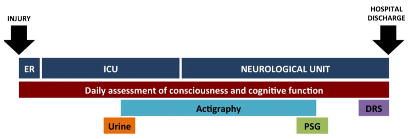
Figure 1. Timeline of study measures in relation to injury and stages of hospitalization…………....………....82 Article 3: Rest-activity cycle disturbances in the acute phase of moderate to severe traumatic brain injury
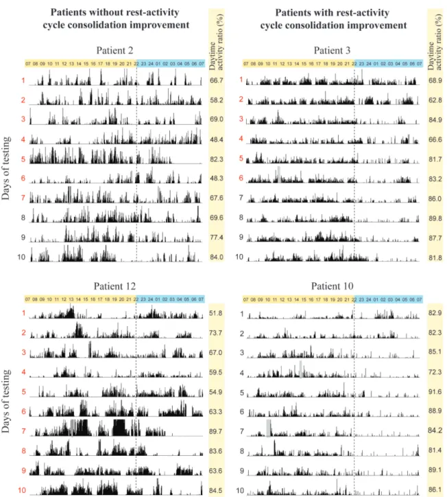
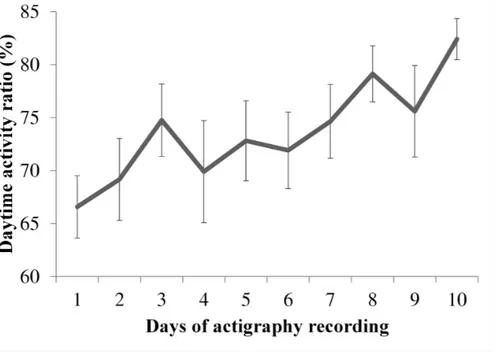
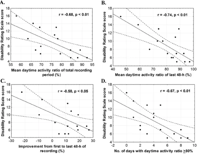
Figure 1. Examples of actigraphy recordings………94 Figure 2. Evolution of the consolidation of rest-activity cycle over ten days………..95 Figure 3. Illustration of the significant Pearson correlations between the daytime activity ratio and the score on the Disability Rating Scale (DRS) at discharge……….98 Supplementary Figures A1, A2, A3. Individual actigraphy recordings……....110-113 Article 4: Evolution of severe sleep-wake cycle disturbances following traumatic brain injury: A case study in both acute and subacute phases post-injury
Figure 1. CT scan at admission………...120 Figure 2. Timeline of injury, hospital stays and actigraphy………125 Figure 3. Actigraphy recordings of the first and second hospital stays………..129 Article 5: Parallel recovery of consciousness and sleep in acute traumatic brain injury
Figure 1. Association between cognitive and consciousness recovery and the sleep-wake cycle………151 Figure 2. Examples of actigraphic findings in relation to RLA scores…………...…152
Article 6: Sleep in the acute phase of severe traumatic brain injury: A snapshot of polysomnography
Figure 1. Flow diagram representing TBI patient recruitment………166 Article 7: Sleep-wake cycle deregulation but normal circadian clock signal in acute traumatic brain injury
Figure 1. Example of actograms of TBI and non-TBI patients………..…192 Figure 2. Rhythm of 6-sulfatoxymelatonin excretion of TBI and non-TBI patients……….………194
List of acronyms
ACTH: adreno-corticotropic hormone ADL: activities of daily living
aMT6s: 6-sulfatoxymelatonin ANCOVA: analysis of covariance ANOVA: analysis of variance AR1: autoregressive
ARAS: ascending reticular activating system
BADS: Behavioral Assessment of the Dysexecutive Syndrome BCAA: branched chain amino acid
CASE: Cognitive Assessment Scale for the Elderly CBT: cognitive behavioural therapy
CO: cerebral oedema CS: compound symmetry CSF: cerebrospinal fluid CT: computed tomography DAI: diffuse axonal injury DAR: daytime activity ratio
DelRS-R98: Delirium Rating Scale – Revised-98 D-KEFS: Delis-Kaplan Executive Function System DRS: Disability Rating Scale
DSM-IV: Fourth Edition of the Diagnostic and Statistical Manual of Mental Disorders EDH: extradural hematoma
EDS: excessive daytime sleepiness EEG: electroencephalography EMG: electromyography EOG: electrooculography ER: emergency room
FOSQ: Functional Outcome Sleep Questionnaire FSH: follicle-stimulating hormone
GABA: gamma-Aminobutyric acid
GCS: Glasgow Coma Scale GH: growth hormone
GOAT: Galveston Orientation and Amnesia Test ICP: intracranial pressure
ICSD: International Classification of Sleep Disorders ICU: Intensive Care Unit
ID: identification
IL-1β: Interleukin 1 beta ISS: Injury Severity Score LH: luteinizing hormone LOS: length of stay
MCS: minimally conscious state MLS: midline shift
MMSE: Mini Mental State Examination MRI: magnetic resonance imaging mNRA: messenger ribonucleic acid MSLT: Multiple Sleep Latency Test mTBI: mild traumatic brain injury MVA: motor vehicle accident
MVPT: Motor-Free Visual Perception Test MWT: Multiple Wakefulness Test
NREM: non-rapid eye movement NS: non significant
OI: orthopaedic injury
OSA: obstructive sleep apnea
PSH: Paroxysmal Sympathetic Hyperactivity PTA: post-traumatic amnesia
REM: rapid eye movement RHD: radial head luxation
RLA: Rancho Los Amigos scale of cognitive functioning SAH: subarachnoid hemorrhage
SCI: spinal cord injury
SCN: suprachiasmatic nucleus SD: standard deviation
SDH: subdural hematoma SEM: strander error of the mean
STROBE: Strengthening the Reporting of Observational Studies in Epidemiology SWA: slow-wave activity
SWD: sleep-wake disturbances SWS: slow-wave sleep
TBI: traumatic brain injury
TIL: Summary Therapy Intensity Level TNF-α: tumor necrosis factor alpha TSH: thyroid-stimulating hormone VLPO: ventrolateral preoptic nucleus WAIS: Wechsler Adult Intelligence Scale WASO: wake after sleep onset
List of abbreviations
a.k.a.: also known ascum.: cumulative
e.g.: such as (exempli gratia) etc. : Et cætera F: female Fig.: Figure fx: fracture g: gram h: hour hrs: hours
i.e.: that is (id est) L: left M: male mg: miligrams min: minute ml: milileter mmHg: milimeters of mercury n.a.: non-applicable nd: number ng: nanogram
ng/ml: nanogram per milileter R: right
À mon fils Liam, né pendant mes études doctorales. Souviens-toi de travailler fort pour atteindre tes rêves, de profiter du parcours qui te mènera à
Acknowledgements
J’aimerais d’abord remercier ma directrice de recherche, Nadia Gosselin, avec qui j’ai eu le plaisir de découvrir la recherche au cours des six dernières années. Merci de m’avoir prise comme étudiante à la maitrise, en 2011, alors que je n’avais aucune formation en science et que je partais de loin. Merci d’avoir cru en mes capacités et de m’avoir fait confiance. J’ai énormément appris de toi, autant sur le plan scientifique que personnel. Tu es pour moi un modèle de la chercheure, superviseure, professeure, collègue, athlète, et maman que je souhaite devenir. Ton calme et ton équilibre sont des sources d’inspiration qui m’ont donné confiance en mes aptitudes et mon désir d’aller plus loin en recherche. Je te remercie pour la place que tu m’as faite dans ton laboratoire, pour ton soutien intellectuel, moral et financier au cours de ces années. Je me compte sincèrement choyée de t’avoir eue comme superviseure et mentore.
Je tiens également à remercier ma co-directrice, Marie Dumont, qui m’a ouvert la porte à la maitrise et au CÉAMS. Marie, merci de m’avoir rencontré, d’avoir considéré mon potentiel malgré mon parcours en anthropologie et développement international qui ne concordait pas avec les exigences du programme. Merci de m’avoir présenté à Nadia et de m’avoir ainsi permise de vivre cette expérience inoubliable. Merci de ton soutien, de ta rigueur scientifique, d’avoir su questionner et pousser nos réflexions plus loin. Ton apport fut indispensable et m’a énormément fait grandir en tant que scientifique. Tu es une grande chercheure et je suis grandement reconnaissante d’avoir pu bénéficier de ta sagesse et de tout ton savoir.
Merci à Hélène Blais, coordonnatrice hors pair. Heureusement que tu étais là, tu m’as vraiment tout appris au début (je n’avais jamais encore vu d’unité de soins intensifs avant mes premières tournées avec toi). Merci pour tout ce que tu as fait dans le recrutement et le suivi des patients. Ton appui quotidien dans la coordination du protocole de recherche fut une aide inestimable. C’était toujours agréable et efficace de travailler avec toi, que demander de mieux!
Baril, Katia Gagnon, Caroline Arbour, Héjar El-Khatib, Solenne Van der Maren, Erlan Sanchez, Marc-André Gareau, Maxime Fortin, Catherine Wiseman-Hakes, Julien Lauzier-Bigué, Sirin Chami, Sabrina Tabet et Liza Pizzimenti. Merci pour votre contribution, de près ou de loin, à mes réflexions et travaux. Merci à Caroline D’Aragon d’avoir épluché autant de dossiers médicaux et d’avoir été présente pour plusieurs polysomnographies sur les unités de soin. Merci aussi aux autres étudiants (présents en anciens) que j’ai eu le plaisir de côtoyer au CÉAMS, notamment Marlène Freyburger, Marie-Pier McSween, Véronique Latreille, Véronique Daneault, Jonathan Dubé, Pauline Brayet, Daphné Génier-Marchand, Pierre-Olivier Gaudreault, Camille Larson-Dupuis, Marianne Jodoin, et j’en passe.
Un grand merci à Jean Paquet pour son précieux soutien au plan statistique, ainsi qu’à Gaétan Poirier et à Sébastien Saucier pour leur soutien technique. Merci à l’équipe de techniciens du laboratoire, Benoît Adam, Nancy Poirier, Mélina-Crécia Babala et Jean-Claude Aubertin, qui nous ont assisté avec nos enregistrement polysomnographiques dans les différentes unités de soins de l’hôpital. Merci à Marie-Josée Quinn pour les dosages de mélatonine. Merci à Renée-Claude Caron, Bridget Gibson, Audrey Lachapelle, Taoufik Terfas, Fanny Boulianne, ainsi que tout le personnel médical et infirmier nous ayant appuyé dans le projet.
Merci à Karine Marcotte, Francis Bernard, Alex Desautels, Julie Carrier, Johannes Frasnelli, Valérie Mongrain, Domnique Petit, Louis de Beaumont, Simon Warby, Gregory Moullec, Marc-André Raymond et François Madore pour vos conseils et votre présence. Vous êtes agréables et inspirants à côtoyer et je vous remercie sincèrement pour vos encouragements tout au long de mon parcours.
Merci à mes amies et amis, qui m’ont soutenu de près ou de loin en me changeant les idées. Je tiens à remercier particulièrement Gabrielle Picard, Laurence Tanguay-Beaudoin et Erika Gaudreault, avec qui j’ai partagé des séances de rédaction et d’études, ou des échanges sur mes recherches et mes choix de carrière. Merci d’avoir été là et d’avoir été vraies.
Merci à mes parents, Martine Simoneau et Denis Duclos, qui m’ont inculqué la valeur de l’éducation et le désir de toujours me pousser un peu plus loin. Maman, merci de toujours être présente pour moi, même quand tu es loin. Merci pour tes petits mots d’encouragement, les moments passés en ta compagnie, les repas en famille, les petites attentions. Merci d’avoir été aussi présente après la naissance de Liam, ce qui m’a permise de faire mes demandes de
bourses, de finaliser des articles. Je n’y serais pas arrivée sans toi. Papa, merci pour ton soutien tout au long de mon parcours scolaire et merci d’avoir cru en moi. Merci à mon frère, Gabriel Duclos, qui est aussi un grand ami. Merci pour ton soutien moral pendant mes études, de m’avoir partagé tes connaissances et certaines parties de ta vie. Je suis heureuse d’avoir pu partager autant de moments d’étude en ta compagnie, que ce soit à la maison, dans un café ou même à la bibliothèque de Concordia! Je te remercie aussi d’être un parrain présent pour Liam. Tes soirées de gardiennage me permettent de demeurer impliquée dans une cause qui me tient à cœur et d’avoir des petits moments en couple. Je t’en remercie!
Merci à mes grands-parents, oncles, tantes, cousins et cousines, si m’encouragent depuis le début. Merci à mes beaux-parents, Johanne Charette et Gaétan Baril pour votre soutien, votre compréhension et nos soupers du dimanche soir.
Un remerciement tout particulier à Marie-Christine Baril pour son appui quotidien et son amour. Merci d’avoir été là depuis le début (ou presque) de ce long parcours, qui fut parsemé de moments plus intenses et exigeants que d’autres. Je sais que je ne suis pas la plus patiente lorsque je suis stressée! Merci d’avoir cru en moi, d’avoir démontré de l’intérêt pour mes recherches et de m’avoir apporté autant de rires et de moments de bonheur à la maison. Merci aussi d’avoir contribué à l’avancement de nos projets de vie pendant ces six dernières années.
Merci aux organismes subventionnaires qui ont financé le projet de recherche et m’ont offert des bourses pendant mes études : les Instituts de recherche en santé du Canada, les Fonds de recherche du Québec – Santé, l’Université de Montréal, la Fondation J.A. de-Sève de l’Hôpital du Sacré-Cœur de Montréal et la Fondation Neurotrauma Marie-Robert.
Enfin, je tiens à remercier tous les patients qui ont participé à notre étude, ainsi qu’à leurs familles. Merci d’avoir collaboré alors que vous étiez souffrants et que votre vie venait de basculer. Votre générosité, votre force et votre résilience m’ont touché et ont donné un sens à mes recherches.
1. General introduction
The hospital environment, in which activity only ceases partially during nighttime, is not conducive to uninterrupted sleep. The care necessary to ensure the health and well-being of patients comes with noise, light, and interventions around the clock. It is therefore not surprising that one of the most stressful experiences of hospitalized patients is their chronic lack of sleep (Novaes et al., 1999; Simini, 1999; Nelson et al., 2001; Park et al., 2014; Beltrami et al., 2015). Unfortunately, despite being a central preoccupation of hospital patients, sleep is often neglected and dismissed as less important than direct medical care and interventions. When we know that sleep is essential to survival of critical illness, optimal health and recovery (Tembo & Parker, 2009), we can wonder what impact sleep disturbances have on the recovery of hospitalized patients. Indeed, poor sleep hinders recovery by lowering the ability to resist infection, by causing or exacerbating neurological problems such as delirium, and prolonging the duration of mechanical ventilation and stay in the Intensive Care Unit (ICU) (Parthasarathy & Tobin, 2004).
The importance of sleep in patients hospitalized with moderate to severe traumatic brain injury (TBI) is especially crucial when considering the role of sleep in learning and neuronal plasticity (Smith, 1996; Walker, 2004; Wilson, 2002; Walker & Stickgold, 2006; Diekelman & Born, 2010), as well as for the generation of new neurons in the adult brain (Roman et al., 2005; Mirescu et al., 2006; Guzman-Marin et al., 2007; Meerlo et al., 2009). Indeed, TBI destroys brain structures and connections that need regeneration. Clinical observations in the acute care setting suggest that sleep-wake disturbances (SWD) appear in the first weeks after TBI, during which patients experience insomnia, inability to stay awake for a few consecutive hours during the day and/or altered sleep-wake cycles, which could point to a disturbance in circadian rhythms. SWD in the acute post-traumatic period probably impede brain recovery, and may contribute to short-term and long-term cognitive, physical, and neurobehavioral impairments resulting from TBI. What is more, SWD, including fatigue, insomnia and hypersomnia, are among the most persistent and debilitating symptoms
3
least 50% of TBI patients in the chronic stage post-injury (Castriotta et al., 2007; Cantor et al., 2008), little is known about their origin, evolution, and consequences.
These preoccupations led us to investigate sleep and circadian rhythms in the acute phase of moderate to severe TBI. More specifically, this thesis aims to characterize SWD in the acute phase, while patients are still hospitalized, to appraise the impact of SWD on short-term cognitive and functional recovery, and to investigate whether these disturbances arise due to a disturbance in the circadian clock. Since sleep is a modifiable behaviour, it is an important therapeutic target with the potential to optimize recovery.
2. Traumatic Brain Injury
Moderate to severe TBI is the principal cause of mortality and lifelong disability among young adults in industrialized countries (Kraus & Chu, 2005; Langlois, Rutland-Brown & Wald, 2006; Roozenbeek, Mass & Menon, 2013). Patients who suffer moderate to severe TBI require an extensive period of hospitalization in the ICU to survive their injury, and generally require several weeks to months of intensive rehabilitation. Ultimately, TBI results in short- and long-term cognitive, psychological and behavioural impairments that interfere with the return to a normal and productive life.
2.1. Characteristics and diagnostic criteria
TBI is an alteration in brain function, or other evidence of brain pathology, caused by an external force (Menon et al., 2010). An alteration in brain function is defined by one of the following signs: a period of decreased level of consciousness, an alteration in mental state at the time of injury (e.g. confusion, disorientation), a loss of memory for events immediately before (retrograde amnesia) or after the injury (post-traumatic amnesia, or PTA), and neurological deficits (e.g. loss of balance, weakness, aphasia, change in vision). Other evidence of brain pathology may include visual, neuroradiological, or laboratory confirmation of brain damage.
The diagnosis of TBI involves an assessment of severity, generally carried out using the Glasgow Coma Scale (GCS), which assesses verbal, ocular and motor functions, immediately following injury (Teasdale & Jennett, 1974). Globally, mild TBI (mTBI) is characterized by a short loss of consciousness (< 30 min), a GCS score between 13 and 15 and a short PTA (< 24 h). Moderate TBI is typically associated with a loss of consciousness of 30 min to 24 h, a GCS between 9 and 12, and PTA of 1 to 14 days. Severe TBI is generally characterized by a loss of consciousness of more than 24 h, a GCS between 3 and 8 and a PTA that persists for several weeks. As opposed to mTBI, patients having suffered a moderate or severe TBI have an extensive loss of consciousness and generally require hospitalization in the ICU to survive their injury. This thesis will focus on moderate to severe TBI.
Varying definitions have been used to describe TBI in the past, which has led to heterogeneity in epidemiological studies. Internationally, the estimated annual incidence of TBI is 295 per 100,000 (Nguyen et al., 2016). In industrialized countries, the annual incidence of moderate and severe TBI is estimated at around 1.2/1000 individuals (Kraus et al., 2005). Though moderate and severe TBI account for approximately 10% of all TBI, they contribute to the majority of deaths, disability and TBI-related costs (McGarry et al., 2002). The most frequent causes of injury are motor-vehicle accidents, falls, assault, recreational injuries, and work accidents (Zygun et al., 2005; Rutland-Brown et al., 2006). Teenagers and young adult males (15-24 years old) have an increased risk of sustaining TBI, while other risk factors include low economic status, low education, and alcohol or drug addiction (Bruns & Hauser, 2003; Nguyen et al., 2016).
2.2. Pathophysiology
Brain lesions resulting from TBI occur in two different time periods. The primary insults occur in the few seconds that follow the TBI and result from the applied biomechanical forces of the injury, including the acceleration, deceleration, and rotational forces concurrent with or secondary to the direct trauma (Greve & Zinc, 2009). They cause focal lesions (i.e. intracranial hematoma, skull fracture, contusions, lacerations) or diffuse axonal injuries.
5
by the primary insult, arise in the hours and days following the initial injury (Raghupathi, 2004; Gennarelli & Graham, 2005; Greve & Zinc, 2009; Petroni et al., 2010; Sandsmark, 2016). Elevated intracranial pressure (ICP) (a.k.a. intracranial hypertension) is the most common form of secondary insult, but others include the production of free radicals, mitochondrial dysfunction, and an inflammatory cascade that ultimately leads to the degeneration of neurons, glial cells, and axons (Holmin et al, 1995). These secondary insults are thus the determining factors in the morbidity and mortality of patients who survive the initial injury (Greve & Zinc, 2009). The long-term damages that arise following moderate to severe TBI are characterized by axonal degeneration throughout the brain (Bendlin et al., 2008; Greenberg et al., 2008; Kumar et al., 2009; Perlbag et al., 2009; Kumar et al., 2010; Kinnunen et al., 2011; Dinkel et al., 2013), and brain atrophy, particularly in the hippocampus and frontal and temporal cortices, as well as the thalamus, basal forebrain, hypothalamus, pituitary stalk, caudate nucleus, and insula (Gale et al., 2005; Gennarelli & Graham, 2005; Salmond et al., 2005; Tasker et al., 2005; Bendlin et al., 2008; Slawik et al., 2009).
2.3. Functional and cognitive consequences of moderate to severe TBI
TBI leads to varying degrees of symptoms and handicaps, depending on mechanism and severity of the injury. In addition to causing both short- and long-term impairments, such as cognitive, behavioural and psychological alterations, TBI also increases the risk of developing psychiatric and neurodegenerative disorders (Vaishnavi, Rao & Fann, 2009; Masel & DeWitt, 2010; Bhalerao et al., 2013), and has been associated with shortened lifespan (Masel & DeWitt, 2010). As such, TBI is no longer considered an event, but a progressive, chronic and heterogeneous disease process (Masel & DeWitt, 2010; Maas, 2016).
In approximately 50% of patients, severe cognitive deficits persist into the chronic phase (Selassie et al., 2008). The most prevalent are attention deficits (Salmond et al., 2005; Salmond et al., 2006; Mathias & Wheaton, 2007), memory impairments (Levin et al., 1988; Curtiss et al., 2001), and executive dysfunctions (i.e. planning, initiation, inhibition, problem solving, mental flexibility, and self-monitoring) (McDonald, Flashman & Saykin, 2002; Spikman & Van der Naalt, 2010). These deficits lead to a reduction in independence and
difficulties regaining a productive lifestyle (Shames et al., 2007). Factors contributing to more severe cognitive deficits include the severity of diffuse axonal injury, the duration of PTA, the extent of cerebral atrophy, and age (Katz & Alexander, 1994).
The period comprising hospitalization and acute rehabilitation following TBI loosely represents the acute and post-acute phases. It is estimated that 85% of cognitive recovery takes place within the first 6-months following TBI (Lippert-Gruner, Lefering & Svestkova, 2007), which suggests that the acute and post-acute phases have an undeniable impact on the long-term functionality and quality of life of patients. Factors that hinder recovery in the first weeks post-injury could determine the course of overall recovery and outcome. This thesis will focus on the hospitalization period of the acute phase.
2.4. The acute phase of TBI
Immediately following the initial injury, most moderate to severe TBI patients are hospitalized in the ICU for several hours to several weeks. This period is marked by an alteration in consciousness, and patients are generally under continuous sedation for several days. Once they reach the awakening stage, patients generally present agitation, confusion, and PTA (Trzepacz & Kennedy, 2005). Neurobehavioural impairments such as impulsivity, irritability, desinhibition, mutism, and apathy can also be observed to varying degrees (Riggio & Wong, 2009). As level of consciousness gradually improves over time, patients may transition through different consciousness levels, transiently or persistently, including unresponsive wake syndrome (UWS; previously known as vegetative state) and minimally conscious state (MCS).
The post-ICU period begins when a patient has reached medical stability, has moved beyond the awakening stage, and has been transferred to a regular or neurological ward within the hospital. Patients generally present impairments in arousal and alertness, reduced information processing speed, impaired memory and executive dysfunctions, impaired language, and reduced self-awareness (McCullagh & Feinstein, 2005). The extent of functional and cognitive deficits observed during this stage is highly variable among patients
7
and depends on several factors, including the severity of diffuse axonal injury, the location of focal lesions, the duration of PTA, age, level of education, and other pre-existing conditions (de Guise et al., 2005; LeBlanc et al., 2006; de Guise et al., 2006; de Guise LeBlanc et al., 2009; Kosch et al., 2010). Patients can remain agitated and disoriented throughout this stage, as some remain in PTA when they are discharged from the hospital and admitted to internal rehabilitation centers. Only a minority of moderate to severe TBI patients are discharged without being sent to acute internal rehabilitation.
Overall, the ICU and post-ICU stages post-injury are challenging research settings, given the fluctuating medical and cognitive states of patients. Most studies having investigated the physiological effects of TBI have taken place prior to the awakening stage, when patients are still mechanically ventilated and continuously sedated, or during the chronic stage of injury.
3. Sleep
3.1. Normal sleep
Sleep is an active and reversible physiological state, which is essential for survival and healthy functioning of the organism (Banks & Dinges, 2007). While the function of sleep is not fully understood, early hypotheses suggest that sleep serves a function of restoration, by restoring depleted energy (Oswald, 1980), and a function of energy conservation (Walker & Berger, 1980). Recent hypotheses link sleep with metabolic waste clearance in the brain (Xie et al. 2013) and synaptic homeostasis (Tononi & Cirelli, 2003; Tononi & Cirelli, 2006), highlighting the role of sleep in learning and plasticity. Though the function of sleep remains somewhat enigmatic, the effects of sleep loss reveal its undeniable role in the maintenance of a healthy body and brain. Indeed, partial or chronic sleep loss causes a range of neurobehavioural and cognitive deficits, as well as endocrine, metabolic, cardiovascular, immune, and inflammatory alterations that hinder general health (Dinges et al., 1994; Spiegel, Leproult & Van Cauter, 1999; Spiegel, Sheridan & Van de Cauter, 2002; Shamsuzzaman et al. 2002; Banks & Dinges, 2007; Goel et al., 2009; Mullington et al., 2009).
Sleep is comprised of various stages that have measurable behavioural and physiological traits, which reflect their underlying functional mechanisms. Polysomnography (PSG) is the most widely used tool to measure sleep, and includes electroencephalography (EEG), electrooculography (EOG), and electromyography (EMG). While EEG is the measurement of the brain’s electrical activity, EOG and EMG measure eye movements and the muscular activity on the chin, respectively. The identification of sleep stages is carried out according to standardized criteria based on the visual analysis of these physiological parameters (Iber, Ancoli-Israel & Quan, 2007).
Sleep architecture is the structural organization of sleep. Sleep stages can be divided into two main categories: rapid eye movement (REM) and non-rapid eye movement (NREM), or slow-wave sleep (SWS). Some bodily movements can accompany NREM, but eye movements are rare. NREM sleep can be divided into three stages that reflect the degree of neuronal synchrony, spanning from lighter sleep (N1 and N2) to deep sleep (N3), where neuronal synchrony is at its peak and slow waves predominate the EEG. REM sleep is the primary stage during which dreaming occurs. It distinguishes itself from NREM sleep through three main characteristics: desynchronized EEG activity, rapid eye movements, and nearly complete muscle atonia, which prevents the enactment of dreams (Siegel, 2011). In the course of one night, the human adult alternates between sleep stages in cycles of approximately 90-100 min in duration. Though stage N3 is most present at the beginning of the night, REM sleep becomes increasingly prevalent as the night progresses.
Although PSG is the gold standard for measuring sleep, it is difficult to use to record sleep and wake states over several consecutive days. Being cumbersome, is it also poorly tolerated by patients hospitalized in critical care and/or in a state of confusion or agitation. Actigraphy is the most-utilized alternative to PSG. The activity monitor, or actigraph, is a small watch-like device worn on the wrist, which records physical motion in all directions, with a sensitivity of 0.05g. Motion is subsequently converted into an electrical signal, which is digitally integrated to derive an activity count (i.e. sum of activity) for each 1-min epoch (Paquet, Kawinska & Carrier, 2007). Actigraphy can easily be used in a hospital setting. With
9
in a clinical setting, and is recognized as an indirect measure of the sleep-wake cycle (Martin & Hakim, 2011). The ability of actigraphy to record the rest-activity cycle continuously for up to several weeks makes it an ideal tool to estimate when sleep and wake arise, how they are distributed over the 24 h day, and how they evolve over the course of several days or weeks. In fact, actigraphy has been successfully used in various clinical populations (Martin & Hakim, 2011).
3.2. Sleep in acute care in non-TBI patients
The acute care setting is not favourable to optimal sleep. Studies on patients hospitalized in the ICU, without TBI, have shown that the sleep-wake cycle and sleep quality are severely altered. In fact, approximately 50% of sleep takes places during the day, and up to 96% of sleep is spent in stages N1 and N2, suggesting a drastic reduction or absence of SWS (N3) (Cooper et al., 2000; Gabor et al., 2003; Friese et al., 2007; Gehlbach et al., 2012). The consequences of sleep disturbances in the ICU are probably similar to the effects of chronic sleep restriction in healthy subjects: for example, they may lead to a decrease in cognitive functioning, a slowing of glucose metabolism, the activation of the hypothalamic-pituitary-adrenal axis and an increase in the inflammatory response (Kamdar, Needham, & Collop, 2012).
For a detailed description of normal sleep, sleep in the hospital setting, and the impact of poor sleep on cognitive and functional outcome of TBI patients in hospital and rehabilitation settings, refer to article 1 (Chapter 1, section 5).
3.3. Sleep in the acute and chronic stages of TBI
Little is known about the nature and evolution of SWD after a TBI, and very few studies have documented SWD in acute settings. The earliest studies investigating sleep following moderate-severe TBI took place in rehabilitation centers, using actigraphy and nurse assessments, and found a high prevalence of sleep disturbances, associated with the resolution of PTA (Makley et al., 2008, Makley et al., 2009) In the ICU, observations by clinical staff suggest that these disturbances are present as early as the awakening stage post-injury, while
patients are still in the ICU. However, no study has yet objectively characterized this phenomenon and its impact on acute recovery.
In order for natural sleep and the sleep-wake cycle to be accurately measured in acute TBI, patients need to have minimally reached a stage where they are able to awaken spontaneously. According to international diagnostic guidelines, sleep-wake cycles among patients with severely altered levels of consciousness (e.g. UWS, MCS) are assessed mainly by means of observation, through the identification of periods of eye-opening and eye-closure (Cruse et al., 2013). Indeed, the return of a sleep-wake cycle is assumed to arise once patients reach UWS. However, little empirical evidence exists to support the existence of normal sleep among patients in UWS. In fact, one study showed that the EEG of patients in UWS remains unchanged between periods of eye-opening and eye-closure despite the preservation of “behavioural sleep” (Landsness et al., 2011), highligthing the unreliability of behavioural assesment for the measure of sleep among UWS patients. In patients having reached MCS however, several EEG characteristics of normal sleep are present, including an alteration between REM and NREM sleep (Landness et al., 2011). Therefore, there remains a lack of consensus as to what sleep objectively is, and how it may vary, among patients whose level of consciousness is severely altered. Moreover, the use of automatic sleep scoring can be misleading at this stage of recovery, given the presence of slower baseline EEG in brain-damaged patients (Landsness et al., 2011).
SWD remain present into the chronic phase post-injury in over 50% of patients (Parcell et al., 2006, Ouellet, Beaulieu-Bonneau, & Morin, 2006; Gosselin et al., 2009; Wiseman-Hakes et al., 2009; Kempf et al., 2010; Beaulieu-Bonneau & Morin, 2012). The most prevalent include excessive daytime sleepiness, insomnia, and hypersomnia (Ouellet, Beaulieu-Bonneau & Morin, 2006; Castriotta et al., 2007; Kempf et al., 2010). For a detailed description of SWD following TBI, refer to article 2 (Chapter 1, section 5). This article provides a narrative review of SWD assessed both subjectively and objectively, spanning all levels of severity and phases post-injury.
11
4. Circadian rhythms
4.1. Processes of sleep regulation
The sleep-wake cycle is regulated by an interaction of the circadian and homeostatic processes (Borbély, 1982). The circadian process involves the rhythmic variation of sleep and wake propensity over 24 h, while the homeostatic process represents the accumulation of sleep pressure during wakefulness, reflected by the amount of EEG slow-wave activity (SWA) during NREM sleep (Achermann et al., 1993; Robillard et al., 2010), and its dissipation during the sleep period (Borbély, 1982). As homeostatic sleep pressure increases with hours spent awake, the circadian wake signal also increases to counterbalance the homeostatic process, reaching its peak approximately 2 h prior to bedtime, which enables us to stay awake during the evening. During nighttime, homeostatic pressure dissipates during sleep, which decreases sleep need as the night progresses. Conversely, the circadian sleep signal reaches its peak approximately 2 h prior to wake time, which enables sleep to be sustained until morning. Therefore, the interaction of these two processes enables the consolidation of wake during the daytime and sleep during nighttime (Borbély & Acherman, 1992; Dijk & Czeisler, 1994; Dijk & Czeisler, 1995), which will henceforth be referred to as a consolidated sleep-wake cycle.
4.2. The circadian system
The importance of the circadian system extends far beyond its implication in sleep-wake regulation. In humans, as in nearly all living organisms, a multitude of biological activities (e.g. hormone secretion, cognitive performance, metabolic functions, muscular strength, cell division) oscillate over a 24 h period and are directly controlled by the endogenous circadian master clock (Takahashi & Zatz, 1982; Moore, 1997; Gronfier, 2009). This master circadian clock is located in the suprachiasmatic nucleus (SCN) of the hypothalamus. Although circadian rhythms are generated endogenously, they are sensitive to the environment, enabling synchronization of the organism to the environmental day. Given that the endogenous period of the circadian clock generally differs slightly from 24 h in humans (Czeisler & Buxton, 2011), this synchronization is crucial to the proper functioning of
the organism in relation to the 24 h day. It enables physiological, cognitive and behavioural functions associated with activity to take place during the daytime, and those associated to rest to occur during nighttime (Czeisler et al., 1999). The main synchronizer of the circadian system is the light-dark cycle, which reaches the SCN through a monosynaptic retino-hypothalamic tract (Takahashi & Zatz, 1982; Moore, 1997; Gronfier, 2009). Non-photic stimuli, such as social interaction and the timing of meals, have also been shown to have some influence on circadian rhythms (Stephan, 2002; Challet et al., 2003; Mistlberger & Skene, 2005).
The rest-activity cycle is the most commonly and most easily measured marker of overall circadian functioning, as it is strongly correlated to the sleep-wake cycle (Barion & Zee, 2007; Martin & Hakim, 2011). However, markers that best reflect the timing of the master circadian clock are those that have a strong endogenous drive. In humans, these include body temperature, melatonin production, and cortisol production, though they may be affected by environment and behaviour when these are not properly controlled. The measurement of these rhythms enables the characterization of the phase (i.e. a specific point within a cycle), period (i.e. a complete cycle, or the elapsed time between successive occurrences of a particular phase), and amplitude (i.e. range of values within a cycle) of the circadian signal (Lemmer & Portaluppi, 1997; Dunlap, Loros, & DeCoursey, 2003; Wirz-Justice, 2007; Czeisler & Gooley, 2007; Mistlberger & Rusak, 2011).
Melatonin production is the best available marker of internal timing of the master circadian clock, given that the timing of onset and offset of melatonin production is tightly controlled by the SCN and that its temporal profile is relatively unaffected by sleep or wake when the environment is constant (Arendt, 2005). Melatonin is a hormone secreted by the pineal gland during the biological night. Its production is driven by the SCN via a multi-synaptic pathway involving the spinal cord and sympathetic nervous system (Teclemariam-Mesbah et al., 1999). Given that melatonin’s principal synchronizer is the light-dark cycle, its production is highly sensitive to light exposure. In fact, melatonin production can be suppressed by exposure to light, partially or completely, depending on the spectrum and
13
circadian clock is properly aligned with the environmental day-night cycle, melatonin levels are elevated in blood, urine and saliva during nighttime, and nearly undectable during the daytime. In healthy individuals, the timing and amplitude of the melatonin rhythm are akin to a hormonal fingerprint, with little variation from day to day, or even week to week, even in the absence of controlled conditions (Arendt, 1988; Klerman et al., 2002; Arendt, 2005). Changes in amplitude and timing can therefore be a powerful indicator of circadian disruption. Though melatonin amplitude varies widely between indivuals, greater amplitude is generally considered to reflect a robust circadian rhythm (Arendt, 2005). However, many factors downstream from the master clock can affect the level of a particular behavioral or physiologic variable, including melatonin (Mitslberger & Rusak, 2011). The timing and duration of melatonin production are thus critical features when using melatonin production as a marker of circadian output.
4.3. Circadian rhythms after TBI
One of the possible factors contributing to SWD following TBI is a disruption of the master circadian clock. In fact, the first manifestation of circadian deregulation is a decline in the consolidation of the sleep-wake cycle, marked by an increase in the daytime sleep, a decrease in nighttime sleep, and an increase in sleep fragmentation (Dijk & Czeisler, 1994; Barion & Zee, 2007). Circadian deregulation occurs when the master clock is no longer synchronized to the environmental day, or when the master clock’s signal is too weak to properly entrain the peripheral clocks located in other regions of the body and brain (Barion & Zee, 2007).
Few studies have investigated the circadian rhythms of acute TBI patients. In a study performed in 11 patients with neurological injury (including three TBI patients), an absence of circadian rhythm was found for plasma melatonin, plasma cortisol and body temperature (Paul & Lemmer, 2007). When results were compared with critically ill patients without neurological injury, circadian rhythm disturbances were more pronounced in patients with neurological injury than in patients without neurological injury. Another study showed a reduction in serum melatonin levels in addition to a disrupted diurnal melatonin rhythm in 8
TBI patients in the ICU, and these alterations were correlated with TBI severity (Paparrigopoulos et al., 2006). An absence of cortisol circadian rhythm was also found in 10 TBI patients using microdialysis, a well-established sampling technique in neurocritically ill patients, which measures analyte concentrations from extracellular fluid in tissue (Llompart-Pou et al., 2010). A fourth study measured serum melatonin levels every 6 h for the first 7 days post-ICU admission in three patient groups: severe TBI, non-TBI trauma, and ICU without trauma or TBI, with the aim to assess the effects of both TBI and the ICU environment on melatonin (Seifman et al., 2014). No group differences were found for mean concentrations of melatonin, but all ICU groups had decreased melatonin concentrations when compared to healthy control subjects. The authors concluded that both TBI and ICU conditions probably affect melatonin production. Taken together, these results suggest that brain injury may be in part responsible for impaired circadian rhythms in the ICU. Indeed, cerebral lesions in the suprachiasmatic region may contribute to changes in circadian rhythms. However, all of these articles had infrequent measures of circadian markers (every 2, 3, 6 and 8 h), as opposed to hourly measures, making it difficult to characterize melatonin rhythm. More importantly, all of these studies were conducted among mechanically ventilated patients who were under continuous sedation, which could potentially alter circadian function (Dispersyn et al., 2008; Gelbach et al., 2012; Korompeli et al., 2017).
The repercussions of disturbed circadian rhythms on acute TBI patients are thus twofold: 1) given the role of circadian rhythms in the proper functioning of the organism, disturbed circadian rhythms can have deleterious effects on overall health by slowing processes of recovery; 2) given the role of sleep in health and recovery, disturbed circadian rhythms can exacerbate TBI sequelae and hinder recovery by deteriorating sleep timing and quality. However, the link between circadian rhythms and SWD in acute TBI has yet to be established.
5. Review articles
15
Article 1: The impact of poor sleep on cognition and activities of daily living
after traumatic brain injury: a review
Catherine Duclos, B.A.,1-2 Marie-Pascale Beauregard, M. Sc. .O.T.3 Carolina Bottari, Ph. D., O.T.3,6 Marie-Christine Ouellet, Ph.D.,4-5 Nadia Gosselin, Ph.D.,1,7
1. Center for Advanced Research in Sleep Medicine, Hôpital du Sacré-Coeur de Montréal, Montreal, Canada
2. Department of Psychiatry, Université de Montréal, Montreal, Canada
3. Occupational Therapy program, School of Rehabilitation, Université de Montréal, Montreal, Canada
4. École de psychologie, Université Laval, Quebec, Canada
5. Institut de réadaptation en déficience physique de Québec, Quebec, Canada
6. Centre for Interdisciplinary Research in Rehabilitation of Greater Montreal, Montreal, Canada
7. Department of Psychology, Université de Montréal, Montreal, Canada
Published in: Australian Occupational Therapy Journal (2015), 62(1): 2-12 doi:10.1111/1440-1630.12164
Contribution: For this article, I conducted the review of the literature, drafted and critically revised the manuscript.
Abstract
Background: Patients frequently report sleep disruptions or insomnia during their hospital stay, particularly after a traumatic brain injury (TBI). The consequences of these sleep disturbances on everyday activities are not well documented and are therefore not considered in the evaluation of independence in activities of daily living (ADLs). The goal of this narrative review is to explore the consequences of poor sleep quality on cognition and ADLs in the acute and subacute stages of a moderate and severe TBI, when patients are in acute care or inpatient rehabilitation.
Methods: We will present an overview of normal sleep and its role in cognitive functioning, and then present the findings of studies that have investigated sleep characteristics in hospital settings and the consequences of sleep disturbances on ADLs.
Results: During hospitalisation, TBI patients present severe sleep disturbances such as insomnia and sleep fragmentation, which are probably influenced by both the medical condition and the hospital or rehabilitation environment. Sleep disruption is associated with several cognitive deficits, including attention, memory and executive function impairments. Poor quality and/or insufficient quantity of sleep in acute TBI probably affect general functioning and ADLs calling for these cognitive functions.
Conclusions and significance: The cognitive impairments present following TBI are probably exacerbated by poor sleep quality and sleep deprivation during hospitalisation, which in turn impact ADLs among this population. Healthcare personnel should further consider sleep disturbances among people with TBI and a sleep protocol should be established.
1. Introduction
Every year in the United States, at least 1.4 million individuals suffer a traumatic brain injury (TBI), and the most high-risk individuals are young adults, particularly men, aged 15 to 24 (Langlois-Oman, Kraus, Zaloshnja & Miller, 2011). In 23 European countries, incidence of hospital admissions for traumatic brain injury are estimated at 235 per 100,000 people (Tagliaferri, Compagnone, Korsic, Servadei & Kraus, 2006), while incidence rates reach 350 and 325 per 100,000 in Brazil and South Africa, respectively (Roozenbeek, Maas & Menon, 2013). In Australia, a rate of 107 TBI-related hospital stays per 100,000 people was reported in 2004-2005, peaking at 300 per 100,000 among 15- to 24-year-olds (Australian Institute of Health and Welfare, 2007). Occupational therapy interventions in acute care consist of evaluating the repercussions of physical, cognitive and behavioural deficits on independence in everyday activities, and their link with decisions about discharge destination as well as the patient’s potential for rehabilitation. Severe sleep-wake cycle disturbances documented in acute and subacute TBI (Duclos et al., 2013; Makley et al., 2009; Nakase-Richardson et al., 2013) probably influence cognitive functioning, particularly learning ability, which by extension impact the patient’s ability to demonstrate optimal levels of functioning in everyday activities. Surprisingly, few studies have examined sleep difficulties and their impact on activities of daily living (ADL) among patients with acute TBI. Consequently, the influence of sleep on general functioning is often not considered when making clinical recommendations on the basis of ADL evaluation results. The aim of this narrative review is to explore the impact of poor sleep quality and sleep deprivation on cognition and ADLs among individuals with moderate and severe TBI, primarily during acute hospitalisation and early inpatient rehabilitation following TBI. We will first present an overview of normal sleep architecture and its role in cognitive functioning. We will then present the findings of studies that have investigated sleep characteristics in hospital settings and the consequences of sleep disturbances on ADLs. Finally, we will describe protocols aimed at improving sleep-wake cycles and the specific role of occupational therapists in their implementation.
1.1 Introduction to normal sleep
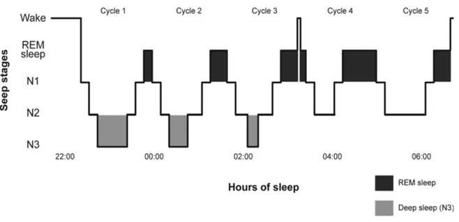
Sleep is composed of slow-wave sleep stages and of the paradoxical sleep stage (see Figure 1 for a schematic representation of a normal night of sleep). Slow-wave sleep is divided into three stages, namely stages N1 and N2, which are light sleep, and N3, also known as stages 3 and 4, or deep sleep (Carskadon & Dement, 2011; Iber, Ancoli-Israel, & Quan, 2007; Roehrs, 2005). These three stages are also referred to as non-rapid eye movement (NREM) sleep. Paradoxical sleep, also known as rapid eye movement (REM) sleep, has a similar electroencephalographic (EEG) activity as wakefulness and is characterised by muscle atonia and marked eye movements. Paradoxical sleep is the primary stage during which dreaming occurs. In the course of one night, the sleeper alternates between the various sleep stages in cycles of 90 to 100 minutes. There are on average four to six cycles per night. At the beginning of the night, stage N3 is the most present of all sleep stages, while stage N2 and REM sleep are the most prevalent stages towards the end of the night.
Whereas adolescents (ages 13-17) generally require 9 hours of sleep, adults sleep an average of 7-8 hours per night (Foley, Ancoli-Israel, Britz, & Walsh, 2004; Mindell, Owens & Carskadon, 1999). Total sleep time decreases with age, whereas the quantity of light sleep increases progressively (Carrier, Monk, Buysse, & Kupfer, 1997; Ohayon, Carskadon, Guilleminault, & Vitiello, 2004). It has also been shown that the decline of deep sleep begins around ages 36-50 (Van Cauter, Leproult, & Plat, 2000). Sleep can be objectively and quantitatively measured by polysomnography (PSG), which minimally includes electroencephalography, electrooculography and electromyography in order to identify each sleep stage. Aside from PSG, other methods are used to evaluate sleep quality, such as actigraphy, an accelerometer that measures movement and is used to monitor the rest-activity cycle, supervision by the nursing staff (when studies occur in hospital settings), and questionnaires.
Figure 1. Sleep architecture of a healthy adult subject
Figure 1 depicts the sleep architecture of a healthy adult subject. Sleep occurs in consecutive cycles of
approximately 90 minutes, characterised by varying proportions of N1, N2, N3, and rapid eye movement (REM) sleep. Stage N3, or deep sleep, occurs mainly in the first sleep cycles, diminishing in proportion as the sleep period advances. Conversely, the proportion of REM sleep increases throughout the night, and is mainly present during later sleep cycles.
1.2. Role of sleep in cognition
It is well established that sleep deprivation affects cognitive performance and this is not only true for acute total sleep deprivation, but also for chronic and partial sleep loss (Goel, Rao, Durmer, & Dinges, 2009). Among the cognitive domains that are simultaneously most affected by poor sleep or sleep loss and most important for ADLs, we find attention, memory, and executive functions (Chee & Choo, 2004; Choo, Lee, Venkatraman, Sheu, & Chee, 2005; Diekelman & Born, 2010; Dinges, 1992; Goel, Rao, Durmer, & Dinges, 2009; Harrison & Horne, 2000; Jones & Harrison, 2001; Mu et al., 2005; Tomasi et al, 2009; Walker & Stickgold, 2006). In this section, we will briefly describe the influence of poor sleep or sleep loss on these specific cognitive domains, within a healthy population.
1.2.1 Attention
Attention is generally defined by its different components, which include vigilance, sustained attention, selective attention, alternating attention, and divided attention (Sohlberg & Mateer, 1987). Most studies have documented the influence of sleep deprivation on vigilance and sustained attention. It has been observed that sleep deprivation leads to behavioural changes characterised by a general slowing of reaction times, an increased number of errors (omission and commission) on tasks requiring detection of randomly occurring stimuli, and an increased time-on-task effect, which means that performance worsens across the course of a cognitive task (see Lim & Dinges, 2008 for a review). These behavioural effects of sleep deprivation are not only observed for acute and complete sleep deprivation, but are also observed following partial sleep deprivation. For example, in a study where sleep was restricted to three hours per night for seven days, performance on the vigilance task was affected, with subjects presenting a high rate of omissions when compared to a non-sleep deprived group (Fafrowicz et al., 2010).
1.2.2. Memory
Two types of memory were found to be very sensitive to poor sleep, namely episodic memory, which refers to the capacity to store and retrieve memories that are associated to a specific time and place, and procedural memory, which enables retention of learned connections between stimuli and responses (Tulving, 1983).
Episodic memory is affected by poor sleep in two ways. Firstly, poor sleep impedes learning of verbal and non-verbal material if learning is preceded by a night of poor sleep (i.e. learning after the night of poor sleep). This has been observed among students, since the consequences of sleep deprivation among this chronically sleep deprived population include learning deficits and inferior academic performances (Curcio, Ferrara, & De Gennaro, 2006). Among healthy subjects and individuals presenting insomnia, a link between total sleep deprivation preceding learning and the presence of memory encoding deficits has also been observed (Fortier-Brochu, Beaulieu-Bonneau, Ivers, & Morin, 2012; Yoo, Hu, Gujar, Jolesz,