UNCORRECTED
PROOF
BBA - Biomembranes xxx (2017) xxx-xxx
Contents lists available at ScienceDirect
BBA - Biomembranes
journal homepage: www.elsevier.com
Changes in biophysical membrane properties induced by the Budesonide/
Hydroxy-β-cyclodextrin complex
Andreia G. dos Santos
a, b, 1, Jules Bayiha
a, 1, Gilles Dufour
c, Didier Cataldo
d, Brigitte Evrard
c, Liana C. Silva
b,
Magali Deleu
e, Marie-Paule Mingeot-Leclercq
a,⁎aUniversité catholique de Louvain, Louvain Drug Research Institute, Cellular and Molecular Pharmacology Unit, Avenue E. Mounier 73, B1.73.05, B-1200 Bruxelles, Belgium bUniversidade de Lisboa, Faculdade de Farmácia, iMed.ULisboa - Research Institute for Medicines, Av. Prof. Gama Pinto, 1649-003 Lisboa, Portugal
cUniversité de Liège, Laboratoire de Technologie Pharmaceutique et Biopharmacie, Avenue de l’Hôpital 3, B-4000 Liège, Belgium
dUniversité de Liège, CHU, Laboratory of Tumor & Development Biology of the Groupe Interdisciplinaire de Génoprotéomique Appliquée, Avenue de l’Hôpital 3, B-4000 Liège,
Belgium
eUniversité de Liège, Gembloux Agro Bio-Tech, Laboratoire de Biophysique Moléculaire aux Interfaces, Passage des Déportés, 2, B-5030 Gembloux, Belgium
A R T I C L E I N F O Article history:
Received 10 February 2017 Received in revised form 1 June 2017 Accepted 16 June 2017 Available online xxx Keywords: Drug-membrane interaction Fluidity Permeability Langmuir Cholesterol Liposomes A B S T R A C T
Budesonide (BUD), a poorly soluble anti-inflammatory drug, is used to treat patients suffering from asthma and COPD (Chronic Obstructive Pulmonary Disease). Hydroxypropyl-β-cyclodextrin (HPβCD), a biocom-patible cyclodextrin known to interact with cholesterol, is used as a drug-solubilizing agent in pharmaceutical formulations. Budesonide administered as an inclusion complex within HPβCD (BUD:HPβCD) required a quarter of the nominal dose of the suspension formulation and significantly reduced neutrophil induced in-flammation in a COPD mouse model exceeding the effect of each molecule administered individually. This suggests the role of lipid domains enriched in cholesterol for inflammatory signaling activation.
In this context, we investigated the effect of BUD:HPβCD on the biophysical properties of membrane lipids. On cellular models (A549, lung epithelial cells), BUD:HPβCD extracted cholesterol similarly to HPβCD. On large unilamellar vesicles (LUVs), by using the fluorescent probes diphenylhexatriene (DPH) and calcein, we demonstrated an increase in membrane fluidity and permeability induced by BUD:HPβCD in vesicles containing cholesterol. On giant unilamellar vesicles (GUVs) and lipid monolayers, BUD:HPβCD induced the disruption of cholesterol-enriched raft-like liquid ordered domains as well as changes in lipid packing and lipid desorption from the cholesterol monolayers, respectively. Except for membrane fluidity, all these effects were enhanced when HPβCD was complexed with budesonide as compared with HPβCD. Since cholesterol-enriched domains have been linked to membrane signaling including pathways involved in inflammation processes, we hypothesized the effects of BUD:HPβCD could be partly mediated by changes in the biophysical properties of cholesterol-enriched domains.
© 2017.
1. Introduction
The concept of biological membranes has evolved from simple physical barriers providing individualization of the cell and subcellu-lar compartments. This concept evolved to encompass cellusubcellu-lar mem-brane complexity [1–5] regarding its (i) composition, including hun-dreds of lipid species, glycolipids and proteins; (ii) organization, in-cluding asymmetry and lateral domains; and (iii) function, e.g. sig-naling cascades, modulation of protein function and folding, cellular communication, pathogen and drug interaction, among many others.
⁎Corresponding author at: FACM/LDRI-UCL - Cellular and Molecular Pharmacology
Unit of the Louvain Drug Research Institute, Université catholique de Louvain, Avenue E. Mounier 73, B1.73.05. B-1200 Bruxelles, Belgium.
Email address: [email protected] (M-P Mingeot-Leclercq)
1Both authors equally contributed to the study.
The presence of non-random domains within the lipid bilayer, e.g. the so-called cholesterol and sphingolipid-enriched lipid rafts [6], fur-ther supports the functional character of the membrane over a sim-ple structural role. Cholesterol and sphingomyelin (SM)-enriched do-mains show particular biophysical properties [7] creating an ordered (liquid ordered, lo) lipid phase within the bulk membrane. Removal of
cholesterol by a randomly methylated-β-cyclodextrin (MeβCD) from the lipid bilayer was shown to induce alterations in membrane bio-physical properties [8]. Lipid rafts have been linked to several mem-brane functions including signaling activation by immune receptors such as TLR4 and CD44 [9] involved in inflammation and cancer [10]. Furthermore, changes in lipid membrane composition and/or biophys-ical properties leading to significant membrane reorganization have been linked to consequent disruption of cell signaling [11].
Cyclodextrins (CD) are cyclic oligosaccharides consisting of six (αCD), seven (βCD) or eight (γCD) glucopyranose units linked with
http://dx.doi.org/10.1016/j.bbamem.2017.06.010 0005-2736/© 2017.
UNCORRECTED
PROOF
α-1,4 glycosidic linkages [12]. The toroidal shape and hydrophobiccavity of cyclodextrins allows the formation of inclusion complexes with hydrophobic molecules of adequate size and shape through non covalent interactions [13]. Hydroxypropyl-β-cyclodextrin (HPβCD) is an FDA/EMA approved β-cyclodextrin derivative with increased wa-ter solubility and low toxicity [14]. HPβCD is able to form complexes with surfactants [15] and polymers [16] as well as with drug mole-cules, such as curcumin [17] and budesonide [18].
Budesonide (BUD), a well-known anti-inflammatory drug is a glu-cocorticoid commercially available as Pulmicort®. Budesonide is rec-ommended for the treatment of asthma [19,20], acute onset of Chronic Obstructive Pulmonary Disease (COPD) [21], allergic rhinitis [22] and Crohn’s disease [23] among others, acting through the direct in-hibition of expression of pro-inflammatory mediators [24]. Unfortu-nately, with a logP of 3.2, budesonide is practically insoluble in water at physiological pH, leading to low pulmonary deposition [25–27] and reduced bioavailability, requiring the use of relatively high doses in clinical use.
In patients with mild to moderate persistent asthma, budesonide administered as an inclusion complex within HPβCD (BUD:HPβCD) required a quarter of the nominal dose of the suspension formulation due to a marked reduction in nebulization time [25,28]. Moreover, co-administration of budesonide solubilized within HPβCD has been shown to significantly reduce neutrophil induced inflammation in a COPD mouse model exceeding the effect of each molecule adminis-tered individually (Rocks et al., unpublished data).
In this context, a clear understanding of the molecular mechanism of action of the BUD:HPβCD complex is essential to design tailored and optimized therapeutic formulations.
This work focused on studying the effect of the BUD:HPβCD complex on biophysical properties of lipid membrane. Since the BUD:HPβCD complex is envisaged for aerial administration by nebu-lization, lung epithelial cells (A549) are used for the study of its cellu-lar toxicity and cholesterol extraction potential.
The effects of BUD:HPβCD on membrane biophysical proper-ties were evaluated using membrane model systems. Large unilamel-lar vesicles (LUVs) were used to study the interaction with mem-brane cholesterol, via a fluorescent analogue of cholesterol (DHE), and changes in membrane fluidity and permeability, by using the flu-orescent probes diphenylhexatriene (DPH) and calcein, respectively. Giant unilamellar vesicles (GUVs) were used to visualize the effect on lateral phase separation and lipid organization using fluorescence mi-croscopy. Finally, Langmuir studies characterized the effect on lipid packing and desorption from the lipid monolayer.
2. Experimental procedures
2.1. Chemicals
The L-α-phosphatidylcholine (PC - Egg, Chicken), 1,2-dipalmi-toyl-sn-glycero-3-phosphocholine (POPC), 1,2-dioleoyl-sn-glyc-ero-3-phosphocholine (DOPC), egg sphingomyelin (SM - Egg, Chicken), N-palmitoyl-D-erythro-sphingosyl phosphoryl choline (pSM - 16:0 SM d18:1/16:0), L-α-phosphatidylinositol (PI - Liver,
Bovine), cholesterol (Chol - ovine wool),
er-gosta-5,7,9(11),22-tetraen-3ß-ol (DHE - dehydroergosterol), 1,2-di-oleoyl-sn-glycero-3-phosphoethanolamine-N-(lissamine rhodamine B sulfonyl) (ammonium salt) (18:1 Liss Rho-PE) and 1,2-dipalmi-toyl-sn-glycero-3-phospho ethanolamine-N-(biotinyl) (sodium salt) (16:0 Biotinyl PE) were purchased from Avanti Polar Lipids (Al-abaster, AL, USA). N-(7-Nitrobenz-2-Oxa-1,3-Diazol-4-yl)-1,2-Di-hexadecanoyl-sn-Glycero-3-Phospho ethanolamine, Triethy
lammonium Salt) (NBD-PE) was purchased from Life Technologies (Leusden, Netherlands). 1,6-diphenyl-1,3,5-hexatriene (DPH), avidin from egg white, 16,17-Butylidenebis(oxy)-11,21-dihydrox-ypregna-1,4-diene-3,20-dione (Budesonide, BUD), Fluores-cein-bis(methyliminodiacetic acid) (Calcein), and Sephadex® G-50 were purchased from Sigma-Aldrich (St. Louis, MO-USA). Methyl β-cyclodextrin (MeβCD, Crysmeb®) and Hydroxypropyl-β-cyclodex-trin (HPβCD, Kleptose® Oral Grade) were purchased from Roquette (Lestrem, France).
Lipids and lipid probes were dissolved in chloroform, except DPH, which was dissolved in tetrahydrofuran (THF), and were kept at − 20 °C. The cyclodextrins were solubilized in PBS (NaCl 137 mM, KCl 2.7 mM, Na2HPO4 9.6 mM and KH2PO4 1.15 mM, pH 7.4) at
their maximal concentration of 30 mM for MeβCD and 250 mM for HPβCD. Budesonide was firstly dissolved in DMSO at 100 mM and then diluted to 0.1 mM in a PBS solution (DMSO 0.1% v/v). All or-ganic solvents used were Spectronorm grade from VWR (Radnor, PA, USA) or Emsure grade from Merck (Darmstadt, Germany).
2.2. Preparation of the Budesonide-Cyclodextrin complex
The Budesonide-Cyclodextrin complex (BUD:HPβCD) was pre-pared by adding budesonide to a HPβCD solution in PBS during 48 h under magnetic agitation or 2 h using a T-25 Ultra-Turrax® labora-tory mixer from IKA (Staufen, Germany). The amount of budesonide effectively encapsulated was determined using HPLC-MS quantifica-tion as described by Dufour et al. [26].
2.3. A549 cell culturing, cytotoxicity assay and cholesterol dosage
A549 cells were cultured in DMEM medium – from Thermo-Fisher Scientific (Waltham, MA-USA) – supplemented with 10% of Fetal Bovine Serum (FBS) at 37 °C and under 5% CO2.
A549 cells, grown to 80% confluence in 96-well plates, were ex-posed to MeβCD, HPβCD, BUD:HPβCD complex and budesonide in low-serum conditions (1% FBS). Cell death was inferred from cell membrane permeabilization to cytoplasmic Lactate Dehydrogenase (LDH). LDH activity was measured in triplicate using the Cytotoxic-ity Detection Kitplus(LDH) version 06 from Sigma-Aldrich (St. Louis, MO-USA).
The amount of protein was determined using the DCTMProtein As-say Kit from Bio-Rad (Hercules, CA-USA).
After extraction, total cholesterol [29] was quantified using the Amplex® Red Cholesterol Assay Kit from Thermo-Fisher Scientific (Waltham, MA-USA).
2.4. Preparation of large unilamellar vesicles (LUVs)
Large unilamellar vesicles (LUVs) were prepared by extrusion from multilamellar vesicles (MLVs). Lipids were mixed at the mo-lar ratios of PC:SM:PI (4:4:3) and PC:SM:PI:Chol (4:4:3:5.5) with a probe-to-lipid ratio of 1:100 for DHE and 1:300 for DPH and a final lipid concentration of 10 mM. A lipid film was obtained after solvent evaporation over 2 h, using a R-210 rotavapor from Buchi (Flawil, Switzerland) coupled to a vacuum pump HZ 2C from Vacu-ubrand (Wertheim, Germany), followed by minimum 2 h in an exsic-cator under vacuum. The lipid film was hydrated in Tris-HCl buffer (Tris-HCl 10 mM, NaCl 135 mM, pH 7.4). MLVs were obtained by repeated cycles (× 7) of vortex/freeze/thawing. LUVs were obtained by MLV extrusion (× 21) using a mini-extruder system from Avanti Polar Lipids (Alabaster, AL, USA) with a 100 nm pore size polycar
UNCORRECTED
PROOF
bonate Nuclepore Track Etch membrane filter from Whatman® (GEHealthcare, Little Chalfont, UK). Total lipids were quantified using the method from Rouser [30] and diluted to the desired final concen-tration in PBS.
2.5. Vesicle size and ζ-potential determinations
LUV mean size and ζ-potential were determined using a Zetasizer Nano SZ equipment from Malvern Instruments (Grovewood Road, UK) with patented NIBS (non-invasive back scatter) technology and the recommended software. Particle size distribution and the poly-dispersion index (PdI) measurements were performed by Dynamic Light Scattering (DLS) technology using 12 mm square polystyrene cuvettes in a thermostated chamber at 25 °C. Particle charge (ζ-poten-tial) was measured using Dynamic Electrophoretic Mobility (DEM) using a disposable folded capillary cell in a thermostated chamber at 25 °C.
2.6. Fluorescence spectroscopy measurements
All fluorescence measurements were carried out with a LS55 spec-trofluorimeter from Perkin Elmer (Waltham, MA-USA) in right angle geometry. Temperature was stabilized at 25 °C using a C25P Phoenix II thermostating water bath from Thermo Scientific (Waltham, MA-USA).
2.7. Dehydroergosterol (DHE) Spectroscopy
The ability of BUD:HPβCD and HPβCD to bind to cholesterol was investigated using DHE fluorescence spectroscopy.
DHE (10 μM) was prepared in PBS pH 7.4 containing 0.1% DMSO [31]. Fluorescence emission spectra of DHE in buffer solu-tion were recorded at increasing concentrasolu-tions of BUD:HPβCD or HPβCD, and the intensity of the monomeric versus microcrystalline peak ratio was plotted against log10 concentration. The excitation monochromator was set at 328 nm, and the emission spectra were recorded from 340 to 545 nm [32,33]. The influence of DMSO and DHE concentration was controlled.
To probe the interaction between BUD:HPβCD or HPβCD with cholesterol in a lipid environment, 1 mol% of DHE was incorporated in LUVs composed of PC:SM:PI:Chol (4:4:3:5.5). Maximal emission of DHE was observed around 372, 404 and 424 nm, as described pre-viously in membrane systems [34]. LUVs (5 μM) were incubated with increasing BUD:HPβCD or HPβCD concentrations for 3 h at 25 °C.
2.8. Diphenylhexatriene fluorescence polarization
Molecule polarization was quantified using steady state fluores-cence anisotropy, < r >, measurements calculated using Eq. (1):
where the different intensities IIJare the steady state polarized
verti-cal and horizontal components of fluorescence emission with excita-tion vertical (IVVand IVH) and horizontal (IHVand IHH) to the emission
axis. The latter pair of components was used to calculate the G factor (G = IHV/ IHH).
DPH concentration was determined by UV spectroscopy and ad-justed to 100 μM in tetrahydrofuran. Final lipid concentration was
adjusted to 50 μM in PBS pH 7.4. LUVs were incubated with BUD:HPβCD or HPβCD for 60 min at 25 °C shielded from light.
2.9. Calcein release
Changes in the membrane permeability were followed by deter-mining the leakage of entrapped calcein at self-quenching concen-trations, from liposomes [35]. Briefly, the dried lipid films were hy-drated with a solution of purified calcein (73 mM) in Tris-HCl buffer at pH 7.4 and osmolarity of 404 mOsm/kg. The un-encapsulated dye was removed by the mini-column centrifugation technique using Sephadex® G-50 [36]. The liposomes were diluted to a final lipid con-centration of 5 μM in an isosmotic Tris-HCl (Tris 10 mM and NaCl 188 mM) pH 7.4 buffer and stabilized for 10 min at 25 °C. Values were recorded for 30 s before addition of BUD:HPβCD or HPβCD at increasing final concentrations of 10 and 20 mM. After the addi-tion of the compounds, the fluorescence intensities were continuously recorded as a function of time for up to 500 s. The percentage of cal-cein released was determined according to Eq. (2):
where Ftis the fluorescence signal measured at a time t in the presence
of compounds, Fcontris the fluorescence signal measured at the same
time t for control liposomes, and Ftotis the total fluorescence signal
obtained after complete disruption of the liposomes by 0.02% Triton X-100.
2.10. Preparation of giant unilamellar vesicles (GUVs)
Giant unilamellar vesicles (GUVs) were prepared using the elec-troformation method [37–39]. In brief, mixtures of DOPC:pSM (1:1) and DOPC:pSM:Chol (1:1:3) with biotinylated lipid-to-lipid ratio of 1:106Biotinyl-PE and probe-to-lipid ratio of 1:750 for Rho-DOPE
and 1:250 for NBD-PE were prepared. A small volume (4 μl) of lipid mixture (4 mM) was evenly spread on the surface of an ITO coated glass lamella and the solvent was allowed to evaporate over 5 min. A 1 mm thick silicon gasket was used to form a sealed reaction cham-ber. Sucrose-Tris (475 μL) was added and a second ITO covered glass lamella was overlaid. The GUVs were formed at 60 °C over a 2 h ex-posure to a sinusoidal signal with a peak-to-peak intensity of 1 V and frequency of 500 Hz. The GUVs were used within the day.
2.11. Fluorescence microscopy measurements
GUVs were used to visualize the lipid lateral segregation and phase separation. GUVs were placed in a μ-Slide 8-well chamber from Ibidi (Martinsried, Germany) previously coated with avidin 0.1% for a minimum of 2 h. GUVs were observed using an Axio Ob-server Z1 inverted microscope (Carl Zeiss, Jena, Germany) equipped with a model CSU-X1 spinning disk (Yokogawa Electric Corporation, Tokyo, Japan) and a Plan-Apochromat 100 ×/1.40 Oil DIC M27 ob-jective (Carl Zeiss, Jena, Germany). Images were recorded and ana-lyzed with an AxioCamMR3 camera using Carl Zeiss AxioVision® 4.8.2 software. The red channel was used for Rho-DOPE (excitation/ emission at 561/617 nm) and in the green channel for NBD-PE (exci-tation/emission at 488/530 nm).
(1)
UNCORRECTED
PROOF
2.12. Surface pressure–area (π-A) compression isotherms
To examine the effect of BUD:HPβCD and HPβCD, on lipid pack-ing, surface pressure–area (π-A), compression isotherms were recorded with an automated Langmuir trough (KSV Mini-trough KSV Instruments Ltd., Helsinki, Finland-width = 7.5 cm, length = 37 cm), two hydrophilic Delrin mobile barriers (symmetric compression), a platinum Wilhelmy plate, and a temperature probe. The system was enclosed in a Plexiglas® box, and the temperature was maintained at 22.0 ± 1.0 °C.
The cleanliness of the surface was ensured by aspiration of the sub-phase surface before each experiment. Once the temperature was stabilized, the barriers were fully closed and reopened and, if a varia-tion in surface pressure of less than 0.5 mM/m was observed, the lipid was deposed on the air-liquid interface surface with a micro-syringe (Hamilton, USA). The platinum plate was cleaned by rinsing with iso-propanol and heating to red glow in-between experiments. PBS pH 7.4 was used as the subphase. Lipids (Chol, SM, POPC, or the nega-tively-charged lipid, PI) were dissolved at a concentration of 2 mM in CHCl3:MeOH (2:1 v/v) and were spread at the liquid/air interface with
a micro-syringe (Hamilton, USA). The volume was chosen in order to obtain an optimal isotherm compression curve (starting at 0 mN/m and showing a collapse at the end of the compression).
After an equilibration time of 15 min, the film was compressed at a rate of 10 mm/min. BUD:HPβCD (0.04:1 mM:mM) or HPβCD (1 mM) were solubilized in the subphase before spreading the lipid us-ing the same amount of lipid as for the control assays. The same pro-cedure as the one used for experiments without cyclodextrin was ap-plied. Each compression isotherm was repeated at least two times; the relative standard deviation in surface pressure and area was ≤ 5%.
2.13. Surface pressure–time (π-t) adsorption isotherms
BUD:HPβCD or HPβCD effect on lipid molecular area were as-sessed by measuring lipid molecular area over time upon incubation with the compounds (π-t isotherms). To this end, the same set-up as described for the surface pressure–area (π-A) compression isotherms was used with a different automated Langmuir trough (KSV Mini-trough KSV Instruments Ltd., Helsinki, Finland-width = 7.5 cm, length = 20 cm).
The lipids (Chol, SM, POPC, or PI) were added until a surface pressure of 30 mN/m was achieved and, after a stabilization period of 15 min, surface pressure over time was recorded. After the acquisi-tion of a 200 s baseline to verify the stability of the monolayer, the compounds were injected into the subphase using specialized injection supports to a final concentration of 0.04:1 mM:mM of BUD:HPβCD or 1 mM of HPβCD. Surface pressure was recorded until a plateau was observed.
The obtained curves were analyzed by a fitting on a 2-phase expo-nential regression from where the estimated plateau values were ex-tracted.
2.14. Statistical analysis
All data manipulation, graphical presentation and statistical analy-sis was performed using Microsoft® Excel® (2016, Microsoft®, Red-mond – Washington USA) and GraphPad Prism® (version 4.03 for Windows, GraphPad Software Inc., La Jolla - California USA, www. graphpad.com).
3. Results
3.1. Cell toxicity and cell cholesterol depletion
We first evaluated the cytotoxicity of the complex BUD:HPβCD in comparison with the highly hydrophobic anti-inflammatory drug, budesonide (BUD) and HPβCD.
The cytotoxic effect was determined by following LDH release on lung epithelial cells (A549). For concentrations in cyclodextrin vary-ing from 0 to 10 mM and after 4 h of incubation (Fig. 1.a), the cy-totoxicity induced by BUD:HPβCD or HPβCD was lower (ca. 10%) as compared with budesonide. At 25 mM in cyclodextrin and
Fig. 1. Cell toxicity and effect on cholesterol of BUD:HPβCD, HPβCD, and BUD on
lung epithelial cells. Percentage of LDH released from A549 lung epithelial cells (a) at 4 h for BUD:HPβCD (squares) 0.04:1 to 2.03:50 mM:mM, HPβCD (circles) 1 to 50 mM in cyclodextrin, or BUD (triangles) 0.04 to 2.03 mM and (b) for BUD:HPβCD 0.41:10 mM:mM (squares), HPβCD 10 mM (circles), or BUD 0.41 mM (triangles) at 1, 2, 4 and 24 h. (c) Total cellular cholesterol (normalized to total amount of proteins) for A549 lung epithelial cells treated with BUD:HPβCD 0.41:10 mM:mM, HPβCD 10 mM, or MeβCD 5 mM, for 45 min normalized to untreated A549 cells.
UNCORRECTED
PROOF
over, significant toxicity (ca. 40%) was observed. Regardless thecon-centrations in cyclodextrin (from 0 to 25 mM), the cytotoxicity in-duced by the complex (BUD:HPβCD) was lower as compared to that observed with budesonide alone. At 50 mM in cyclodextrin, cyto-toxicity was comparable for HPβCD, BUD:HPβCD and budesonide. Regarding the time dependency cytotoxic effect induced by BUD:HPβCD (0.41:10 mM:mM), budesonide (0.41 mM), and HPβCD (10 mM) (Fig. 1.b), we didn't have any effect for 2 h of in-cubation. After 4 h, LDH release started and after 24 h of incubation, BUD:HPβCD and budesonide, showed more than 35% of LDH re-lease. In comparison, HPβCD induced less than 10% LDH rere-lease.
Selecting non-toxic conditions, we quantified the extraction of cholesterol from cells (Fig. 1.c). MeβCD (5 mM) was used as a pos-itive control. For BUD:HPβCD (0.41:10 mM:mM) and HPβCD (10 mM), after 45 min of incubation, the amount of total cholesterol (normalized to total protein) was reduced by ca. 45%. HPβCD was able to significantly extract cholesterol from the membrane regardless of previous complexation with budesonide.
3.2. Interaction with membrane model systems
The effect of the extraction of cholesterol induced by BUD:HPβCD and HPβCD on membrane biophysical properties was further characterized in membrane model systems.
To determine the concentration range of HPβCD without vesicle destabilization, vesicle mean size and ζ-potential were measured upon incubation with increasing concentration of HPβCD (up to 100 mM, Fig. S1). Briefly, at ratios of HPβCD:lipid exceeding 5000:1 (HPβCD 25 mM to lipids 5 μM), the fraction of vesicles within the diameter range of the control samples became significantly reduced. Therefore, a 25 mM threshold of HPβCD was set to avoid experimental artefacts and/or skewed results.
3.3. Interaction with sterols in aqueous solution
The interaction of BUD:HPβCD and HPβCD with cholesterol was studied using a cholesterol analogue presenting similar behavior in aqueous solution and biological membranes [33,34,40], the dehy-droergosterol (DHE). Especially, it shows similar properties regard-ing lateral phase separation compared to cholesterol [41]. Lateral in-teractions between sterols are responsible for an increase in DHE fluo-rescence quantum yield at higher wavelengths of emission [32,42,43]. DHE is a self-quenching molecule and fluorescence emission of the monomeric peak (I372) occurs upon the dissolution of
cholesterol-en-riched domains [44] or desorption of DHE from the lipid bilayer [45] by βCDs. DHE aggregates in solution can be quantified using the fluo-rescence emission ratio I372/I424[34], corresponding to the monomeric
species over the aggregate forms, such as DHE microcrystals in so-lution [34,42]. The spectral properties of DHE, namely fluorescence intensity and peak ratios (I372/I424), are used to infer upon the
microen-vironment of cholesterol, in agreement with results showing compara-ble, although slightly faster, extraction of DHE in mixed monolayers (sterols:POPC 30:70) by HPβCD, to that of cholesterol [45].
The interaction of increasing concentrations of BUD:HPβCD and HPβCD with DHE in solution was characterized (Fig. 2) and com-parative control studies were performed using MeβCD (Fig. S2, a, b). An increase of the intensity of the monomeric peak at 372 nm (Fig. 2.a), concomitant with an increase in the ratio of the monomeric over the aggregate form of DHE (Fig. 2.b) was observed. The effect started at 1 mM HPβCD and a plateau value was reached at 10 mM. Regarding the ratio of the monomeric over the aggregate form of
Fig. 2. Fluorescence of DHE in aqueous solution upon interaction with BUD-HPβCD,
and HPβCD. (a) Fluorescence emission intensity at 372 nm and (b) ratio of peak inten-sity between 372 and 424 nm upon addition of BUD:HPβCD (squares; dotted line), or HPβCD (circles; solid line). The lines correspond to a non-linear fitting of a non-loga-rithmic sigmoidal Hill growth function to the data.
DHE (I372/I424), the effect was observed at slightly lower
concentra-tions of HPβCD when complexed with the budesonide. No effect was observed for budesonide alone (not shown). Overall, the increase in fluorescence of the DHE monomeric peak indicates the solubilisation of DHE by the HPβCDs in a concentration dependent manner, which is likely due to the formation of HPβCD-sterol complexes as previ-ously described [45].
3.4. Interaction with sterols in a lipid membrane
In order to study the effect of increasing concentrations of cy-clodextrin (HPβCD and BUD:HPβCD) on the microenvironment of cholesterol within lipid bilayers, a small fraction of DHE was incor-porated into LUVs mimicking the lipid composition of plasma mem-brane (PC:SM:PI:Chol 4:4:3:5.5) (Fig. 3) and comparative control studies were performed using MeβCD (Fig. S2, c, d).
Fig. 3 presents the fluorescence intensity at the 372 nm sion peak of DHE monomeric form (Fig. 3.a) and the ratio of emis-sion at 372 nm over 424 nm (Fig. 3.b) upon interaction with the BUD-HPβCD complex and HPβCD. An increase in fluorescence in-tensity of DHE (Fig. 3.a) was observed, which was less pronounced as compared with DHE microcrystals in solution. This can be due to the lower amount of DHE within the membranes (200 × less con-centrated). Moreover, DHE microenvironment can affect βCD poten-tial for complexing with cholesterol [45], the dissolution of an aggre-gate form in solution being likely facilitated over the extraction of DHE stabilized within a lipid membrane. The results showed that, re-gardless of complexation with budesonide, HPβCD was able to in-crease the fluorescence of DHE monomeric peak. Regarding the ratio of the monomeric over the aggregate form of DHE within the bilayer (Fig. 3.b), it was comparable to that of DHE microcrystals solubilized
UNCORRECTED
PROOF
Fig. 3. Fluorescence of DHE within LUVs upon interaction with BUD:HPβCD, and
HPβCD. (a) Emission at the monomeric 372 nm peak and (b) ratio of emission at 372 nm over 424 nm upon interaction with BUD-HPβCD (squares; dotted line), or HPβCD (circles; solid line), with LUVs composed of PC:SM:PI:Chol 4:4:3:5.5. DHE was present at 1 mol% of total lipid concentration. Measurements were performed at 25 °C in triplicate. The lines correspond to a non-linear fitting of a non-logarithmic sig-moidal Hill growth function to the data.
by the HPβCD (Fig. 2.b). No major difference was observed when the effect of BUD-HPβCD was compared to that of HPβCD.
DHE de-quenching can be due to either the disruption of choles-terol-enriched domains [34] or to extraction from membrane by the HPβCD [45]. Measurements of emission intensity over time (data not shown) showing an instantaneous endpoint de-quenching of DHE, suggest DHE extraction from the membrane instead of the rearrange-ment of lipid lateral organization, which would occur over several minutes. Overall, increase in DHE monomeric peak emission by BUD:HPβCD or HPβCD demonstrated changes of the sterol environ-ment in agreeenviron-ment with DHE extraction from the membrane [44]. Be-cause cholesterol is a main modulator of membrane fluidity, its ex-traction from membrane by BUD:HPβCD and HPβCD could modify membrane fluidity.
3.5. Effect on membrane fluidity
Cholesterol increases the fluidity of very ordered domains and in-creases the rigidity of very disordered domains. Moreover, cholesterol and sphingolipid-enriched domains, i.e. raft-like domains, present de-creased fluidity and hydration when compared to the bulk membrane. The lipid dynamics of acyl lipid chains can be monitored using diphenylhexatriene (DPH), a dye that probes the hydrophobic core of the membrane [46]. The degree of polarization of DPH, measured by its fluorescence anisotropy (< r >), increases as membrane fluidity de-creases. Fig. 4 shows the variation of DPH anisotropy in LUVs lack-ing cholesterol (PC:SM:PI 4:4:3) and LUVs containlack-ing cholesterol (PC:SM:PI:Chol 4:4:3:5.5) upon interaction with the BUD:HPβCD complex or HPβCD.
Fig. 4. Membrane fluidity measurements of LUVs containing and lacking cholesterol
upon interaction with BUD:HPβCD, and HPβCD. Measurement of DPH anisotropy (< r >) upon addition of BUD:HPβCD complex (grey bars), or HPβCD (black bars) to LUVs composed of (a) PC:SM:PI (4:4:3) or (b) PC:SM:PI:Chol (4:4:3:5.5) containing DPH (1:300 molar ratio).
For vesicles lacking cholesterol, the results (Fig. 4.a) show a fluid membrane (ld) in the absence of cholesterol (< r > of ca. 0.15), as is
expected for a lipid mixture containing phospholipid:sphingolipid at a molar ratio of 7:4 at 25 °C [47]. Adding cholesterol (Fig. 4.b) reduced membrane fluidity (< r > of ca. 0.25), indicating the presence of a liq-uid ordered (lo) phase typical of a mixture containing ca. 33 mol% of
cholesterol and 25 mol% of SM at 25 °C [47].
Upon incubation of cholesterol-free vesicles with HPβCD (Fig. 4.a), an increase in DPH anisotropy to values suggesting a gel phase (< r > of ca. 0.30) was observed at the lower concentration of HPβCD (10 mM). Increasing HPβCD concentration caused a concentration de-pendent increase in membrane fluidity back to control values. These results are in agreement with literature [48,49,50]. Since we excluded increase in average size of liposomes (Fig. S1) as suggested in litera-ture [48,49], the formation of supra-molecular cyclodextrin struclitera-tures on the surface of the lipid bilayer is likely the mechanism involved in decrease in membrane fluidity [50].
The incubation of BUD:HPβCD complex or HPβCD with cho-lesterol-enriched membranes (Fig. 4.b) resulted in a dose-dependent decrease of anisotropy indicating an increase in membrane fluidity. This effect reached a plateau at a value of anisotropy indicative of a very fluid cholesterol-free membrane (< r > of ca. 0.10) at 30 mM of CD. Moreover, the cholesterol depleted membrane by HPβCD became more fluid (< r > of ca. 0.10) than the control mixture without choles-terol (< r > of ca. 0.15). As for the cholescholes-terol-free LUVs, the extrac-tion of other membrane rigidifying lipids, such as the high Tm
sphin-golipid, sphingomyelin also located within the lipid raft, is likely. De-spite a larger affinity of βCD towards cholesterol, other lipids may also be extracted from the membrane with varying affinities [45].
UNCORRECTED
PROOF
As was observed for DHE extraction from the membrane, HPβCDcomplexation with budesonide did not show any effect on the induced changes to membrane fluidity as compared with HPβCD. The pres-ence of free budesonide did not change the membrane fluidity.
In conclusion, BUD:HPβCD or HPβCD incubation with choles-terol-enriched membranes lead to increased membrane fluidity. This is in agreement with the extraction of cholesterol and destabilization of the liquid ordered cholesterol- and sphingolipid-enriched raft-like do-mains. Altogether, this suggests a lateral reorganization of the lipids, which could be associated with increased membrane permeability.
3.6. Effect on membrane permeability
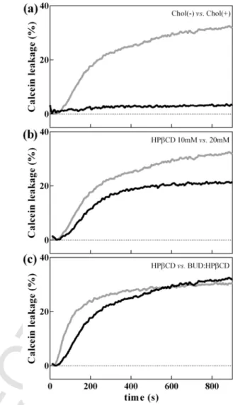
Permeation of the plasma membrane is often the first barrier for drug entry into the cell, as is the case for budesonide. Therefore, in-creased membrane permeability may be one of the possible modu-lators of drug bioavailability and efficacy. The permeabilization of the membrane can be determined by quantifying the increase in flu-orescence emission of the self-quenching calcein upon release from permeabilized membranes. Calcein fluorescence intensity was mea-sured upon interaction of BUD-HPβCD (0.41:10 mM:mM and 0.82:20 mM:mM) or HPβCD (10 and 20 mM) with vesicles lack-ing cholesterol (PC:SM:PI 4:4:3) and vesicles containlack-ing cholesterol (PC:SM:PI:Chol 4:4:3:5.5) (Fig. 5 and Fig. S3).
In the absence of cholesterol, BUD:HPβCD (20 mM in cyclodex-trins) did not induce calcein leakage. In the presence of cholesterol, the extent of permeabilization increased to reach a plateau value (around 30% of calcein release) after 200 s (Fig. 5.top). The effect was dependent upon the concentration of cyclodextrins (10 mM < 20 mM in cyclodextrins) (Fig. 5.middle). These results were consistent with the increase in lipid extraction and consequent leakage of encapsulated calcein.
As compared to HPβCD alone, incubation with the BUD:HPβCD increased the rate of calcein release (Fig. 5.bottom) without affecting the percentage of calcein released at equilibrium. The increase in rate of membrane permeabilization may be explained by either (i) perme-ation of budesonide through the membrane or (ii) increase in HPβCD affinity towards the membrane induced by budesonide.
3.7. Effect on membrane phase separation
The interaction of BUD:HPβCD and HPβCD with cholesterol may lead to the disruption of the lipid raft domains, typically composed of cholesterol and sphingolipids.
Confocal fluorescence microscopy was used to observe the changes in lipid phase separation over time upon interaction with BUD:HPβCD or HPβCD. Control GUVs and lipid-raft model GUVs, composed of DOPC:pSM (1:1) and DOPC:pSM:Chol (1:1:3) [50], were labeled with Rho-DOPE (red channel) and NBD-PE (green chan-nel) for the liquid disordered (ld) and liquid ordered (lo) domains,
re-spectively.
Typically, the GUV population was relatively heterogeneous re-garding vesicle size with diameters centered on ca. 15 μm (± 5 μm). Overall, no effect was observed by incubation with budesonide.
While the majority of the GUVs immediately presented changes in lipid phase separation upon interaction with the HPβCD, a subpopula-tion, mostly comprised of the very small GUVS (under 10 μm in diam-eter) began to exhibit observable changes only after more than 30 min of incubation or remained apparently unaffected.
In the absence of cholesterol (Fig. 6-left), the DOPC:pSM 1:1 membranes show a ld/so(red/dark) phase separation as expected [47].
Fig. 5. Calcein leakage from LUVs containing and lacking cholesterol upon
interac-tion with BUD:HPβCD, and HPβCD. Comparison of leakage of calcein (top) from PC:SM:PI (black line) and PC:SM:PI:Chol (gray line) vesicles in the presence of BUD:HPβCD 0.82:20 mM:mM; (middle) from PC:SM:PI:Chol vesicles in the presence of BUD:HPβCD 0.41:10 mM:mM (black line) and BUD:HPβCD 0.82:20 mM:mM (gray line); and (bottom) from PC:SM:PI:Chol vesicles upon interaction with BUD:HPβCD 0.82:20 mM:mM (gray line) and 20 mM of HPβCD 20 mM (black line). The curves are representative of three independent experiments.
Interaction with either BUD:HPβCD or HPβCD removed any micro-scopic phase separation and a single ldphase (red) was observed.
As was observed for the kinetics of membrane permeabilization (Fig. 5.c), the effect occurred at earlier incubation times for the BUD:HPβCD (under 5 min) when compared with the HPβCD alone (ca. 15 min).
In the presence of cholesterol (Fig. 6-right), no microscopic ld/lo
phase separation was visible as expected for a mixture containing 60 mol% of cholesterol. Interaction with the HPβCD, and BUD:HPβCD alike, caused the appearance of observable lodomains
after 5 min of incubation, congruent with a decrease in the molar frac-tion of cholesterol. Longer incubafrac-tion times showed further decrease of the lophase giving yield to lddomains (eventually a single ldphase
was visible). Within 45 min, a ld/sophase separation was observed,
in-dicating a negligible amount or the absence of cholesterol within the membrane.
The decrease and, ultimately, the disappearance, of the liquid or-dered phase (lo), a phase enriched in cholesterol and sphingomyelin,
UNCORRECTED
PROOF
Fig. 6. Confocal fluorescence microscopy imaging of membrane phase separation in GUVs upon incubation with BUD:HPβCD, and HPβCD. Imaging of membrane domains in
GUVs composed of (left) DOPC:pSM (1:1) and (right) DOPC:pSM:Chol (1:1:3) before (top, control) and after (descending) 5, 15 and 45 min with the BUD:HPβCD complex or HPβCD. DOPC:pSM vesicles were labeled with Rho-DOPE (red channel) to visualize the liquid disordered (ld)/solid ordered (so) phase separation in red/dark. DOPC:pSM:Chol were
labeled with Rho-DOPE (red channel) and NBD-PE (green channel) to visualize the liquid disordered (ld)/liquid ordered (lo) phase separation in red/green channels, respectively. The
absence of fluorescent labelling indicates a solid ordered phase (so) and the co-localization of both probes (yellow) indicates a lack of observable phase separation.
is consistent with removal of cholesterol from the membrane by inter-action with HPβCD [51].
3.8. Effect on lipid mean molecular area
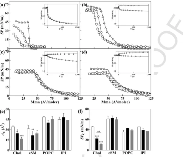
To further characterize the effect of BUD:HPβCD and HPβCD on the biophysical membrane properties, we compared the isotherms of lipid monolayers spread with BUD:HPβCD or HPβCD aqueous solu-tions to those deposited on PBS buffer (Fig. 7).
In presence of BUD:HPβCD or HPβCD, a long plateau at a non-zero surface pressure was observed at large molecular areas what-ever the composition of the monolayer. It suggests that BUD:HPβCD or HPβCD are able to adsorb to the lipid monolayer in a gaseous state [52,53] despite the fact that they do not change the surface pressure by themselves in the absence of lipids.
For all the lipids, further compression of monolayers in presence of BUD:HPβCD and HPβCD induced a progressive increase of the surface pressure indicating the formation of a liquid-expanded mono
layer. Finally, at low molecular areas, the isotherms showed either a final plateau at constant surface pressure or a sharp decrease in surface pressure corresponding to the collapse of the monolayer. In the case of cholesterol (Fig. 7.a) and SM (Fig. 7.b) monolayers and in a lesser extent for PI (Fig. 7.d) monolayers, the profile of the lipid isotherm in the presence of BUD:HPβCD or HPβCD is different from that ex-pected if the lipid molecules were simply removed from the interface. The lipid/ BUD:HPβCD or HPβCD interactions leads to the formation of new system at the interface.
The molecular area and surface pressure at which the membrane collapsed (referred to as ACand SPC, respectively) were used to
quan-titatively compare the interfacial behavior of lipids in presence or in absence of BUD:HPβCD or HPβCD (Fig. 7.e and f).
In the case of cholesterol monolayers (Fig. 7.a and e), a decrease in AC(Fig. 7.a and e) was observed with BUD:HPβCD to a higher
extent as compared with HPβCD. A reduction of the lipid molecular area in presence of exogenous drug can arise from desorption of the interfacial material into the subphase and/or from a reorganization of
UNCORRECTED
PROOF
Fig. 7. Effect of BUD:HPβCD, and HPβCD on Chol, eSM, POPC and PI monolayers. Surface pressure-area (Π-A) compression isotherms of the pure lipids (a) Chol, (b) eSM, (c)
POPC and (d) PI with a subphase composed of PBS (triangles), BUD:HPβCD 0.2:5 mM:mM (squares), or HPβCD 5 mM (circles). (inset) Surface pressure-time (Π-t) curves for the lipids with a subphase composed of PBS with BUD:HPβCD 0.04:1 mM (squares), or HPβCD 1 mM (circles) fitted with a non-linear regression curve. (e) Molecular area at the sur-face pressure onset (A0) and (f) two-dimensional compressibility factor (Cs) for the higher compression for the pure lipid (light grey) and in the presence of BUD:HPβCD (dark grey)
or HPβCD (grey) calculated from the surface pressure-area (Π-A) isotherms as described by [61]. The curves were recorded at 22 °C (± 1 °C) and are representative of replicated assays.
the interfacial monolayer (domain packing, nucleation) [53]. The sig-nificant decrease in SPC(Fig. 7.f) in presence of HPβCD and even
much more in presence of BUD:HPβCD means that the interaction of HPβCD destabilizes the liquid-condensed phase of the cholesterol monolayer, which is in accordance with the fluidification effect shown by DPH fluorescence polarization.
The injection of BUD:HPβCD or HPβCD beneath the cholesterol monolayer initially spread at the air-water interface until a surface pressure of 30 mN/m and maintained at a constant spreading surface gave rise to a rapid and important decrease of the surface pressure (Fig. 7.a-inset). These results are in favor of cholesterol depletion from the interface, as also shown for βCD in previous studies [45,52,53].
HPβCD and BUD:HPβCD also reduces the ACof SM monolayers
(Fig. 7.b and e) without significantly affecting SPC(Fig. 7.f)
suggest-ing some lipid desorption from the interface without changsuggest-ing lipid packing. The extraction of SM from the interface by HPβCD was in agreement with the decrease of surface pressure in the time-de-pendence surface pressure experiments. In contrast, BUD:HPβCD in-creased the surface pressure when it is injected under the SM mono-layer. This suggests that two parallel phenomena occurs when BUD:HPβCD interacts with the SM monolayer, (i) a limited
depletion of SM from the monolayer and (ii) an adsorption of BUD:HPβCD molecules to the interface.
In the case of POPC monolayers (Fig. 7.c, d, e), the presence of HPβCD decreased AC(Fig. 7e) but in a lesser extent than in the case of
cholesterol and SM (reduction of ~ 17% for POPC vs ~ 30% for cho-lesterol and SM). No effect on ACwas observed for the BUD:HPβCD
complex with POPC or for either compound with PI monolayer. As for SM monolayer, HPβCD or BUD:HPβCD did not greatly affect the interfacial stability of POPC or PI monolayers (Fig. 7.f). However, while the HPβCD caused lipid desorption from the monolayer (Fig. 7.b–d-insets), the BUD:HPβCD complex increased slightly the sur-face pressure when injected under the POPC or PI monolayers. Inser-tion of budesonide and/or BUD:HPβCD into the monolayer can also occurs in these cases.
Overall, the BUD:HPBCD complex shows i) increased extent of destabilization of cholesterol monolayers and ii) adsorption into the phospholipid monolayers when compared to the free HPBCD. This might be due to i) a possible increase in affinity/efficacy of the HPβCD regarding cholesterol-containing membranes due to the pres-ence of budesonide and/or ii) a possible insertion of budesonide into the monolayer.
UNCORRECTED
PROOF
4. Discussion
Membrane cholesterol has several important properties including the lateral segregation into cholesterol and sphingolipid-enriched do-mains known as lipid rafts [6]. These ordered dodo-mains have been shown to be essential for creating an appropriate microenvironment for signal reception and transduction. Lipid rafts are able to stabilize and cluster the receptor structures. This provides a sorting mechanism, and co-localizing receptors and cofactors thus being responsible for the fine-tuning of signal transduction [54]. Cyclodextrins and β-cy-clodextrin are able to form inclusion complexes with cholesterol. They are commonly used to extract or insert cholesterol from membranes [55–58]. In addition, βCDs are also known to form inclusion com-plexes with several hydrophobic drugs [17,18,59,60] including budes-onide (BUD).
Interestingly, clinical studies [14] showed a lower cellular toxi-city of budesonide when budesonide was complexed with HPβCD. The aim of the present study was therefore to understand the po-tential effect of the extraction of cholesterol in lipid-raft domains in relation with regulation of the inflammatory response induced by the BUD:HPβCD complex. Thus, we characterized the interaction of BUD:HPβCD and HPβCD with lipid model membranes containing and lacking cholesterol and determined changes in biophysical mem-brane properties.
In model membranes containing cholesterol, we demonstrated an effect of BUD:HPβCD with cholesterol-enriched membranes leading to changes in membrane biophysical properties in agreement with cho-lesterol extraction, such as increased membrane fluidity and perme-ability, changes in lipid packing and lipid desorption from the lipid in-terface as well as the disruption of cholesterol-enriched raft-like liquid ordered domains. In comparison with HPβCD and except for mem-brane fluidity, all these effects were enhanced and/or observed earlier with the complex BUD:HPβCD.
The molecular mechanisms involved in cholesterol extraction by HPβCD and BUD:HPβCD was unknown but as demonstrated by Lopez et al. [50], the distribution of the cyclodextrins on the surface of the monolayer could play a critical role. Spontaneous cholesterol extraction on a nanosecond time scale might be related with a suit-ably oriented dimer. Moreover, free energy calculations reveal that the cyclodextrins have a strong affinity to bind to the membrane surface, and, by doing so, destabilize the local packing of cholesterol mole-cules making their extraction favorable [50].
For the model systems lacking cholesterol, the BUD:HPβCD and HPβCD caused an increase in DPH anisotropy for the lowest con-centrations and the disappearance of liquid disordered/solid ordered phase separation in GUVs. The increase of membrane rigidity is sur-prising but can reasonably be ascribed to the formation of a rela-tively thick polymer layer around the phospholipid bilayers [48]. In the same line, Lopez et al. [50], reported from simulations studies, the formation of supra-molecular cyclodextrin structures on the sur-face of cholesterol monolayers. The mechanism of interaction of cy-clodextrins with the lipid membrane is suggested to depend on the molecular ratio of cyclodextrins to lipid. Since cyclodextrins possess a greater affinity towards cholesterol when compared to phospho-lipids, it is possible that the observed increase in membrane rigid-ity observed for the lowest HPβCD:lipid ratio might reflect non-spe-cific stabilizing cyclodextrin interaction with the surface of the choles-terol-free membrane. For higher HPβCD concentrations the increase in membrane fluidity coming back to values similar to those ob-tained for controls might be explained by extraction of lipids other than cholesterol, namely sphingomyelin. Moreover, at higher HPβCD
concentrations, HPβCD:HPβCD interactions could be promoted in comparison with HPβCD:membrane interactions explaining why no global effect was observed on membrane fluidity. Regarding the ef-fect on membrane phase separation, in absence of cholesterol, BUD:HPβCD affected membrane phase separation without increasing membrane permeability and affecting (or only slightly) the stability of SM, POPC or PI monolayers.
A critical question is the potential competition between budesonide and lipid for the HPβCD cavity which may occur at the interface. Focusing on the mechanism involved, the BUD:HPBCD complex in-duced a greater destabilization of the cholesterol monolayer with-out affecting HPBCD cholesterol extraction potential (Fig. 7a + in-set). It resulted in an increase in surface pressure over time in mono-layers composed of phospholipids. These results suggest an insertion of budesonide into the air:liquid interface, an exchange between the budesonide and cholesterol in favor of cholesterol. This could be re-lated to the increased kinetics of membrane permeabilization to cal-cein induced by the BUD:HPβCD complex when compared with the HPβCD.
Altogether, the results showed that BUD:HPβCD and HPβCD can effectively induce significant changes in the composition and biophys-ical properties of cholesterol-enriched raft-like domains in model sys-tems. Destabilization of these domains might explain the anti-inflam-matory effect observed for the HPβCD (preliminary data), probably by modifying the lipid environment of receptors involved in inflam-matory processes. The co-administration of budesonide and HPβCD might provide a higher therapeutic effect acting through complemen-tary anti-inflammatory mechanisms. Thus HPβCD could play a criti-cal role for administration of poorly soluble drug like budesonide by increasing cellular delivery of budesonide in vivo as well as for reg-ulation of critical biophysical membrane properties of cholesterol-en-riched domains where immune receptors are located. HPβCD can be both a targeted delivery vehicle and an anti-inflammatory agent by own.
Further studies about the modulation of the inflammatory response, focusing particularly on the relevance of lipid rafts in signal activation, are required to evaluate the mechanism involved in BUD:HPβCD and HPβCD anti-inflammatory properties.
Supplementary data to this article can be found online at http://dx. doi.org/10.1016/j.bbamem.2017.06.010.
Conflict of interest
The authors declare that they have no conflicts of interest with the contents of this article.
Author contributions
AGS and MPML wrote the manuscript and designed experiments. JB contributed for the membrane fluidity and performed the mem-brane permeability assays.
GD, DC and BE prepared and characterized the BUD:HPβCD complex used in this study.
MD provided the expertise, equipment and resources to perform the surface pressure–area compression isotherms- and surface pres-sure–time (π-t) adsorption isotherms-measurements.
All authors discussed the results.
Transparency document
The Transparency document associate with this article can be found, in online version.
UNCORRECTED
PROOF
Acknowledgments
AGS thanks GD, DC and BE for the BUD:HPβCD used in this study.
AGS thanks PVDS for providing the equipment and training re-quired for microscopy imaging.
AGS thanks Lucas Vanderavero (Agro-Bio Tech from the Univer-sity of Liège – ULg) for his help with the surface pressure–area com-pression isotherm measurements.
M.-C. Cambier, and V. Mohymont provided dedicated technical assistance.
LCS is supported by Investigador FCT 2014 (IF/00437/2014) from Fundação para a Ciência e a Tecnologia, Portugal.
MD is Senior Research Associate for the Fonds National de la Recherche Scientifique (FRS-FNRS).
This work was supported by Walloon Region (AEROGAL).
References
[1] S.J. Singer, G.L. Nicolson, The fluid mosaic model of the structure of cell mem-branes, Science 175 (1972) 720–731.
[2] G. van Meer, Lipid traffic in animal cells, Annu. Rev. Cell Biol. 5 (1989) 247–275.
[3] K. Simons, E. Ikonen, Functional rafts in cell membranes, Nature 387 (1997) 569–572.
[4] G.L. Nicolson, The fluid-mosaic model of membrane structure: still relevant to understanding the structure, function and dynamics of biological membranes af-ter more than 40 years, Biochim. Biophys. Acta 2014 (1838) 1451–1466. [5] F.M. Goni, The basic structure and dynamics of cell membranes: an update of
the Singer-Nicolson model, Biochim. Biophys. Acta 2014 (1838) 1467–1476. [6] R. Schroeder, E. London, D. Brown, Interactions between saturated acyl chains
confer detergent resistance on lipids and glycosylphosphatidylinositol (GPI)-an-chored proteins: GPI-an(GPI)-an-chored proteins in liposomes and cells show similar be-havior, Proc. Natl. Acad. Sci. U. S. A. 91 (1994) 12130–12134.
[7] J.D. Nickels, X. Cheng, B. Mostofian, C. Stanley, B. Lindner, F.A. Heberle, S. Perticaroli, M. Feygenson, T. Egami, R.F. Standaert, et al., Mechanical proper-ties of nanoscopic lipid domains, J. Am. Chem. Soc. 137 (2015) 15772–15780. [8] M.P. Besenicar, A. Bavdek, A. Kladnik, P. Macek, G. Anderluh, Kinetics of
cholesterol extraction from lipid membranes by methyl-beta-cyclodextrin—a surface plasmon resonance approach, Biochim. Biophys. Acta 2008 (1778) 175–184.
[9] T. Murai, Lipid raft-mediated regulation of hyaluronan-CD44 interactions in in-flammation and cancer, Front. Immunol. 6 (2015) 1–9.
[10] P. Varshney, V. Yadav, N. Saini, Lipid rafts in immune signaling: current progress and future perspective, Immunology 149 (2016) 13–24.
[11] M.G. Sorci-Thomas, M.J. Thomas, Microdomains, inflammation, and athero-sclerosis, Circ. Res. 118 (2016) 679–691.
[12] T. Loftsson, M.E. Brewster, Pharmaceutical applications of cyclodextrins: basic science and product development, J. Pharm. Pharmacol. 62 (2010) 1607–1621. [13] S.S. Jambhekar, P. Breen, Cyclodextrins in pharmaceutical formulations I:
structure and physicochemical properties, formation of complexes, and types of complex, Drug Discov. Today 21 (2016) 356–362.
[14] S. Gould, R.C. Scott, 2-Hydroxypropyl-beta-cyclodextrin (HP-beta-CD): a toxi-cology review, Food Chem. Toxicol. 43 (2005) 1451–1459.
[15] A.J. Valente, O. Soderman, The formation of host-guest complexes between surfactants and cyclodextrins, Adv. Colloid Interf. Sci. 205 (2014) 156–176. [16] H. Wei, C.Y. Yu, Cyclodextrin-functionalized polymers as drug carriers for
can-cer therapy, Biomater. Sci. 3 (2015) 1050–1060.
[17] C.S. Mangolim, C. Moriwaki, A.C. Nogueira, F. Sato, M.L. Baesso, A.M. Neto, G. Matioli, Curcumin-beta-cyclodextrin inclusion complex: stability, solubility, characterisation by FT-IR, FT-Raman, X-ray diffraction and photoacoustic spectroscopy, and food application, Food Chem. 153 (2014) 361–370. [18] G. Dufour, W. Bigazzi, N. Wong, F. Boschini, P. de Tullio, G. Piel, D. Cataldo,
B. Evrard, Interest of cyclodextrins in spray-dried microparticles formulation for sustained pulmonary delivery of budesonide, Int. J. Pharm. 495 (2015) 869–878. [19] D. Hodgson, K. Mortimer, T. Harrison, Budesonide/formoterol in the treatment
of asthma, Expert Rev. Respir. Med. 4 (2010) 557–566.
[20] I.M. Adcock, G. Caramori, P.A. Kirkham, Strategies for improving the efficacy and therapeutic ratio of glucocorticoids, Curr. Opin. Pharmacol. 12 (2012) 246–251.
[21] G. Caramori, P. Casolari, A. Barczyk, A.L. Durham, A. Di Stefano, I. Adcock, COPD immunopathology, Semin. Immunopathol. 38 (2016) 407–515. [22] N.Z. Fabbri, E. Abib-Jr, Z.R. de Lima, Azelastine and budesonide (nasal
sprays): effect of combination therapy monitored by acoustic rhinometry and clinical symptom score in the treatment of allergic rhinitis, Allergy Rhinol. (Providence) 5 (2014) 78–86.
[23] A. Rezaie, M.E. Kuenzig, E.I. Benchimol, A.M. Griffiths, A.R. Otley, A.H. Steinhart, G.G. Kaplan, C.H. Seow, Budesonide for induction of remission in Crohn's disease, Cochrane Database Syst. Rev. 6 (2015), CD000296. [24] P.J. Barnes, Pathophysiology of allergic inflammation, Immunol.
Rev. 242 (2011) 31–50.
[25] K. Basu, A. Nair, P.A. Williamson, S. Mukhopadhyay, B.J. Lipworth, Airway and systemic effects of soluble and suspension formulations of nebulized budes-onide in asthmatic children, Ann Allergy Asthma Immunol 103 (2009) 436–441.
[26] G. Dufour, B. Evrard, P. de Tullio, Rapid quantification of 2-hydrox-ypropyl-beta-cyclodextrin in liquid pharmaceutical formulations by (1)H nu-clear magnetic resonance spectroscopy, Eur. J. Pharm. Sci. 73 (2015) 20–28. [27] T. Kinnarinen, P. Jarho, K. Jarvinen, T. Jarvinen, Pulmonary deposition of a
budesonide/gamma-cyclodextrin complex in vitro, J. Control. Release 90 (2003) 197–205.
[28] P.A. Williamson, D. Menzies, A. Nair, A. Tutuncu, B.J. Lipworth, A proof-of-concept study to evaluate the antiinflammatory effects of a novel solu-ble cyclodextrin formulation of nebulized budesonide in patients with mild to moderate asthma, Ann Allergy Asthma Immunol 102 (2009) 161–167. [29] E.G. Bligh, W.J. Dyer, A rapid method of total lipid extraction and purification,
Can. J. Biochem. Physiol. 37 (1959) 911–917.
[30] G. Rouser, S. Fkeischer, A. Yamamoto, Two dimensional then layer chromato-graphic separation of polar lipids and determination of phospholipids by phos-phorus analysis of spots, Lipids 5 (1970) 494–496.
[31] J. Lorent, C.S. Le Duff, J. Quetin-Leclercq, M.P. Mingeot-Leclercq, Induction of highly curved structures in relation to membrane permeabilization and bud-ding by the triterpenoid saponins, alpha- and delta-Hederin, J. Biol. Chem. 288 (2013) 14000–14017.
[32] F. Schroeder, Y. Barenholz, E. Gratton, T.E. Thompson, A fluorescence study of dehydroergosterol in phosphatidylcholine bilayer vesicles, Biochem-istry 26 (1987) 2441–2448.
[33] K.H. Cheng, J. Virtanen, P. Somerharju, Fluorescence studies of dehydroergos-terol in phosphatidylethanolamine/phosphatidylcholine bilayers, Biophys. J. 77 (1999) 3108–3119.
[34] L.M. Loura, M. Prieto, Dehydroergosterol structural organization in aqueous medium and in a model system of membranes, Biophys. J. 1997 (72) (1997) 2226–2236.
[35] J.N. Weinstein, S. Yoshikami, P. Henkart, R. Blumenthal, W.A. Hagins, Lipo-some-cell interaction: transfer and intracellular release of a trapped fluorescent marker, Science 195 (1977) 489–492.
[36] P.I. Lelkes, Liposome Technology, CRC Press, Boca Raton, FL, 1984225–246. [37] M.I. Angelova, S. Soléau, P. Méléard, J.F. Faucon, P. Bothorel, Preparation of
giant vesicles by external AC electric fields. Kinetics and applications, in: C. Helm, M. Lösche, H. Möhvald (Eds.), Trends in Colloid and Interface Science VI, Steinkopff, 1992, pp. 127–131.
[38] D.J. Estes, M. Mayer, Giant liposomes in physiological buffer using electrofor-mation in a flow chamber, Biochim. Biophys. Acta 2005 (1712) 152–160. [39] N. Rodriguez, F. Pincet, S. Cribier, Giant vesicles formed by gentle hydration
and electroformation: a comparison by fluorescence microscopy, Colloids Surf. B: Biointerfaces 42 (2005) 125–130.
[40] D. Wustner, Fluorescent sterols as tools in membrane biophysics and cell biol-ogy, Chem. Phys. Lipids 146 (2007) 1–25.
[41] M.G. Benesch, R.N. Lewis, R.N. McElhaney, A calorimetric and spectroscopic comparison of the effects of cholesterol and its immediate biosynthetic precur-sors 7-dehydrocholesterol and desmosterol on the thermotropic phase behavior and organization of dipalmitoylphosphatidylcholine bilayer membranes, Chem. Phys. Lipids 191 (2015) 123–135.
[42] A.L. McIntosh, A.M. Gallegos, B.P. Atshaves, S.M. Storey, D. Kannoju, F. Schroeder, Fluorescence and multiphoton imaging resolve unique structural forms of sterol in membranes of living cells, J. Biol. Chem. 278 (2003) 6384–6403.
[43] M.A. Soto-Arriaza, C. Olivares-Ortega, F.H. Quina, L.F. Aguilar, C.P. So-tomayor, Effect of cholesterol content on the structural and dynamic membrane properties of DMPC/DSPC large unilamellar bilayers, Biochim. Biophys. Acta 2013 (1828) 2763–2769.
[44] M. Seras, J. Gallay, M. Vincent, M. Ollivon, S. Lesieur, Cholesterol assemblies induced by octyl lgucoside: a time-resolved fluorescence study of dehydroergos-terol, J. Colloid Interfaces B 167 (1994) 159–171.
UNCORRECTED
PROOF
[45] H. Ohvo-Rekila, B. Akerlund, J.P. Slotte, Cyclodextrin-catalyzed extraction of fluorescent sterols from monolayer membranes and small unilamellar vesicles, Chem. Phys. Lipids 105 (2000) 167–178.
[46] M. Shinitzky, Y. Barenholz, Fluidity parameters of lipid regions determined by fluorescence polarization, Biochim. Biophys. Acta 515 (1978) 367–394. [47] R.F. de Almeida, A. Fedorov, M. Prieto, Sphingomyelin/phosphatidylcholine/
cholesterol phase diagram: boundaries and composition of lipid rafts, Biophys. J. 85 (2003) 2406–2416.
[48] I. Puskas, L. Barcza, L. Szente, F. Csempesz, Features of the interaction be-tween cyclodextrins and colloidal liposomes, J. Incl. Phenom. 54 (2006) 89–93. [49] I. Puskas, F. Csempesz, Influence of cyclodextrins on the physical stability of
DPPC-liposomes, Colloids Surf. B: Biointerfaces 58 (2007) 218–224. [50] C.A. Lopez, A.H. de Vries, S.J. Marrink, Molecular mechanism of cyclodextrin
mediated cholesterol extraction, PLoS Comput. Biol. 7 (2011) e1002020. [51] S.L. Veatch, S.L. Keller, Seeing spots: complex phase behavior in simple
mem-branes, Biochim. Biophys. Acta 2005 (1746) 172–185.
[52] J. Mascetti, S. Castano, D. Cavagnat, B. Desbat, Organization of beta-cyclodex-trin under pure cholesterol, DMPC, or DMPG and mixed cholesterol/phospho-lipid monolayers, Langmuir 24 (2008) 9616–9622.
[53] M. Flasinski, M. Broniatowski, J. Majewski, P. Dynarowicz-Latka, X-ray graz-ing incidence diffraction and Langmuir monolayer studies of the interaction of beta-cyclodextrin with model lipid membranes, J. Colloid Interface Sci. 348 (2010) 511–521.
[54] D. Lingwood, K. Simons, Lipid rafts as a membrane-organizing principle, Sci-ence 327 (2010) 46–50.
[55] T. Irie, K. Fukunaga, J. Pitha, Hydroxypropylcyclodextrins in parenteral use. I: lipid dissolution and effects on lipid transfers in vitro, J. Pharm. Sci. 81 (1992) 521–523.
[56] Y. Ohtani, T. Irie, K. Uekama, K. Fukunaga, J. Pitha, Differential effects of al-pha-, beta- and gamma-cyclodextrins on human erythrocytes, Eur. J. Biochem. 186 (1989) 17–22.
[57] H. Ohvo, J.P. Slotte, Cyclodextrin-mediated removal of sterols from monolay-ers: effects of sterol structure and phospholipids on desorption rate, Biochem-istry 35 (1996) 8018–8024.
[58] A.E. Christian, M.P. Haynes, M.C. Phillips, G.H. Rothblat, Use of cyclodextrins for manipulating cellular cholesterol content, J. Lipid Res. 38 (1997)
2264–2272.
[59] N. Rocks, S. Bekaert, I. Coia, G. Paulissen, M. Gueders, B. Evrard, J.C. Van Heugen, P. Chiap, J.M. Foidart, A. Noel, et al., Curcumin-cyclodextrin com-plexes potentiate gemcitabine effects in an orthotopic mouse model of lung can-cer, Br. J. Cancer 107 (2012) 1083–1092.
[60] J.E. Kim, H.J. Cho, D.D. Kim, Budesonide/cyclodextrin complex-loaded lyophilized microparticles for intranasal application, Drug Dev. Ind. Pharm. 40 (2014) 743–748.
[61] M. Eeman, G. Francius, Y.F. Dufrene, K. Nott, M. Paquot, M. Deleu, Effect of cholesterol and fatty acids on the molecular interactions of fengycin with stra-tum corneum mimicking lipid monolayers, Langmuir 25 (2009) 3029–3039.