M
INI
-
REVIEW
The minor collagens in articular cartilage
Yunyun Luo
1,2&,
Dovile Sinkeviciute
1,3, Yi He
1, Morten Karsdal
1, Yves Henrotin
4, Ali Mobasheri
5,6,
Patrik Önnerfjord
3, Anne Bay-Jensen
11
Biomarkers
& Research, Nordic Bioscience A/S, Herlev, Denmark
2
Faculty of Healthy and Medical Sciences, University of Copenhagen, Copenhagen, Denmark
3Department of Clinical Sciences, Medical Faculty, Lund University, Lund, Sweden
4
Bone and Cartilage Research Unit, Institute of Pathology, Level 5, Arthropole Liège, University of Liège, CHU Sart-Tilman,
4000 Liège, Belgium
5
Faculty of Health and Medical Sciences, University of Surrey, Guildford, Surrey GU2 7XH, UK
6Arthritis Research UK Centre for Sport, Exercise and Osteoarthritis, Arthritis Research UK Centre for Musculoskeletal Ageing
Research, Queen
’s Medical Centre, Nottingham, NG7 2UH, UK
& Correspondence: [email protected] (Y. Luo)
Received November 8, 2016
Accepted January 25, 2017
ABSTRACT
Articular cartilage is a connective tissue consisting of a
specialized extracellular matrix (ECM) that dominates the
bulk of its wet and dry weight. Type II collagen and
aggrecan are the main ECM proteins in cartilage. However,
little attention has been paid to less abundant molecular
components, especially minor collagens, including type IV,
VI, IX, X, XI, XII, XIII, and XIV, etc. Although accounting for
only a small fraction of the mature matrix, these minor
collagens not only play essential structural roles in the
mechanical properties, organization, and shape of
articu-lar cartilage, but also ful
fil specific biological functions.
Genetic studies of these minor collagens have revealed
that they are associated with multiple connective tissue
diseases, especially degenerative joint disease. The
pro-gressive destruction of cartilage involves the degradation
of matrix constituents including these minor collagens.
The generation and release of fragmented molecules
could generate novel biochemical markers with the
capacity to monitor disease progression, facilitate drug
development and add to the existing toolbox for
in vitro
studies, preclinical research and clinical trials.
KEYWORDS
collagen, biomarker, arthritis
ARTICULAR CARTILAGE
Articular cartilage is the most widespread load-bearing
car-tilage in adults. It is a highly specialized and mechanically
resilient connective tissue found on the surface of
sub-chondral bone in diarthrodial joints. Cartilage contains
specialized cells called chondrocytes. These cells occupy
1%
–3% of the total tissue volume in fully developed tissue
and their surrounding extracellular matrix (ECM) is a
com-plex network made up of water, collagen, proteoglycans, and
other noncollagenous proteins. Other four types of cartilage
are
fibroelastic cartilage, fibrocartilage, elastic cartilage, and
epiphyseal cartilage.
COLLAGENS
Collagens are the most abundant family of ECM proteins,
which account for two-thirds of the dry mass of adult articular
cartilage (Eyre,
2004
). Numerous collagen subtypes have
been identi
fied in articular cartilage, such as type II, IX, X, XI,
VI, XII, and XIV collagen (Van der Rest,
1987
). Articular
cartilage collagen
fibrils mostly consist of type II collagen
accompanied with a lesser amount of minor collagens, which
provide cartilage with tensile strength and contribute to the
physical properties of the mature matrix (Heinegård and
Saxne,
2011
; Ichimura et al.,
2000
). However, little is known
about the processing of these minor collagens and how their
turnover is affected by the progression of osteoarthritis (OA).
New knowledge about turnover of those minor collagens will
lead to deeper understanding of the dynamics of cartilage
turnover, thereby facilitating the development of novel
biomarkers that re
flect joint health and drug discovery in OA.
In this review, we present an outline of minor collagens in
articular cartilage, focusing on the link between these
extracellular matrix proteins to OA. Finally, we elaborate on
how knowledge of these associations can be used to
develop new biomarkers, which provide insight into the
Protein
&
translational medicine of OA. Such biomarkers also indicate
the effect of a drug on cartilage metabolism and the mode of
action. Even though a number of biomarkers already exist,
there is a clear medical need for new biomarkers for
per-sonalized healthcare (PHC) in OA, biomarkers that more
accurately reflect biological activity in different phenotypes of
the disease as well as serve as tools in diagnosis and
prognosis. This may assist in identi
fication of patients that
are in foremost need of treatment and may respond
opti-mally, with the highest ef
ficacy and lowest safety concerns,
to a given treatment. Moreover, biomarkers aid
pharma-ceutical companies develop better targeted therapeutic
strategies for selected subpopulations of OA patients.
Biomarkers can also enable early decision-making and
benchmarking. It is becoming increasingly clear, that one
simple marker is insuf
ficient for improved diagnosis, and
thus multiple makers that reflect different aspects of the
pathophysiology and clinical phenotypes may most likely be
needed in combination (Kraus et al.,
2015
; Karsdal et al.,
2014
; Henrotin et al.,
2016
).
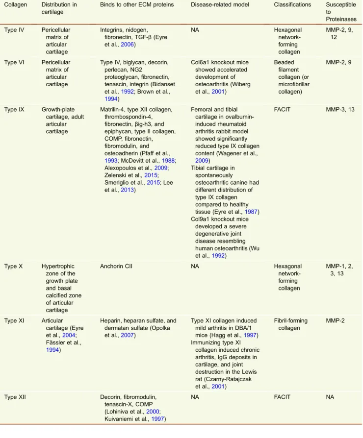
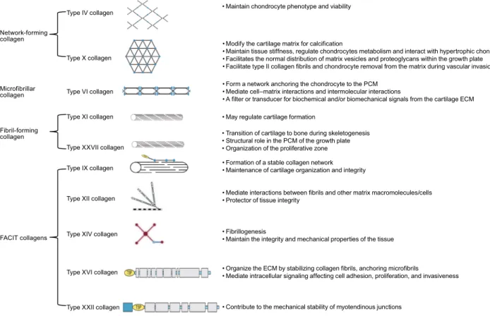
The various types of minor collagens found in articular
cartilage are listed in Table
1
and schematically illustrated in
Fig.
1
.
DISTRIBUTION, STRUCTURE, AND FUNCTION OF
MINOR COLLAGENS
Type VI collagen
—a microfibrillar collagen
Although it only makes up 1% of total collagen in adult
articular cartilage (Eyre et al.,
2006
), type VI collagen is
mainly enriched in the pericellular matrix (PCM), involving
the attachment and integrity of chondrocytes. Type VI
col-lagen is able to bind to a wide variety of ECM proteins,
including type II collagen (Bidanset et al.,
1992
), type XIV
collagen (Brown et al.,
1994
), matrilin-1 (Wiberg et al.,
2001
), and decorin (Bidanset et al.,
1992
), thereby forming
a network that anchors the chondrocyte to the PCM in
articular cartilage. Due to the high af
finity with numerous
ECM components and cell membrane (Bidanset et al.,
1992
; Wiberg et al.,
2001
), type VI collagen has been
hypothesized to play important roles in mediating cell
–ma-trix interactions and intermolecular interactions (Pfaff et al.,
1993
).
The precise role of collagen VI has not yet been clearly
defined. However, type VI collagen may serve as a filter or
transducer for biochemical and/or biomechanical signals
from the cartilage ECM. Type VI collagen with lower
molecular weight was evident in the pathological
osteoar-thritic dogs sacri
ficed 3, 5, and 7 months after surgery in
comparison to the controls (McDevitt et al.,
1988
). It
indi-cated that degradation products of larger type VI chains
might be signi
ficant in the role this molecule plays in
osteoarthritis. The type VI collagen-deficient mice (Col6a1)
exhibited accelerated development of hip osteoarthritis, a
delayed secondary ossi
fication process, and a loss of the
stiffness of the articular cartilage PCM (Alexopoulos et al.,
2009
). Type VI collagen demonstrated an important role in
regulating the physiology of the synovial joint. In another
context, the de
ficiency of type VI collagen in mice resulted in
decreased stiffness and increased chondrocyte swelling
(Zelenski et al.,
2015
). These
findings suggest that type VI
collagen has essential roles in transmitting mechanical and
osmotic stresses from the ECM to the chondrocytes
(Ze-lenski et al.,
2015
). The soluble type VI collagen was
reported to promote chondrocyte proliferation under both
healthy and osteoarthritic conditions. However, proliferation
was not observed upon treatment of immobilized type VI
collagen in chondrocytes, indicating that soluble type VI
collagen can be applied for autologous chondrocyte
implantation to expand chondrocytes (Smeriglio et al.,
2015
).
It was reported that a variant in the human Col6a4 gene is
associated with knee OA in Japanese and Chinese
popu-lations, but not found in a Korean population (Lee et al.,
2013
) nor in European OA individuals (Wagener et al.,
2009
). The contradictory
findings could be explained by
either the ethnic differences in OA susceptibility genes or the
differences of criteria in OA selection.
Type IX, XII, XIV, XVI, and XXII collagen
—the FACIT
collagens
These collagens are members of the
fibril-associated
colla-gen with interrupted triple helix (FACIT), which do not form
fibrils by themselves, but are associated with the surface of
various
fibrils.
Type IX collagen
Type IX collagen simply makes up 1%
–5% of total collagen
in adult articular cartilage and 10% of that in fetal cartilage
(Eyre et al.,
2006
). It is usually found in tissues containing
type II collagen, like growth plate cartilage and adult articular
cartilage (Eyre et al.,
1987
). It forms the unique hetero
fibril
network in the matrix of cartilage via association with type II
and type IX collagen (Wu et al.,
1992
). Type IX collagen is
extensively cross-linked with type II collagen through the
lysyl oxidase mechanism (Wu et al.,
1992
). Eyre et al.
dis-covered that the covalent cross-linking formed at the
N-telopeptide of
α1(II) chain and the COL1 in all three chains
of type IX collagen in both human and bovine cartilage (Eyre
et al.,
2004
). Additionally, Eyre et al. also observed the
binding inter type IX collagen molecules at the COL2 domain
and the non-collagenous globular domain (NC1) domain
(Wu et al.,
1992
).
Mice with a completely inactivated Col9a1 gene showed
no detectable abnormalities at birth but thereafter had a
severe degenerative joint disease resembling human OA at
4-months or older (Fässler et al.,
1994
). In a different
con-text, the knockout of type IX collagen altered the time course
of callus differentiation during bone fracture healing, and
delayed the maturation of cartilage matrix (Opolka et al.,
Protein
&
Table 1. Minor collagen overview
Collagen Distribution in cartilage
Binds to other ECM proteins Disease-related model Classifications Susceptible to Proteinases Type IV Pericellular matrix of articular cartilage Integrins, nidogen, fibronectin, TGF-β (Eyre et al.,2006) NA Hexagonal network-forming collagen MMP-2, 9, 12 Type VI Pericellular matrix of articular cartilage
Type IV, biglycan, decorin, perlecan, NG2
proteoglycan,fibronectin, tenascin, integrin (Bidanset et al.,1992; Brown et al.,
1994)
Col6a1 knockout mice showed accelerated development of osteoarthritis (Wiberg et al.,2001) Beaded filament collagen (or microfibrillar collagen) MMP-2, 9 Type IX Growth-plate cartilage, adult articular cartilage
Matrilin-4, type XII collagen, thrombospondin-4, fibronectin, βig-h3, and epiphycan, type II collagen, COMP,fibronectin, fibromodulin, and osteoadherin (Pfaff et al.,
1993; McDevitt et al.,1988; Alexopoulos et al.,2009; Zelenski et al.,2015; Smeriglio et al.,2015; Lee et al.,2013)
Femoral and tibial cartilage in ovalbumin-induced rheumatoid arthritis rabbit model showed significantly reduced type IX collagen content (Wagener et al.,
2009)
Tibial cartilage in spontaneously
osteoarthritic canine had different distribution of type IX collagen compared to healthy tissue (Eyre et al.,1987) Col9a1 knockout mice
developed a severe degenerative joint disease resembling human osteoarthritis (Wu et al.,1992) FACIT MMP-3, 13 Type X Hypertrophic zone of the growth plate and basal calcified zone of articular cartilage
Anchorin CII NA Hexagonal
network-forming collagen MMP-1, 2, 3, 13 Type XI Articular cartilage (Eyre et al.,2004; Fässler et al., 1994)
Heparin, heparan sulfate, and dermatan sulfate (Opolka et al.,2007)
Type XI collagen induced mild arthritis in DBA/1 mice (Hagg et al.,1997) Immunizing type XI
collagen induced chronic arthritis, IgG deposits in cartilage, and joint destruction in the Lewis rat (Czarny-Ratajczak et al.,2001)
Fibril-forming collagen
MMP-2
Type XII Decorin,fibromodulin, tenascin-X, COMP (Lohiniva et al.,2000; Kuivaniemi et al.,1997) NA FACIT NA
Protein
&
Cell
2007
). A de
ficiency of α1(IX) in mice has been shown to lead
to instability of hyaline cartilage (Hagg et al.,
1997
). Other
studies suggested that mutations of
α1(IX)
(Czarny-Rata-jczak et al.,
2001
),
α2(IX), and α3(IX) (Lohiniva et al.,
2000
)
can elicit multiple epiphyseal dysplasia, a heterogeneous
skeletal disorder with early-onset OA as a manifestation.
Mutations in the Col9a2 are also linked to multiple
epiphy-seal dysplasia characterized by symptoms ranging from pain
and stiffness in joints to OA (Kuivaniemi et al.,
1997
). In
another experiment, the mutated type IX collagen had a mild
chondrodysplasia (Nakata et al.,
1993
). In a rabbit model of
ovalbumin-induced rheumatoid arthritis, the NC4 domain of
type IX collagen content was reduced in femoral and tibial
cartilage, revealing early damage of type IX collagen in
articular cartilage following induction of joint in
flammation
(Kojima et al.,
2001
). The immunohistochemical staining of
type IX collagen in normal mature and spontaneously
osteoarthritic canine tibial cartilage revealed that changes in
type IX collagen distribution played crucial role in the
chon-dron remodeling and chondrocyte cluster formation
associ-ated with osteoarthritic degeneration (Poole et al.,
1997
). All
these
findings highlight that type IX collagen may play
important roles in the pathogenesis of arthritis diseases, the
formation of a stable collagen network and in the
mainte-nance of cartilage organization and integrity. In humans,
Col9a1 has been identi
fied as a susceptibility locus for
female hip OA (Mustafa et al.,
2000
; Loughlin et al.,
2002
;
Alizadeh et al.,
2005
), suggesting that Col9a1 is involved in
hip OA. The decreased expression of type IX collagen in the
cartilage may render the matrix more subject to mechanical
forces, thereby resulting in the pathogenesis of human OA.
Type IX collagen is also used to induce chronic arthritis in the
DBA/1 mice (Boissier et al.,
1990
).
Taken together, type IX collagen is crucial for the
main-tenance of cartilage matrix and formation of collagen
meshwork. Turnover of type IX collagen by proteases is an
early event in degenerative joint disease. The reduced level
of type IX collagen may contribute to the pathogenesis of
OA.
Type XII collagen
Type XII collagen shares structural homologies with type IX
and type XIV collagens (Yamagata et al.,
1991
). Additionally,
in common with several other FACITs, the length of
α1(XII)
chains is affected by complex alternative splicing of type XII
collagen primary transcripts. As a result, two distinct forms of
type XII collagen—short(XIIB) and long(XIIA)—are
gener-ated. Although both forms of type XII collagen are present in
cultured
fibroblasts—the expression of long or short form
being determined by whether cells grow in monolayer or 3D
culture
—the long transcript variant predominates. The long
form is also the only type XII collagen variant expressed in
human fetal chondrocytes (Keene et al.,
1991
).
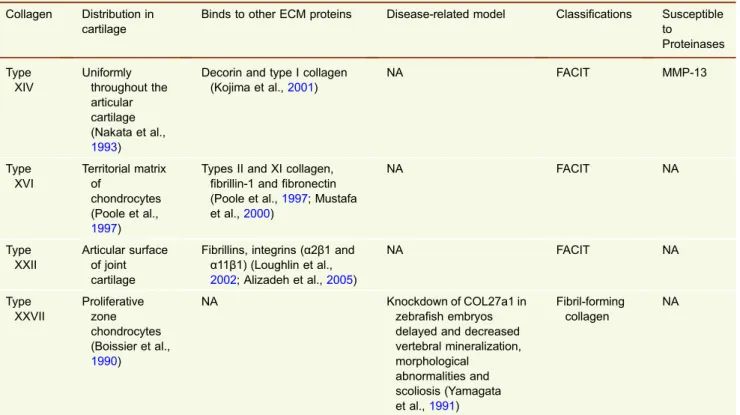
Table 1 continued
Collagen Distribution in cartilage
Binds to other ECM proteins Disease-related model Classifications Susceptible to Proteinases Type XIV Uniformly throughout the articular cartilage (Nakata et al., 1993)
Decorin and type I collagen (Kojima et al.,2001) NA FACIT MMP-13 Type XVI Territorial matrix of chondrocytes (Poole et al., 1997)
Types II and XI collagen, fibrillin-1 and fibronectin (Poole et al.,1997; Mustafa et al.,2000) NA FACIT NA Type XXII Articular surface of joint cartilage
Fibrillins, integrins (α2β1 and α11β1) (Loughlin et al., 2002; Alizadeh et al.,2005) NA FACIT NA Type XXVII Proliferative zone chondrocytes (Boissier et al., 1990) NA Knockdown of COL27a1 in zebrafish embryos delayed and decreased vertebral mineralization, morphological abnormalities and scoliosis (Yamagata et al.,1991) Fibril-forming collagen NA
FACIT = Fibril-associated collagens with interrupted triple helices. COMP = cartilage oligomeric matrix protein. NA = Not available.
Protein
&
In terms of biological function, type XII collagen has been
implicated in
fibril formation, cell adhesion, fibrosis and
osteogenesis, and in areas of high mechanical stress may
serve as a protector of tissue integrity (Chiquet et al.,
2014
;
Arai et al.,
2008
). Immunohistochemistry staining and
fibril-logenesis studies show that type XII collagen can be
incor-porated into type I collagen
fibrils in dense connective
tissues and bone. It potentially helps to mediate interactions
between
fibrils and other matrix macromolecules/cells, or
acts as a
‘shock-absorber’ similar to proteoglycans in
carti-lage (Arai et al.,
2008
; Taylor et al.,
2014
). Type XII collagen
associates with articular cartilage and growth plate region
during rat forelimb development, and may be necessary for
microenvironment that supports hyaline cartilage formation
(Taylor et al.,
2014
; Gregory et al.,
2001
). Type XII collagen
also has been shown to be present in the secretome of
human passaged chondrocytes (Polacek et al.,
2011
),
however, not in the secretome of cartilage explants (Taylor
et al.,
2014
).
Type XIV collagen
Type XIV collagen is a large, non-fibrillar ECM protein,
structurally similar to type XII collagen. In cartilage, a
pop-ulation of type XIV collagen exists a chondroitin sulfate
proteoglycan, since it was sensitive to chondroitinases ABC
and AC treatments (Watt et al.,
1992
).
Type XIV collagen is prevalent within connective tissues
that contain large amounts of
fibrillar collagens, where it
localizes near the surface of banded collagen
fibrils
(Nish-iyama
et
al.,
1994
).
Immuno
fluorescence localization
showed that type XIV collagen was prominent at the
liga-ment-bone junction, and in bovine cartilage. Type XIV
col-lagen localizes relatively uniformly throughout the articular
cartilage, but is absent from growth plate regions (Watt et al.,
1992
).
In addition to reported interactions with type I, II, V, and VI
collagens, type XIV collagen also interacts with heparin,
CD44, and cartilage oligomeric matrix protein (COMP)
(Giry-Lozinguez et al.,
1998
). Type XIV collagen is predominantly
expressed in differentiated tissues and late embryonic
development. Ruehl et al. postulated that it is involved in
tissue differentiation, and particularly, its
first FN-III domain
are potent inducers of reversible cellular quiescence and
differentiation in human and mouse mesenchymal cells
(Ruehl et al.,
2005
). Their study saw reduction of de novo
DNA synthesis without alterations to cell numbers and
via-bility and restoration of maximal proliferation upon serum
supplementation. Similarly to type XII collagen, type XIV
collagen is often found in areas of high mechanical stress
Type IV collagen Type VI collagen Network-forming collagen Type X collagen Microfibrillar collagen • • • • • • • • Fibril-forming collagen Type XI collagenType XXVII collagen
• • • • Type IX collagen FACIT collagens
Type XII collagen
Type XIV collagen
Type XVI collagen
Type XXII collagen
• • • • • • • • •
Form a network anchoring the chondrocyte to the PCM Mediate cell–matrix interactions and intermolecular interactions
A filter or transducer for biochemical and/or biomechanical signals from the cartilage ECM Maintain chondrocyte phenotype and viability
Modify the cartilage matrix for calcification
Maintain tissue stiffness, regulate chondrocytes metabolism and interact with hypertrophic chondrocytes Facilitates the normal distribution of matrix vesicles and proteoglycans within the growth plate Facilitate type II collagen fibrils and chondrocyte removal from the matrix during vascular invasion
May regulate cartilage formation
Transition of cartilage to bone during skeletogenesis Structural role in the PCM of the growth plate Organization of the proliferative zone
Formation of a stable collagen network Maintenance of cartilage organization and integrity
Mediate interactions between fibrils and other matrix macromolecules/cells Protector of tissue integrity
Fibrillogenesis
Maintain the integrity and mechanical properties of the tissue
Contribute to the mechanical stability of myotendinous junctions Organize the ECM by stabilizing collagen fibrils, anchoring microfibrils
Mediate intracellular signaling affecting cell adhesion, proliferation, and invasiveness
Figure 1. The schematic of minor collagens in articular cartilage.Reproduced with permission from Richard-Blum, S. The
collagen family. Cold Spring Harb Perspect Biol 2011;3:a004978. PCM: pericellular matrix; ECM: extracellular matrix.
Protein
&
(Hemmavanh et al.,
2013
), and has roles in
fibrillogenesis
and maintaining the integrity and mechanical properties of
the tissue (Chen et al.,
2015
).
Type XVI and XXII collagen
Type XVI collagen has been identi
fied in the territorial matrix
of the chondrocytes, associating with thin weakly banded
collagen
fibrils containing types II and XI collagen (Kassner
et al.,
2003
). In cartilage, type XVI collagen is a component
of small heterotypic D-banded
fibrils (Kassner et al.,
2003
)
and is strongly expressed in differentiating chondrocytes (Lai
and Chu,
1996
). Type XVI collagen may be incorporated into
structurally and functionally discrete matrix aggregates in
cartilage. Its main function is to organize the ECM by
stabi-lizing collagen
fibrils, anchoring microfibrils, mediating
intracellular signalling affecting cell adhesion, proliferation,
invasiveness as well as the formation of focal adhesions.
It has been shown that N-terminal processing of type XVI
collagen results in 182 kDa and 78 kDa fragments (Kassner
et al.,
2004
), whereas C-terminal and subsequent N-terminal
processing results in 150 kDa, 110 kDa, 50 kDa, and 35 kDa
fragments, respectively (Grässel et al.,
1996
). Such
frag-ments may be utilized as targets of biochemical markers for
cartilage biology.
Type XXII collagen is expressed at the junction between
synovial
fluid and surface of articular cartilage (Koch et al.,
2004
) and associated with the extra
fibrillar matrix in
carti-lage. Type XXII collagen is also detectable in human arthritic
joints, but the immuno
fluorescence staining pattern is
broadened and fuzzy. Unlike other FACIT collagens, type
XXII collagen interacts with micro
fibrils, such as fibrillins or
type VI collagens instead of collagen
fibrils (Koch et al.,
2004
). Its function remains unknown, but may contribute to
the mechanical stability of myotendinous junctions (Koch
et al.,
2004
; Zwolanek et al.,
2014
). Type XXII collagen could
serve as a marker to explore pathologic processes of joint
diseases and to study tissue junction formation during
development and regeneration of cartilage due to its
expression location.
Type IV and X collagen
—network-forming collagen
Type IV collagen is a network forming collagen, which is
exclusively found in the pericellular matrix of normal
articular cartilage, and osteoarthritic articular cartilage in
human and goat (Foldager et al.,
2014
; Jeng et al.,
2013
;
Kvist et al.,
2008
). However, controversially it was
repor-ted to be absent in any human cartilage subtypes including
hyaline,
fibrous, and elastic cartilage (Wachsmuth et al.,
2006
). Overexpression of regulator of MMP-13 increased
the expression of type IV collagen in chondrocytes (Wang
et al.,
2015
). Type IV collagen may be involved in
main-taining chondrocyte phenotype and viability and provide
clues to the progression of degenerative joint disorders
(Kvist et al.,
2008
).
Fragments originating from type IV collagen released by
protein remodeling have been thoroughly investigated for
their uses as biomarkers. Several formation and degradation
biomarkers, e.g. C4M, C4M2 (Karsdal et al.,
2015
), C4M3a
(Sand et al.,
2016
), C4M12a1, C4M12a3 (Sand et al.,
2013
),
P4NP 7S (Leeming et al.,
2013
), and Tumstatin (Hamano
et al.,
2003
) have been developed, indicating the role of type
IV collagen turnover in most connective tissue diseases.
Type X collagen is a homotrimeric collagen, which
con-sists of three identical
α1(X) chains with 3 domains each, a
short triple helix, an NC1 at C-terminus, and a short
non-helical at N-terminus (NC2) (Shen,
2005
). These structures
are believed to play a key role in modifying the cartilage
matrix for the subsequent bone formation during
endo-chondral ossi
fication (Kwan et al.,
1991
). Type X collagen is
inclined to be cleaved by interstitial collagenase, gelatinase,
human neutrophil elastase, pepsin, and trypsin (Frischholz
et al.,
1998
).
Type X collagen constitutes about 1% of total collagen in
adult articular cartilage (Eyre,
1991
). It is revealed that 45%
of the total collagens produced by mature hypertrophic
chondrocytes are type X collagen (Shen,
2005
). As a
speci
fic collagen in cartilage, type X collagen is synthesized
by hypertrophic chondrocytes and is found exclusively in the
hypertrophic cartilage and the calcified zone of articular
cartilage (Gannon et al.,
1991
). Increased expression has
been observed in arthritis as the chondrocytes become
hypertrophic (Shen,
2005
; Kwan et al.,
1991
; Frischholz
et al.,
1998
; Eyre,
1991
; Gannon et al.,
1991
; van der Kraan
and van den Berg,
2012
). Hypertrophic chondrocytes
express a variety of proteins and enzymes including type X
collagen, matrix metalloproteinase 13, alkaline phosphatase,
which do not seem to exist in normal proliferating
chondro-cytes (Steinert et al.,
2009
; D
’Angelo et al.,
2000
). As the
most widely used marker for chondrocyte hypertrophy
(Al-varez et al.,
2000
), type X collagen is normally expressed in
human OA cartilage especially in the vicinity of lesions, but
not in human healthy articular cartilage (Brew et al.,
2010
).
Interestingly, Fukui et al. observed that the expression of
Col10a1 was lower in the more degenerated OA cartilage
than in the less degenerated area (Fukui et al.,
2008
,
2008
).
The expression of type X collagen has been reported to be
upregulated in experimental animal OA models (Matsumoto
et al.,
2009
; Huebner et al.,
2009
) and human OA cartilage
as well (Walker et al.,
1995
). However, other studies have
shown that the expression of type X collagen in late stage
osteoarthritic cartilage was not significantly elevated in
human and rat OA (Brew et al.,
2010
; Appleton et al.,
2007
).
A possible explanation for this discrepancy could be that
chondrocyte hypertrophy-like change possibly only exists in
a subset of human OA patients.
The biological function of type X collagen is thought to
maintain tissue stiffness, regulate chondrocytes metabolism
and interact with hypertrophic chondrocytes (Luckman et al.,
2003
). It also facilitates the process of calcification, the
normal distribution of matrix vesicles and proteoglycans
Protein
&
within the growth plate. Mutations in the type X collagen
have been found in patients with Schmid metaphyseal
chondrodysplasia, an autosomal dominant cartilage disorder
with symptoms of coxa vara, short stature, and a waddling
gait (Kuivaniemi et al.,
1997
). Given its restricted localization
in the hypertrophic zone of the growth plate, type X collagen
appears to support endochondral bone growth and
devel-opment during the degradation of ECM in cartilage. It also
participates in the matrix calci
fication, facilitate type II
colla-gen
fibrils removal and chondrocyte removal from the matrix
during vascular invasion (Schmid and Linsenmayer,
1985
).
Type XI and XXVII collagen
—the fibril-forming collagen
Type XI collagen is primarily cross-linked to each other in
cartilage. The cross-linkages result in the formation of
mature type XI collagen
fibers with the help of type II and IX
collagen. It is broadly distributed in articular cartilage,
ten-dons, trabecular bone, and skeletal muscle (Mio et al.,
2007
). Like the other
fibril-forming collagens, type XI
colla-gen is synthesized as a procollacolla-gen which is subsequently
degraded to the mature form depositing into the ECM
(Sussman et al.,
1984
). The absence in the
α chain of type
XI collagen leads to abnormally thickened cartilage
fibril
(Hida et al.,
2014
) and OA (Rodriguez-Fontenla et al.,
2014
;
Jakkula et al.,
2005
). It has been shown that a type XI
col-lagen mutation results in increased degradation of type II
collagen in articular cartilage (Lu et al.,
2002
).
Type XI collagen accounts for 3% to 10% of total collagen
in adult articular cartilage and fetal cartilage, respectively
(Eyre,
2002
). It is preferentially retained at the chondrocyte
surface and involved in the organization of the pericellular
matrix via interaction with cartilage proteoglycans (Smith
et al.,
1989
). In embryonic cartilage, type XI collagen has a
uniform diameter of
∼20 nm and diameter control is
regu-lated by the proportion of collagen II and XI while collagen IX
strongly increase the ef
ficiency of fibril formation (Blaschke
et al.,
2000
). The thin
fibrils in embryonic cartilage are
con-structed from a 10 + 4 micro
fibrillar arrangement (central
core of 2 micro
fibrils each of type II and type XI collagen)
(Holmes and Kadler,
2006
). This arrangement explains why
the narrow
fibrils are lacking in collagen XI knockout animals.
The mutation of type XI collagen in mice leads to
Stick-ler’s syndrome, an autosomal dominant disorder with
symptoms of mild spondyloepiphyseal dysplasia, OA, and
sensorineural hearing loss (Kuivaniemi et al.,
1997
). In other
experiments, the mice lacking type XI collagen exhibited
age-dependent OA-like changes in knee and
temporo-mandibular joints of heterozygous cho/+ mice (Xu et al.,
2003
,
2005
). Mutations in Col11a1 and Col11a2 have also
been shown to result in relatively mild chondrodysplasias
associated with OA (Myllyharju and Kivirikko,
2001
). In
addition, two single-nucleotide polymorphisms (SNPs) in
Col11a1 showed signi
ficant association with hip OA in a
meta-analysis of nine genome-wide association studies
(Rodriguez-Fontenla et al.,
2014
). Type XI collagen is often
used to induce chronic arthritis in the DBA/1 mouse and rat
(Cremer et al.,
1994
). Interestingly, type XI collagen was
shown to be arthritogenic in Adderley Park rats but not in
Sprague-Dawley rats, although type II collagen-induced
arthritis in both strains (Morgan et al.,
1983
). Lu et al.
observed that immunization of rats with homologous type XI
collagen led to chronic and relapsing arthritis with different
genetics and joint pathology than arthritis induced with
homologous type II collagen (Lu et al.,
2002
).
Although the role of type XI collagen in the formation of
cartilage collagen
fibrils remains unclear, type XI collagen
may regulate cartilage formation in that it is the
first cartilage
collagen deposited by mesenchymal stem cells undergoing
chondrogenic differentiation (Xu et al.,
2008
).
Type XXVII collagen is prominently located at sites of
transition from cartilage to bone (Pace et al.,
2003
;
Boot-Handford et al.,
2003
) and in the matrix surrounding
prolif-erative chondrocytes in the epiphyseal growth plate (Plumb
et al.,
2011
). The expression of type XXVII collagen is
reg-ulated by factors SOX9 and Lc-Maf in chondrocytes (Mayo
et al.,
2009
; Jenkins et al.,
2005
).
In developing endochondral bone, type XXVII collagen
plays a role in the transition of cartilage to bone during
skeletogenesis (Hjorten et al.,
2007
). It is also believed to
play a key structural role in the pericellular extracellular
matrix of the growth plate and is required for the organization
of the proliferative zone (Plumb et al.,
2011
).
MINOR COLLAGEN METABOLITES AS
BIOCHEMICAL MARKERS OF JOINT DISEASE
Extracellular matrix remolding (ECMR) is a delicate
equilib-rium and a prerequisite for maintenance of a healthy tissue,
in which old proteins continuously are degraded and new
proteins are formed (Karsdal et al.,
2013
). This delicate
balance may be disturbed in connective tissues disease,
resulting in an altered turnover of both formation and
degradation, leading to a tissue imbalance. Irreversible
degradation in the cartilage collagen network is believed to
be a critical event involved in the pathophysiological
pro-gress of arthritis. During tissue remodeling, proteases
release small protein fragments into the circulation that may
be used as serological biomarkers of tissue degradation
(Karsdal et al.,
2013
). A sub-set of pathological proteases
are over-expressed in the affected tissue area, resulting in
release of protease speci
fic fragments of signature proteins
of the arthritis ECM (Karsdal et al.,
2010
). These fragments
may be utilized as early diagnostic or prognostic serological
markers, as they originate from the structure of cartilage,
which in part is the consequence of disease.
Although accounting for only a small fraction of the
mature matrix, minor collagens not only play structural roles
in the mechanical properties, organization, and shape of
articular cartilage, but also have speci
fic biological functions.
Genetic studies of these minor collagens in articular cartilage
Protein
&
reveal they are associated with degenerative joint disease.
The progressive destruction of cartilage involves the
degra-dation of matrix constituents including these minor collagens.
We speculate that the release of fragmented molecules from
minor collagen could be potential complementary biomarkers
of the existing one. It has been shown that pro-peptides of type
VI collagen are released during collagen synthesis (Sun et al.,
2015
; Sand et al.,
2015
). However, whether pro-peptides of
other minor collagens exist is still unknown. Many minor
col-lagens of articular cartilage have been shown to be
suscepti-ble to degradation by MMPs (Eckhard et al.,
2016
), e.g. IV
(Karsdal et al.,
2013
), VI, IX, and X collagen (He et al.,
2014
;
Schmid et al.,
1986
). The degradation products of type IX
collagen have been investigated in in vitro, ex vivo, and in vivo
cartilage models. An MMP-3 cleavage site within NC2 domain
was revealed in vitro (Wu et al.,
1991
). D. Heinegård and
colleagues observed two MMP-13 cleavage sites within NC4
and COL3 domain respectively, in a bovine nasal cartilage
ex vivo induced by interleukin-1 (IL-1) (Danfelter et al.,
2007
).
They claimed that these degradation events precede the
major loss of type II collagen. This cleavage, which released
NC-4 fragments into synovial
fluid and serum of patients with
OA or rheumatic arthritis (RA), caused the collagen network
swelling seen in articular cartilage in early experimental OA.
Type X collagen is subject to interstitial collagenase and
gelatinase cleavage at two distinct sites within triple helix
domain (Goldring et al.,
2013
). He et al. reported that C-Col 10,
which is a C-terminal fragment of the NC1 domain in type X
collagen, signi
ficantly elevated in OA patients compared to
healthy subjects (He et al.,
2014
; Gudmann et al.,
2016
). Type
XI collagen is resistant to collagenase but hydrolysed by
gelatinases resulting in a number of degradation products.
These events were believed to play a vital role in the turnover
of articular cartilage in health and disease states. Type VI
collagen was reported to be susceptible to degradation by
MMP2 and MMP9 (Veidal et al.,
2011
).
The collagen of articular cartilage is a co-polymeric
net-work of different types of collagen that interact speci
fically at
the molecular level. Types II, IX, and XI collagen are
cross-linked together, forming the extracellular framework of the
tissue. Cross-linking plays an important role in the ECM
meshwork, especially for the
fibrillar collagens (types I–III)
and minor collagens (types IV
–XIV), and thereby in tissue
integrity. Type XII and XIV collagen can be extracted without
proteolysis, so they appear not to be covalently polymerized
in the matrix (Watt et al.,
1992
), but are thought to bind
physically to collagen
fibril surfaces via their COL1/NC1
domains. It is vital for collagen to be able to cross-link with
the neighboring collagen and/or other ECM components
(Reiser et al.,
1992
). Understanding the details of ECM
remodeling mechanisms in cartilage is critical for knowing
the pathological process of joint diseases. ECMR is a
con-tinuous and dynamic process of cartilage development,
maintenance, and pathogenesis. It results in uniquely
mod-ified proteins during the pathogenesis of disease. Specific
proteolytic activities are required for a range of cellular
functions and interactions with the ECM. However, in
pathological condition, proteolysis of collagen framework is
integral to the process of cartilage destruction and joint
fail-ure. So in theory, a range of type II, IX, and XI collagen
metabolites could be exploited as molecular biochemical
markers in arthritis.
PERSPECTIVES
Our understanding of the biology of joint disease has been
hampered by the lack of well-characterised biomarkers that
perform well in clinical studies. Imaging markers, e.g.
radio-graphs, which are the traditional method of de
fining clinical
arthritis, can only detect advanced, relatively gross changes in
joint anatomy and joint space narrowing only after signi
ficant
deterioration has already taken place. According to the FDA
critical path, there is an unmet need for the development of
novel diagnostic and prognostic OA biomarkers for use in
clinical trials (Karsdal et al.,
2009
). A strong prognostic or
burden of disease biomarker for osteoarthritis would be of
great value to healthcare all over the world, as the prevalence
of OA is continuously increasing.
Therefore biochemical markers are receiving increased
attention for their capability to detect earlier stages of the
disease process, monitor the progress of destruction and
prognose the development of arthritis, accurately and
rela-tively quickly assess the ef
ficacy of therapy. Recently the US
National Institutes of Health (NIH)-industry partnership
fun-ded by the OA Biochemical Markers Network (Bauer et al.,
2006
; van Spil et al.,
2010
) proposed the BIPED (Burden of
disease, Investigative, Prognostic, Ef
ficacy of intervention,
and Diagnostic) classi
fication system. It seems unlikely that
any single marker can offer sufficient sensitivity and
speci-ficity to predict the progression of arthritis and detect
response to medical treatment. This classi
fication system
will help to improve the capability to develop and analyze
arthritis biomarkers (Henrotin et al.,
2016
; Bay-Jensen et al.,
2016
; Kraus et al.,
2015
).
The release of protease degradation products provides
exciting opportunities for monitoring disease progression in
arthritis patients, and to investigate whether these fragments
are involved in facilitating the existing pathology, for
exam-ple, by inducing in
flammation. As the fragmented molecules
of type II collagen have shown promise as molecular
mark-ers of joint disease, it is likely that identi
fication of cleavage
fragments and other post-translational modi
fications (PTMs),
including cross-linking and isomerization from various minor
collagens in cartilage may produce unique joint
disease-speci
fic biomarkers.
Development
of
simple
and
reliable
non-invasive
biomarkers of OA is an important goal in clinical rheumatology
and will facilitate the design and evaluation of clinical trials on
disease modifying osteoarthritis drugs (DMOADs).
Biomark-ers that measure the stages and phenotypes of OA and,
ide-ally, predict risk of joint-related outcomes would significantly
improve decision-making in terms of dosing, treatment time,
Protein
&
risk/bene
fit ratio, and transfer knowledge to label. By
imple-menting biochemical markers in all stages of drug
develop-ment, novel drug candidates may be identi
fied at early
decision points and potential safety issues may be addressed
in a timely way, thereby increasing ef
ficiency, reducing costs
and prompting efficient allocation of limited resources. Thus,
there is a need for different types of biochemical markers for
different stages of drug development in OA.
In conclusion, development of biomarkers assessing the
turnover of minor collagens may provide novel and
transla-tional diagnostic tools for investigating the effect of known
drug targets on cartilage in preclinical or clinical settings,
thereby providing proof of principle for test of those drugs in
OA clinical trials.
ACKNOWLEDGEMENTS
The research leading to these results has received funding from the Danish Science Foundation (Den Danske Forskningsfond), and the Swedish Foundation for Strategic Research (SSF). YH, AM, and ACBJ are members of the D-BOARD Consortium. The D-BOARD Consortium is funded by European Commission Framework 7 pro-gramme (EU FP7; HEALTH.2012.2.4.5-2, project number 305815, Novel Diagnostics and Biomarkers for Early Identification of Chronic Inflammatory Joint Diseases). AM is a member of the Arthritis Research UK Centre for Sport, Exercise, and Osteoarthritis, funded by Arthritis Research UK (Grant Reference: 20194). MK, YH, AM, and ACBJ are members of the Applied Public-Private Research enabling OsteoArthritis Clinical Headway (APPROACH) consortium, a 5-year project funded by the European Commission’s Innovative Medicines Initiative (IMI). APPROACH is a public-private partnership directed towards osteoarthritis biomarker development through the establishment of a heavily phenotyped and comprehensively ana-lyzed longitudinal cohort. The research leading to these results has received partial support from the Innovative Medicines Initiative (IMI) Joint Undertaking under grant agreement No. 115770, resources of which are composed offinancial contribution from the European Union’s Seventh Framework programme (FP7/2007-2013) and EFPIA companies’ in kind contribution.
ABBREVIATIONS
BIPED, burden, investigatory, prognostic, efficacy and diagnostic; CTX-II, C-terminal telopeptide of type II collagen; CIIM, MMP-generated neo-epitope of type II collagen; COMP, cartilage oligomeric protein; DMOADs, disease modifying osteoarthritis drugs; ECM, extracellular matrix; ECMR, extracellular matrix remodeling; FACIT, fibril-associated collagen with an interrupted triple helix; MMP, metalloproteinase; MRI, Magnetic resonance imaging; OA, osteoarthritis; PHC, Personal Health Care; PIIANP, type IIA procol-lagen N-terminal peptide; PIIBNP, type IIB procolprocol-lagen N-terminal peptide; PTMs, post-translational modifications.
COMPLIANCE WITH ETHICS GUIDELINES
The authors declare no potential conflicts of interest with respect to research, authorship, and/or publication of this article.
OPEN ACCESS
This article is distributed under the terms of the Creative Commons Attribution 4.0 International License (http://creativecommons.org/ licenses/by/4.0/), which permits unrestricted use, distribution, and reproduction in any medium, provided you give appropriate credit to the original author(s) and the source, provide a link to the Creative Commons license, and indicate if changes were made.
REFERENCES
Alexopoulos LG, Youn I, Bonaldo P, Guilak F (2009) Developmental and osteoarthritic changes in Col6a1-knockout mice: biomechan-ics of type VI collagen in the cartilage pericellular matrix. Arthritis Rheum 60(3):771–779
Alizadeh BZ, Njajou OT, Bijkerk C, Meulenbelt I, De Wildt SC, Hofman A, Pols HAP, Slagboom PE, Van Duijn CM (2005) Evidence for a role of the genomic region of the gene encoding for theα1 chain of type IX collagen (COL9A1) in hip osteoarthritis: a population-based study. Arthritis Rheum 52(5):1437–1442 Alvarez J, Balbin M, Santos F, Fernandez M, Ferrando S, Lopez JM
(2000) Different bone growth rates are associated with changes in the expression pattern of types II and X collagens and collagenase 3 in proximal growth plates of the rat tibia. J Bone Miner Res 15(1):82–94
Appleton CTG, Pitelka V, Henry J, Beier F (2007) Global analyses of gene expression in early experimental osteoarthritis. Arthritis Rheum 56(6):1854–1868
Arai K, Nagashima Y, Takemoto T, Nishiyama T (2008) Mechanical strain increases expression of type XII collagen in murine osteoblastic MC3T3-E1 cells. Cell Struct Funct 33(2):203–210 Bauer DC, Hunter DJ, Abramson SB, Attur M, Corr M, Felson D,
Heinegård D, Jordan JM, Kepler TB, Lane NE, Saxne T, Tyree B, Kraus VB, For the Osteoarthritis Biomarkers Network (2006) Classification of osteoarthritis biomarkers: a proposed approach. Osteoarthritis Cartil 14(8):723–727
Bay-Jensen A-C, Henrotin Y, Karsdal M, Mobasheri A (2016) The need for predictive, prognostic, objective and complementary blood-based biomarkers in osteoarthritis (OA). EBioMedicine 7:4–6
Bidanset DJ, Guidry C, Rosenberg LC, Choi HU, Timpl R, Hook M (1992) Binding of the proteoglycan decorin to collagen type VI. J Biol Chem 267(8):5250–5256
Blaschke UK, Eikenberry EF, Hulmes DJS, Galla HJ, Bruckner P (2000) Collagen XI nucleates self-assembly and limits lateral growth of cartilagefibrils. J Biol Chem 275(14):10370–10378 Boissier MC, Chiocchia G, Ronziere MC, Herbage D, Fournier C
(1990) Arthritogenicity of minor cartilage collagens (types IX and XI) in mice. Arthritis Rheum 33:1–8
Boot-Handford RP, Tuckwell DS, Plumb DA, Farrington Rock C, Poulsom R (2003) A novel and highly conserved collagen (proα1 (XXVII)) with a unique expression pattern and unusual molecular characteristics establishes a new clade within the vertebrate fibrillar collagen family. J Biol Chem 278(33):31067–31077 Brew CJ, Clegg PD, Boot-Handford RP, Andrew JG, Hardingham T
(2010) Gene expression in human chondrocytes in late
Protein
&
osteoarthritis is changed in both fibrillated and intact cartilage without evidence of generalised chondrocyte hypertrophy. Ann Rheum Dis 69(1):234–240
Brown JC, Golbik R, Mann K, Timpl R (1994) Structure and stability of the triple-helical domains of human collagen XIV. Matrix Biol 14 (4):287–295
Chen S, Mienaltowski MJ, Birk DE (2015) Regulation of corneal stroma extracellular matrix assembly. Exp Eye Res 133:69–80 Chiquet M, Birk DE, Bönnemann CG, Koch M (2014) Collagen XII:
protecting bone and muscle integrity by organizing collagen fibrils. Int J Biochem Cell Biol 53:51–54
Cremer MA, Ye XJ, Terato K, Owens SW, Seyer JM, Kang AH (1994) Type XI collagen-induced arthritis in the Lewis rat. Characterization of cellular and humoral immune responses to native types XI, V, and II collagen and constituent alpha-chains. J. Immunol. 153:824–832
Czarny-Ratajczak M, Lohiniva J, Rogala P, Kozlowski K, Perälä M, Carter L, Spector TD, Kolodziej L, Seppänen U, Glazar R, Królewski J, Latos-Bielenska A, Ala-Kokko L (2001) A mutation in COL9A1 causes multiple epiphyseal dysplasia: further evidence for locus heterogeneity. Am J Hum Genet 69:969–980
D’Angelo M, Yan Z, Nooreyazdan M, Pacifici M, Sarment DS, Billings PC, Leboy PS (2000) MMP-13 is induced during chondrocyte hypertrophy. J Cell Biochem 77(4):678–693 Danfelter M, Önnerfjord P, Heinegård D (2007) Fragmentation of
proteins in cartilage treated with interleukin-1: Specific cleavage of type IX collagen by matrix metalloproteinase 13 releases the NC4 domain. J Biol Chem 282(51):36933–36941
Eckhard U, Huesgen PF, Schilling O, Bellac CL, Butler GS, Cox JH, Dufour A, Goebeler V, Kappelhoff R, dem Keller UA, Klein T, Lange PF, Marino G, Morrison CJ, Prudova A, Rodriguez D, Starr AE, Wang Y, Overall CM (2016) Active site specificity profiling of the matrix metalloproteinase family: Proteomic identification of 4300 cleavage sites by nine MMPs explored with structural and synthetic peptide cleavage analyses. Matrix Biol 49:37–60 Eyre DR (1991) The collagens of articular cartilage. Semin Arthritis
Rheum 21(3):2–11
Eyre D (2002) Collagen of articular cartilage. Arthritis Res. 4(1):30– 35
Eyre DR (2004) Collagens and cartilage matrix homeostasis. Clin Orthop Relat Res 427(Suppl):S118–S122
Eyre DR, Apon S, Wu JJ, Ericsson LH, Walsh KA (1987) Collagen type IX: evidence for covalent linkages to type II collagen in cartilage. FEBS Lett 220(2):337–341
Eyre DR, Pietka T, Weis MA, Wu JJ (2004) Covalent cross-linking of the NC1 domain of collagen type IX to collagen type II in cartilage. J Biol Chem 279(4):2564–2568
Eyre DR, Weis MA, Wu JJ (2006) Articular cartilage collagen: an irreplaceable framework? Eur Cells Mater 12:57–63
Fässler R, Schnegelsberg PN, Dausman J, Shinya T, Muragaki Y, McCarthy MT, Olsen BR, Jaenisch R (1994) Mice lacking alpha1 (IX) collagen develop noninflammatory degenerative joint dis-ease. Proc Natl Acad Sci USA 91(1):5070–5074
Foldager CB, Toh WS, Gomoll AH, Olsen BR, Spector M (2014) Distribution of basement membrane molecules, laminin and collagen type IV, in normal and degenerated cartilage tissues. Cartilage 5(2):123–132
Frischholz S, Beier F, Girkontaite I, Wagner K, Pöschl E, Turnay J, Mayer U, Von Der Mark K (1998) Characterization of human type X procollagen and its NC-1 domain expressed as recombinant proteins in HEK293 cells. J Biol Chem 273(8):4547–4555 Fukui N, Miyamoto Y, Nakajima M, Ikeda Y, Hikita A, Furukawa H,
Mitomi H, Tanaka N, Katsuragawa Y, Yamamoto S, Sawabe M, Juji T, Mori T, Suzuki R, Ikegawa S (2008a) Zonal gene expression of chondrocytes in osteoarthritic cartilage. Arthritis Rheum 58(12):3843–3853
Fukui N, Ikeda Y, Ohnuki T, Tanaka N, Hikita A, Mitomi H, Mori T, Juji T, Katsuragawa Y, Yamamoto S, Sawabe M, Yamane S, Suzuki R, Sandell LJ, Ochi T (2008b) Regional differences in chondrocyte metabolism in osteoarthritis: a detailed analysis by laser capture microdissection. Arthritis Rheum 58 (1):154–163
Gannon JM, Walker G, Fischer M, Carpenter R, Thompson RC, Oegema TR (1991) Localization of type X collagen in canine growth plate and adult canine articular cartilage. J Orthop Res 9 (4):485–494
Giry-Lozinguez C, Aubert-Foucher E, Penin F, Deléage G, Dublet B, Van Der Rest M (1998) Identification and characterization of a heparin binding site within the NC1 domain of chicken collagen XIV. Matrix Biol 17(2):145–149
Goldring SR, Purdue PE, Crotti TN, Shen Z, Flannery MR, Binder NB, Ross FP, McHugh KP (2013) Bone remodelling in in flam-matory arthritis. Ann Rheum Dis 72(Suppl 2):ii52–ii55
Grässel S, Timpl R, Tan EM, Chu ML (1996) Biosynthesis and processing of type XVI collagen in humanfibroblasts and smooth muscle cells. Eur J Biochem 242:576–584
Gregory KE, Keene DR, Tufa SF, Lunstrum GP, Morris NP (2001) Developmental distribution of collagen type XII in cartilage: association with articular cartilage and the growth plate. J Bone Miner Res 16(11):2005–2016
Gudmann NS, Munk HL, Christensen AF, Ejstrup L, Sørensen GL, Loft AG, Karsdal MA, Bay-Jensen A-C, He Y, Siebuhr AS, Junker P (2016) Chondrocyte activity is increased in psoriatic arthritis and axial spondyloarthritis. Arthritis Res Ther 18(1):141 Hagg R, Hedbom E, Möllers U, Aszódi A, Fässler R, Mo U, Aszo A,
Fa R (1997) Absence of theα1(IX) chain leads to a functional knock-out of the entire collagen IX protein in mice. J Biol Chem 1 (33):20650–20654
Hamano Y, Zeisberg M, Sugimoto H, Lively JC, Maeshima Y, Yang C, Hynes RO, Werb Z, Sudhakar A, Kalluri R (2003) Physiolog-ical levels of tumstatin, a fragment of collagen IVα3 chain, are generated by MMP-9 proteolysis and suppress angiogenesis via αVβ3 integrin. Cancer Cell 3(6):589–601
He Y, Siebuhr AS, Brandt-hansen NU, Wang J, Su D, Zheng Q, Simonsen O, Petersen KK, Arendt-nielsen L, Eskehave T, Hoeck HC, Karsdal MA, Bay-jensen AC (2014) Type X collagen levels are elevated in serum from human osteoarthritis patients and associated with biomarkers of cartilage degradation and in flam-mation. BMC Musculoskelet Disord 15:309
Heinegård D, Saxne T (2011) The role of the cartilage matrix in osteoarthritis. Nat Rev Rheumatol 7(1):50–56
Hemmavanh C, Koch M, Birk DE, Espana EM (2013) Abnormal corneal endothelial maturation in collagen XII and XIV Null mice. Investig Ophthalmol Vis Sci 54(5):3297–3308
Protein
&
Henrotin Y, Sanchez C, Bay-Jensen AC, Mobasheri A (2016) Osteoarthritis biomarkers derived from cartilage extracellular matrix: current status and future perspectives. Ann Phys Rehabil Med 59(3):145–148
Hida M, Hamanaka R, Okamoto O, Yamashita K, Sasaki T, Yoshioka H, Matsuo N (2014) Nuclear factor y (NF-Y) regulates the proximal promoter activity of the mouse collagenα1(XI) gene (Col11a1) in chondrocytes. In Vitro Cell Dev Biol Anim 50(4):358–366 Hjorten R, Hansen U, Underwood RA, Telfer HE, Fernandes RJ,
Krakow D, Sebald E, Wachsmann-Hogiu S, Bruckner P, Jacquet R, Landis WJ, Byers PH, Pace JM (2007) Type XXVII collagen at the transition of cartilage to bone during skeletogenesis. Bone 41 (4):535–542
Holmes DF, Kadler KE (2006) The 10+4 microfibril structure of thin cartilagefibrils. Proc Natl Acad Sci USA 103(46):17249–17254 Huebner JL, Johnson KA, Kraus VB, Terkeltaub RA (2009)
Trans-glutaminase 2 is a marker of chondrocyte hypertrophy and osteoarthritis severity in the Hartley guinea pig model of knee OA. Osteoarthritis Cartil 17(8):1056–1064
Ichimura S, Wu JJ, Eyre DR (2000) Two-dimensional peptide mapping of cross-linked type IX collagen in human cartilage. Arch Biochem Biophys 378(1):33–39
Jakkula E, Melkoniemi M, Kiviranta I, Lohiniva J, Räinä SS, Perälä M, Warman ML, Ahonen K, Kröger H, Göring HHH, Ala-Kokko L (2005) The role of sequence variations within the genes encoding collagen II, IX and XI in non-syndromic, early-onset osteoarthritis. Osteoarthritis Cartil 13(6):497–507
Jeng L, Hsu H-P, Spector M (2013) Tissue-engineered cartilaginous constructs for the treatment of caprine cartilage defects, including distribution of laminin and type IV collagen. Tissue Eng Part A 19 (19–20):2267–2274
Jenkins E, Moss JB, Pace JM, Bridgewater LC (2005) The new collagen gene COL27A1 contains SOX9-responsive enhancer elements. Matrix Biol 24(3):177–184
Karsdal MA, Henriksen K, Leeming DJ, Mitchell P, Duffin K, Barascuk N, Klickstein L, Aggarwal P, Nemirovskiy O, Byrjalsen I, Qvist P, Bay-Jensen AC, Dam EB, Madsen SH, Christiansen C (2009) Biochemical markers and the FDA Critical Path: how biomarkers may contribute to the understanding of pathophysi-ology and provide unique and necessary tools for drug develop-ment. Biomarkers 14(3):181–202
Karsdal MA, Henriksen K, Leeming DJ, Woodworth T, Vassiliadis E, Bay-Jensen A-C (2010) Novel combinations of Post-Translational Modification (PTM) neo-epitopes provide tissue-specific bio-chemical markers—are they the cause or the consequence of the disease? Clin Biochem 43(10–11):793–804
Karsdal MA, Nielsen MJ, Sand JM, Henriksen K, Genovese F, Bay-Jensen A-C, Smith V, Adamkewicz JI, Christiansen C, Leeming DJ (2013a) Extracellular matrix remodeling: the common denom-inator in connective tissue diseases. Possibilities for evaluation and current understanding of the matrix as more than a passive architecture, but a key player in tissue failure. Assay Drug Dev Technol 11(2):70–92
Karsdal MA, Bay-Jensen AC, Leeming DJ, Henriksen K, Chris-tiansen C (2013b) Quantification of ‘end products’ of tissue destruction in inflammation may reflect convergence of cytokine
and signaling pathways—implications for modern clinical chem-istry. Biomarkers 18(5):375–378
Karsdal MA, Christiansen C, Ladel C, Henriksen K, Kraus VB, Bay-Jensen AC (2014) Osteoarthritis—a case for personalized health care? Osteoarthritis Cartil 22(1):7–16
Karsdal MA, Genovese F, Madsen EA, Manon-Jensen T, Schuppan D (2015) Collagen and tissue turnover as a function of age: implications forfibrosis. J Hepatol 64:103–109
Kassner A, Hansen U, Miosge N, Reinhardt DP, Aigner T, Bruckner-Tuderman L, Bruckner P, Grässel S (2003) Discrete integration of collagen XVI into tissue-specific collagen fibrils or beaded microfibrils. Matrix Biol 22(2):131–143
Kassner A, Tiedemann K, Notbohm H, Ludwig T, Mörgelin M, Reinhardt DP, Chu ML, Bruckner P, Grässel S (2004) Molecular structure and interaction of recombinant human type XVI colla-gen. J Mol Biol 339(4):835–853
Keene DR, Lunstrum GP, Morris NP, Stoddard DW, Burgeson RE (1991) Two type XII-like collagens localize to the surface of banded collagenfibrils. J Cell Biol 113(4):971–978
Koch M, Schulze J, Hansen U, Ashwodt T, Keene DR, Brunken WJ, Burgeson RE, Bruckner P, Bruckner- L (2004) A novel marker of tissue junctions, collagen XXII. J Biol Chem 279(21):22514– 22521
Kojima T, Mwale F, Yasuda T, Girard C, Poole AR, Laverty S (2001) Early degradation of type IX and type II collagen with the onset of experimental inflammatory arthritis. Arthritis Rheum 44(1):120– 127
Kraus VB, Blanco FJ, Englund M, Karsdal MA, Lohmander LS (2015a) Call for standardized definitions of osteoarthritis and risk stratification for clinical trials and clinical use. Osteoarthritis Cartil 23(8):1233–1241
Kraus VB, Blanco FJ, Englund M, Henrotin Y, Lohmander LS, Losina E, Önnerfjord P, Persiani S (2015b) OARSI clinical trials recommendations: soluble biomarker assessments in clinical trials in osteoarthritis. Osteoarthritis Cartilage 23(5):686–697 Kuivaniemi H, Tromp G, Prockop DJ (1997) Mutations in fibrillar
collagens (types I, II, III, and XI),fibril-associated collagen (type IX), and network-forming collagen (type X) cause a spectrum of disease of bone, cartilage, and blood vessels. Hum Mutat 9 (4):300–315
Kvist AJ, Nyström A, Hultenby K, Sasaki T, Talts JF, Aspberg A (2008) The major basement membrane components localize to the chondrocyte pericellular matrix—a cartilage basement mem-brane equivalent? Matrix Biol 27(1):22–33
Kwan APL, Cummings CE, Chapman JA, Grant ME (1991) Macromolecular organization of chicken type X collagen in vitro. J Cell Biol 114(3):597–604
Lai CH, Chu ML (1996) Tissue distribution and developmental expression of type XVI collagen in the mouse. Tissue Cell 28 (2):155–164
Lee SJ, Kim MJ, Kee SJ, Song SK, Kweon SS, Shin MH, Park DJ, Park YW, Lee SS, Kim TJ (2013) Association study of the candidate gene for knee osteoarthritis in Koreans. Rheumatol Int 33(3):783–786
Leeming DJ, Karsdal MA, Rasmussen LM, Scholze A, Tepel M (2013) Association of systemic collagen type IV formation with
Protein
&
survival among patients undergoing hemodialysis. PLoS ONE 8 (8):e71050
Lohiniva J, Paassilta P, Seppänen U, Vierimaa O, Kivirikko S, Ala-Kokko L (2000) Splicing mutations in the COL3 domain of collagen IX cause multiple epiphyseal dysplasia. Am J Med Genet 90:216–222
Loughlin J, Mustafa Z, Dowling B, Southam L, Marcelline L, Räinä SS, Ala-Kokko L, Chapman K (2002) Finer linkage mapping of a primary hip osteoarthritis susceptibility locus on chromosome 6. Eur J Hum Genet 10(9):562–568
Lu S, Carlsen S, Hansson AS, Holmdahl R (2002) Immunization of rats with homologous type XI collagen leads to chronic and relapsing arthritis with different genetics and joint pathology than arthritis induced with homologous type II collagen. J Autoimmun 18:199–211
Luckman SP, Rees E, Kwan APL (2003) Partial characterization of cell-type X collagen interactions. Biochem J 372(Pt 2):485–493 Matsumoto T, Cooper GM, Gharaibeh B, Meszaros LB, Li G, Usas A,
Fu FH, Huard J (2009) Cartilage repair in a rat model of osteoarthritis through intraarticular transplantation of muscle-derived stem cells expressing bone morphogenetic protein 4 and soluble Flt-1. Arthritis Rheum 60(5):1390–1405
Mayo JL, Holden DN, Barrow JR, Bridgewater LC (2009) The transcription factor Lc-Maf participates in Col27a1 regulation during chondrocyte maturation. Exp Cell Res 315(13):2293–2300 McDevitt CA, Pahl JA, Ayad S, Miller RR, Uratsuji M, Andrish JT (1988) Experimental osteoarthritic articular cartilage is enriched in guanidine soluble type VI collagen. Biochem Biophys Res Commun 157:250–255
Mio F, Chiba K, Hirose Y, Kawaguchi Y, Mikami Y, Oya T, Mori M, Kamata M, Matsumoto M, Ozaki K, Tanaka T, Takahashi A, Kubo T, Kimura T, Toyama Y, Ikegawa S (2007) A functional polymor-phism in COL11A1, which encodes the alpha 1 chain of type XI collagen, is associated with susceptibility to lumbar disc herni-ation. Am J Hum Genet 81(6):1271–1277
Morgan K, Evans HB, Firth SA, Smith MN, Ayad S, Weiss JB, Lennox PJ (1983) Holt, 1 Alpha 2 alpha 3 alpha collagen is arthritogenic. Ann Rheum Dis 42(6):680–683
Mustafa Z, Chapman K, Irven C, Carr AJ, Clipsham K, Chitnavis J, Sinsheimer JS, Bloomfield VA, McCartney M, Cox O, Sykes B, Loughlin J (2000) Linkage analysis of candidate genes as susceptibility loci for osteoarthritis-suggestive linkage of COL9A1 to female hip osteoarthritis. Rheumatology (Oxford) 39(3):299– 306
Myllyharju J, Kivirikko KI (2001) Collagens and collagen-related diseases. Ann Med 33(1):7–21
Nakata K, Ono K, Miyazaki J, Olsen BR, Muragaki Y, Adachi E, Yamamura K, Kimura T (1993) Osteoarthritis associated with mild chondrodysplasia in transgenic mice expressing alpha-1(IX) collagen chains with a central deletion. Proc Natl Acad Sci USA 90(7):2870–2874
Nishiyama T, McDonough AM, Bruns RR, Burgeson RE (1994) Type XII and XIV collagens mediate interactions between banded collagen fibers in vitro and may modulate extracellular matrix deformability. J Biol Chem 269(45):28193–28199
Opolka A, Ratzinger S, Schubert T, Spiegel HU, Grifka J, Bruckner P, Probst A, Grässel S (2007) Collagen IX is indispensable for
timely maturation of cartilage during fracture repair in mice. Matrix Biol 26(2):85–95
Pace JM, Corrado M, Missero C, Byers PH (2003) Identification, characterization and expression analysis of a new fibrillar collagen gene, COL27A1. Matrix Biol 22(1):3–14
Pfaff M, Aumailley M, Specks U, Knolle J, Zerwes HG, Timpl R (1993) Integrin and Arg-Gly-Asp dependence of cell adhesion to the native and unfolded triple helix of collagen type VI. Exp Cell Res 206(1):167–176
Plumb DA, Ferrara L, Torbica T, Knowles L, Mironov A, Kadler KE, Briggs MD, Boot-Handford RP (2011) Collagen XXVII organises the pericellular matrix in the growth plate. PLoS ONE 6(12): e29422
Polacek M, Bruun J-A, Elvenes J, Figenschau Y, Martinez I (2011) The secretory profiles of cultured human articular chondrocytes and mesenchymal stem cells: implications for autologous cell transplantation strategies. Cell Transplant 20(9):1381–1393 Poole CA, Gilbert RT, Herbage D, Hartmann DJ (1997)
Immunolo-calization of type IX collagen in normal and spontaneously osteoarthritic canine tibial cartilage and isolated chondrons. Osteoarthritis Cartil 5:191–204
Reiser K, McCormick RJ, Rucker RB (1992) Enzymatic and nonenzymatic cross-linking of collagen and elastin. FASEB J 6 (7):2439–2449
Rodriguez-Fontenla C, Calaza M, Evangelou E, Valdes AM, Arden N, Blanco FJ, Carr A, Chapman K, Deloukas P, Doherty M, Esko T, Garcés Aletá CM, Gomez-Reino Carnota JJ, Helgadottir H, Hofman A, Jonsdottir I, Kerkhof HJM, Kloppenburg M, McCaskie A, Ntzani EE, Ollier WER, Oreiro N, Panoutsopoulou K, Ralston SH, Ramos YF, Riancho JA, Rivadeneira F, Slagboom PE, Styrkarsdottir U, Thorsteinsdottir U, Thorleifsson G, Tsezou A, Uitterlinden AG, Wallis GA, Wilkinson JM, Zhai G, Zhu Y, Felson DT, Ioannidis JPA, Loughlin J, Metspalu A, Meulenbelt I, Stefansson K, Van Meurs JB, Zeggini E, Spector TD, Gonzalez A (2014) Assessment of osteoarthritis candidate genes in a meta-analysis of nine genome-wide association studies. Arthritis Rheumatol. 66(4):940–949
Ruehl M, Erben U, Schuppan D, Wagner C, Zeller A, Freise C, Al-Hasani H, Loesekann M, Notter M, Wittig BM, Zeitz M, Dieterich W, Somasundaram R (2005) The elongatedfirst fibronectin type III domain of collagen XIV is an inducer of quiescence and differentiation infibroblasts and preadipocytes. J Biol Chem 280 (46):38537–38543
Sand JM, Larsen L, Hogaboam C, Martinez F, Han M, Larsen MR, Nawrocki A, Zheng Q, Karsdal MA, Leeming DJ (2013) MMP mediated degradation of type IV collagen alpha 1 and alpha 3 chains reflects basement membrane remodeling in experimental and clinical fibrosis—validation of two novel biomarker assays. PLoS ONE 8(12):1–12
Sand JMB, Knox AJ, Lange P, Sun S, Kristensen JH, Leeming DJ, Karsdal MA, Bolton CE, Johnson SR (2015) Accelerated extra-cellular matrix turnover during exacerbations of COPD. Respir Res 16(1):69
Sand JM, Martinez G, Midjord AK, Karsdal MA, Leeming DJ, Lange P (2016) Characterization of serological neo-epitope biomarkers reflecting collagen remodeling in clinically stable chronic obstruc-tive pulmonary disease. Clin Biochem 49(15):1144–1151