Full Terms & Conditions of access and use can be found at
https://www.tandfonline.com/action/journalInformation?journalCode=ljfp20
International Journal of Food Properties
ISSN: (Print) (Online) Journal homepage: https://www.tandfonline.com/loi/ljfp20
Structure and thermal properties of arachin from
six varieties: effect of 35.5 kDa subunit
Li Liu , Fei Xu , Magali Deleu & Qiang Wang
To cite this article: Li Liu , Fei Xu , Magali Deleu & Qiang Wang (2020) Structure and thermal properties of arachin from six varieties: effect of 35.5 kDa subunit, International Journal of Food Properties, 23:1, 908-917, DOI: 10.1080/10942912.2020.1769648
To link to this article: https://doi.org/10.1080/10942912.2020.1769648
© 2020 Li Liua, Fei Xua, Magali Deleub, and Qiang Wanga. Published with license by Taylor & Francis Group, LLC
Published online: 16 Jun 2020.
Submit your article to this journal
Article views: 135
View related articles
Structure and thermal properties of arachin from six varieties: effect
of 35.5 kDa subunit
Li Liua, Fei Xua, Magali Deleub*, and Qiang Wanga*
aInstitute of Food Science and Technology, Chinese Academy of Agriculture Sciences, Beijing, China; bAgricultureis Life Plateform and Laboratoire de Biophysique Moléculaire Aux Interfaces, Gembloux Agro-Bio Tech - University of Liege, Gembloux, Belgium
ABSTRACT
The subunit composition, and thermal and molecular conformation proper-ties of arachin from six peanut varieproper-ties are characterized and compared. The thermal and molecular conformation properties including the thermal stabi-lity, steady flow properties, surface hydrophobicity, and sulfhydryl content were determined. Two groups of arachin were identified: one containing the 35.5 kDa subunit and the other one without this specific subunit. The results showed that arachin with the 35.5 kDa subunit was more heat-sensitive (Tonset <100°C), had a significant lower initial denaturation temperature, less disulfide bonds, and more hydrophobic groups. The arachin without the 35.5 kDa subunit was less heat-sensitive to temperature (Tonset >100°C), and a more compact globular structure. The presence or absence of 35.5 kDa subunit in arachin significantly influenced the thermal and molecular con-formation properties of arachin. Furthermore, the 35.5 kDa subunit was sequenced by Q-TOF and identified as an isoform of Ara h3. This study can provide useful information for processing peanut protein products with good thermal stability and hypoallergenic properties.
ARTICLE HISTORY
Received 26 December 2019 Revised 27 April 2020 Accepted 11 May 2020
KEYWORDS
Arachin; Subunit; Thermal properties; Identification
Introduction
Peanut (Arachis hypogaea) is an important legume crop and its proteins represent 11% of the world protein consumption. It is one of the most important sources of plant proteins.[1,2] Peanut proteins are composed of two major components, arachin and conarachin, both belonging to globulins.[3,4] Among them, arachin, is the predominant component, accounting for 63% of total peanut seed proteins.[5,6] Similar to other legumin-type globulin, such as pea legumin, [7] soybean glycinin, [8] arachin is composed of different subunits. By SDS-PAGE, arachin was confirmed to be composed by four polypeptides bands with molecular weight of 40.5–42 kDa, 37.5–39, 35.5–36 kDa, and 22–23.5 kDa.[4,9,10]
Over the last decades, numerous works have confirmed that the discrepancy in subunit composition in globulins such as glycinin had a profound influence on their functional properties in globulins,[11–13] β-conglycinin,[2,14] and legumin.[15,16] Group I (A1aB1b, A1bB2, A2B1a) and group IIb (A3B4) of soybean protein significantly affect gel firmness while group IIa (A5A4B3) has a negative effect on tofu quality.[12] The difference in the subunit composition of β-conglycinin resulted in different thermal stabilities, which are mainly controlled by the homologous core region structure.[2] As for peanut, most studies focus on the physicochemical properties modification[6,17] and hypoallergenicity of arachin,[18,19] In the study of Wang,[20] it is mentioned that different
CONTACT Qiang Wang wangqiangcaas@163.com Institute of Food Science and Technology CAAS, Chinese Academy of Agricultural Sciences, No. 2 Yuanmingyuan west Rd., Haidian District, Beijing, 100193; Magali Deleu magali.deleu@uliege.be
Gembloux Agro-Bio Tech - Université de Liège(Belgium), 2 passage des déportés, B-5030 Gembloux, Belgium
*These authors contributed equally to this article. INTERNATIONAL JOURNAL OF FOOD PROPERTIES 2020, VOL. 23, NO. 1, 908–917
https://doi.org/10.1080/10942912.2020.1769648
© 2020 Li Liua, Fei Xua, Magali Deleub, and Qiang Wanga. Published with license by Taylor & Francis Group, LLC
This is an Open Access article distributed under the terms of the Creative Commons Attribution License (http://creativecommons.org/licenses/by/4.0/), which permits unrestricted use, distribution, and reproduction in any medium, provided the original work is properly cited.
protein subunit compositions contribute to different protein solubilities, especially for the 35.5 kDa, for which its content can vary up to 62.14% in arachin. Subunit of 35.5 kDa is one of the major components of storage proteins in peanuts. The presence or absence of this subunit may influence the quality of peanut protein products. Therefore, understanding the structure-thermal properties will be useful for peanut protein quality improvement. In the study of KELLA,[21] it is shown that sugars would decrease the thermal unfolding of arachin, which strengthens the intra- protein hydrophobic and hydrogen bonding. However, there are only very few studies on the characterization of the structure and thermal properties of arachin with different subunit compositions.
The main objective of the present work was to investigate the effect of subunit composition on the structure and functionality of arachin. The subunit composition, thermal properties, and molecular conformation of arachin from six different peanut varieties were investigated. Then, the 35.5 kDa subunit was identified by 2-D gel and Q-TOF. This character, presence or absence of the 35.5 kDa subunit in a peanut variety, could be used as an important criterion in the assessment of the quality of peanuts for processing purposes.
Materials and methods
Materials
Six peanut cultivars, Baisha No. 1016, Baihuasheng, Xuhua No. 13, Shuangji No. 2, Hongguan, Quanhua 551, representing the major market types, were used in this study. All seeds were obtained from Chinese Academy of Agricultural Sciences. Seeds were harvested until full maturity. After harvest, seeds were dried below 5% water content and stored at −20°C before use.
Arachin preparation
Arachin was prepared according to the method of Yamada and Aibara[9,22] with few modifica-tions. Defatted groundnut flour was extracted with phosphate buffer (10 mM, pH 7.9) containing 0.5 M NaCl (1:10, w/v) for 3 hours and centrifuged at 10000 g for 30 min. After removing the precipitate, ammonium sulfate was added to 40% saturation and the mixture was kept at 4°C for 3 h and centrifuged. The precipitate was resolved in the minimum volume of extraction buffer, dialyzed extensively against distilled water at 4°C and freeze-dried to obtain arachin powder.
The extracted arachin patterns of selected peanut varieties were determined by reducing SDS- PAGE according to Feng.[6] The discontinuous system, using a Tris-HCl polyacrylamide stacking gel (T = 5%, pH 6.8) and running gel (T = 13%, pH 8.8) was performed in a Bio-Rad mini-protein electrophoresis system (Bio-Rad Laboratories, Hercules, CA). One milligram (1.0 mg) sample was dissolved in 0.5 mL reduced buffer and then heated in boiled water for 7 min and then centrifuged at 8000 g for 10 min before electrophoresis. Four microliters (4.0 μL) supernatant was loaded into each well. Gels were immediately stained using Coomassie blue R-250 for 30 min and detained in 10% (v/v) acetic acidic. The content of each band was expressed as integral optical density (IOD) determined by Gel Image System Ver. 3.75 (Sage Creation Science Co., China).
Thermal properties
Differential scanning calorimetry (DSC): The thermal stability of arachin was determined using a TA-Q200 DSC (TA Instruments, Inc., New Castle, USA). Approximately 2.0 mg of arachin sample was weighed in an aluminum pan and 10 μL of 0.1 M phosphate buffer (pH7.9) was added. The pan along with the solution was hermetically sealed using an aluminum cap. Thermal scanning was carried out from 20°C to 120°C at a heating rate of 10°C/min. The initial denaturation temperature (Tonset), midpoint denaturation tempera-ture (Td value) and the amount of heat flow (enthalpy of transition, ΔH) were determined from the
thermograms by the accompanied software (Universal Analysis 2000, Version 4.1D, TA Instruments– Waters LLC). All experiments were conducted in triplicate for each sample.
Steady flow properties as a function of temperature: Arachin dispersion was prepared at 15% wt by dissolving 3 g of arachin powder in PBS (10 mM, pH 7.9) at 25°C under continuous stirring. The steady flow properties were measured using a rotational rheometer (Physica MCR 301, Anton Paar, Austria). The temperature scan was carried out from 25°C to 90°C at a scanning rate of 2°C/min. The apparent viscosity values were recorded every 1°C at a set shear rate of 50 s−1.
Surface hydrophobicity
Surface hydrophobicity (H0) was determined using 1,8-anilinonaphthalenesulfonate (ANS), as a fluorescent probe. Protein suspension was adjusted to 1 mg•mL−1 using Lowry’s method at pH 7.9, and then diluted to 0.015, 0.075, 0.038, and 0.019, respectively. Twenty microliter ANS (8.0 mM) in PBS was added to 4 mL of each diluted suspension. The fluorescence intensity (FI) was measured at 470 nm (emission) and 390 nm (excitation) at 25 ± 5°C using an F-2500 fluorescence spectro-photometer (Hitachi Co.). The initial slope of the FI versus protein concentration plot was calculated by linear regression analysis and used as an index of H0.
Sulfhydryl and S-S contents
Sulfhydryl and S-S contents of arachin were determined according to Gong[23] with some modifica-tions. Ellman’s reagent was prepared by dissolving 4 mg of 5,5′-dithiol-bis 2-nitrobenzoic acid (DTNB) reagent in 1 mL of Tris–glycine buffer (0.086 M Tris, 0.09 M glycine, 4 mM EDTA, pH 8.0). Fifteen milligrams of samples were suspended in 5.0 mL of Tris–glycine buffer with (total sulfhydryl) or without 8 M urea (exposed sulfhydryl). Then, 50 μL of the Ellman’s reagent was added. The resultant suspension was incubated for 1 h at room temperature, with occasional shaking, then centrifuged at 13600 × g for 10 min. The absorbance of the supernatant was determined at 412 nm against the reagent buffer as blank. The sulfhydryl contents were calculated by using the extinction coefficient of 2-nitro-5-thiobenzoate (NTB) at 412 nm (13 600 M/cm) and expressed as μM/g of protein. S-S contents were calculated using equation, (Ht-Hf)/2.
Identification of arachin subunit
Two dimensional electrophoresis: The protein pellet was solubilized in LB-TT [7 M Urea, 2 M thiourea, 4% (w/v) CHAPS, 18 mM Tris-HCl (pH 7.9), 14 mM Trizma base, 0.2% (v/v) Triton X-100 and 50 mM DTT in autoclaved MQ water]. After centrifugation at 12,000 g for 15 min (4°C), the protein in the supernatant was quantified using a Bradford protein assay kit (PIERCE, Rockford, IL), and stored in aliquots at −80°C. The 2-D electrophoresis was carried out using pre-cast IPG strip (pH 4–7; 24 cm) gels on an IPGphor unit (Hoefer Inc.). The volume carrying 80 mg total soluble protein was mixed with LB-TT containing 0.5% (v/v) pH 4–7 IPG buffer to a final volume of 450 mL. The IPG strips were allowed to passively rehydrate with protein for 12 h. This was followed by overlaying the IPG strips with cover fluid (mineral oil), which was directly linked to a five-step focusing protocol. The entire procedure was performed at 20°C, and a total of 80000 Vh was reached for isoelectric focusing (IEF). The focused strips were equilibrated immediately for 30 min in 10 mL SDS equilibration buffer (60 mM pH6.8 Tris-HCl, 2% SDS, 10% glycerol, and 0.05% Bromophenol blue). After equilibration, the gels were embedded in a 1% agarose solution at the top of the 2-D gel. The second dimension was run on 15% polyacrylamide gels in a Hoefer SE900 second dimension electrophoresis system (Hoefer Inc.), 1 W/gel for 8 h. The gels were stained with Coomassie Brilliant Blue R250.
Q-TOF MS identification: Protein spot was excised from stained gels and subjected to in-gel trypsin digestion. The excised gel plugs were washed for 10 min in 1 mL Milli-Q water twice before washing for 10 min in 1 mL in-gel digestion bleaching solution (50% acetonitrile, 25 mmol•L−1 NH4HCO3
twice and shrunk in 1 mL of 100% acetonitrile. The proteins were reduced by 10 mmol•L−1 DTT for 1 h at 56°C. The gel piece was then washed with 25 mmol•L − 1 NH4HCO3 and dehydrated with 100% acetonitrile. The trypsin solution, 1 μg•μL−1 of trypsin dissolved in 25 mmol•L−[1] of NH4HCO3 was added to the dry gel pieces and incubated on ice for 1 h; then the unabsorbed buffer was removed and replaced with 25 mmol•L−1 NH4HCO3. The proteins were digested overnight at 37°C and acidified by 0.1% trifluoroacetic acid (TFA). Finally, the peptides were directly pointed on to the LC-MS/MS target. Protein identification was performed using an LC-MS/MS instrument (Thermo Q-Exactive; Thermo Scientific, Waltham, MA, USA). The raw spectral data from the analyzer were extracted as peak lists using Thermo Scientific, Thermo Proteome Discoverer 1.3.0.339.
The peak lists were searched against the Uniprot Database using the MASCOT search engine with the following search criteria parameters: enzyme was trypsin; maximum missed cleavage was 1; fixed modification was carbamidomethyl (C); variable modification was Gln→pyro-Glu (N-term Q), oxidation (M); mass values were monoisotopic; peptide mass tolerance was set to 15 mg• L−.1 For all protein assignments, at least two unique peptides should be identified for one protein.
Statistical analysis
The statistical significance was assessed by one-way ANOVA followed by Turkey test at a probability (p) of 0.05 using the SAS software; version: SAS 9.2. The treatments were performed in triplicate. All data are presented as mean ± standard deviation.
Results and discussion
Subunit composition of arachin
Varietal differences of arachin profiles were investigated by SDS-PAGE analysis (Fig. 1), which revealed significant differences in selected varieties. According to the content of the four arachin bands, the six varieties were categorized into two groups: one containing the 35.5 kDa subunit and the other one without this specific subunit. Lane M on the gel represents the marker protein; Lane 1: Baihuasheng; Lane 2: Xuhua No. 13; Lane 3: Hongguan; Lane 4: Baisha No. 1016; Lane 5: Quanhua No. 551; Lane 6: Shuangji No. 2; AS: Acidic subunit; BS: Basic.
By optical density analysis, the content of 35.5 kDa subunit was evaluated in the first group and was about 24%±1.68% of peanut protein. This 35.5 kDa band was reported as a 36 kDa protein by different researchers.[24] The slight inconformity of molecular weight revealed by SDS-PAGE may be due to the difference in experimental conditions. The varieties without the special subunit showed poor blanchability.
This study of 40 varieties by SDS-PAGE and analyzed by PCA have shown that the 35.5 kDa subunit plays a very important role in the content of arachin.[20] The present work will evaluate the importance of this specific subunit for the functional properties of arachin by comparing samples with and without the 35.5 kDa subunit.
Thermal properties
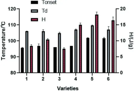
The thermal properties of arachin with different subunit compositions were evaluated by DSC. The Tonset and Td of both two groups are summarized in Fig. 2. The Tonset value was the temperature from which the unfolding of the globular protein structure is initiated. The Td value is the midpoint denaturation temperature. A higher value usually indicates a high thermal stability for globular protein.[25] The Td values of arachin are from 104.95°C to 109.74°C, which are consistent with previous studies, [6,26] and the Tonset value did not vary significantly for both groups of arachin. On the other hand, an interesting feature observed in Fig. 2 is that the Tonset values of arachin without 35.5 kDa subunit were significantly higher (>100°C) than the ones in presence of 35.5 kDa subunit, suggesting that arachin without 35.5 kDa subunit was less heat-sensitive. The enthalpy change (H) of the endothermic peak of arachin without 35.5 kDa was significantly (P < .05) higher than that of arachin with 35.5 kDa. The H reflects the proportion of undenatured protein in a sample, or extent of ordered structure.[27] Thus, this data suggests that the extent of ordered structure in arachin without 35.5 kDa was higher than that in arachin with 35.5 kDa. In other words, arachin without 35.5 kDa has a more stable structure compared with arachin with 35.5 kDa upon temperature increase. This result indicated that the presence or absence of the35.5 kDa subunit did not change the denaturation temperature, but it can influence the unfolding rate of arachin.
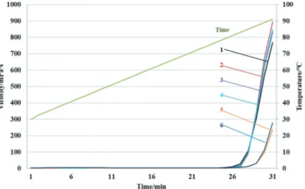
The steady flow behavior of arachin was investigated as shown in Fig. 3. Steady flow curves indicated that viscosity depended on the temperature. It was noticed that the presence or absence of the 35.5 kDa subunit significantly influenced the viscosity of the arachin with temperature increase. As
Figure 2. Thermal parameters of arachin of the two groups. 1, 2, 3, 4 – varieties contain the 35.5 kDa subunit; 5 and 6 – varieties do not contain the 35.5 kDa subunit.
expected, the viscosity only slightly changes before 80°C. However, in a range from 80°C to 90°C, an apparent shear-thick behavior is observed for both arachin groups. These changes in viscosity of protein under heat treatment are approximately in accordance with those previously reported for soybean protein isolates with high concentrates (>6%).[28] It could be explained by the fact that increase in temperature affects the thermal motion of molecules, increases the intermolecular inter-action which is reflected in changes of viscous and viscoelastic behavior of proteins.[29] Upon temperature increase proteins in solution can form aggregates or a protein network via protein interaction before being totally denatured. The rise rate of the viscosity upon temperature increase was significantly higher for arachin with the 35.5 kDa. It suggests that the unfolding of this arachin group occurs faster upon temperature increase than the group without the 35.5 kDa subunit. It may facilitate the formation of aggregate with other subunits, or its complex with the special subunit due to strong hydrophobic interactions between these subunits. These results confirmed that the thermal property of arachin was affected by the difference of its subunit constituent.
Surface hydrophobicity
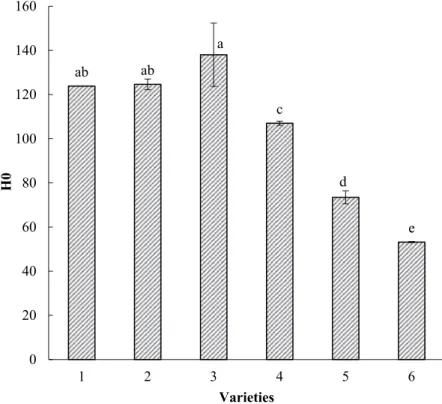
The surface hydrophobicity is one of the important properties of proteins, which is closely related to the functional properties such as emulsifying, foaming abilities, and surface stability. The H0 values of arachin with different subunit compositions are shown in Fig. 4. The values of the group without 35.5 kDa (73.45, 53.17) are significantly lower than the ones with the 35.5 kDa (123.82, 124.61, 138.01, p < .05), indicating that there is a higher number of hydrophobic segments in arachin with 35.5 kDa subunit. The group without 35.5 kDa has probably lower dispensability and a less availability and exposure of hydrophobic groups to the surface.[6] According to Nakai,[30] the surface hydrophobicity is related to the arithmetic mean of the hydrophobic residues composing the subunit. The higher surface hydrophobicity of the 35.5 kDa subunit may be due to a higher proportion of hydrophobic amino acids. Sulfhydryl and S–S contents
Free sulfhydryl groups and S–S bonds have significant influence on the functional properties of food proteins, and can play an important role in the formation of relatively rigid structures in protein conformation.[31] The total and exposed sulfhydryl, disulfide bond of the two arachin groups were summarized in Table 1. The exposed sulfhydryl content of the group containing the 35.5 kDa was
Figure 3. The viscosity of arachin of the two groups. 1, 2, 3, 4 – varieties contain the 35.5 kDa subunit; 5 and 6 – varieties do not contain the 35.5 kDa subunit.
significantly higher than the one without it. The S–S content of the group with 35.5 kDawas less than 1.61 μmol/g, while the one without it was nearly double (2.68 μmol/g) or more (3.55 μmol/g). According to the data, the exposed sulfhydryl was the main part of the total sulfhydryl, suggesting that most cysteine residues exist as free –SH groups instead of S–S bonds. A higher content of disulfide bond indicated a more compact globular structure These results revealed that arachin without 35.5 kDa was more structurally stable than the one with 3535 kDa, which was consistent with the trend observed in the thermal stability study.
Identification of the 35.5 kDa subunit
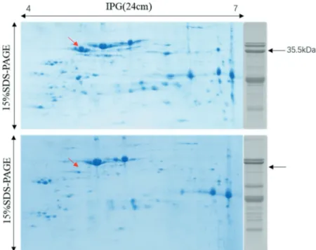
To further identify the 35.5 kDa subunit, we used 2-D gel electrophoresis to separate it at a higher resolution. Representative stained gel is shown in Fig. 5. Comparing the 1-D and 2-D gels, we found
ab ab a c d e 0 20 40 60 80 100 120 140 160 1 2 3 4 5 6 H 0 Varieties
Figure 4. Surface hydrophobicity of arachin of the two groups. 1, 2, 3, 4 – varieties contain the 35.5 kDa subunit; 5 and 6 – varieties do not contain the 35.5 kDa subunit.
Table 1. Sulfhydryl and disulfide contents of two arachin groups. Sulfhydryl content (μmol/g)
Sample Ht Hf S–S (μmol/g) 1 42.14 ± 1.07b 40.88 ± 2.11b 0.63 ± 0.11d 2 43.21 ± 0.31b 39.99 ± 1.08 b 1.61 ± 0.06c 3 45.04 ± 1.01a 44.52 ± 0.97a 0.26 ± 0.17e 4 44.28 ± 0.15ab 42.75 ± 0.11b 0.77 ± 0.01d 5 38.13 ± 1.22c 32.77 ± 0.89c 2.68 ± 0.03b 6 38.37 ± 0.37c 31.28 ± 1.93c 3.55 ± 0.07a
1, 2, 3, 4 – varieties contain the 35.5 kDa subunit; 5 and 6 – varieties do not contain the 35.5 kDa subunit.
Ht: total sulfhydryl contents; Hf: free sulfhydryl contents; S–S: disulfide contents.
the target spot representing the 35.5 kDa subunit pointed by arrow in Fig. 5. The identification of the target peptide sequence was conducted by Q-TOF. The information of the target spot is summarized in Table 2, including the name of the identified protein, the accession number, the theoretical Mw, the sequence coverage, the MOWSE score, and the total acid amino residues. According to the NCBI database, the cDNA of this spot was first partially sequenced (accession number AY618460) by Kang and Gallo, [32] and the full-length cDNA (accession number DQ855115) was later deposited in GenBank by Liang.[33] The sequence was first recognized as an Ara h3-related cDNA (ara h3-im) through genomic Southern blot and reported to display potentially decreased allergenicity due to its lacking in critical residues for IgE-bingding of E4.[32]
Conclusion
In this paper, arachin fraction from six different varieties of peanut was extracted by ammonium sulfate precipitation method. The profiles revealed by SDS-PAGE showed a significant difference between subunit constituents, categorized into two groups: one containing the 35.5 kDa subunit and the other one without this specific subunit. To investigate whether the 35.5 kDa subunit affects the structure and properties of arachin fraction, thermal parameters, surface hydrophobicity, and sulfhy-dryl content were evaluated. The results led to the following conclusions: (1) arachin with the 35.5 kDa was much easier denatured upon temperature (2) arachin without 35.5 kDa subunit was less sensitive to temperature and showed a more compact globular structure. The sequence of 35.5 kDa correspond-ing to an isoform of Ara h3 was also confirmed by 2-D electrophoresis and Q-TOF-MS. Arachin
Figure 5. 2-D gel profiles of arachin in selected varieties. Arachin (80 μg) were separated on a 24 cm IPG (pH 4–7) strip in the first dimension followed by SDS-PAGE on 15% large-format gel in the second dimension. The band on the right was the SDS-PAGE (15%) profiles of the same variety. The red arrows show the target 35.5 kDa subunit in the gel. The upper one is the variety (Baisha No. 1016) of arachin containing 35.5 kDa, the other one is the variety (Shuangji No. 2) of arahcin without 35.5 kDa.
Table 2. Basic information of identified 35.5 kDa subunit by Q-TOF mass spectrometry.
Protein name Accession No. MW(Da)/pI Sequence coverage Score Total length in aa Iso-Ara h3 Q0GM57 58570/5.41 16 1044 512
without 35.5 kDa could be a potential source for processing low allergenic products. Globally, this research can also be useful to peanut cultivar breeding. The presence or absence of a specific subunit in peanut proteins can be key parameter for the development of peanut-based products with specific functionality.
Funding
This work was supported by the National Natural Science Foundation of China [(31801487)]; M. D. thanks the Fund for National Scientific Research in Belgium (FNRS) for her position has Senior Research Associate.
References
[1] Kottapalli, K. R.; Payton, P.; Rakwal, R.; Agrawal, G. K.; Shibato, J.; Burow, M.; Puppala, N. Proteomics Analysis of Mature Seed of Four Peanut Cultivars Using Two-dimensional Gel Electrophoresis Reveals Distinct Differential Expression of Storage, Anti-nutritional, and Allergenic Proteins. Plant Sci. 2008, 175, 321–329. DOI: 10.1016/j.plantsci.2008.05.005.
[2] Maruyama, Y.; Maruyama, N.; Mikami, B.; Utsumi, S. Structure of the Core Region of the Soybean β-conglycinin α′ Subunit. Acta Crystallogr. Sect. D. 2004, 60, 289–297. DOI: 10.1107/S0907444903027367.
[3] Qiang, W.;. Peanuts: Processing Technology and Product Development; Beijing, China: Academic Press, 2016. [4] Freitas, R. L.; Teixeira, A. R.; Ferreira, R. B. Vicilin-type Globulins Follow Distinct Patterns of Degradation in
Different Species of Germinating Legume Seeds. Food Chem. 2007, 102, 323–329. DOI: 10.1016/j. foodchem.2006.05.023.
[5] Chun, J.-Y.; (2002). Vitamin E Content and Stability in Peanuts and Peanut Products during Processing and Storage. PhD Dissertation, University of Georgia, Athens.
[6] Feng, X.-L.; Liu, H.-Z.; Shi, A.-M.; Liu, L.; Wang, Q.; Adhikari, B. Effects of Transglutaminase Catalyzed Crosslinking on Physicochemical Characteristics of Arachin and Conarachin-rich Peanut Protein Fractions.
Food Res. Int. 2014, 62, 84–90. DOI: 10.1016/j.foodres.2014.02.022.
[7] O’kane, F. E.; Happe, R. P.; Vereijken, J. M.; Gruppen, H.; Van Boekel, M. A. Heat-induced Gelation of Pea Legumin: Comparison with Soybean Glycinin. J. Agric. Food Chem. 2004, 52, 5071–5078. DOI: 10.1021/ jf035215h.
[8] Adachi, M.; Takenaka, Y.; Gidamis, A. B.; Mikami, B.; Utsumi, S. Crystal Structure of Soybean Proglycinin A1aB1b Homotrimer. J. Mol. Biol. 2001, 305, 291–305. DOI: 10.1006/jmbi.2000.4310.
[9] Yamada, T.; Aibara, S.; Morita, Y. Isolation and Some Properties of Arachin Subunits. Agric Biol Chem. 1979a, 43, 2563–2568. DOI: 10.1080/00021369.1979.10863848.
[10] Zhao, G.; Liu, Y.; Zhao, M.; Ren, J.; Yang, B. Enzymatic Hydrolysis and Their Effects on Conformational and Functional Properties of Peanut Protein Isolate. Food Chem. 2011, 127, 1438–1443. DOI: 10.1016/j. foodchem.2011.01.046.
[11] Hopkins, C.;. Vibration Transmission between Coupled Plates Using Finite Element Methods and Statistical Energy Analysis. Part 1: Comparison of Measured and Predicted Data for Masonry Walls with and without Apertures. Appl. Acoustics. 2003, 64, 955–973. DOI: 10.1016/S0003-682X(03)00062-8.
[12] Poysa, V.; Woodrow, L.; Yu, K. Effect of Soy Protein Subunit Composition on Tofu Quality. Food Res. Int. 2006,
39, 309–317. DOI: 10.1016/j.foodres.2005.08.003.
[13] Bainy, E. M.; Tosh, S. M.; Corredig, M.; Woodrow, L.; Poysa, V. Protein Subunit Composition Effects on the Thermal Denaturation at Different Stages during the Soy Protein Isolate Processing and Gelation Profiles of Soy Protein Isolates. J. Am. Oil Chem. Soc. 2008, 85, 581–590. DOI: 10.1007/s11746-008-1238-6.
[14] Mohamad Ramlan, B. M. S.; Maruyama, N.; Takahashi, K.; Yagasaki, K.; Higasa, T.; Matsumura, Y.; Utsumi, S. Gelling Properties of Soybean β-conglycinin Having Different Subunit Compositions. Biosci., Biotechnol.,
Biochem. 2004, 68, 1091–1096. DOI: 10.1271/bbb.68.1091.
[15] Fukuda, T.; Prak, K.; Fujioka, M.; Maruyama, N.; Utsumi, S. Physicochemical Properties of Native Adzuki Bean (Vigna Angularis) 7S Globulin and the Molecular Cloning of Its cDNA Isoforms. J. Agric. Food Chem. 2007, 55, 3667–3674. DOI: 10.1021/jf063205l.
[16] Tang, C.-H.; Sun, X. A Comparative Study of Physicochemical and Conformational Properties in Three Vicilins from Phaseolus Legumes: Implications for the Structure–function Relationship. Food Hydrocolloids. 2011, 25, 315–324. DOI: 10.1016/j.foodhyd.2010.06.009.
[17] Govindaraju, K.; Srinivas, H. Studies on the Effects of Enzymatic Hydrolysis on Functional and Physico-chemical Properties of Arachin. LWT - Food Sci. Technol. 2006, 39(1), 54–62. DOI: 10.1016/j.lwt.2004.11.001.
[18] Blanc, F.; Vissers, Y. M.; Adel-Patient, K.; Rigby, N. M.; Mackie, A. R.; Gunning, A. P.; Wellner, N. K.; Skov, P. S.; Przybylski-Nicaise, L.; Ballmer-Weber, B.;; et al. Boiling Peanut Ara H 1 Results in the Formation of Aggregates with Reduced Allergenicity. Mol. Nutr. Food Res. 2011, 55(12), 1887–1894.
[19] Vanga, S. K.; Singh, A.; Raghavan, V. Effect of Thermal and Electric Field Treatment on the Conformation of Ara H 6 Peanut Protein allergen[J]. Innovative Food Sci. Emerg. Technol. 2015, 30, 79–88. DOI: 10.1016/j. ifset.2015.03.003.
[20] Wang, L.; Liu, H.; Liu, L.; Wang, Q.; Li, Q.; Du, Y.; Zhang, J. Protein Contents in Different Peanut Varieties and Their Relationship to Gel Property. Int. J. Food Prop. 2014, 17(7), 1560–1576.
[21] Kella, N. K. D.; Rao, M. N. Effect of Sodium Dodecyl Sulfate on the Heat Denaturation and Aggregation of Arachin at pH 3.6. Int. J. Pept. Protein Res. 1985, 25(3), 308–315.
[22] Yamada, T.; Aibara, S.; Morita, Y. Dissociation-association Behavior of Arachin between Dimeric and Monomeric Forms. Agric Biol Chem. 1979b, 43(12), 2549–2556. DOI: 10.1080/00021369.1979.10863847. [23] Gong, K. J.; Shi, A. M.; Liu, H. Z.; Liu, L.; Hu, H.; Adhikari, B.; Wang, Q. Emulsifying Properties and Structure
Changes of Spray and Freeze-dried Peanut Protein Isolate. J. Food Eng. 2016, 170, 33–40. DOI: 10.1016/j. jfoodeng.2015.09.011.
[24] Shokraii, E. H.; Esen, A.; Mozingo, R. W. Relation of a 36,000-dalton Arachin Subunit to Blanchability in Peanuts (Arachis Hypogaea L.). J. Agric. Food Chem. 1985, 33, 1114–1116. DOI: 10.1021/jf00066a024.
[25] Tang, C.-H.; Sun, X.; Yin, S.-W. Physicochemical, Functional and Structural Properties of Vicilin-rich Protein Isolates from Three Phaseolus Legumes: Effect of Heat Treatment. Food Hydrocolloids. 2009, 23, 1771–1778. DOI:
10.1016/j.foodhyd.2009.03.008.
[26] He, X. H.; Liu, H. Z.; Liu, L.; Zhao, G. L.; Wang, Q.; Chen, Q. L. Effects of High Pressure on the Physicochemical and Functional Properties of Peanut Protein Isolates. Food Hydrocolloids. 2014, 36, 123–129. DOI: 10.1016/j. foodhyd.2013.08.031.
[27] Wang, X. S.; Tang, C. H.; Yang, X. Q.; Gao W. R. Characterization, Amino Acid Composition and in Vitro Digestibility of Hemp (Cannabis Sativa L.) Proteins. Food Chem. 2008, 107(1), 11–18.
[28] Shaojun, T.; Jipeng, L.; Axin, S. Rheological Properties and Viscosity Mathematical Model of Soybean Proteins.
J. Chin. Cereal. Oils Assoc. 2005, 20(2), 53–56.
[29] Barnett, G. V.; Qi, W.; Amin, S.; Neil Lewis, E.; Roberts, C. J. Aggregate Structure, Morphology and the Effect of Aggregation Mechanisms on Viscosity at Elevated Protein Concentrations. Biophys. Chem. 2015, 207, 21–29. DOI: 10.1016/j.bpc.2015.07.002.
[30] Nakai, S.;. Structure-function Relationships of Food Proteins: With an Emphasis on the Importance of Protein Hydrophobicity. J. Agric. Food Chem. 1983, 31, 676–683. DOI: 10.1021/jf00118a001.
[31] Nishinari, K.; Fang, Y.; Guo, S.; Phillips, G. O. Soy Proteins: A Review on Composition, Aggregation and Emulsification. Food Hydrocolloids. 2014, 39, 301–318. DOI: 10.1016/j.foodhyd.2014.01.013.
[32] Kang, I. H.; Gallo, M. Cloning and Characterization of a Novel Peanut Allergen Ara H 3 Isoform Displaying Potentially Decreased Allergenicity. Plant Sci. 2007, 172, 345–353. DOI: 10.1016/j.plantsci.2006.09.014. [33] Liang, X. Q.; Luo, M.; Holbrook, C. C.; GUO, B. Z. Storage Protein Profiles in Spanish and Runner Market Type