HAL Id: tel-01342525
https://tel.archives-ouvertes.fr/tel-01342525
Submitted on 6 Jul 2016HAL is a multi-disciplinary open access
archive for the deposit and dissemination of sci-entific research documents, whether they are pub-lished or not. The documents may come from teaching and research institutions in France or abroad, or from public or private research centers.
L’archive ouverte pluridisciplinaire HAL, est destinée au dépôt et à la diffusion de documents scientifiques de niveau recherche, publiés ou non, émanant des établissements d’enseignement et de recherche français ou étrangers, des laboratoires publics ou privés.
cryptosporidium : de la souris à l’homme
Marwan Osman
To cite this version:
Marwan Osman. Caractérisation génétique et phénotypique de cryptosporidium : de la souris à l’homme. Médecine humaine et pathologie. Université du Droit et de la Santé - Lille II; Univer-sité libanaise, 2015. Français. �NNT : 2015LIL2S068�. �tel-01342525�
Pour l’obtention du grade de Docteur délivré par
L’Université de Lille 2
et
L’Ecole Doctorale Sciences et Technologies
(Université Libanaise)
Spécialité : Parasitologie
Présentée et soutenue publiquement par
OSMAN Marwan
Le 30 septembre 2015
Caractérisation génétique et phénotypique de Cryptosporidium :
de la souris à l’homme
Membres du Jury
Président du Jury : Dr. Eric VISCOGLIOSI (Centre d'Infection et d'Immunité de Lille, IPL, Lille, France) Directeurs de thèse : Dr. Gabriela CERTAD (Centre d'Infection et d'Immunité de Lille, IPL, Lille, France)
Pr. Fouad DABBOUSSI (Université Libanaise, Tripoli, Liban) Rapporteurs : Pr. Loïc FAVENNEC (Université de Rouen, Rouen, France)
Pr. Ghassan MATTAR (American University of Beirut, Beyrouth, Liban) Examinateurs : Pr. Sima TOKAJIAN (Lebanese American University, Byblos, Liban)
Pr. Sebastien MONCHY (Université du Littoral Côte d’Opale, Wimereux, France)
Pr. Nabil HADDAD (Université Libanaise, Beyrouth, Liban) Professeur invité : Pr. Monzer HAMZE (Université Libanaise, Tripoli, Liban)
La thèse n’a pas été seulement un projet de recherche, elle a été également un projet de vie. Ce projet m’a donné la possibilité non seulement de parfaire ma formation scientifique mais aussi de bénéficier d’un riche échange culturel ce qui m’a fait grandir. Je serai toujours reconnaissant à toutes les personnes ici citées qui ont contribuées à l’enrichissement de ce projet, d’un point de vue professionnel, mais aussi personnel.
Je tiens tout d’abord à remercier ma directrice de thèse en France, Dr. Gabriela Certad, avec qui j’ai eu l’énorme plaisir de travailler pendant ces trois années de thèse. Je la remercie pour la confiance qu’elle m’a accordée, ses remarques et ses conseils judicieux. Je la remercie pour le temps qu’elle a consacré à diriger ce projet et à m’aider à rédiger les articles et la thèse. Je lui suis infiniment reconnaissant pour sa grande disponibilité et ses qualités humaines d'écoute et de compréhension tout au long de cette thèse.
Je remercie également mon directeur de thèse au Liban Pr. Fouad Dabboussi pour son humanité, son dynamisme et ses connaissances scientifiques qui ont permis d’enrichir cette thèse. Je te serai toujours reconnaissant pour ton soutient pendant les moments difficiles de la thèse.
J’adresse mes remerciements aux Pr. Sébastien Monchy, Pr. Loïc Favennec, Pr.
Ghassan Matar, Pr. Nabil Haddad et Pr. Sima Tokajian d’avoir accepté de participer à
mon jury de thèse et de juger ce travail en tant que rapporteurs et examinateurs. Je leur suis très reconnaissant pour leurs judicieuses suggestions.
Je tiens à remercier le directeur de l’Ecole Doctorale de l’Université Libanaise, le Pr.
Fawaz El Omar, le directeur de l’Ecole Doctorale Biologie Santé de Lille, le Pr. Bernard Sablonnière et le directeur du Centre AZM pour la Recherche en Biotechnologie et ses
Applications (EDST-UL) au Liban, le Pr. Mohamad Khalil pour leurs disponibilités.
Je tiens à remercier le Pr. Monzer Hamze pour ses qualités humaines qui m’ont aidé à me sentir intégré et à trouver ma place de doctorant. Ses connaissances infinies de la bibliographie en microbiologie ainsi que son ouverture d’esprit ont fait de cet
encadrement un véritable soutien. Merci pour ces conversations d’égal à égal, merci pour votre compréhension lors des difficultés que chacun peut rencontrer au cours de sa vie de doctorant. J’ai beaucoup appris grâce à vous.
Je tiens à remercier le Dr. Eric Viscogliosi que j’ai vu pour la première fois le 26 avril à Tripoli. J’ai été très rapidement touché par sa gentillesse, impression qui n’a fait que se confirmer lors de notre rencontre en France. Définitivement, merci beaucoup pour toute ta gentillesse, pour tes conseils, pour tes enseignements, pour ton aide pendant la rédaction des articles et de cette thèse et pour ton amitié.
Je tiens à remercier fortement le Dr. Sadia Benamrouz, avec qui j’ai travaillé tout au long de la thèse. Je la remercie pour sa pédagogie, pour sa patience, pour m’avoir fait réfléchir, pour toutes les informations scientifiques qui m’ont beaucoup apporté au cours de cette thèse et pour m’avoir aidé à bien comprendre mon projet de doctorat. Je te serai toujours infiniment reconnaissant pour tes efforts et pour ton soutient pendant les moments difficiles de la thèse.
Je tiens à remercier le Dr. Khaled El Omari, que j’ai vu pour la première fois le Mai 2014 à Lille. Je vous remercie pour votre amitié, vos encouragements, votre soutien affectif sans faille et vos multiples conseils toujours très pertinents qui m'ont permis de faire cette thèse dans de bonnes conditions.
Je tiens à remercier le Dr. Emilie Fréalle. Je te remercie pour ta gentillesse et la confiance que tu m’as témoignée. Je t’assure de mon respect et de ma profonde reconnaissance.
Je tiens à remercier le Pr. El Moukhtar Aliouat pour vos conseils et votre soutient durant les trois années de la thèse, je vous en serai toujours reconnaissant.
Je tiens à remercier Madame Karine Guyot pour m’avoir appris beaucoup des choses et pour son aide précieuse pendant ma thèse, surtout pour la partie Crypto-Cancer.
Je tiens à remercier le Pr. Michel Simonet, le Dr. Olivier Gaillot, le Pr. Françoise
Norel-Bozouklian et le Pr. Christophe Beloin pour la formation scientifique de qualité
qu’ils m’ont donnée. Leur grande rigueur scientifique et leur passion pour la science m’ont beaucoup appris.
Je tiens à remercier tous les autres membres de l’équipe du laboratoire BDPEE pour leur sollicitude et pour la bonne ambiance : Dr. Laurence Delhaes, Dr. Magali Chabé,
Nausicaa Gantois, Dr. Cécile Marie Aliouat, Dr. Annie Vitse et Muriel Poittier.
Je tiens à remercier également tous les membres du laboratoire Microbiologie, Environnement et Santé au centre AZM qui ont toujours été disponible pour m’aider. Un Grand Merci à Taha Abdo, Mariam Yehya, Majd Mzawak, Iman Darwich, Sara
Amrieh, Farah Charrouf Obeid et Houssam Khaled.
Je tiens à remercier Monsieur Jean-Jacques Hauser, Madame Laurence Fofana et
Monsieur François Delcroix pour votre disponibilité mais également pour avoir
toujours été à l’écoute de mes problèmes administratifs.
Je remercie également mes amis doctorants, chercheurs et techniciens, que j’ai eu le grand plaisir de rencontrer au laboratoire ou aux autres instituts de recherche, pour les discussions scientifiques et/ou pour les agréables moments que nous avons partagés ensemble : Dr. Frederic Delbac, Dr. Hicham El Alaoui, Dr. Philippe Poirier, Dr. Ivan
Wawrzyniak, Dr. Nawaf Jurdi, Dr. Albert Aoun, Pr. Colette Creusy, Pr. Pierre Gosset, Dr. Pilar Viscogliosi, Dr. Dionigia Meloni, Dr. Dima El Safadi, Baptiste Delaire, Amandine Cian, Laurine Vandervield, Yuwalee Seesao, Linh Nguyen, Dr. Sara Khalife et Clara Khairallah. Je ne vous oublierai jamais!
A mon frère, Ahmad Kashef, je te remercie pour ton soutien depuis ma licence. Je suis chanceux d’avoir un ami comme toi.
Je remercie aussi mes amis qui m’ont motivé durant mon doctorat : Khalil Haydar,
Raghdan Abdallah, Mohamad Kanjo, Adnan Awad, Ali Kahwaji, Mohamad Salma, Dr. Ahmad Kamaleddine, Ahmad Atrouni, Mohamad Diab, Dr. Imad Al Kassaa, Dr. Bashar Ismaiil, Dr. Rayane Rafei, Dr. Michel Wehbe, Dr. Charbel El Bayssari, Marianne Ecco, Maher Masri, Oussama Bakir, Josephine Chaar, Oubayda Saiid, Mohamad Ammoun, Hassan Al Sahmarani et Dr. Thomas Vanneste.
Cette thèse n’aurait pas pu se faire sans le soutien financier du Conseil National de la
Recherche Scientifique CNRS Libanais et de l’association AZM et SAADE. Je
remercie l’association LASeR, la chambre de commerce, d’industrie et
d’agriculture du Nord de Liban et la société SFM pour le soutien financier de certains
congrès.
Enfin, je tiens à remercier ma famille, mon père Bassam Osman, ma mère Dalal
Naboulsi et ma belle Halima El Cheikh, avec qui j’ai initié ce projet de vie. Nous avons
rêvé tous les jours ensemble. Je les remercie de m’avoir encouragé dans les moments difficiles et pour toutes les expériences merveilleuses que nous avons vécues pendant ces trois ans.
Merci aussi d’avoir écouté mes présentations, de les avoir critiqué, et aussi financé. Je vous aime! Et je vous dédie cette thèse.
Mon travail de thèse a été valorisé pour le moment par les articles suivants :
Osman M, El Safadi D, Benamrouz S, Guyot K, Dei-Cas E, Aliouat el M, Creusy C, Mallat H, Hamze M, Dabboussi F, Viscogliosi E, Certad G. Initial data on the
molecular epidemiology of cryptosporidiosis in Lebanon. PLoS One. 2015
7;10(5):e0125129. doi: 10.1371/journal.
Fréalle E, El Safadi D, Cian A, Aubry E, Certad G, Osman M, Wacrenier A, Dutoit E, Creusy C, Dubos F, Viscogliosi E. Acute blastocystis-associated appendicular
peritonitis in a child, Casablanca, Morocco. Emerg Infect Dis. 2015;21(1):91-4.
Benamrouz S, Conseil V, Chabé M, Praet M, Audebert C, Blervaque R, Guyot K, Gazzola S, Mouray A, Chassat T, Delaire B, Goetinck N, Gantois N, Osman M, Slomianny C, Dehennaut V, Lefebvre T, Viscogliosi E, Cuvelier C, Dei-Cas E, Creusy C, Certad G. Cryptosporidium parvum-induced ileo-caecal adenocarcinoma
and Wnt signaling in a mouse model. Dis Model Mech. 2014;7(6):693-700.
El Safadi D, Meloni D, Poirier P, Osman M, Cian A, Gaayeb L, Wawrzyniak I, Delbac F, El Alaoui H, Delhaes L, Dei-Cas E, Mallat H, Dabboussi F, Hamze M, Viscogliosi E. Molecular epidemiology of Blastocystis in Lebanon and correlation
between subtype 1 and gastrointestinal symptoms. Am J Trop Med Hyg.
2013;88(6):1203-6.
Wawrzyniak I, Courtine D, Osman M, Hubans-Pierlot C, Cian A, Nourrisson C, Chabe M, Poirier P, Bart A, Polonais V, Delgado-Viscogliosi P, El Alaoui H, Belkorchia A, Van Gool T, Tan K, Ferreira S, Viscogliosi E, Delbac F (2015).
Genome sequence of the intestinal parasite Blastocystis subtype 4-isolate WR1.
Genomics Data (4), 22–23.
Certad G, Dupouy-Camet J, Gantois N, Hammouma-Ghelboun O, Pottier M, Guyot K, Benamrouz S, Osman M, Delaire B, Creusy C, Viscogliosi E, Dei-Cas, Aliouat-Denis CM, Follet J. (2015) Identification of Cryptosporidium species in fish from
Lake Geneva (Lac Léman) in France. Plos One (sous presse).
A cette liste de publications s’ajoute un chapitre de livre sous presse :
A) Certad G, Osman M, Benamrouz S. (2015) Pathogenesis of Cryptosporidium in
humans. In: Emerging and Re-Emerging Human Infections. Edited by John Wiley
Les articles suivants ont été soumis :
Osman M†, Bories J†, El Safadi D†, Poirel M, Gantois N, Benamrouz-Vanneste S, Delhaes L, Hugonnard M, Certad G, Zenner L, Viscogliosi E. Prevalence and
genetic diversity of the intestinal parasites Blastocystis sp. and Cryptosporidium spp. in household dogs in France and evaluation of zoonotic transmission risk.
Article soumis à Veterinary Parasitology (en révision).
Osman M, El Safadi D, Benamrouz S, Nourrisson C, Poirier P, Pereira B, Pinon A, Lambert C, Wawrzyniak I, Dabboussi F, Delbac F, Hamze M, Viscogliosi Eric, Certad G. Prevalence and Risk Factors for Intestinal Protozoan Infections with
Cryptosporidium, Giardia, Blastocystis and Dientamoeba among Schoolchildren in Tripoli, Lebanon. Article soumis à Plos Neglected Tropical Diseases (en révision).
Trois autres articles et une revue sont également en cours de finalisation pour une prochaine soumission.
Mon travail de thèse a été valorisé pour l’instant par les communications suivantes dans des congrès nationaux et internationaux :
1) Osman et al, 2015. 11ème Congrès National de la Société Française de Microbiologie, Paris, France. (23-25 March 2015). New insights into the molecular
epidemiology and transmission dynamics of Cryptosporidium spp. and Blastocystis sp. in North Lebanon.
2) Osman et al, 2014. 4ème Forum Doctoral-EDST-UL, Beyrouth, Liban (27 Novembre 2014). Prevalence, genetic diversity and risk factors of Cryptosporidium and Giardia infections among school children in Lebanon.
3) Osman et al, 2014. 9th Dubai International Food Safety Conference, Dubai, UAE. (9-11 Novembre 2014). Identification of Cryptosporidium species in fresh water and marine fish in France.
4) Osman et al, 2014. 9th Dubai International Food Safety Conference, Dubai, UAE. (9-11 Novembre 2014). New insights into the molecular epidemiology and transmission dynamics of Cryptosporidium spp. and Blastocystis sp. in North Lebanon.
5) Osman et al, 2014. 14ème journée André Verbert, Lille, France. (11 Septembre
2014). Prévalence de Giardia duodenalis et Cryptosporidium spp. et facteurs de risque chez des écoliers à Tripoli
6) Osman et al, 2014. 5th International Giardia & Cryptosporidium Conference, Uppsala, Suède. (27-30 Mai 2014). Prevalence, genetic diversity and risk factors of Cryptosporidium and Giardia infections among schoolchildren in Lebanon.
7) Osman et al, 2014. Congrès de la Société Française de Parasitologie et de
Mycologie Médicale, Reims, France. (21-24 May 2014). Epidémiologie moléculaire
de la cryptosporidiose au Liban.
8) Osman et al, 2014. 20th LAAS International Science Conference, Beyrouth, Liban. (27-29 March 2014). Epidémiologie moléculaire de la cryptosporidiose au Liban.
9) Osman et al, 2013. 1ères Journées Franco-Maghrébines de Parasitologie et Mycologie, Rabat, Maroc. (23-26 Octobre 2013). Epidémiologie moléculaire de la
10) Osman et al, 2013. 3ème Forum Doctoral-EDST-UL, Beyrouth, Liban (25 Juin 2013). Caractérisation génétique et phénotypique de Cryptosporidium: de la souris à l’homme.
A cette liste de communications s’ajoute des participations à d’autres congrès :
A) The international standards and regulations for food safety (Bioteck Industry, Dbayeh, Liban (28 Mai 2015).
B) 9th Arab Conference for Antimicrobial Agents, ArAPUA, Beyrouth, Liban (15-17 Novembre 2012).
A ces deux listes on ajoute la contribution dans l’organisation du séminaire « l’Antibiogramme aux Normes Européennes », Université Libanaise, 20 et 21
Durant ma thèse, j’ai eu la chance de participer aux formations suivantes :
1) Diplôme Universitaire en Microbiologie générale – Université Paris Diderot et Institut Pasteur de Paris. Paris, France (1 Septembre – 30 Octobre 2014).
2) Diplôme en Résistance bactérienne aux antibiotiques – Institut Pasteur de Paris.
Paris, France. (23 Février – 28 Février et 1 Juin – 5 Juin 2015).
3) Stage à mi-temps dans le service de Bactériologie – Hygiène de CHRU de Lille.
Lille, France. (3 – 20 Mars 2015).
4) Bio-informatique – Clermont Université, Université Blaise Pascal-Université d’Auvergne-CNRS, UMR 6023 Laboratoire Microorganismes : Génome et Environnement, Clermont-Ferrand, France. (9 – 13 Juin 2014).
5) Notions fondamentales en statistiques incluant spécificité petits échantillons – Université Lille 2 Droit et Santé. Lille, France. (12 – 15 Mai 2014).
6) Doctoriales 2014 – Université Lille Nord de France. Lille, France. (6 – 11 Avril
2014).
7) Expérimentation animale niveau II – Institut Pasteur de Lille. Lille, France. (16 –
Liste des abréviations………...15
I. Résumé………..17
II. Abstract………...19
III. Introduction………..21
IV. Généralités………25
1. Pathogenesis of Cryptosporidium in humans ………26
1. Pathogen………..26
1.1. History………..26
1.2. Life cycle………..27
1.3. Cryptosporidium species………...31
1.4. Genomics of Cryptosporidium species……….34
2. Epidemiology………...37
3. Clinical features………...41
3.1. Clinical manifestations associated to infecting Cryptosporidium species…....43
3.2. Cryptosporidium and cancer (some clinical evidences)………...44
4. Pathogenesis and Immunity……….45
4.1. Adherence to and invasion of epithelial host cells………...46
4.2. Epithelial cellular processes initiated by Cryptosporidium infection………..48
4.3. Cryptosporidium and cancer (some experimental evidences)……….51
5. Diagnosis………53
5.1. Staining methods……….53
5.2. Immunological methods………..54
5.3. Molecular tools………57
6. Treatment………60
7. Control and prevention………...61
8. References………..63
2. Epidémiologie moléculaire de la cryptosporidiose au Moyen Orient…..…………80
3. Cryptosporidium et cancer………97
V. Objectifs et Stratégies………...104
VI. Résultats……….107
1. Axe 1 : Premières données d’épidémiologie moléculaire et facteurs de risque liés à l’infection par Cryptosporidium spp. au Liban………...108
1. Article 1 : Initial data on the molecular epidemiology of cryptosporidiosis in Lebanon………...109
2. Article 2 : Prevalence and Risk Factors for Intestinal Protozoan Infections with Cryptosporidium, Giardia, Blastocystis and Dientamoeba among Schoolchildren in
Tripoli, Lebanon……….………124
2. Axe 2 : Etude de la prévalence de Cryptosporidium spp. dans les échantillons animaux et l’évaluation du pouvoir zoonotique du parasite………..157
1. Article 3 : Cryptosporidiosis in humans and cattle in a rural area of Northern Lebanon………..158
2. Article 4 : Prevalence and genetic diversity of the intestinal parasites Blastocystis sp. and Cryptosporidium spp. in household dogs in France and evaluation of zoonotic transmission risk……….171
3. Prévalence et caractérisation moléculaire de Cryptosporidium chez plusieurs groupes d’animaux des parcs zoologiques français………..193
4. Article 5 : Identification of Cryptosporidium species in fish from Lake Geneva (Lac Léman) in France………210
3. Axe 3 : Etude de la pathogénicité de Cryptosporidium spp.……...………..235
1. Article 6 : High Association of Cryptosporidium infection with Digestive Cancer in Lebanese patients………236
2. Article 7 : Cryptosporidium parvum-induced ileo-caecal adenocarcinoma and WNT signaling in a rodent model……….255
VII. Discussion………...…285
VIII. Conclusions et Perspectives………..301
IX. Annexe………305
1. Travaux présentés aux congrès scientifiques……….…….……..306
2. Autres articles publiés dans des journaux internationaux………….………..……...320
1. Article A : Short report: Molecular epidemiology of Blastocystis in Lebanon and correlation between subtype 1 and gastrointestinal symptoms………...320
2. Article B : Acute Blastocystis-Associated Appendicular Peritonitis in a Child, Casablanca, Morocco………..……331
3. Article C : Draft genome sequence of the intestinal parasite Blastocystis subtype 4-isolate WR1……….339
ADN Acide Desoxyribonucléique
AEECL Association Européenne pour l'Étude et la Conservation des Lémuriens
ANOFEL Association française des enseignants et praticiens hospitaliers titulaires de parasitologie et mycologie médicale
APC Adenomatous Polyposis Coli
ARNr 18S Acide Ribonucléique de la petite sous unité ribosomique ARNt ARN de transfert
ATP Adénosine triphosphate CCR Cancer colorectal
C. hominis Cryptosporidium hominis C. parvum Cryptosporidium parvum C. muris Cryptosporidium muris
DAPI 4',6'‐diamidino‐2‐phénylindole EBV Epstein barr virus
ELISA Enzyme Linked ImmunoSorbent Assay
HAART Highly active antiretroviral therapy ; Traitement antirétroviral hautement actif HSP Heat shock protein
IARC International Agency for Research on Cancer
IBS Irritable Bowel Syndrome ou Syndrome du Côlon Irritable Ig Immunoglobuline
IL Interleukine
IMDM Iscove’s modified Dulbecco’s medium ITS Internal Transcribed Spacer
kb Kilobase pb Paires de bases
MLO Mitochondrion-Like Organelle MET: Microscopie électronique à transmission NFκB : Facteur nucléaire κB
OMS Organisation Mondiale de la Santé PI3K Phosphatidyl inositol 3‐ kinase PBS Phosphate‐buffered saline PCR Polymerase chain reaction
SCID‐D Severe combined immunodepression (avec traitement à la dexaméthasone) SIDA Syndrome de l’immunodéficience acquise
PI-IBS IBS post-infectieux
RFLP Restriction Fragment Length Polymorphism TRAP Thrombospondin‐related adhesive protein VHB Virus de l’hépatite B
VHC Virus de l’hépatite C
VIH Virus de l’immunodéficience humaine sp. Espèce
ssrRNA Small subunit ribosomal RNA ST Sous-type
Les parasites du genre Cryptosporidium comprennent des espèces infectant le tractus gastro-intestinal ou respiratoire d’un grand nombre de vertébrés y compris l'homme. Ces protistes intracellulaires sont les agents d’une zoonose cosmopolite à transmission oro-fécale, la cryptosporidiose. Au vu des travaux réalisés dans notre laboratoire, nous savons à présent que Cryptosporidium parvum est également capable d’induire des néoplasies digestives chez un modèle murin SCID (Severe Combined Immunodeficiency mice), traitées ou pas par la dexaméthasone. Alors que C. muris, une autre espèce de Cryptosporidium, induit une infection chronique non associée à des transformations néoplasiques.
Pour toutes ces raisons, il nous est apparu intéressant d’effectuer un travail de thèse articulé autour de trois axes principaux, l’épidémiologie, la transmission et la pathogénicité du parasite Cryptosporidium. Nous nous sommes intéressés dans un premier temps à l’épidémiologie moléculaire et la biodiversité génétique de Cryptosporidium dans des populations humaines de la région du Nord-Liban. Ceci nous a permis de mettre en évidence une prévalence de 5% de Cryptosporidium chez la population générale avec une prédominance de C. hominis. Ce qui constituait les premières données épidémiologiques de la cryptosporidiose au Liban. Ensuite d’autres études nous ont permis de montrer que cette prévalence pouvait atteindre même 10% chez les patients symptomatiques et les enfants. Dans un second temps, nous avons voulu étudier le mode de transmission du parasite et les facteurs de risque pouvant y être associés. Pour ce faire, une recherche du parasite a été réalisée aussi bien au Liban qu’en France chez des animaux d’élevage, sauvages, de compagnie et en captivité. Une première étude a été réalisée chez des patients et des bovins du Nord-Liban. L’ensemble des données rapportées nous permettent de suggérer un mode de transmission de la cryptosporidiose majoritairement anthroponotique au Liban, mais les résultats du génotypage ne permettent pas d’exclure la présence d’une transmission zoonotique. D’autres études réalisées en France, notamment sur des échantillons de selles collectées auprès des zoos de la Palmyre (à Royan, Charente-Maritime) et de Lille (Nord de la France) ont montré un taux de prévalence de Cryptosporidium spp inférieur à 1%. Ces animaux ne semblent donc pas être un réservoir potentiel de cette infection. Alors que chez les poissons sauvages du Lac Léman (France), nous avons pu identifier la présence entre autre de l’espèce zoonotique C. parvum dans l’estomac et l’intestin des poissons. Ceci nous permet de considérer les poissons comme étant une source de contamination potentiel pour l’homme, l’animal mais également pour l’environnement.
Enfin le troisième axe avait pour but d’étudier la pathogénicité de ce parasite. Pour commencer nous avons voulu étudier l’association entre la pathologie cancéreuse et le parasitisme par Cryptosporidium chez l’homme. Une recherche du parasite a donc été réalisée dans des biopsies coliques et gastriques inclues en paraffine appartenant à des patients atteints ou non de cancers digestifs. Une différence significative a été rapportée entre la prévalence de la cryptosporidiose retrouvée chez la population de patients présentant des lésions cancéreuses (17%) et celle du groupe control constitué de patients non cancéreux mais présentant des symptômes (7%) p-value = 0.03. Ensuite nous avons voulu explorer les mécanismes de la cancérogénèse induite par la souche IOWA de C. parvum au niveau de la région iléocæcale des souris SCID traitées par la dexaméthasone. Pour ce faire nous nous sommes intéressés à quatre marqueurs de voies de signalisation cellulaires impliquées dans la survenue de cancers colorectaux (APC, Bêta-caténine, P53 et K‐ras). Nous avons ainsi pu montrer que la voie Wnt était impliquée dans ce processus. L’ensemble de ces données obtenues chez l’animal et chez l’homme montre que ce parasite a un impact important en santé humaine et animale.
Parasites of the genus Cryptosporidium comprise species infecting the gastrointestinal or respiratory tract of a wide variety of vertebrates including humans. These intracellular protists are the agents of a cosmopolitan zoonosis, with féco-oral transmission, cryptosporidiosis. Recent work from our laboratory, showed that the zoonotic species Cryptosporidium parvum is capable to induce digestive neoplasia in a SCID Severe Combined Immunodeficiency Mice (SCID) model, treated or not with dexamethasone. However C. muris, another species of Cryptosporidium, induces chronic infection in this rodent model but is not associated with neoplastic transformation.
For all these reasons, it seemed interesting to carry out a thesis project articulated around three different axes: epidemiology, transmission and pathogenesis of the Cryptosporidium infection. We focused initially on the molecular epidemiology and genetic biodiversity of this parasite among human populations in North Lebanon. We found a Cryptosporidium prevalence of 5% among the general population, being C. hominis the predominant species. This prevalence could reach until 10% in symptomatic patients and children. This is the first epidemiological data about cryptosporidiosis in this country.
Secondly, we studied the transmission routes and the main risk factors associated with the transmission of this parasite. To do this, a first study was conducted in parallel among animal populations in North Lebanon and France. The reported data suggest a predominance of an anthroponotic route of transmission for cryptosporidiosis in Lebanon, but the results of genotyping does not exclude the presence of zoonotic transmission. Other studies conducted in France, especially based on collection of stool samples in the zoos of Palmyre (Royan, Charente-Maritime) and Lille showed that Cryptosporidium spp were present in less than 1% of captivity animals. The low prevalence strongly demonstrates that these animals play a negligible role as potential reservoirs of infection. While in wild fish, we could identify the presence of C. parvum, a zoonotic species, in the stomach and the gut of fish. These data suggest that the fish could be a natural host of C. parvum and a potential source of contamination for humans, animals but also for the environment.
The third topic aimed to study the pathogenicity of this parasite. Firstly, we wanted to study the association between digestive cancer and parasitism with Cryptosporidium in humans. Cryptosporidium molecular detection was therefore carried out in colonic and gastric biopsies belonging to patients with and without digestive cancers of recent diagnosis collected in North
Lebanon. A statistically significant difference was observed between the prevalence of cryptosporidiosis found among the population of patients with digestive cancer (17%) and that of the control group consisting of non-cancer patients but with digestive symptoms (7%) (p-value = 0.03). All these data obtained in animals and humans strengthens the importance of this parasite in public health. Finally, we explored metabolic pathways potentially involved in the development of C. parvum-induced ileo-caecal oncogenesis in the SCID model treated with dexamethasone. We searched for alterations in genes or proteins commonly involved in cell cycle, differentiation or cell migration, such as β-catenin, Apc, E-cadherin, Kras and p53. We were able to show that the Wnt pathway was involved in this process.
La cryptosporidiose est une zoonose opportuniste cosmopolite causée par diverses espèces appartenant au genre Cryptosporidium. Un Apicomplexa qui se multiplie dans les cellules épithéliales du tractus gastro-intestinal et du système respiratoire d’un grand nombre de vertébrés, y compris l’homme (Chalmers and Katzer, 2013; Ryan, Fayer, and Xiao, 2014). Les oocystes sont la forme de résistance et de dissémination du parasite. Le mode de transmission de cette infection est oro-fécal, soit indirectement par ingestion d’oocystes contaminants l’eau ou les aliments soit par contact direct avec un sujet infecté. Ce dernier peut être un homme ou un animal, la maladie peut donc se répandre par la voie anthroponotique ou zoonotique (Chalmers and Katzer, 2013; Ryan, Fayer, and Xiao, 2014). Actuellement, plus d’une vingtaine d’espèces ont été validés pour le genre Cryptosporidium. Les espèces qui parasitent le plus fréquemment l’homme sont Cryptosporidium parvum (C. parvum) et C. hominis, même si des infections par C. meleagridis, C. cuniculus, C. andersoni, C. felis, et C. canis ont été rapportées mais le plus souvent chez des patients à risques (Chalmers and Katzer, 2013; Xiao, 2010).
Cette parasitose émergente a un impact considérable chez le patient immunodéprimé, chez qui elle peut devenir chronique voire létale. L’infection est toutefois possible chez les sujets immunocompétents, de façon sporadique ou associée à des épidémies, mais cela se traduit par des diarrhées auto-résolutives, généralement sans complications. Cryptosporidium est à l’origine de plus de 165 épidémies d’origine hydrique (Chalmers, 2012). La plus importante fut celle de Milwaukee (Wisconsin, États-Unis) en 1993, qui contamina près de 403 000 personnes via le réseau de distribution d’eau (Mac Kenzie et al., 1994). De plus, des données récentes ont montré que Cryptosporidium est parmi les principales causes de diarrhée infantile modérée à sévère chez les enfants âgés de moins de 2 ans dans les pays en développement (Kotloff et al., 2013; Striepen, 2013). De même, selon l’Organisation Mondiale de la Santé (OMS), la cryptosporidiose est inclue dans la liste des maladies négligées qui constituent un frein au développement socio-économique dans les pays en voie de développement et pour lesquelles des études de terrain sont indispensables à leur compréhension et à l’établissement de conduites permettant leur contrôle (Savioli, Smith, and Thompson, 2006). Cette maladie est un véritable problème de santé publique.
Le Liban, à l’instar de la plupart des pays en développement, est très touché par les parasitoses intestinales (Hamze et al., 2004; Hamze, Naja, and Mallat, 2008). Malgré la virulence de ce protiste intracellulaire et son impact socio-économique et en santé publique
non négligeable, aucune information n’était disponible concernant la situation de la cryptosporidiose dans ce pays avant ce travail de thèse. Ces informations sont indispensables afin que ce pays puisse définir des stratégies de prévention, de contrôle et de lutte contre cette parasitose. La France, par contre, semble peu touchée par la cryptosporidiose (ANOFEL, 2010). Cependant, peu d’études ont été réalisées sur des populations animales autres que les animaux d’élevages (Follet et al., 2011; Rieux et al., 2013; Rieux et al., 2014).
D’autre part, l’implication de C. parvum dans l’induction de néoplasies digestives a été démontrée en 2007 dans le laboratoire Biologie et Diversité des Pathogènes Eucaryotes Emergents (BDPEE) chez un modèle murin d’infection chronique (i.e. le modèle souris SCID traité ou non à la Dexaméthasone) (Certad et al., 2007). Il a ainsi pu être mis en évidence que l'infection par C. parvum induisait chez ces souris, le développement d'adénocarcinomes digestifs invasifs (Benamrouz et al., 2012a; Benamrouz et al., 2012b; Certad et al., 2012; Certad et al., 2010a; Certad et al., 2010b). Cependant, C. muris, une autre espèce de Cryptosporidium, n'a pas induit ce type de transformation épithéliale et les raisons de ces différences dans l’expression de la maladie restent inconnues (Certad et al, 2007).
L’ensemble de ces données expérimentales chez l’animal ainsi que d’autres données clinico-épidémiologiques chez l’homme suggère fortement que Cryptosporidium pourrait avoir vraisemblablement un pouvoir carcinogène qui s'exprime dans le tractus gastro-intestinal. Avant de présenter en détail les différents résultats obtenu durant ma thèse, je commencerai tout d’abord par faire un état de l’art de ce que l’on sait sur Cryptosporidium, son épidémiologie et enfin sa pathogénicité. Ce chapitre, nommé « Généralités », bien que non exhaustif permettra :
- Dans un premier temps, de donner une vue d’ensemble de ce qu’est Cryptosporidium et son impact en santé humaine. Pour ce faire, j’ai intégré à cette partie un chapitre de livre intitulé « Pathogenesis of Cryptosporidium in Humans » que j’ai rédigé avec d’autres membres de mon laboratoire d’accueil Français. Ce dernier aborde notamment, la biologie du parasite, les manifestations cliniques et la réaction immunitaire qu’il induit chez son hôte, ses facteurs de virulences, les outils de diagnostiques ainsi que les traitements dont nous disposons à l’heure actuelle pour lutter contre cette maladie.
- Dans un second temps, de dresser un état des lieux des connaissances actuelles concernant l’épidémiologie de Cryptosporidium. En effet, l’étude de l’épidémiologie du parasite est nécessaire à une meilleure connaissance de sa diversité génétique et à une meilleure compréhension de son mode de transmission et ainsi à l’établissement de moyens de prévention. C’est pourquoi, j’ai intégré au manuscrit, un travail bibliographique qui rassemble les données publiées sur l’épidémiologie de Cryptosporidium dans divers pays du globe et plus particulièrement au Moyen Orient.
- Et enfin, dans un troisième temps, cet état de l’art me permet de revenir sur une découverte faite dans mon laboratoire d’accueil Français, qui a permis de mettre en évidence pour la première fois que l’espèce C. parvum était dotée d’un pouvoir carcinogène.
1. Pathogenesis of Cryptosporidium in humans
Certad G, Osman M, Benamrouz S
(Chapitre de livre accepté pour publication in: Emerging and Re-Emerging Human Infections. Edited by John Wiley & Sons/Wiley Blackwell Press (sous press))
1. Pathogen 1.1. History
The genus Cryptosporidium is a member of the phylum Apicomplexa. It is composed of protozoan parasites that infect epithelial cells in the microvillus border of the gastrointestinal tract of all classes of vertebrates, including humans. Cryptosporidium was described for the first time by Ernest Tyzzer in 1907 who isolated the parasite from the gastric mucosa of mice (Mus musculus) and named Cryptosporidium muris (C. muris) (Tyzzer, 1910). In 1912, E. Tyzzer described again this parasite but this time in the small intestine of mice. Smaller in size than the parasite described in the stomach, it looked like a different species that was named Cryptosporidium parvum (C. parvum) (Tyzzer, 1912). It was not until the 1970s that first cases of human cryptosporidiosis in patients with severe watery diarrhea were reported (Nime et al., 1976). However, it is only in the early 1980’s that clinical significance and pathogenicity of this parasite were recognized, especially due to a high morbidity and mortality found in immunocompromised patients (Tzipori and Widmer, 2008). In particular, at this time cryptosporidiosis emerged as a frequent cause of diarrhea in HIV/AIDS patients, and the link with AIDS was so strong that the infection became one of the defining features of the syndrome (Colford et al., 1996; Mwachari et al., 1998).
Cryptosporidium has been also considered as the infectious agent responsible of more than 60% of waterborne outbreaks (Baldursson and Karanis, 2011). The best known example of a waterborne outbreak caused by this parasite was described in the spring of 1993 in Milwaukee (United States of America) due to increased contamination of source water and a breakdown in the water filtration process at a water treatment plant in the city (Naumova et al., 2003). This outbreak affected more than 400,000 people who drank tap water contaminated with oocysts (Mac Kenzie et al., 1994). The resistance of Cryptosporidium oocysts in water and the environment, the lack of treatment or vaccination in humans and animals, as well as its socio-economic implications, have led the World Health Organization (WHO) to include this parasite in the neglected disease initiative of the WHO (Savioli, Smith,
and Thompson, 2006) and to use Cryptosporidium as a "reference pathogen" for the faecal-orally transmitted protozoa in the design and implementation of WHO Guidelines for Drinking Water Quality (Medema, 2009; WHO, 2011). Monitoring the presence of oocysts in water is now part of the surveillance to support water safety plans (WHO, 2011).
The final step in our evolving perception of the public health significance of Cryptosporidium parasites has been the inclusion of C. parvum and C. hominis in the list of category B infectious agents due to their bioterrorism potential (Widmer and Sullivan, 2012). In fact, the low infectious dose, the resistance of the oocyst to many disinfectants and the potentially severe symptoms caused by the parasite could motivate the malicious contamination of centralized water supply systems with Cryptosporidium oocysts.
1.2. Life cycle
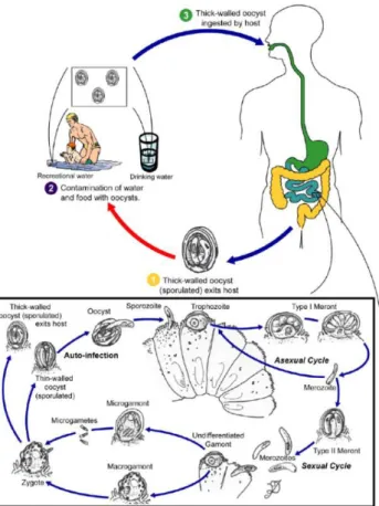
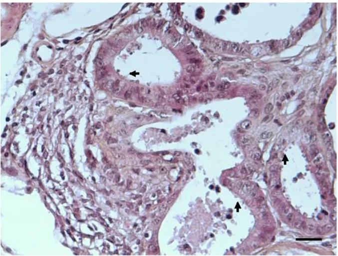
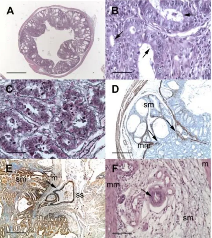
The life cycle of Cryptosporidium begins with the ingestions of the sporulated, environmentally resistant oocysts by the susceptible host (Figure 1).
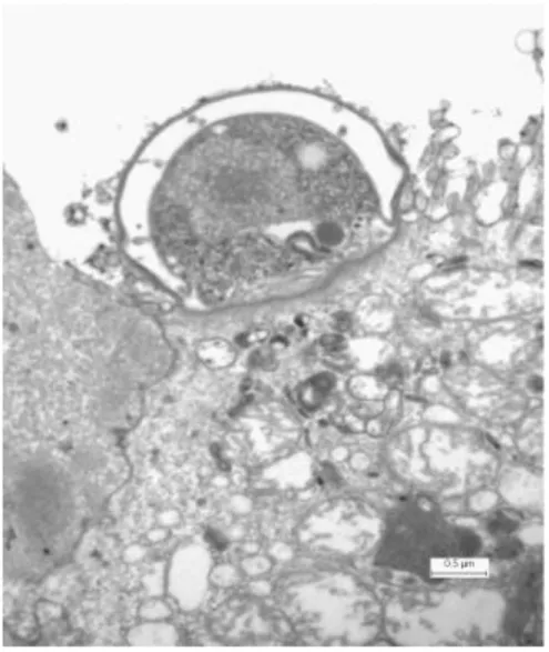
Oocysts undergo excystation and release four infective sporozoites in the intestinal lumen upper small intestine. The excystation of oocysts has been reported to be triggered by various factors including temperature, pancreatic enzymes, bile salt, and carbon dioxide (Fayer and Leek, 1984; O'Donoghue, 1995; Reduker and Speer, 1985). After excystation, the released sporozoites penetrate the mucus layer and emerge to invade the microvilli at the brush border of the mucosal. The sporozoites possess an apical complex composed of micronemes, roptries and dense granules which are also present in other apicomplexa parasites and are involved in host cells attachment and invasion. Then a parasitophorous vacuole is formed around the parasite which develops into spherical trophozoite. An unusual feature of the parasitophorous vacuole is that it is located within the host cell plasma membrane, but outside the host cell cytoplasm, it is separated from the latter by a feeder organelle and cytoskeletal elements (Figure 2).
Figure 2. Transmission electron micrograph showing intracellular Cryptosporidium
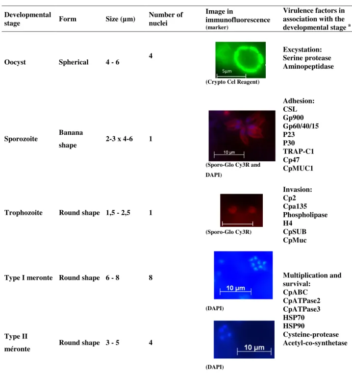
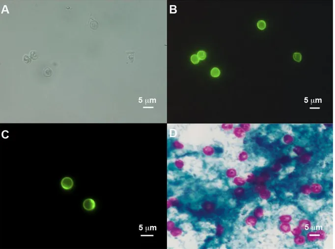
The trophozoites undergo asexual division (merogony) and form type I meronts containing 6 to 8 merozoites. Like sporozoites, merozoites escape the parasitophorous vacuole and attach again to the surface of epithelial cells, establishing amplified asexual infectious cycles. They undergo merogony once more and form either a further type I meront or a type II meront. The type II meront produce 4 merozoites which attach again to epithelial cells, but instead of developing into further meronts, they initiate gametogony. Individual merozoites produce either microgamonts or macrogamonts. Sixteen or more microgametes from the microgamont are released and each of these can fertilize a macrogamont to form a diploid zygote, which differentiates to an oocyst. Meiosis then results in the formation of four sporozoites. Most of the zygotes develop into oocysts with a thick-wall and are released into the lumen of the intestine, and then they are excreted into the environment. These thick-walled oocysts are immediately infective, allowing the spread of infection to other susceptible hosts. The minority develop into thin-walled oocysts which facilitate an auto-infective cycle that maintains infection without further ingestion of thick-walled oocysts (Chen et al., 2002). Characteristics of different Cryptosporidium life cycle stages can be observed in Table 1.
Table 1. Different life cycle stages of Cryptosporidium
a Data compiled from Okhuysen and Chappell, 2002; O’Hara and Chen, 2011; Bouzid et al., 2013.
Developmental
stage Form Size (µm) Number of nuclei
Image in
immunofluorescence
(marker)
Virulence factors in association with the
developmental stage a
Oocyst Spherical 4 - 6 4
(Crypto Cel Reagent)
Excystation: Serine protease Aminopeptidase
Sporozoite Banana
shape 2-3 x 4-6 1
(Sporo-Glo Cy3R and DAPI) Adhesion: CSL Gp900 Gp60/40/15 P23 P30 TRAP-C1 Cp47 CpMUC1
Trophozoite Round shape 1,5 - 2,5 1
(Sporo-Glo Cy3R) Invasion: Cp2 Cpa135 Phospholipase H4 CpSUB CpMuc
Type I meronte Round shape 6 - 8 8
(DAPI) Multiplication and survival: CpABC CpATPase2 CpATPase3 HSP70 HSP90 Cysteine-protease Acetyl-co-synthetase Type II
méronte Round shape 3 - 5 4
(DAPI)
10µm 5µm
1.3. Cryptosporidium species
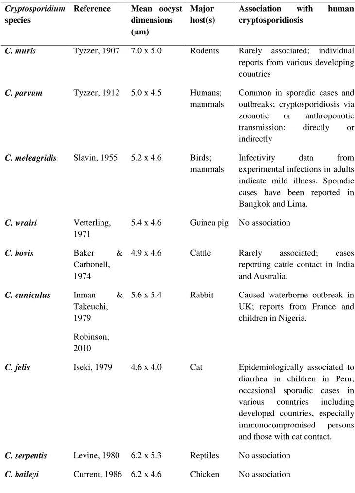
To date, they are 26 validated species of Cryptosporidium spp. (Chalmers and Katzer, 2013; Slapeta, 2012). These species have been found to infect mammals, birds, reptiles, amphibians and fish. The two species that most commonly infect humans (>90%) are C. hominis and C. parvum, and while the former species seems to be mainly limited to humans, the latter has a wide range of hosts, including most major domestic livestock animal species (Fayer, 2010; Xiao, 2010). Other species that have less commonly been associated with human disease include C. meleagridis, C. felis, C. canis, and C. cuniculus. Some species of Cryptosporidium are also reported in sporadic human cases such as C. andersoni, C. bovis, C. fayeri, C. muris, C. scrofarum, C. suis, C. tyzzeri, C. ubiquitum and C. viatorum (Chalmers and Katzer, 2013). Some species of Cryptosporidium infect many host species, whereas others appear to be restricted to groups such as rodents or ruminants, and still others are known to infect only one host species. Some species primarily infect the stomach, whereas others primarily infect the intestine (Table 2).
Table 2. Cryptosporidium species and their association with human cryptosporidiosis
Cryptosporidium species
Reference Mean oocyst dimensions (μm)
Major host(s)
Association with human cryptosporidiosis
C. muris Tyzzer, 1907 7.0 x 5.0 Rodents Rarely associated; individual
reports from various developing countries
C. parvum Tyzzer, 1912 5.0 x 4.5 Humans;
mammals
Common in sporadic cases and outbreaks; cryptosporidiosis via zoonotic or anthroponotic transmission: directly or indirectly
C. meleagridis Slavin, 1955 5.2 x 4.6 Birds;
mammals
Infectivity data from experimental infections in adults indicate mild illness. Sporadic cases have been reported in Bangkok and Lima.
C. wrairi Vetterling,
1971
5.4 x 4.6 Guinea pig No association
C. bovis Baker &
Carbonell, 1974
4.9 x 4.6 Cattle Rarely associated; cases reporting cattle contact in India and Australia.
C. cuniculus Inman &
Takeuchi, 1979 Robinson, 2010
5.6 x 5.4 Rabbit Caused waterborne outbreak in UK; reports from France and children in Nigeria.
C. felis Iseki, 1979 4.6 x 4.0 Cat Epidemiologically associated to
diarrhea in children in Peru; occasional sporadic cases in various countries including developed countries, especially immunocompromised persons and those with cat contact.
C. serpentis Levine, 1980 6.2 x 5.3 Reptiles No association C. baileyi Current, 1986 6.2 x 4.6 Chicken No association
C. varanii Pavlásek, 1995 4.8 x 4.7 Reptiles No association C. galli Pavlásek, 1999 8.3 x 6.3 Chicken No association C. andersoni Lindsay, 2000
7.4 x 5.5 Cattle Rarely associated; individual reports from UK, Australia, and Malawi.
C. canis Fayer, 2001 5.0 x 4.7 Dog Epidemiologically linked to
diarrhea in children in Peru; occasional sporadic cases in various, especially non-industrialized countries; some cases report contact with dogs.
C. hominis
Morgan-Ryan, 2002
4.9 x 5.2 Humans Common cause of diarrheal disease in sporadic cases and outbreaks.
C. molnari
Sijta-Bobadilla, 2002
4.7 x 4.5 Fish No association
C. suis Ryan, 2004 4.6 x 4.2 Pig Rarely associated; individual
reports from UK and Peru involving contact with pig
C. scophthalmi Alvarez
Pellitero, 2004
4.4 x 3.9 Fish No association
C. fayeri Ryan, 2008 4.9 x 4.3 Marsupials Rarely associated; one case
reported in Australia of a person in contact with marsupials
C. ryanae Fayer, 2008 3.7 x 3.2 Cattle No association C. fragile Jirku, 2008 6.2 x 5.5 Black
spined toad
No association
C. macropodum Power, 2008 5.4 x 4.9 Eastern
grey kangaroo
No association
C. ubiquitum Fayer, 2010 5.0 x 4.7 Mammals Sporadic cases in various
countries possibly involving untreated water supplies contaminated by animals hosts
C. tyzzeri Zhao, 2012
Invalid by Slapeta et al, 2012
4.6 x 4.2 Mice Rarely associated, individual reports from Czech Republic involving contact with wild mice
C. viatorum Elwin, 2012 5.4 x 4.7 Humans Sporadic cases in UK and
Sweden
C. scrofarum Kvac, 2013 5.2 x 4.8 Pig Rarely associated, reports from
Czech Republic involving contact with pigs
Data compiled from Slapeta, 2012 and Chalmers and Katzer 2013
1.4. Genomics of Cryptosporidium species
The era of Cryptosporidium genomic research began in the late nineties. The National Institute of Allergy and Infectious Diseases funded a consortium of three universities (University of Minnesota, Virginia Commonwealth University and Tufts University) to sequence the genome of two species of Cryptosporidium: C. parvum and C. hominis species responsible for the majority of human infections. The primary motivation for subsequent sequencing efforts was the public health importance of C. parvum and C. hominis, the lack of effective drugs to treat cryptosporidiosis, and the lack of research tools (Woods, Nesterenko, and Upton, 1996). The genome sequencing became important in order to address a wide range of questions, from basic biology to the identification of drug targets, from taxonomy to population biology (Striepen et al., 2004).
The widely distributed C. parvum IOWA isolate and C. hominis TU502 isolate from a Ugandan child were chosen for sequencing. The genome sequences of C. parvum and C. hominis isolates, each comprising eight chromosomes, showed similar genome sizes of 9.11 and 9.16 Mb, respectively, with 95-97% DNA sequence identity and ~30% GC content, with no large indels (insertion or deletion of bases in the DNA) or evident rearrangements when C.
hominis contigs were mapped to the C. parvum genome (Abrahamsen et al., 2004; Xu et al., 2004). Their sets of protein and RNA coding genes are essentially identical; thus phenotypic differences between them such as host range are presumed to be due to subtle polymorphisms (e.g., in micro- and mini-satellite repeat lengths (Tanriverdi and Widmer, 2006)) and differential gene regulations (Xu et al., 2004). Comparing Cryptosporidium genome to other eukaryotes, including the apicomplexans, a streamlining is achieved by a reduction in gene number, number of introns, and intergenic lengths. In addition to this, the genome sequencing and assembly of C. muris strain RN66 (unpublished but deposited into public databases) (http://CryptoDB.org/) are essentially complete.
Cryptosporidium genome analysis has revealed extremely streamlined metabolic pathways and a lack of many cellular structures and particular metabolic pathways found in general in eukaryotes or more specifically in Apicomplexa (Wanyiri and Ward, 2006). Energy metabolism is largely dependent from glycolysis, and both aerobic and anaerobic metabolisms are available, thus conferring environmental flexibility (Barta and Thompson, 2006). Limited biosynthetic capabilities and minimal metabolism have been reported, suggesting a large dependence on nutrient acquisition from the host (Rider and Zhu, 2010). Cryptosporidians have completely lost their apicoplast, a non-photosynthetic plastid, though few of its proteomic relicts can be identified in the nuclear genome by their homology to algal and cyanobacterial genes. C. parvum and C. hominis have a degenerate ‘mitosome’ instead of a mitochondrion, and have lost the mitochondrial genome and nuclear genes for many mitochondrial proteins, including those required for the Krebs cycle, oxidative phosphorylation, and fatty acid oxidation (LaGier et al., 2003). However, the existence of a relict mitochondrion was subsequently confirmed by ultra-structural studies (Keithly et al., 2005). Genes for de novo biosynthesis of amino acids, nucleotides, and sugars, as well as mechanisms for splicing RNA and gene silencing are also absent. The loss of these genes could mean that C. parvum and C. hominis rely heavily on scavenging nutrients from the host for energy production (Abrahamsen et al., 2004; Huang et al., 2004; Templeton et al., 2010; Toso and Omoto, 2007; Xu et al., 2004).
Nutrient uptake is accomplished by a lineage-specific expanded array of transporters of sugars, amino acids, and fatty acids (Abrahamsen et al., 2004; Xu et al., 2004). At least, 31 genes are very likely to have been acquired via intracellular transfer from organelle genomes or via horizontal transfer from bacteria (Huang et al., 2004), including a few that are not
conserved in other apicomplexans, e.g., genes encoding α-amylase, Inosine monophosphate dehydrogenase and thymidine kinase (Striepen et al., 2004).
A comprehensive genome database, CryptoDB (http://CryptoDB.org/), serves as an online public interface for Cryptosporidium genome sequences (Puiu et al., 2004). This website offers access to sophisticated tools which enable the identification of genes based on text, sequence similarity, and motif queries (Striepen and Kissinger, 2004). CryptoDB has recently been incorporated into a more comprehensive assembly of parasite databases under EuPathDB (Aurrecoechea et al., 2007). The genome sequences for C. parvum, C. hominis and C. muris have also been produced and provide a valuable comparison between them. An additional metabolic resource for Cryptosporidium which is also accessible through CryptoDB, is the CryptoCyc database (http://apicyc.apidb.org/CPARVUM/server.html) and is useful for comparison with predicted metabolism pathways in other organisms (Caspi et al., 2012).
The sequencing of Cryptosporidium genomes has revealed a vast amount of information, contributing to a better knowledge of microbial biology, pathogenicity, evolution and virulence. The quest for the molecular basis of virulence has exploited these genomic data to search for genes that may ultimately unravel the regulation of virulence, host range, and transmissibility at the genetic level (Casadevall and Pirofski, 2001).
Several comparative genomics studies have been performed since the completion of genome sequences from apicomplexan parasites of medical and veterinary importance. Phylogenomics analyses of C. parvum genes provided a preliminary estimate of the number of loci that are likely to have originated through lateral gene transfer (Huang et al., 2004). Templeton and colleagues (Templeton et al., 2004) showed that Cryptosporidium spp. and Plasmodium spp. share over 150 ancestral “apicomplexan” proteins, involved mainly in interactions with eukaryotic host cells and in the biogenesis of the apical complex. Gordon and Sibley (Gordon and Sibley, 2005) used genome sequences of Toxoplasma gondii, Plasmodium spp., Cryptosporidium spp., and Theileria spp. to show the conservation of actin-like proteins among these parasites, which rely on actin-based motility for cell invasion, while comparative genomics of Plasmodium spp., Cryptosporidium spp., and Toxoplasma gondii revealed that calcium-regulated proteins (plant-like pathways for calcium release channels and calcium-dependent kinases) were also conserved (Nagamune and Sibley, 2006). In addition to conserved genes, comparative genomics can identify unique, novel, and
uncharacterized virulence genes. Kuo et al. compared the genome sequences of three apicomplexan parasites (Plasmodium, Theileria, and Cryptosporidium) and showed that as many as 45% of the Cryptosporidium genes could be considered genus specific (Kuo, Wares, and Kissinger, 2008). Although the predicted gene counts were not identical, the papers reporting the genomes of C. parvum (3,952 genes) and C. hominis (3,994 genes) found no evidence for unique genes between the species, noting that the variation that appeared to be present reflected primarily gaps in one genome or the other, and speculating that the phenotypic differences likely arose from polymorphisms in coding regions and from differences in gene regulation. However, the comparative genomics study by Kuo et al., identified 334 putative C. hominis-specific genes and 178 putative C. parvum specific genes by interrogations of the Cryptosporidium database into which the gene sequences had been placed (Kuo, Wares, and Kissinger, 2008). Furthermore, a study by Widmer and colleagues, which also aimed to investigate the genetic basis of Cryptosporidium host specificity, used genome-wide comparisons of C. parvum zoonotic, C. parvum anthroponotic, and C. hominis isolates. Those authors reported that for some genetic loci, there was actually more sequence similarity between C. parvum anthroponotic and C. hominis strains than there was between C. parvum anthroponotic and C. parvum zoonotic strains (Widmer et al., 2012).
In the post-genomic era, the discovery and validation of protozoan virulence factors in particular have been accelerated by the application of technological advances, including comparative genomics, transcriptomics, microarrays, and reverse genetics including gene replacement and small interfering RNA (siRNA). In conclusion, the era of cheap sequencing promises to advance our understanding of the evolution of Cryptosporidium.
2. Epidemiology
Cryptosporidiosis is a cosmopolitan infection reported on every continent except Antarctica, with the incidence and prevalence varying around the world. The global prevalence of human cryptosporidiosis is between 0.5 and 2% in industrialized countries and can exceed 10% in developing countries (Guyot, Sarfati, and Derouin, 2012).
Worldwide environmental and veterinary surveillance data reveal the presence of Cryptosporidium spp. in water treatment systems, which represents an unacceptable health risk, particularly in sensitive (elderly, pregnant women, children) and immunocompromised
populations (HIV-positive and transplanted patients). However, the greatest burden of cryptosporidiosis occurs in developing countries because of the disseminated status of malnutrition in children and the highest world prevalence of HIV infection in certain geographic areas of this region, especially in Africa (Fergusson and Tomkins, 2009). It is very difficult to quantify as estimates vary widely, even among studies in the same geographic regions, as a result of differences in study design, sample size, age range, immunity status, severity of the disease, and sensitivity of the diagnostic methods. Nevertheless, it is recognized in most epidemiological studies that the prevalence of cryptosporidiosis is probably underestimated because infection with this parasite can be asymptomatic and difficult to detect in immunocompetent people. Studies of children in daycare centers, suggest that asymptomatic excretion of oocysts is not uncommon. For instance, in Trujillo State, Venezuela, Cryptosporidium oocyst shedding was reported in 89% of healthy children attending day-care centers (Miller et al., 2003). Recent publications agree on the fact that the incidence of cryptosporidiosis is significantly increasing worldwide (Chalmers et al., 2011b; Fournet et al., 2013; Yoder, Harral, and Beach, 2010; Yoder et al., 2012).
The epidemiology of cryptosporidiosis is influenced by difference factors: the infective dose is low (one to ten oocysts); (ii) oocysts are immediately infectious when excreted in the environment; (iii) oocysts are stable and highly resistant to chlorine disinfection and survive for months in the environment; and (iv) environmental dispersal can lead to the contamination of drinking water and food (Caccio et al., 2005).
Cryptosporidium oocysts are most commonly transmitted by the fecal-oral route through direct host to host contact, and indirect contamination of water or food. Direct and indirect transmission between infected hosts and susceptible individuals is favored by high population densities and by close contact (Caccio et al., 2005). Cases of human-to-human transmission have been reported between family members, sexual partners, children, and in hospitals. Zoonotic transmission has been confirmed by epidemiological studies concerning farm and companion animals, and veterinary workers. Food handled by a contaminated or infected person, and food that has been exposed to contaminated water have been sources for food-borne cryptosporidiosis. Food grown in soil fertilized with manure could also be considered a potential source of infection. However, contaminated water represents the major source of infections for humans (Ramirez, Ward, and Sreevatsan, 2004).
During a period of almost hundred years, more than 300 waterborne protozoan parasitic outbreaks have been reported worldwide being Cryptosporidium spp. the main etiological agent (present in 60.3% of waterborne outbreaks) (Karanis, Kourenti, and Smith, 2007) followed by other parasites such as Giardia spp. and Toxoplasma gondii (Baldursson and Karanis, 2011). In particular, more than 160 waterborne outbreaks of cryptosporidiosis have been reported in the last decade implicating contaminated drinking and recreational water (Yoder, Harral, and Beach, 2010; Yoder et al., 2012).
C. hominis and C. parvum are known as the major agents causing human cryptosporidiosis both in immunocompetent and in immunocompromised individuals and their prevalence varies in different regions of the world. Epidemiological studies showed that C. hominis is more prevalent in Australia, West Europe, India, and Africa (Fournet et al., 2013; Helmy et al., 2013; O'Brien, McInnes, and Ryan, 2008; Sharma et al., 2013), whereas C. parvum human cases are more prevalent in Chile, Turkey, Middle East and Thailand (Alyousefi et al., 2013; Iqbal, Khalid, and Hira, 2011; Neira et al., 2012; Nuchjangreed et al., 2008; Taghipour et al., 2011; Usluca and Aksoy, 2011). Additionally, C. meleagridis has been confirmed as an emerging human pathogen, being responsible for 10% of cases in Peru, where its prevalence is as high as the prevalence of C. parvum (Cama et al., 2008). Geographic variations in the distribution of C. parvum and C. hominis can also occur within a country. For example, C. parvum is more common than C. hominis in rural states of Ireland (Zintl et al., 2009).
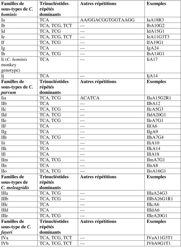
Molecular tools have provided new insights into Cryptosporidium taxonomy helping to understand its epidemiology. Subtyping tools have been used extensively in studies of the transmission of C. hominis in humans, and C. parvum in humans and ruminants. The DNA sequence analysis of the 60 kDa glycoprotein (gene) is one of the most employed subtyping tools. The gene has tandem repeats of the serine-coding trinucleotide TCA, TCG or TCT at the 5’ end of the gene. In addition to variations in the number of trinucleotide repeats, there are extensive sequence differences in the non-repeat regions, which categorize C. parvum and C. hominis into several subtype families (Xiao, 2010).
Nowadays it is known that most of the outbreaks around the world were caused by the anthroponotic species C. hominis and an intra-specific characterization using this marker allowed the identification of the C. hominis subtype family Ib in most of the disease cases. The subtype Ib had very different subtypes from which the investigations have identified the
subtypes IbA9G3 and IbA10G2 commonly found in the majority of areas in the world (Xiao, 2010).
The IbA9G3 is usually observed in humans in Kenya, India, and Australia, and IbA10G2 is commonly seen in South Africa, Peru, USA, Canada, Australia, and European countries, as France, UK, Portugal, Spain and Ireland (Alves et al., 2006a; Cama et al., 2008; Chalmers et al., 2005; Fournet et al., 2013; Gatei et al., 2006b; Leav et al., 2002). IbA10G2 is responsible for more than half of the waterborne outbreaks in USA, and Canada (Boehmer et al., 2009). Most notably, C. hominis IbA10G2 subtype was the causal agent associated to the 1993 Milwaukee waterborne cryptosporidiosis outbreak (Corso et al., 2003). Other subtypes of C. hominis exist such as IbA13G3 subtype, which was seen in Peru (Cama et al., 2007), and several otherwise rare subtypes, IbA6G3, IbA19G2, IbA20G2, IbA21G2 and IbA23G2, found in Jordan and China (Feng et al., 2009; Hijjawi et al., 2010). Likewise, in most developing countries, humans with the subtype family Ie are mostly infected with IeA11G3T3, excepting Jamaica and China where IeA12G3T3 is seen (Alyousefi et al., 2013; Feng et al., 2009; Gatei et al., 2008; Sulaiman et al., 2005). As well, other rare subtypes such as IdA14 (Sulaiman et al., 2005), IdA19 (Trotz-Williams et al., 2006), Id A20 (Nazemalhosseini-Mojarad et al., 2011), IdA21 and IdA24 (Hijjawi et al., 2010) exist. IfA22G1 subtype is also described in Iran (Nazemalhosseini-Mojarad et al., 2011; Taghipour et al., 2011). Much less genetic diversity of C. hominis is observed in industrialized nations. In European countries and Australia, most C. hominis infections are caused by the Ib subtype family (Alves et al., 2006b; O'Brien, McInnes, and Ryan, 2008; Waldron, Ferrari, and Power, 2009; Wielinga et al., 2008; Xiao, 2010; Zintl et al., 2009). The majority of these cases had the IbA10G2 subtype and less commonly the IbA9G3. In addition, several unusual Ib subtypes (IbA18G1, IbA5G2T3 and IbA9G2T1) were also occasionally found (Jex et al., 2008; Jex et al., 2007; O'Brien, McInnes, and Ryan, 2008).
On the other hand, the C. parvum IIa subtype family is responsible of the majority of cryptosporidiosis outbreaks due to this specie. Particularly, the subtypes IIaA15G2R1, IIaA16G1R1, IIaA17G2R1, and IIaA19G2R1 are responsible for zoonotic cryptosporidiosis and have been found in both humans and ruminants. These subtypes are described in many geographic areas of the world, especially the subtype IIaA15G2R1 which is known as the major subtype of C. parvum in the world. It dominates in most of the countries around the world such as in USA, Canada, UK, Germany, Netherlands, Spain, Portugal, Slovenia, India, Japan and Yemen. Whereas other subtypes dominates in other countries such as IIdA20G1 in