UNIVERSITÉ DE MONTRÉAL
IDENTIFICATION OF IN SITU PROGENITOR CELL FEATURES CONTRIBUTING TO CARTILAGE REPAIR AND STRATEGIES FOR CARTILAGE REPAIR AUGMENTATION
GARIMA DWIVEDI
INSTITUT DE GÉNIE BIOMÉDICAL ÉCOLE POLYTECHNIQUE DE MONTRÉAL
THÈSE PRÉSENTÉE EN VUE DE L’OBTENTION DU DIPLÔME DE PHILOSOPHIAE DOCTOR
(GÉNIE BIOMÉDICAL) AVRIL 2017
UNIVERSITÉ DE MONTRÉAL
ÉCOLE POLYTECHNIQUE DE MONTRÉAL
Cette thèse intitulée:
IDENTIFICATION OF IN SITU PROGENITOR CELL FEATURES CONTRIBUTING TO CARTILAGE REPAIR AND STRATEGIES FOR CARTILAGE REPAIR AUGMENTATION
présentée par : DWIVEDI Garima
en vue de l’obtention du diplôme de: Philosophiae Doctor a été dûment acceptée par le jury d’examen constitué de : M. VIRGILIO Nick, Ph. D., président
M. BUSCHMANN Michael, Ph. D., membre et directeur de recherche Mme HOEMANN Caroline, Ph. D., membre et codirectrice de recherche M. NANCI Antonio, Ph. D., membre
DEDICATION
To my husband, My brother
& My parents
ACKNOWLEDGEMENTS
It was summer of 2012 and I was new and slightly apprehensive in a new country, trying to adapt to an unfamiliar environment. At this point, my research project provided me the stability and purpose I needed. For this, I thank my Ph.D. research supervisor, Professor Michael D Buschmann for providing me the opportunity to expand my learning in the beloved field of cartilage repair. I thank him for placing trust in me, and letting me grow as an individual in science and in life. I feel extremely fortunate to have him as a guide to tide over the most challenging and most fruitful turning point of my professional life. Over a period of time, his ideas and thoughts have moulded me into a better researcher and enabled me to develop a genuine appreciation for good science. His vision and structured thinking have made tremendous contribution in driving my research and writing. I am thankful for several opportunities to attend and present at international conferences which added to my growth as a professional. During the course of my study, I encountered some difficult times and I am grateful for his immense support, understanding and patience. I am certain that his wise words will continue to guide me in the future.
I am also thankful to my co-supervisor Professor Caroline Hoemann, whose wisdom and experience has made invaluable contribution in the progress of my research. Her eye for detail and desire for painstaking precision have left me in awe on several occasions and inspired me to be assiduous and meticulous in my research. I can recall multiple instances where our discussions were extremely stimulating and provided valuable breakthroughs at difficult junctures in my research. Her inputs have always helped me in improving the quality of my writing as well as presentations. Through her actions, she constantly conveys that there is no short cut to success. Her motivation and guidance has made the journey of PhD more interesting and fulfilling.
This note of thanks would be incomplete without recognizing the immense contribution of Anik Chevrier in my research. She has been a guide, a confidante and a friend-all bundled into one. I cannot thank her enough for several occasions when she was beside me helping in the time of predicament-be it the nerve-wracking rabbit surgeries or the tricky statistical procedures for my papers. Through her actions, she has taught me the importance of team work, patience and kindness. I am grateful to her for always being there in my difficult times-both professional and
personal- and her eagerness to help in whatever way she could. I will forever cherish our fun moments on our several conference trips.
I extend a special note of thanks to Catherine Trudeau and Thomas Clinton. Guiding them through their internship was an enriching experience for me and I am grateful for their contribution in my research.
I would be remiss in not recognizing the contributions of my colleagues, Julie Tremblay, Jun Sun, Genevieve Picard, Vincent Darras, Marc Lavertu and Chi-Yuan Chang. I have deep appreciation for their time, ideas and assistance in helping me accomplish my project. Their support made my everyday life more proficient and resourceful.
My time at Ecole Polytechnique was made enjoyable in large part due to my friends and colleagues at the lab. I thank my roommate and fellow student, Almas Siddiqui for being a wonderful and supportive friend. Life would have been dull without friends like Sotcheadt, Mohamad-Gabriel, Colleen, Leili, David, Ashkan, Nic and Tanushri. Time spent with them was always filled with fun and stimulating conversations. I am grateful that I could always rely on them in the time of need.
I would not have been able to reach the stage I have today, if not for the sacrifices and efforts of my parents-Sudha and Sushil K Dwivedi. My growth was always a priority for them and despite limited means; they never hesitated to provide me and my brother with the best. I thank them for believing in me even when I did not and motivating me to push my boundaries and aim for higher goals. Undoubtedly, I owe my success and achievements to them. I would also like to thank my brother, Prateek Dwivedi, whose support and calmness has kept me grounded and helped me push through several challenges in life. Finally, I thank my loving and supporting husband, Rajesh Shukla, in whom I found a friend, a guide. His unwavering faith and encouragement have been the biggest support in the final stages of my research. His cheerful and positive nature has inspired me to stay calm and keep moving forward. Thank you all.
RÉSUMÉ
Le cartilage articulaire hyalin recouvre l’extrémité des os longs. Sa capacité de guérison intrinsèque est faible puisqu’il n’est pas vascularisé. Suite à une blessure, la guérison n’a généralement pas lieu, ou demeure de faible amplitude. Une dégénérescence des lésions peut également survenir suite au relargage de cytokines et autres facteurs inflammatoires. Un déséquilibre entre les activités anaboliques et cataboliques peut éventuellement induire l’érosion du cartilage, l’exposition de l’os sous-jacent et l’ostéoarthrite.
Les techniques de stimulation de la moelle osseuse sont une façon de stimuler la guérison du cartilage en perçant l’os situé sous les lésions du cartilage. Les canaux percés permettent aux cellules progénitrices qui se trouvent dans la moelle osseuse (bone marrow progenitor cells en anglais) de migrer vers les lésions. Ces cellules progénitrices se différencient et synthétisent un tissue de granulation qui sera remodelé en tissue de guérison. Les techniques de stimulation de la moelle osseuse sont beaucoup utilisées en clinique puisqu’elles sont simples et peu coûteuses à effectuer, que les patients se rétablissent rapidement après la chirurgie et qu’elles sont efficaces pour diminuer la douleur. Cependant, les bienfaits ressentis ne sont pas durables puisque le tissu de guérison est un tissue fibrocartilagineux qui ne possède pas les propriétés biologiques et mécaniques du cartilage hyalin. Les inconvénients les plus communs observés au cours d’études pré-cliniques et cliniques portant sur les techniques de stimulation de la moelle osseuse sont une guérison incomplète, une grande variabilité inter-individu, et des résultats insatisfaisants, surtout chez les individus plus âgés. Il a déjà été démontré dans des études pré-cliniques que l’emplacement de la lésion ainsi que l’âge de l’animal sont deux paramètres qui affectent grandement la guérison. Plus spécifiquement, chez le lapin, les lésions situées sur la trochlée guérissent mieux que les lésions situées sur le condyle fémoral médial, ce qui suggère que les cellules progénitrices de la moelle osseuse présentes aux deux emplacements ne possèderaient pas les mêmes propriétés biologiques intrinsèques.
L’objectif de la première étude de cette thèse était de déterminer l’effet de l’emplacement et de l’âge de l’animal sur les propriétés biologiques intrinsèques des cellules progénitrices de la moelle osseuse. Pour ce faire, les propriétés biologiques des cellules isolées à partir de la trochlée et des condyles chez des lapins jeunes et âgés ont été caractérisées. Ensuite, puisque de
précédentes études chez le lapin ont démontré qu’un perçage profond à 6 mm favorise la guérison du cartilage dans la trochlée en comparaison à un perçage peu profond, les propriétés biologiques de cellules progénitrices isolées à partir de la moelle métaphysaire (région profonde) et de la moelle épiphysaire (région peu profondes) ont été caractérisées. Deux méthodes d’isolation des cellules progénitrices ont été utilisées, soit la digestion enzymatique à la collagénase et la culture d’explants osseux. Pour chaque population de cellules progénitrices, le rendement cellulaire, le potentiel clonogénique et l’expression de différents marqueurs ont été déterminés in vitro. Le potentiel chondrogénique a, quant à lui, été déterminé en stimulant les cellules progénitrices avec un milieu de culture chondrogénique contenant de l’ascorbate, ITS et TGF-III. Enfin, le potentiel ostéogénique a été déterminé en stimulant les cellules progénitrices avec un milieu de culture ostéogénique contenant du dexamethasone, de la β-glycerophosphate et de l’ascorbate. Une comparaison entre les propriétés biologiques obtenues pour les cellules progénitrices provenant des condyles, de la trochlée profonde, ou de la trochlée peu profonde, pour les lapins jeune ou âgés et pour les cellules isolées par digestion enzymatique ou par culture d’explants a ensuite été effectuée. Selon les critères établis par l’International Society for Cell Therapy, les cellules progénitrices isolées lors de l’étude pouvaient être décrites comme une population de cellules souches mésenchymateuses. Les condyles avaient un faible rendement cellulaire en comparaison à la trochlée et les cellules progénitrices isolées à partir des condyles avaient également de faibles potentiels clonogénique et chondrogénique. En revanche, les cellules progénitrices isolées à partir de la trochlée possédaient un haut rendement cellulaire, des potentiels clonogénique et chondrogénique élevés et exprimaient beaucoup de glycosaminoglycanes et de collagène type II, deux molécules présentes dans le cartilage articulaire hyalin. Le site d’isolation ou l’emplacement était donc un facteur déterminant pour les propriétés biologiques des cellules progénitrices, ce qui suggère que le potentiel de guérison élevé observé dans la trochlée du lapin serait dû à la présence d’une population nombreuse de cellules progénitrices ayant un fort potentiel chondrogénique. L’âge des lapins s’est aussi avéré être un facteur important puisque le rendement cellulaire, l’expression de marqueurs pour cellules souches et la différenciation chondrogénique et ostéogénique étaient meilleurs chez les lapins jeunes que chez les lapins âgés. Enfin, il n’y avait pas de différence entre les cellules isolées par digestion enzymatique ou par culture d’explants.
L’objectif de la deuxième étude de cette thèse était de déterminer s’il existe une corrélation entre les propriétés biologiques intrinsèques des cellules progénitrices de la moelle osseuse et la capacité de guérison du cartilage sus-jacent. Pour ce faire, un modèle bilatéral a été effectué chez 8 lapins. Pour chaque lapin, des lésions cartilagineuses ont été créées sur un genou à deux emplacements (la trochlée et le condyle fémoral médial) puis traitées par microperçage, tandis que l’autre genou demeurait intact. Après 3 semaines de guérison, les lapins ont été sacrifiés et des sections histologiques ont été récoltées du genou traité par microperçage afin d’évaluer la guérison. Parallèlement, les cellules progénitrices du genou intact ont été récoltées et leurs propriétés biologiques caractérisées tel que décrit ci-haut. Chez tous les lapins, la guérison induite par stimulation de la moelle osseuse était compromise dans le condyle fémoral médial, où très peu de tissu de guérison était présent après 3 semaines. En revanche, une guérison modérée à excellente était apparente dans la trochlée de 6 lapins sur 8, alors que 2 lapins ont été classifiés comme faisant partie d’un groupe ayant un plus faible potentiel de guérison. Tout comme pour la première étude, les cellules progénitrices de la trochlée avaient un meilleur rendement cellulaire et de forts potentiels clonogénique et chondrogénique que les cellules des condyles. Une variabilité inter-individu était également présente dans le cas des propriétés biologiques des cellules progénitrices. Les tissus de guérison de la trochlée ainsi que la matrice extracellulaire synthétisée par les cellules progénitrices isolées à partir de la trochlée contenaient une grande quantité de glycosaminoglycanes et de collagène type II. Des analyses statistiques ont permis de déterminer qu’il existait de fortes corrélations positives entre les propriétés intrinsèques des cellules progénitrices de la moelle osseuse et la capacité de guérison du cartilage sus-jacent. Les propriétés biologiques ayant le plus d’impact sur la guérison ont été identifiées.
L’objectif de la troisième étude de cette thèse était de développer un modèle animal représentatif de la situation clinique, et d’y tester une nouvelle méthode de guérison. En clinique, les lésions du cartilage sont rarement détectées rapidement, ce qui complique le traitement. Une grande variabilité de la réponse et une guérison compromise sont également observées chez les patients plus âgées souffrant de lésions dégénératives chroniques. La rétraction du caillot sanguin qui remplit les lésions cartilagineuses suite au microperçage et sa perte subséquente sont une raison qui pourrait expliquer les résultats insatisfaisants observés en clinique. Il a été démontré que le chitosane permet d’inhiber la rétraction du caillot sanguin et, par le fait même, qu’il promeut la guérison du cartilage en ayant des effets sur le recrutement cellulaire, la vascularisation and le
remodelage osseux. Le plasma riche en plaquettes (PRP) contient une concentration élevée de facteurs de croissance capables de stimuler le recrutement cellulaire et la différenciation chondrogénique. Il a été démontré que le chitosane favorise la stabilité du PRP et le relargage de facteurs de croissance. Un modèle de lésions cartilagineuses chroniques a d’abord été développé chez le lapin. Deux chirurgies était nécessaire pour effectuer ce modèle. Au cours de la première chirurgie, des lésions cartilagineuses bilatérales ont été induites dans la trochlée de 8 lapins et laissées sans traitement pendant 4 semaines afin qu’elles dégénèrent à un stade chronique. Au cours de la deuxième chirurgie, ces lésions cartilagineuses ont été traitées par microperçage et par l’implantation de chitosane lyophilisé solubilisé dans du PRP autologue, ou par injection de PRP seul, comme contrôle. Le tissu de guérison a été récolté après 2 mois et des sections histologiques ont permis d’évaluer la quantité et la qualité du tissu de guérison. La guérison osseuse ainsi que le remodelage osseux ont également été caractérisées par analyse micro-CT. Les implants chitosane-PRP favorisaient la guérison des lésions cartilagineuses chroniques comparées au PRP seul. La composition biochimique de la matrice cartilagineuse ressemblait plus à du cartilage hyalin en présence des implants chitosane-PRP. Finalement, l’analyse micro-CT démontrait que les implants chitosane-PRP favorisent le remodelage osseux mais que le phénomène est toujours en cours après 2 mois de guérison.
En résumé, le contenu de cette thèse nous a permis d’identifier certaines variables liées à l’emplacement, au donneur et à l’âge qui expliqueraient partiellement la variabilité inter-individu et les résultats décevants parfois obtenus suite à une stimulation de la moelle osseuse. Le modèle de lésions chroniques décrit dans cette thèse contribuera grandement au développement de nouvelles techniques et de nouveaux produits pour favoriser la guérison du cartilage articulaire. Enfin, les implants chitosane-PRP étudiés au cours de cette thèse constituent une approche prometteuse qui pourrait éventuellement servir à soulager des patients souffrant de lésions chroniques ou dégénératives.
ABSTRACT
Articular cartilage is a thin layer of hyaline cartilaginous tissue covering the ends of long bones. Due to its avascular nature, it possesses very limited regeneration potential. In the event of an injury, the repair is either very limited or not initiated at all. As a result, cartilage lesions degenerate extensively under the influence of damaging cytokines and proinflammatory factors released. An imbalance in anabolic and catabolic activities continues to erode the cartilaginous surface exposing subchondral bone leading to osteoarthritis.
Bone marrow stimulation (BMS) initiates repair by fracturing or drilling into subchondral bone. The channels created provide access to the underlying subchondral progenitor cells that are recruited into the defect. Granulation tissue thus formed undergoes a complex cascade of events to generate a repair tissue. BMS is considered a gold standard of cartilage repair strategies since it is easy, relatively less invasive with swift recovery, more economical and appreciable short-mid term relief. However, long term outcome is generally discouraging due to poor durability of repair tissue. The quality of repair tissue is generally fibrocartilagenous with inferior mechanical and biological characteristics compared to hyaline cartilage. The most common drawbacks of the procedure include incomplete regeneration, high inter-individual variability and poor repair outcome especially in older individuals in preclinical as well as clinical studies. Earlier studies have shown that defect location and animal age affect the cartilage repair outcome observed in vivo, suggesting a strong influence of biological characteristics of progenitor cells (Bone Marrow stem cells; BMSCs or mesenchymal stem cells; MSCs) present in subchondral bone. Specifically, rabbit trochlea showed a superior repair and chondrogenic potential compared to the medial femoral condyle. Inspired by these observations, we carried out a study for comprehensive analysis of progenitor cells present in the subchondral bone of condyle and trochlea in young and old rabbits in order to determine the influence of location and age on the properties of progenitor cells. Earlier we had found that drilling to 6 mm improves the repair outcome in rabbit trochlea possibly by providing access to metaphyseal marrow and thus higher progenitor cell population. As a result, MSCs were isolated from epiphyseal (trochlea upper) and metaphyseal (trochlea lower) regions of the trochlea. Collagenase-derived MSCs originated from collagenase digest of bone chips while the digested explants were subsequently cultured to obtain explant-derived MSCs via cell outgrowth. In vitro characterization assays were used to determine the cell yield, clonogenic potential and surface marker expression profile. Chondrogenic differentiation was
stimulated using pellets of condylar and trochlear MSCs in the presence of chondrogenic media, rich in chondrogenic stimulating factors such as ascorbate, insulin-transferrin-selenium (ITS) and TGF-βIII. Finally, MSCs were differentiated into osteogenic lineage in presence of osteogenic media containing osteogenic stimulants such as dexamethasone, β-glycerophosphate and ascorbate. The aforementioned MSC characteristics were compared for trochlea vs. condyle, old vs. young and collagenase- vs. explant-derived cultures. MSCs isolated from distal femur epiphyseal bone had stem cell characteristics and fulfilled the stem cell criteria outlined by International Society for Cell Therapy. Location was an important factor influencing the properties of MSCs indicated by low cell yield, clonogenic potential and inferior chondrogenic potential of condylar cells. Results showed that rabbit trochlear vs. condylar subchondral bone yielded a greater number of progenitors with superior cartilaginous matrix expression under chondrogenic conditions suggesting higher intrinsic capacity for cartilage repair compared to condylar subchondral bone. Trochlear MSCs displayed superior chondrogenic differentiation evidenced by higher glycosaminoglycan (GAG) and collagen type II deposition. Age of the donor was an additional factor with significant bearing on the inherent properties of MSCs since growth rate, cell yield, expression of stem cell markers and osteogenic differentiation were found to be significantly superior in younger animals. As expected, no significant difference was observed between collagenase- and explant-derived cultures.
Continuing our exploration of the mechanisms underlying variability in BMS procedure, we carried out the next study to verify if the cartilage repair outcome in condyle and trochlea correlated with the pre-existing properties of progenitor cells present in these two sites. A bilateral rabbit model was used to test this hypothesis. For each rabbit, full thickness acute defects were created in one knee and treated by BMS by microdrilling, while the second knee was left intact. Animals were sacrificed 3 weeks later and transverse sections of repair tissue from the treated knee were used to analyze the quality and quantity of repair macroscopically and by safranin- O (Saf-O) and immunostaining. At the time of animal sacrifice, distal femurs were separated and MSCs were isolated from condylar and trochlear regions of the intact contralateral knee for biological characterization of cell yield, clonogenic potential, cell surface marker profile and differentiation potency. BMS induced inferior cartilage repair in condyles of all donors studied further underlining the implication of location in influencing repair outcome. Macroscopic examination revealed extremely poor fill in condylar defects. In contrast, moderate
to excellent fill was observed in six out of eight trochlear defects. This led us to identify a donor-dependent variability in repair outcome and the donors were classified as good trochlear responders and poor trochlear responders. In another observation, trochlear MSCs were characterized by increased cell yield, higher clonogenic potential and superior chondrogenic potential. Histopathological analysis indicated better matrix composition, rich in GAG and collagen type II in trochlea both in vivo and in vitro. Donor related variability in repair outcome was also observed in vitro through biological characterization of MSCs. Statistical analysis provided substantial evidence to identify strong, positive location- and donor-dependent correlations between inherent properties of MSCs and cartilage repair outcome in condyle and trochlea and factors with maximum influence on the repair outcome were identified.
By this stage, it became imperative to seek after a clinically relevant application of our understanding based on current and past findings. Cartilage lesions are rarely detected in early stages, unfortunately reducing the efficacy of BMS procedure. Unpredictable, highly variable repair of poor quality is observed in degenerative defects, especially in older patients. Platelet-mediated retraction of the blood clot that fills cartilage lesions following BMS is believed to be one underlying cause of inferior quality and durability of repair tissue. Chitosan inhibits this blood clot shrinkage and leads to the formation of a voluminous, adherent and stable clot and thereby enhances cartilage repair by promoting cell recruitment, transient vascularization and subchondral bone remodeling. Platelet-rich-plasma (PRP) contains a 2-10 fold concentration of growth factors and cytokines and has been shown to improve recruitment and chondrogenic potential of subchondral MSCs. Chitosan increases the stability of PRP and promotes the release of platelet-derived growth factors in a more sustained manner. Using a more clinically relevant chronic defect model, we tested the efficacy of implants composed of freeze-dried chitosan solubilized in PRP in improving marrow stimulated cartilage repair outcome compared to BMS augmented with just PRP. Two surgeries were performed in skeletally mature rabbits to create a bilateral trochlear defect model. Defects created in the first surgery were allowed to degenerate over 4 weeks and repaired in a second surgery by BMS. Treatments included BMS augmented with chitosan/PRP implants in one knee and BMS with PRP in the other knee. Repair tissue was harvested 2 months later and transverse sections were used to characterize the quality and quantity of repair by assessing the GAG and coll-II deposition. Subchondral bone repair was analysed by micro-CT. Augmentation of BMS with freeze-dried chitosan/PRP implants in
chronic defects improved cartilage repair compared to PRP by increasing quantity and quality of repair tissue and promoting subchondral bone repair. Matrix composition was improved in presence of chitosan/PRP indicated by increased GAG and collagen deposition. MicroCT analysis showed further evidence of increased, albeit incomplete, subchondral bone remodeling induced by chitosan/PRP.
In summary, these studies led us to recognize significant donor-, age- and location-dependent variability which might help in understanding the mechanism underneath the unpredictability and inter-individual variability observed with BMS. Needless to say, these observations would be extremely valuable in improving the consistency, durability and quality of BMS repair procedure. Moreover, the innovative chronic model studied here is expected to make significant contributions in progress of cartilage repair field. Finally, chitosan/PRP implants investigated in this research are a promising approach to treat chronic cartilage lesions and could eventually provide relief to osteoarthritic patients with challenging, degenerative defects.
TABLE OF CONTENTS
DEDICATION ... iii
ACKNOWLEDGEMENTS ... iv
RÉSUMÉ ... vi
ABSTRACT ... x
TABLE OF CONTENTS ... xiv
LIST OF TABLES ... xviii
LIST OF FIGURES ... xix
LIST OF SYMBOLS AND ABBREVIATIONS... xxvii
CHAPTER 1 INTRODUCTION ... 1
1.1 Introduction ... 1
1.2 Research Hypotheses and Objectives ... 5
1.2.1 General objective ... 5
1.2.2 Study 1 ... 5
1.2.3 Study 2 ... 6
1.2.4 Study 3 ... 7
CHAPTER 2 LITERATURE REVIEW ... 8
2.1 Articular Cartilage ... 8
2.1.1 Overview ... 8
2.1.2 Composition of articular cartilage ... 8
2.1.3 Development of articular cartilage ... 9
2.1.4 Articular cartilage defects ... 10
2.2 Osteoarthritis ... 11
2.3 Bone marrow stem cells (BMSCs)/Mesenchymal stem cells (MSCs) ... 14
2.3.1 Introduction and history ... 14
2.3.2 Isolation of BMSCs ... 16
2.3.3 Colony forming unit assay (CFU-f) ... 19
2.3.4 BMSC surface marker expression ... 20
2.3.5 Heterogeneity of BMSCs ... 20
2.4 Bone marrow stimulation for cartilage repair ... 29
2.5 Influence of ageing on proliferation and differentiation potential of BMSCs ... 33
2.6 Influence of location of defect on the marrow stimulated cartilage repair outcome: Condyles v/s trochlea... 38
2.7 Application of chitosan in improving cartilage repair ... 41
2.8 Application of PRP in improving cartilage repair outcome ... 46
2.9 Considerations for suitable animal model ... 50
CHAPTER 3 ORGANIZATION OF ARTICLES ... 53
CHAPTER 4 ARTICLE 1: BONE MARROW PROGENITOR CELLS ISOLATED FROM YOUNG RABBIT TROCHLEA ARE MORE NUMEROUS AND EXHIBIT GREATER CLONOGENIC, CHONDROGENIC AND OSTEOGENIC POTENTIAL THAN CELLS ISOLATED FROM CONDYLES ... 55
4.1 Introduction ... 58
4.2 Materials and Methods ... 60
4.2.1 Necropsy ... 60
4.2.2 Isolation of BMPCs ... 60
4.2.3 Histology of bone chips ... 61
4.2.4 Cell Yield ... 61
4.2.5 Flow cytometry ... 61
4.2.6 CFU-f ... 62
4.2.7 Chondrogenic differentiation assay ... 62
4.2.8 Histology of pellets ... 62
4.2.9 Osteogenic differentiation assay ... 63
4.2.10 Statistical Analysis ... 63
4.3 Results ... 64
4.3.1 BMPCs can be isolated from different locations in rabbit femur ... 64
4.3.2 Cell yield, clonogenic potential and matrix production are highest in trochlear segments; and decrease with age ... 66
4.3.3 Chondrogenic potential displays high inter-individual variability, is on average superior for trochlear segments and decreases with age ... 71
4.3.4 Osteogenic potential is higher in trochlear segments and decreases with age ... 73
4.4 Discussion ... 77
CHAPTER 5 ARTICLE 2: QUALITY OF CARTILAGE REPAIR FROM MARROW
STIMULATION CORRELATES WITH CELL NUMBER, CLONOGENIC, CHONDROGENIC AND MATRIX PRODUCTION POTENTIAL OF UNDERLYING BONE MARROW
STROMAL CELLS ... 85
5.1 Introduction ... 88
5.2 Materials and methods ... 90
5.2.1 Study design and rabbit surgical model ... 90
5.2.2 Characterization of cartilage repair ... 91
5.2.3 Isolation and in vitro characterization of condylar and trochlear BMSCs ... 92
5.2.4 Correlation of cartilage repair to BMSC properties ... 92
5.2.5 Statistical Analyses ... 93
5.3 Results ... 94
5.3.1 Repair of trochlear defects was superior to condylar defects- and correlated with in vitro properties of BMSCs as function of location and donor ... 94
5.3.2 Cell yield, clonogenic potential and expression of stem cell markers are higher in trochlear vs condylar BMSCs ... 101
5.3.3 Moderate to strong positive correlations were observed between in vitro properties of collagenase-derived BMSCs and in vivo early repair responses and strong influence of cell yield, CFU, % Coll-II was observed ... 103
5.4 Discussion ... 115
5.5 Conclusion ... 118
CHAPTER 6 ARTICLE 3: FREEZE-DRIED CHITOSAN/PRP IMPLANTS IMPROVE MARROW STIMULATED CARTILAGE REPAIR IN A CHRONIC DEFECT RABBIT MODEL ... 123
6.1 Introduction ... 126
6.2 Materials and Methods ... 128
6.2.1 Preparation of freeze-dried chitosan formulation and PRP isolation ... 128
6.2.2 Experimental design and rabbit surgical model for cartilage repair in chronic lesions ………128
6.2.3 Characterization of repair ... 131
6.2.4 MicroCT analysis of subchondral bone repair ... 132
6.2.5 Statistical Analysis ... 132
6.3 Results ... 134
6.3.1 Freeze dried chitosan/PRP implants induced inflammatory and wound bloom repair responses in chronic cartilage defects ... 134
6.3.2 Chitosan/PRP implants solidified quickly in situ and improved the macroscopic
repair appearance in chronic defects ... 136
6.3.3 Histological assessment showed superior repair in defects treated with chitosan/PRP implants ... 137
6.3.4 Chitosan/PRP implants induced bone remodeling in BMS-treated defects ... 143
6.4 Discussion ... 146
6.5 Conclusions ... 148
CHAPTER 7 GENERAL DISCUSSION ... 157
CHAPTER 8 CONCLUSIONS AND RECOMMENDATIONS ... 166
LIST OF TABLES
Table 4.1: Influence of location, age and isolation method on the different in vitro biological properties assessed……….76 Table 5.1S: Summary of average cell yield and CFU-f from good and poor trochlear responders for collagenase- and explant-derived BMSCs, highlighting the difference between good and poor trochlear responders………...………..102 Table 5.1: GLM analysis of dependent variables to determine the variables with maximum influence on repair outcome. First column denotes the variables with least influence on the repair outcome, removed sequentially in the order of significance. The resulting r2 value after removal of each variable is outlined in column 3. Significant variables providing the maximum explanation for variability in repair outcome parameters are summarized in column 4 with their respective p values in column 5. ……….113 Table 5.2S: Individual pair-wise correlation coefficients between independent and dependent variables with corresponding p values for condyles, trochlea upper and trochlea lower. Values highlighted in green denote significant correlations between in vivo parameters - ICRS score, O’Driscoll score, Saf-O RT (repair tissue) and Coll-II RT (repair tissue) – and in vitro parameters including cell yield, CFU-f, pellet size, Saf-O pellets and Coll-II pellets. (Saf-O and Coll-II denote % Saf-O and % Coll-II respectively)………...114 Table 6.1: Number of defects in each repair category for both treatments. Macroscopic repair scored according to the ICRS system………..137
LIST OF FIGURES
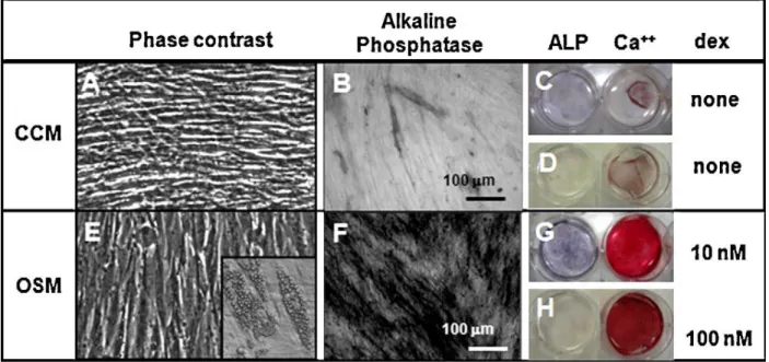
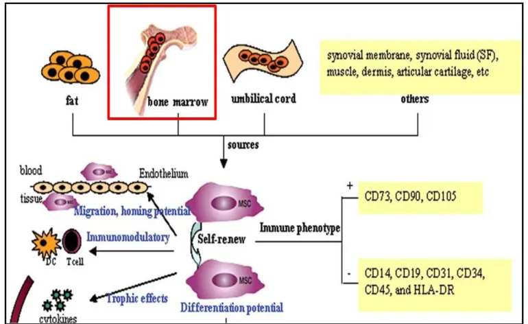
Figure 2.1: An arthroscopic view of the knee joint revealing a full thickness cartilage lesion on medial femoral condyle leading to exposure of subchondral bone………..……….….11 Figure 2.2: Schematic diagram depicting the inhibitory role of inflammatory mediators- IL-1beta, TNF-and IL-17 in chondrogenic differentiation of BMSCs. BMP-Bone morphogenetic protein, TGF-Transforming growth factor, Runx-Runt related gene, Sox-SRY box….…….…...13 Figure 2.3: Schematic representing the properties and role of stem cells. ………….…………...16 Figure 2.4.: In vitro characterization of BMSCs depicting their surface marker profile and differentiation potential. ………18 Figure 2.5: Schematic illustrating the hierarchical program of lineage specification and evolution of tri-, bi- and unipotent progenitors. ………....21 Figure 2.6: Schematic of stages in condensation phase of embryonic limb formation. Adapted from Maeda et al. ………...24 Figure 2.7: Molecular regulation of mesenchymal stem cell differentiation programs. Differentiation is transduced as a result of interaction between extracellular molecular signaling and mechanical inducers and putative receptors, channels, and/or other cell-surface-associated mechanisms. The downstream lineages arise from crosstalk of signaling pathways, including that between distinct mitogen-activated protein kinases (MAPKs) and R-Smads. GDF- growth and differentiation factor; TGF- transforming growth factor; BMP, bone morphogenetic protein; FA- fatty acid; βcat- β-catenin; PPAR- peroxisome proliferator-activated receptor; MSK- mitogen- and stress-activated protein kinase; PCAF- p300/CBP-associated factor; Ac- acetyl; c- chondroblast; o- osteoblast; a- adipoblast; m- myoblast; cm- cardiomyoblast; t- tenoblast……..25 Figure 2.8: Representative osteogenesis cultures cultured in control media (CCM) and osteogenic media (OSM) containing dexamethasone. The osteogenic phenotype was represented by cuboidal cells and detected by Alizarin Red S and alkaline phosphatase staining………...…28
Figure 2.9: Stages in BMS procedure. A. Arthroscopic image of a trochlear defect. B. Debridement of the calcified layer in the defect using a curette. C. BMS procedure using an awl to create multiple perforations in the subchondral bone. D. Second look at 1.5 years after BMS showing asymptomatic lesion in the same patient………..29 Figure 2.10: Cellular morphology of (A) early and (B) senescent BMSCs. (A). Cells are narrow and spindle shaped in younger passage. (B). Large and flattened senescent BMSCs in late passage are shown by arrows………..34 Figure 2.11: Figure illustrating the relationship between adult stem cell and mesenchymal progenitor theories of aging and free radicals, telomeres,and accumulation of DNA damage…..38 Figure 2.12: Schematic of structure of chitosan- cationic polymer of partly acetylated glucosamine………42 Figure 2.13: Chitosan inhibits the platelet mediated retraction of blood clot leading to negligible loss in volume (b) compared to > 50% loss in volume in clot formed by blood alone (a)…….…42 Figure 2.14: Comparison of cartilage repair outcome in groups treated “early” and “late”. (a). Repair response was better in acute defect group characterized by hyaline staining of matrix and evidence of SC bone remodeling. (b). Chronic defects were characterized by imcomplete fill, hypertrophy, uneven staining and poor bonding with adjacent cartilage……….…..52 Figure 4.1: Processing of rabbit femur. Femoral end secured on sample holder of Isomet saw while condylar segment is being sectioned off (a). Femoral end while distal trochlear segment is being sectioned off (b). Femoral end while proximal trochlear segment is being sectioned off (c). Three segments post-sectioning: Condyles, distal trochlea and proximal trochlea (d). Separation of a trochlear segment into Upper and Lower trochlea above and below the growth plate (in the young) and epiphyseal line (in the old) (e) All segments were fragmented with flat blades to yield 3 groups: Condyles, Upper trochlea (pooled from distal and proximal segments) and Lower trochlea (pooled from distal and proximal segments) (f). H&E stained paraffin sections of bone fragments were collected at pre-digestion (g-i) and post-digestion after 4 days of explant culture (j-l). Marrow spaces are initially occupied by cells. Arrows point to the bone lining cells in pre-digested samples (g-i). In post-digestion samples, marrow spaces are partly void, bone-lining cells are mostly absent. Dashed arrows point to empty bone lacunae in post-digestion samples (j-l). Bone marrow progenitor cells growing as colonies from Collagenase-derived cultures (m) and Explant-derived cultures (n). Arrow in n points to an explant bone fragment……….65
Figure 4.2: Cell yield observed in young animals (a, n = 8 knees) versus old animals (b, n = 3 knees) for collagenase- and explant-derived cultures. Cell yield was higher for Trochlea Upper in both collagenase- and explant-derived cultures in the young animals (a). Cell yield was also higher for Trochlea Upper in collagenase-derived samples in older animals (b). Data is presented as mean (circle); median (line); Box: 25th and 75th percentile; Whisker: Box to the most extreme point within 1.5 interquartile range. Horizontal lines show significant differences between pairs. Greater expression of stem cell marker CD44 was observed in young animals (c) versus old animals (d). CD34 was not expressed in young (e) or old (f) animals………...67 Figure 4.3: Clonogenic colonies stained with 1% (w/v) Crystal Violet stain show increased colony formation in the case of young (panels a to f) versus old animals (panels g to l). Higher CFU-f was observed for both trochlear segments versus condyles in collagenase-derived cultures in young animals (m, n = 8 knees), but not in old animals (n, n = 3 knees). CFU-f was consistently higher for collagenase-derived cultures compared to explant-derived cultures (m & n). Data is presented as mean (circle); median (line); Box: 25th and 75th percentile; Whisker: Box to the most extreme point within 1.5 interquartile range. Horizontal lines show significant differences between pairs………...68 Figure 4.4: Gross structure of pellets show increase in size upon culture in presence versus absence of TGF-βIII in young animals (a-f) but not in old animals (g to l). Pellets derived from collagenase-derived cultures are shown here. Pellets from trochlea upper samples were larger compared to pellets from condyles in young animals (m&n, n = 6 knees), and significantly so in the case of explant-derived cultures (n). All samples show low pellet diameter in old animals for collagenase- (o, n = 5 knees), and even lower in explant-derived cultures (p, n=2 knees). Data is presented as mean (circle); median (line); Box: 25th and 75th percentile; Whisker: Box to the most extreme point within 1.5 interquartile range. Horizontal lines show significant differences between pairs, unless otherwise indicated. ………...….70 Figure 4.5: Safranin O/Fast Green staining of pellets derived from collagenase- and explant-derived cultures, showing a high responder (panels a&b), medium responder (panels c&d) and low responder (panels e&f) in the young, as well as pellets derived from an older animal (panels g&h). TGF-βIII treatment only induced chondrogenesis in the young animals, as revealed by the chondrogenesis score (compare panels i&j to panels k&l). Cells derived from the trochlear upper
sample displayed increased chondrogenic potential in both collagenase-derived and explant-derived cultures in the young (i&j). Data is presented as mean (circle); median (line); Box: 25th and 75th percentile; Whisker: Box to the most extreme point within 1.5 interquartile range. Horizontal lines show significant differences between pairs………...…..72 Figure 4.6: Osteogenic cultures stained with Alizarin Red S (AR) showed increased matrix synthesis in presence (panels b-d&f-h) versus absence (a&e) of dexamethasone in young animals. Poorer osteogenic matrix synthesis was found in old animals (i-p). In young animals (n = 4 knees), AR accumulation was higher in upper trochlea samples in both collagenase-(q) and explant-derived cultures (r). Inferior AR accumulation was found in old animals (s&t, n=3 knees). Data is presented as mean (circle); median (line); Box: 25th and 75th percentile; Whisker: Box to the most extreme point within 1.5 interquartile range. Horizontal lines show significant differences between pairs……….…..74 Figure 5.1: Schematic representing the study design. Condylar and trochlear cartilage repair outcome in one knee from each animal was correlated with in vitro biological properties of BMSCs isolated from the condyle, upper trochlea and lower trochlea from the contralateral knee………....91 Figure 5.2: Macroscopic appearance of repair tissue in good and poor trochlear responders. Acute trochlear defects treated with BMS were largely restored with repair tissue in six of eight donors-good responders (a) while two donors had extremely poor repair outcome-poor responders (b). Condylar defects were incompletely filled with distinct holes and defect margins in all 8 donors (inserts c,d are from corresponding condylar repair outcomes in good and poor trochlear responders). Scale bar=1mm. Significantly higher mean macroscopic ICRS scores indicate superior early repair response in trochlea versus condyles (e)………....94 Figure 5.3: Representative Saf-O/Fast Green (a-h) staining of repair response in sections collected between holes (a,b,e,f) and through holes (c,d,g,h) in good (a-d) and poor (e-f) trochlear responders. Depleted Saf-O staining was evident in all condyles. Among good trochlear responders, abundant GAG deposition was observed, more evident in sections from between holes. Expression was reduced in poor trochlear responders. Arrow points to subchondral cyst observed in the condyle of a poor responder. Scale bar=1mm. Significantly higher O’Driscoll
scores indicate superior early repair response in trochlea versus condyles (i). Trochlear matrix was more abundant in GAG indicated by significantly higher %Saf-O (j) in repair tissues…….95 Figure 5.4: Representative Coll-II (a-h) staining of repair response in sections collected between holes (a,b,e,f) and through holes (c,d,g,h) in good (a-d) and poor (e-h) trochlear responders. Reduced coll-II staining was observed in all condyles. Increased collagen expression was observed in good vs poor trochlear responders, more evident in sections taken from between holes. The arrow points to a subchondral cyst observed in condyle of poor responder. Scale bar=1mm. Trochlear matrix was more abundant in collagen type-II indicated by significantly higher % Coll-II (i) in repair tissues………..97 Figure 5.5: Safranin-O/fast-green and collagen type II staining of pellets from good [a-n] and poor [a’-n’] trochlear responders. Negligible GAG and collagen type II expression was evident in condyles indicating inferior chondrogenic potential in vitro. GAG deposition was low in trochlea lower pellets in trochlea poor responders. a-h, a’-h’). Collagenase-derived; i-n, i’-n’). Explant-derived BMSCs. Scale bar=500 µm……….99 Figure 5.1-S: Summary of in vitro biological properties of collagenase- (a-e) and explant-derived BMSCs (f-j). Higher values were observed for trochlea versus condyles for following properties characterized-P0 cell yield (a,f); CFU-f (b,g); pellet size (c, h); % Saf-O_Pellets (d,i) and % Coll-II_Pellets (e,j)………..…100 Figure 5.2-S: Gross structure of pellets show an increase in size upon culture in presence of TGFβ-III. Pellets from trochlea upper were largest compared to condyles in good and poor trochlear responders. Trochlea lower pellets were smaller in poor trochlear responders. a). Collagenase-derived; b). Explant-derived BMSCs. Scale bar=1 mm……….101 Figure 5.3-S: CD44 expression was reduced in trochlea lower in collagenase-derived BMSCs (a vs b) and in all samples in explant-derived BMSCs (e vs f). CD34 expression was consistently absent (c,d,g,h)……….103 Figure 5.6: Strong, positive correlation between in vitro biological properties of BMSCs-Cell yield, CFU, pellet size, % Saf-O (pellets), % Coll-II (pellets) and repair response measured by ICRS macroscopic scoring and O’Driscoll Score in trochlea vs. condyles. N=8. C.I-95%;
p<0.0001-0.0068. (RT-repair tissues. Blue circles-Condyles, Red triangles-Trochlea Upper, Black squares-Trochlea Lower)………...105 Figure 5.7: Strong, positive correlation between in vitro biological properties of BMSCs-Cell yield, CFU, pellet size, % Saf-O (pellets), % Coll-II (pellets) and repair response measured by % Saf-O (RT) and % Coll-II (RT) in trochlea vs. condyles. N=8. C.I-95%; p<0.0001-0.0068. (RT-repair tissues. Blue circles-Condyles, Red triangles-Trochlea Upper, Black squares-Trochlea Lower)……….……….107 Figure 5.4-S: Weak, positive correlation between in vitro biological properties of Explant-derived BMSCs-Cell yield, CFU, pellet size, % Saf-O (pellets), % Coll-II (pellets)- and repair response measured by ICRS macroscopic scoring and O’Driscoll Score in trochlea vs. condyles. N=8. C.I-95%; p<0.0001-0.0068. (RT-repair tissues. Blue circles-Condyles, Red triangles-Trochlea Upper, Black squares-triangles-Trochlea Lower)………....109 Figure 5.5-S: Weak, positive correlation between in vitro biological properties of explant-derived BMSCs-Cell yield, CFU, pellet size, % Saf-O (pellets), % Coll-II (pellets)- and repair response measured by % Saf-O (RT) and % Coll-II (RT) in trochlea vs. condyles. N=8. C.I-95%; p<0.0001-0.0068. (RT-repair tissues. Blue circles-Condyles, Red triangles-Trochlea Upper, Black squares-Trochlea Lower)………..……….111 Figure 6.1: Procedure of surgical manipulation to create and treat chronic defects using BMS+CS/PRP implants (left panels) or BMS+PRP (right panels). a,b: Creation of 4 × 4 mm defects by debriding all non-calcified cartilage from trochlea; c,d: Appearance of chronic defects four weeks after creation at the time of second surgery; e,f: Treatment of defects by debriding spontaneous repair tissue (when present) and calcified cartilage and drilling 4 holes measuring 0.9 mm in width and 6 mm deep; g: Application of CS/PRP implant at defect site; h: Application of recalcified PRP at defect site………131 Figure 6.2: Macroscopic and histopathological assessment of fresh chronic defect (a,e,i), chronic defect after 4 weeks development (b,f,j), chronic defect treated with BMS+CS/PRP implant (c,g,k) and chronic defect treated with BMS alone (d,h,l). (e,i): Debridement was not homogenous and varying levels of calcified cartilage (CC) and debrided bone (DB) were seen in freshly debrided defects. (f,j): After 4 weeks, chronic defects showed evidence of partial spontaneous repair (SR) in some areas along with tufts of calcified cartilage (CC). (g,k):
Granulation tissue formation (GT) and enlarged drill holes were seen in presence of CS/PRP implants. (h,l): Fibrocartilagenous repair and endochondral ossification (EC) process were seen in presence of BMS alone, associated with chondrocyte hypertrophy (HT) and vascular invasion (VI). Red dotted lines in g&h represent original drill holes- hole enlargement and wound bloom effect is apparent in defect treated with BMS+CS/PRP (g). Scale bar (e-h):1 mm, (i-l): 100 µm………...……..135 Figure 6.3: Best (a,b) and worst (c,d) repair response in defects treated with BMS+CS/PRP (a,c) and BMS+PRP (b,d). Scale bar=1 mm. (e): Mean macroscopic ICRS score was higher (non-significant) in defects treated with BMS+CS/PRP versus defects treated with BMS+PRP…...136 Figure 6.4. Comparison of histopathological assessment of best and worst repair tissues generated by BMS+CS/PRP and BMS+PRP. (a-h): Saf-O staining for best (a,b,e,f) and worst (c,d,g,h) repair outcomes; (i-p): Coll-II immunostaining for best (i,j,m,n) and worst (k,l,o,p) repair outcomes; Scale bars 2.5mm (a-d & i-l) and 500 µm (e-h & m-p)……….…….138 Figure 6.5: Representative sections of repair tissues generated by BMS+CS/PRP and BMS+PRP. (a,b): Restoration of surface and structural integrity was better in presence of CS/PRP (b) versus PRP (a) (defect margins flanked by solid black arrows); (c,d): Missing repair tissue in BMS+PRP versus more uniform tissue in BMS+CS/PRP (d); (e,f): Comparison of adjacent cartilage (AC) showing improved appearance in the case of BMS+CS/PRP; (g,h): Best sections, (i,j): worst sections- All sections from same animal. Black arrows indicate zones of hypocellularity, yellow arrows indicate cell clusters, both more frequent in BMS+PRP. Cartilage–bone interface is marked by black dotted line (g). Scale bars=a,b: 1 mm, e-f: 250 µm, g-j: 100 µm………...…..140 Figure 6.6: (a) Mean O’Driscoll score was significantly higher for repair tissues in defects treated with BMS+CS/PRP versus defects treated with BMS+PRP. (b). Significant differences (*) were observed between treatments, and scores for adjacent cartilage (p=0.004), cellular changes (p=0.002), cell clusters (p=0.009), structural integrity (p=0.0001), surface integrity (0.05) and thickness of repair tissue (p=0.002) were significantly higher for defects treated with BMS+CS/PRP. (c). Criteria used in modified O’Driscoll scoring with respective score range……….141
Figure 6.7: a). Mean %Coll-II was significantly higher for repair tissues in defects treated with BMS+CS/PRP versus defects treated with BMS+PRP. b). Mean % Saf-O was higher for repair tissues in defects treated with BMS+CS/PRP versus defects treated with BMS+PRP, although this difference was not significant………...…….142 Figure 6.1-S: MicroCT 3-D analysis showed differences in structural parameters between defects treated with BMS+CS/PRP versus defects treated with BMS+PRP. Although the results were not significant, the values for bone surface density (a), bone surface (b), connectivity density (c), and trabecular number (f) were trending high for BMS+CS/PRP group, suggesting an increase in bone remodeling. i. Schematic representing the region of interest (ROI) for micro CT analysis……….144
LIST OF SYMBOLS AND ABBREVIATIONS α Alpha β Beta κ Kappa Ac Acetyl AC Articular cartilage
ACI Autologous Chondrocyte Implantation ADSC Adipose derived stem cell
AGE Advanced glycation end product APase Alkaline Phosphatase
ARS Alizarin Red S
BM Bone Marrow
BMP Bone Morphogenetic Proteins BMPC Bone marrow progenitor cell BMSC Bone marrow stem cell BMS Bone Marrow stimulation BS Bone surface area
BSA Bovine Serum Albumin
BV Bone volume
CaCl2 Calcium chloride cAMP cyclic AMP CC Calcified cartilage CCM Control media
Cfkh1 Chicken Forkhead 1
CFUf Colony Forming Unit-fibroblastic Col-I Type I collagen
Col-II Type II collagen Col-IV Type IV collagen Col-X Type X collagen Col-XI Type XI collagen
COMP Cartilage oligomatrix protein CPC Cetylpyridinium Chloride
CS Chitosan
CT Computed tomography DB Bone debridement DDA Degree of deacetylation
DMEM Dulbecco’s Modified Eagle’s Medium DNA Deoxyribonucleic acid
DRL Drill
ECM Extracellular matrix
EDTA Ethylene Diamine Tetraacetate EGF Epithelial Growth Factor EO Endochondral ossification FBS Fetal Bovine Serum FGF Fibroblast Growth Factor
FG Fast Green
GDF Growth and differentiation factor GF Growth factor
GLM General Linear Model GT Granulation tissue βGP β-Glycerophosphate HA Hyaluronic acid
HBSS Hank’s Buffered Salt Solution HCl Hydrochloric Acid
H&E Hematoxylin and Eosin HLA Human Leukocyte Antigen
Hu Human
HSC Hematopoetic stem cell HT Chondrocyte Hypertrophy IGF Insulin Growth Factor
ICRS International Cartilage Repair Society ISCT International Society of Cell Therapy IHH Indian Hedgehog
IL Interleukin
ISCT International Society of Cell Therapy ITS Insulin Transferrin Selenium
JNK Jun amino-terminal kinases kDa kilodalton
kV kilovolt
µ Micro
M Molar
MAPK Mitogen Activated Protein Kinase MEM Minimum Essential Medium MfH1 Mesenchymal Fork head-1 MFx Microfracture
MFC Medial femoral condyle
miR micro RNA
MMP Matrix Metalloproteinase MPC Mesenchymal Progenitor Cell MSC Mesenchymal stem cell
MSK Mitogen- and stress-activated protein kinase NBF Normal Buffered Formalin
OA Osteoarthritis OSM Osteogenic media
P0 Passage 0
PCAF p300/CBP-associated factor PCL Poly caprolactone
PDGF Platelet Derived Growth Factor PGE2 Prostaglandin E2
PLLA Poly-L-Lactic acid PMN Polymononuclear cells
PPAR Peroxisome proliferator-activated receptor PRP Platelet-Rich Plasma
P-S Penicillin Streptomycin PTH Parathyroid Hormone
PTHrP Parathyroid Hormone related Peptide PTOA Post traumatic osteorthritis
r Pearson Correlation coefficient
r2 Square of Pearson correlation coefficient
Rb Rabbit
RNA Ribonucleic acid RT Repair tissue
Runx Runt-related transcription factor Saf-O Safranin-O
SDF Stromal-derived factor SR Spontaneous repair TE Tissue Enginneering
TGF Transforming Growth Factor
TIMP Tissue inhibitors of metalloproteinase TNF Tumour Necrosis Factor
TV Total volume
VEGF Vascular Endothelial Growth Factor VCAM Vascular cell adhesion molecule VI Vascular invasion
Wnt Wingless int w/v Weight by volume
CHAPTER 1
INTRODUCTION
1.1 Introduction
The ends of long bones such as femurs are covered by a thin layer of cartilaginous tissue known as articular cartilage [1]. It is a highly specialized skeletal tissue and derives its unique biomechanical and physiological properties from an intact matrix comprised of proteoglycans and collagen type II [2]. Since this tissue lacks blood, nervous and lymphatic supply, in the event of an injury, the regeneration process is either not initiated or is insufficient to restore healthy tissue. Chondrocytes, the primary cell type of cartilage- are embedded in copious amount of ECM and are unable to migrate to populate the region of defect. Negligible blood supply further aggravates the problem since the stimulatory signals required to drive a repair process fail to propagate [3]. Full thickness lesions which reach sub-chondral bone undergo a limited repair process due to the access to the progenitor cells in the bone marrow stroma, though the quality of the repair tissue is mainly fibrocartilagenous with inferior mechanical and physiological properties On the other hand, partial thickness defects are not repaired at all [4, 5].
The knee joint is the most commonly affected joint and accounts for 75% of all lesions observed in articular cartilage. In an early study involving over 31,000 patients, it was found that while 63% knees had chondral lesions, 20% suffered from full thickness lesions [6]. It is expected that 60% of the patients who develop cartilage lesions will have osteoarthritis (OA) within 20 years of generating an articular cartilage defect [6]. Cartilage loss generally occurs due to age-related wear and tear and the likelihood of its degradation increases with increasing age. In a population with increasing life expectancy, up to 40% of population above 70 could be expected to be affected by OA leading to a burden of 5-20 billion dollars on the Canadian government [7]. However, trauma related osteoarthritis is not uncommon and although focal at onset, may progress into OA if not addressed swiftly. Cartilage degeneration is multifactorial and involves femoral, patellar and synovial compartments. Catabolic activities mediated by pro-inflammatory cytokines such as IL-1 and TNF-α are not balanced by anabolic growth factors like IGF-1 and TGF-β and anti-inflammatory cytokines like IL-4 and IL-10 [8, 9]. The disrupted equilibrium leads to degeneration of matrix exposing the subchondral bone and causing patient discomfort [1-5].
Bone marrow stimulation (BMS) initiates cartilage repair by fracturing or drilling into subchondral bone at the base of a debrided cartilage defect, typically leading to the formation of a fibrocartilagenous repair tissue providing varied levels of clinical relie-f. It is relatively easy, economical, associated with swift recovery and provides appreciable short to mid term relief. Due to advantages offered by the procedure, BMS is adopted as first line of treatment globally. However, repair outcomes are affected by defect location and age, suggesting a strong influence of the structural and biological factors in the knee joint. These differences may include anatomical differences in the structure of knee as well as biological properties of mesenchymal progenitor cells in the subchondral bone. It is well known that human trochlear defects are rarer but harder to treat compared to condyles [10], while animal models present varying scenarios. In an earlier study, rabbit trochlea showed a superior repair and chondrogenic potential compared to medial femoral condyle [11, 12]. On the other hand, quality of repair was better in condyle than trochlea in a sheep model [13]. Taken together, these studies emphasize the need to address the inconsistent repair outcomes arising from location-dependent factors. Unfortunately, BMS procedures are also severely affected by increasing age leading to incomplete regeneration, high inter-individual variability and poorer outcome in older individuals. Therefore, identification of mechanism for reduced potential of progenitor cells in cartilage repair is required in order to understand fundamental causes of poor repair in older individuals. The exact mechanism of donor-, location- and age-dependent variability still eludes us since our knowledge about underlying factors such as bone structure and mechanics, load-bearing conditions and the role of subchondral bone progenitor cells is still extremely limited. A better understanding of the underlying causes and mechanism of variability in the marrow stimulated repair outcomes will enable us to make the approach more predictable, clinically efficient and personalized to patient’s needs. In addition, it will be pave the way to develop strategies to enhance the quality of repair, especially in hard to treat cases.
BMS provides access to the underlying bone marrow stroma rich in bone marrow progenitor cells (BMPCs)/bone marrow stem cells (BMSCs)/mesenchymal stem cells (MSCs) by creating channels in the subchondral bone [14]. BMSCs are characterized by a unique potential of self renewal and ability to differentiate into cells of multiple lineages [15]. When recruited to the site of defect, they differentiate into chondrogenic phenotype leading to the formation of a repair tissue with varying amounts of hyaline and fibrous cartilage [16]. Since BMS mediated repair of
soft and hard tissues relies on recruitment of underlying BMSCs, it is logical to explore the properties of these progenitors in order to provide important mechanistic insights into variability present in marrow stimulated repair outcomes. It is reasonable to assume that differences in number, stemness and differentiation potential of BMSCs will influence the chondrogenesis in vitro and cartilage repair in vivo. Biological characterization can be carried out in vitro by means of several assays including determination of cell yield, CFU-f, expression of cell surface markers and multilineage differentiation assays. In a previously published study, our group demonstrated superior repair when trochlea was drilled to provide access to metaphyseal marrow [17]. Due to likely differences in the progenitor cell population in the epiphyseal and metaphyseal regions of bone, and their subsequent influence on repair response, the cells derived from these locations need to be characterized individually. Cells participating in BMS include cortico-spongious bone and marrow spaces and therefore characterization of cells originating from both these sources is warranted.
Though repair response induced by BMS along with characterization of BMSCs has been investigated in animal models before [18-21], a study designed to correlate the repair response in different anatomic locations with biological properties of BMSCs is lacking. Needless to say, inherent differences in the properties of BMSCs can be expected to influence cartilage repair outcomes in a profound manner. A statistically sound study designed to investigate the influence of location- and donor-dependent factors on cartilage repair outcomes is more likely to elucidate the underlying mechanism of inter-individual variability observed in cartilage repair. Inferences from such an analysis will probably aid clinicians in developing repair strategies on a more patient-relevant basis.
Another reason for poor quality of BMS repair is the compromised residency and stability of marrow-derived blood clot that fills cartilage lesions following bone fracturing [13, 18, 19]. Due to platelet-mediated clot retraction and serum exudation, the clot can lose more than half of its original volume. A retracted clot will eventually lead to early detachment of repair tissue from bone bed undermining the efficacy of this procedure. In contrast, a voluminous, physically adherent and stable clot is generated when chitosan - a biocompatible and biodegradable polymer comprised of glucosamine and N-acetyl glucosamine residues - is mixed with autologous blood prior to clotting and leads to an improved osteochondral repair [13]. Platelet-rich-plasma (PRP)
containing concentrated platelets is a rich source of growth factors, cytokines and chemokines [22-24] and has been shown to provide adequate stimulation for recruitment and chondrogenic differentiation of BMSCs [25, 26]. However, PRP clots are even more prone to retraction which might lead to insufficient chondrogenesis and hyaline cartilage restoration as reported by some authors [27]. A combination of freeze-dried chitosan and PRP would increase the stability and bioactivity of PRP likely improving cartilage repair outcomes in challenging cases. Liquid chitosan in aqueous solution suffers from limited shelf life due to poor stability during storage [28]. Therefore, freeze-dried chitosan offers the advantages of improved stability, longer shelf life and easier sterilization.
The principle of augmented marrow-stimulation relies on the enhanced recruitment of mesenchymal progenitor cells to the lesion accompanied by increased stability of growth factors and BMSCs in the defect milieu. It is important to bear in mind that cartilage repair does not occur in isolation and inflammatory mediators in defect milieu are likely to complicate the healing process. Therefore, it becomes imperative to examine cartilage repair strategies in a model which is a more realistic depiction of clinical situation. Interplay of catabolic factors including damaging cytokines and pro-inflammatory factors are more likely to be represented in a model with degenerative, chronic lesions [29, 30]. To ensure successful clinical translation, it is critical to explore the potential of chitosan and PRP in improving the repair outcome in chronic lesions which are harder to treat compared to acute lesions.
The objective of the study presented here is to make significant contributions to enhance the existing understanding of variability and unpredictability in marrow stimulated repair outcomes. The research aims to address the gap in our current comprehension of the underlying mechanisms of donor-, age- and location-dependent inconsistency observed with BMS, likely arising due to inherent properties of mesenchymal progenitor cells. Correlation of intrinsic biological properties of progenitor cells and cartilage repair outcome will eventually enable clinicians to perform BMS in a more patient-specific basis where intrinsic repair capacity is highly variable and unpredictable. Finally, conclusions from analysis of BMS augmented with chitosan/PRP implants in a chronic lesion will likely offer a promising strategy specifically designed to enhance the cartilage repair outcome in demanding cases.
1.2 Research Hypotheses and Objectives
1.2.1 General objective
The research presented here was carried out with an objective to explore the underlying mechanism of variability in marrow stimulated repair outcome by identifying location-, age- and donor-dependent factors influencing the inherent properties of subchondral bone progenitor cells (or mesenchymal stem cells, MSCs). Furthermore, strategies to improve cartilage repair outcome mediated by augmented BMS techniques were investigated in animal studies designed for a more accurate representation of the chronic lesions present in the clinical situation.
1.2.2 Study 1
The aim of this study was to carry out a comprehensive analysis of the progenitor cells present in the subchondral bone of rabbit condyle and trochlea, taking into account location and age revealing a likely mechanism for variability in marrow stimulated cartilage repair seen in vivo.
1.2.2.1 Hypothesis1
We hypothesized that the inherent biological properties of bone marrow progenitor cells (BMPCs; or mesenchymal stem cells, MSCs) including number, clonogenicity, surface marker profile and multilineage differentiation potential will be influenced by location and age while origin of the cells will have no significant bearing on these properties. Specific hypotheses tested through this study included the following:
In-vitro biological properties including P0 cell yield, clonogenic potential, expression of cell surface markers, chondrogenic and osteogenic differentiation potential will be superior for BMPCs/MSCs isolated from rabbit trochlea compared to condyles.
Cell yield, stemness markers, clonogenic, chondrogenic and osteogenic potential of BMPCs/MSCs will decrease with increasing age.
The aforementioned in vitro biological properties of BMPCs/MSCs will not be affected by the method of isolation and will be similar for collagenase- and explant-derived BMPCs/MSCs.


![Figure 2.3: Schematic representing the properties and role of stem cells. [ Copied image -67]](https://thumb-eu.123doks.com/thumbv2/123doknet/2329517.31334/47.918.226.696.103.455/figure-schematic-representing-properties-role-cells-copied-image.webp)
![Figure 2.5: Schematic illustrating the hierarchical program of lineage specification and evolution of tri-, bi- and unipotent progenitors [Image copied from Phinney at al-77]](https://thumb-eu.123doks.com/thumbv2/123doknet/2329517.31334/52.918.177.748.113.573/schematic-illustrating-hierarchical-specification-evolution-unipotent-progenitors-phinney.webp)
![Figure 2.6: Schematic of stages in condensation phase of embryonic limb formation. Adapted from Maeda et al [67]](https://thumb-eu.123doks.com/thumbv2/123doknet/2329517.31334/55.918.137.779.440.859/figure-schematic-stages-condensation-embryonic-formation-adapted-maeda.webp)