Pépite | Propriétés antagonistes et probiotiques de nouvelles bactéries lactiques et levures isolées des matières fécales humaine et animale
223
0
0
Texte intégral
(2) Thèse de Alaa Abdulhussain Al-Seraih, Lille 1, 2016. Acknowledgements I would like to convey my heartfelt gratitude and sincere appreciation to all the people who have helped and inspired me during my doctoral study. This thesis would not have been possible without the support of many people. First of all, I would like to express my deep appreciation and gratitude to my honorable advisor, Professor Dr. Djamel Drider, for the patient guidance and mentorship he provided to me throughout my PhD study. Professor Drider’s intellectual heft is matched only by his genuine good nature and humility, and I am truly fortunate to have had the opportunity to work with him. Also, with a deep sense of honor, I would like to express my sincere gratitude to Professor Dr. Pascal Dhulster, the director of Charles Viollette Institute, for his unceasing and encouraging support. I would like to thank all of the thesis committee, Professor Dr. Véronique Delcenserie, Dr. Nathalie Connil, Dr. John Baah, Dr. Benoit Cudennec, and Dr François Krier, for accepting the evaluation of my PhD work, and for their insightful comments and encouragement. My sincere thanks also go to Dr. Rabah Boukherroub (IEMN, CNRS) and Professor Dr. Sabine Szunerits (IMEN, Lille 1) for their collaboration and SEM assistance. I would like to extend thanks to Dr. Annee Vachee (Roubaix hospital, France) for her cooperation in the antibiotic resistance analysis. I would also like to express my heartfelt gratitude to Dr. Yanath Belguesmia, Dr. Max Béchet, and Dr. Gabrielle Chataigné for their help, support, and encouragement throughout my PhD work. Similar profound gratitude goes to my friends Ahmed, Ameen, Yazen, Hamza, Mahammed, Qassim, Michael, Debarun, Alexandre, Delphine, Juliette and all other members of the QSA and ProBioGEM teams who provided me a family environment for more than four years.. II. © 2016 Tous droits réservés.. lilliad.univ-lille.fr.
(3) Thèse de Alaa Abdulhussain Al-Seraih, Lille 1, 2016. I would like to extend thanks to the Iraqi Ministry of Higher Education and Scientific Research for giving me the opportunity to do my PhD by awarding the doctoral scholarship. Similar deep gratitude goes to Campus France Organization, especially to Mr. Adrien Chalancon, who successfully managed the dossiers of Iraqi students. Also with deep sense of honor, I wish to express my heartily gratitude to the Best Environmental Technologies Company (Alberta, Canada), especially to Mr. Jim G. Watson and Dr. John Baah for all their support of my PhD project. Finally, I have no words to express my profound gratitude and appreciation to my family. My loving mother, father and brothers Bahaa, Osama, Ahmed, and Amir always gave me unconditional love, support, and encouragement during all the stages of my life. To each of them, I would like to say thank you from the bottom of my heart. No words can either express the deepest gratitude and appreciation I have for my loving wife Hiba, who has always supported me and helped me overcome all difficulties during my studies. She has been there for me every minute of every year of my studies and supported me and my work without complaint. I would like to extend my warmest thanks to my dear son, Murtadha. If this work has sometimes prevented us from sharing important moments of life, know that I never stopped thinking about you. You are the most important people in my life, and I dedicate this work to all of you.. III. © 2016 Tous droits réservés.. lilliad.univ-lille.fr.
(4) Thèse de Alaa Abdulhussain Al-Seraih, Lille 1, 2016. Scientific valorization. Publications in peer reviewed journals 1- Al-Seraih A, Flahaut C, Krier F, Cudennec B, Drider D., 2015. Characterization of Candida famata Isolated from Poultry Feces for Possible Probiotic Applications. Probiotics and antimicrobial proteins. 7, 233-41. 2- Al-Seraih A, Belguesmiaa Y, Baah J, Szuneritsc S, Boukherroubc R, Drider D., 2016. Enterocin B3A-B3B produced by LAB collected from infant feces: potential utilization in the food industry for Listeria monocytogenes biofilm management. In Antonie van Leeuwenhoek Journal of Microbiology (Under review). 3- Al-Seraih, Kergourlay G, Cudennec B, Baah J, Belguesmia Y, Drider D., 2016. Enterococcus faecalis B3A-B3B: Genome analysis and experimental evidences to claim its beneficial attributes. (Manuscript in preparation).. Posters and workshops 1- Al-Seraih A, Flahaut C, Krier F, Cudennec B, Drider D. Characterization of Candida famata Isolated from Poultry Feces for Possible Probiotic Applications. Annual Conference of Microbiology Society. 21-24 March 2016, Liverpool, UK. 2- Al-Seraih A, Belguesmia Y, Desriac F, Drider D. Enterocin B3A-B3B from infant origin helps nisin to control growth of the foodborne pathogen Listeria monocytogenes. AMP. 6-8 June 2016. Montpellier, France. 3- 2nd International workshop on (Bioinformatics Tools for PKS and NRPS Discovery from Genomic Data for the Product), University of Lille1. 28-30 October, 2015. Lille, France.. IV. © 2016 Tous droits réservés.. lilliad.univ-lille.fr.
(5) Thèse de Alaa Abdulhussain Al-Seraih, Lille 1, 2016. ABSTRACT In this study, we isolated probiotic yeasts and lactic acid bacteria (LAB) from different microbial sources. Eighty-one (81) yeasts and seventy (70) LAB isolates were randomly selected and identified from fecal samples of poultry feces and healthy Iraqi infants, respectively. The yeast strains were obtained from a farm of broiler chickens located in the city of Lille. They were clustered into 22 groups by GTG5-rep PCR technique, then identified as Debaryomyces hansenii, (teleomorph of Candida famata) species using the biochemical ID-32C system and molecular sequencing of 26S rDNA and ITS1-5.8-ITS2 rDNA region methods. Only one yeast strain, designated as Candida famata Y.5 (C. famata Y.5), exhibited antimicrobial activity against Listeria innocua. For more accurate discrimination, the antagonistic strain C. famata Y.5 was identified by MALDI-TOF-MS technology. Further characterization of this anti-Listeria strain, permitted to unveil its probiotic potential. Thus, C. famata Y.5 appeared to be a non-hemolytic strain. In vitro tests of cytotoxicity and adhesion on human Caco-2 epithelial cells confirmed the safety traits of this strain. C. famata Y.5 displayed good surface properties, especially auto-aggregation, in addition to high survival ability under harsh conditions mimicking those of the gastrointestinal tract (GIT). The LAB strains were isolated from fecal samples of a group of Iraqi children living in the north of France. LAB strains were obtained from six blind donors and then identified as 41 cocci and 29 bacilli. Two strains displayed antagonistic activities against. Gram-positive. bacteria. (GPB). including:. Listeria. monocytogenes,. Staphylococcus aureus, methicillin-resistant Staphylococcus aureus (MRSA), and Clostridium perfringens but not against fungi or Gram-negative bacteria (GNB), except for Salmonella Newport. The biochemical, MALDI-TOF-MS, and molecular (16S rDNA sequencing) methods identified these two strains as Enterococcus faecalis B3A-B3B and B20A-B20B . Bacteriocin produced by strain B3A-B3B, designed as V. © 2016 Tous droits réservés.. lilliad.univ-lille.fr.
(6) Thèse de Alaa Abdulhussain Al-Seraih, Lille 1, 2016. enterocin B3A-B3B, was purified by a simplified two-step procedure including a liquid-liquid. phase. extraction. and. reverse. phase. high-performance. liquid. chromatography (RP-HPLC). The predicted molecular mass of this enterocin consists of two peptides of 5,176.31 Da (B3A) and 5,182.21 Da (B3B). Notably, B3A-B3B hampered the biofilm installation of L. monocytogenes strain grown on AISI 304 stainless steel slides. The treatment of stainless steel with nisin (1 mg. ml-1 or 16 mg. ml-1) diminished the cell numbers by about 2 logs CFU. ml-1, preventing therefore the biofilm formation by L. monocytogenes 162 or by its nisin-resistant variant L. monocytogenes 162R. Further combination of nisin and B3A-B3B enterocin reduced the MIC value needed to inhibit this pathogen about 2 logs CFU. ml-1. To gain insights on the probiotic profile of the E. faecalis B3A-B3B strain, the whole genome was sequenced and in silico analysis was performed and compared with those of clinical strains as E. faecalis MMH594, E. faecalis V583, and E. faecalis OG1RF from humans, and also compared to that of the well-known probiotic E. faecalis Symbioflor1 strain. Even harboring gelE, cpd, efaAfm, ccf, agg, and cob coding for virulence factors, the B3A-B3B strain resulted to be sensitive to most antibiotics tested here, non-cytotoxic, non-hemolytic, and devoid of inflammatory effects. Moreover, B3A-B3B strain showed remarkable hydrophobicity, autoaggregation, adhesion to human Caco-2 cells, viability in simulated GIT conditions, and cholesterol assimilation. These features together introduce the E. faecalis B3AB3B strain as an interesting probiotic candidate.. VI. © 2016 Tous droits réservés.. lilliad.univ-lille.fr.
(7) Thèse de Alaa Abdulhussain Al-Seraih, Lille 1, 2016. RESUME Dans cette étude, nous avons isolé des levures et des bactéries lactiques (BL) potentiellement probiotiques, à partir de différents écosystèmes microbiens. Quatrevingt-une (81) levures, et soixante-dix (70) BL, ont été isolées et identifiées à partir de matières fécales animales (poulet) et humaines (enfants Irakiens en bonne santé). Ainsi, les souches de levures ont été isolées à partir de matières fécales de poulets, dans une ferme située dans la région de Lille (France). Elles ont été regroupées en 22 groupes par la technique de Rep-PCR utilisant une amorce unique 5'GTG5-3', puis identifiées comme appartenant à l’espèce Debaryomyces hansenii (téléomorphe de Candida famata) en utilisant des méthodes biochimique (système ID32C) et moléculaire (séquençage de l'ADNr 26S et les régions ITS1-5.8-ITS2 de l’ADNr). Dans le criblage des activités antibactériennes, seule la souche nommée, Candida famata Y.5, a montré une activité contre Listeria innocua. L'identification de cette souche a été confirmée par la méthode robuste de MALDI-TOF-MS. Une ample caractérisation de cette souche, a permis de révéler son potentiel probiotique. Ainsi C. famata Y.5 est non-hémolytique, non-cytotoxique et présente une capacité d’adhésion remarquable sur les cellules Caco-2 épithéliales. Cette souche s'avère posséder des propriétés de surface intéressantes en particulier les capacités d’auto-agrégation, et de survie dans les conditions du tractus gastro-intestinal. Comme précédemment indiqué, les bactéries lactiques, quant à elles, ont été isolées à partir d'échantillons fécaux, provenant d’un groupe d'enfants irakiens résidant dans le nord de la France. 70 souches lactiques ont été obtenues à partir de six donneurs, celles-ci ont été caractérisées comme étant 41 cocci et 29 bacilles avec une coloration différentielle de Gram positive et une absence de catalase. Le criblage des activités antagonistes, a permis de mettre en évidence une activité contre des bactéries à Gram-positif comprenant Listeria monocytogenes, Staphylococcus aureus, Staphylococcus aureus résistant à la méthicilline (SARM), et Clostridium perfringens, mais pas contre des champignons microscopiques ou les bactéries à Gram-négatif (GNB), à l'exception de Salmonella Newport. L'utilisation de plusieurs méthodes d'identification comme les méthodes biochimiques, et moléculaires (séquençage de l'ADNr 16S et MALDI-TOF-MS) ont permis d'identifier avec certitude ces deux VII. © 2016 Tous droits réservés.. lilliad.univ-lille.fr.
(8) Thèse de Alaa Abdulhussain Al-Seraih, Lille 1, 2016. souches comme appartenant à l’espèce Enterococcus faecalis. Ces souches sont alors nommées E. faecalis B3A-B3B et B20A-B20B. L'activité antagoniste est attribuée à une activité bactériocinogénique. Ainsi, la bactériocine produite par la souche B3AB3B est désignée entérocine B3A-B3B. Celle-ci a été purifiée par une procédure en deux étapes comportant une extraction en phase liquide-liquide, suivie par une chromatographie liquide à haute performance en phase inversée (HPLC-RP). Les masses moléculaires prédites pour les deux peptides composant cette entérocine seraient de 5 176,31 Da (B3A) et 5 182,21 Da (B3B). Par ailleurs, cette entérocine a montré une activité limitant l'installation de biofilms de L. monocytogenes cultivées sur des lames AISI 304 en acier inoxydable. Le prétraitement de ces lames avec de la nisine à 1 ou 16 mg. ml-1 permet de réduire le nombre de cellules bactériennes sur cette surface d'environ 2 logs UFC. ml-1, perturbant ainsi la formation de biofilms par L. monocytogenes 162 ou son variant résistant à la nisine, appelé L. monocytogenes 162R. En plus la combinaison des deux bactériocines (nisine et entérocine B3A-B3B) permet de réduire la valeur de la concentration minimal inhibitrice (CMI) nécessaire pour inhiber ce pathogène d’environ 2 logs CFU. ml-1. Pour déterminer le profil probiotique de la souche E. faecalis B3, son génome a été séquencé et comparé aux génomes des souches cliniques notamment E. faecalis MMH594, E. faecalis V583 et E. faecalis OG1RF d'origine humaine, et au génome de la souche probiotique E. faecalis Symbioflor1. Même si six gènes codant pour des facteurs de virulence, gelE, cpd, efaAfm, ccf, agg, et cob, ont été retrouvés dans l'ADN de notre souche, cell-ci s'est globalement avérée sensible aux antibiotiques utilisés dans cette étude, noncytotoxique, non-hémolytique et dépourvue d’effets inflammatoires sur les lignées cellulaires de type Caco-2. En outre, E. faecalis B3A-B3B a montré des caractéristiques d’hydrophobicité, d’auto-agrégation, d'adhésion aux cellules Caco-2, un taux de survie aux conditions gastro-intestinales simulées, et une capacité d’assimilation du cholestérol remarquables. Les résultats obtenus laissent à penser et considérer la souche E. faecalis B3A-B3B comme une souche avec un potentiel probiotique intéressant.. VIII. © 2016 Tous droits réservés.. lilliad.univ-lille.fr.
(9) Thèse de Alaa Abdulhussain Al-Seraih, Lille 1, 2016. Table of contents Acknowledgements............................................................................................................... II Scientific valorization ........................................................................................................... IV ABSTRACT............................................................................................................................. V List of figures ....................................................................................................................... XI List of tables ....................................................................................................................... XII List of frequently used abbreviations .................................................................................. XIII General Introduction..............................................................................................................1 Chapter 1. Literature Review ..................................................................................................6 1. Microbial community in gastrointestinal tract and feces......................................................6 1.1. Development of gut microbiota .......................................................................................7 1.1.1. Neonate and infancy.....................................................................................................8 1.1.2. Early .............................................................................................................................8 1.1.3. Adult ............................................................................................................................9 1.1.4. Elderly ..........................................................................................................................9 1.2. Factors affecting microbiota development and composition .............................................9 1.2.1 Maternal microbiota.................................................................................................... 10 1.2.2 Mode of delivery ......................................................................................................... 11 1.2.3 Mode of feeding .......................................................................................................... 11 1.2.4. Geographical location and environment ...................................................................... 12 1.2.5 Antibiotics, probiotics and prebiotics ........................................................................... 13 1.2.6. Other factors .............................................................................................................. 14 2. Host microbe interactions ................................................................................................ 14 2.1. Host–microbiota cross-talk ............................................................................................ 17 2.2. Microbial adhesion and interaction with pathogens ....................................................... 17 3. Probiotics......................................................................................................................... 18 3.1. What are probiotics? ..................................................................................................... 18 3.2. Probiotic criteria and safety assessment ........................................................................ 19 3.3. Probiotics and their health effects ................................................................................. 23 4. Fecal origin microbes with probiotic potential................................................................... 25 4.1. Yeasts as probiotics ....................................................................................................... 26 4.1.1 General characteristics of yeast ................................................................................... 26 4.1.2. Saccharomyces cerevisiae var. boulardii ...................................................................... 28 IX. © 2016 Tous droits réservés.. lilliad.univ-lille.fr.
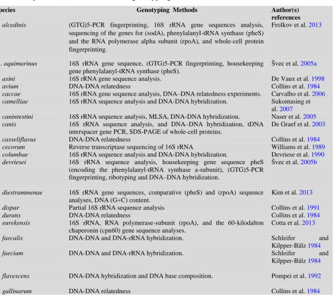
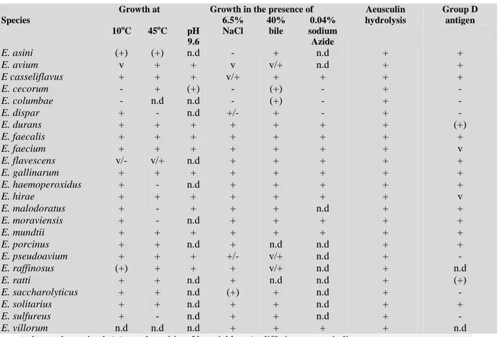
(10) Thèse de Alaa Abdulhussain Al-Seraih, Lille 1, 2016. 4.1.3. Debaryomyces hansenii (Candida famata) .................................................................. 29 4.1.3.1. Taxonomy, morphology and physiology ................................................................... 29 4.1.3.2. Biochemical properties with industrial importance and applications ......................... 31 4.1.4. Probiotic and antagonistic potential of Debaryomyces hansenii ................................... 34 4.2. Lactic acid bacteria as probiotics .................................................................................... 36 4.2.1. General characteristics of lactic acid bacteria .............................................................. 36 4.2.2. Taxonomy of the genus Enterococcus .......................................................................... 36 4.2.3. Phenotypic characteristics of Enterococcus genus ........................................................ 38 4.2.4. Genotypic characterization of the genus Enterococcus ................................................. 40 4.2.5. Physiological properties .............................................................................................. 44 4.2.6. Biochemical properties with industrial importance and applications ............................ 45 4.2.6.1. Lactic acid production .............................................................................................. 46 4.2.6.2. Proteolytic activity ................................................................................................... 47 4.2.6.3. Lipolytic activity....................................................................................................... 48 4.2.6.4. Citrate and pyruvate metabolism ............................................................................. 48 4.2.6.5. Production of volatile compounds ............................................................................ 50 4.2.6.6. Bacteriocin production............................................................................................. 51 4.2.6.6.1 Class I bacteriocins (Lantibiotics) ............................................................................ 52 4.2.6.6.2 Class II bacteriocins ................................................................................................ 54 4.2.6.6.3 Class IIa bacteriocins (pediocin-like peptides) ......................................................... 54 4.2.6.6.4 Class IIb bacteriocins (two-peptide bacteriocins)..................................................... 56 4.2.6.6.5 Class IIc bacteriocins (circular bacteriocins) ............................................................. 60 4.2.6.6.6 Class IId bacteriocins (unmodified, linear, non-pediocin-like bacteriocins) ............... 60 4.3 Enterococci as probiotics ................................................................................................ 61 Chapter 2. Yeasts from fecal samples of chicken displayed anti-Listeria activities and further probiotic properties ............................................................................................................. 65 Chapter 3.Enterocin B3A-B3B produced by LAB collected from infant feces: potential utilization in the food industry for Listeria monocytogenes biofilm management .................. 94 Chapter 4. Bacteriocinogenic LAB from the feces of Iraqi infants and their potential as probiotics .......................................................................................................................... 139 General Conclusions and Prospects .................................................................................... 176 References......................................................................................................................... 181. X. © 2016 Tous droits réservés.. lilliad.univ-lille.fr.
(11) Thèse de Alaa Abdulhussain Al-Seraih, Lille 1, 2016. List of figures Figure 1. Distribution and variations in microbial numbers and composition across the length of the GIT. Figure 2. Factors influencing the development of infant, adult, and elderly gut microbiota. Figure 3. Pattern recognition receptors (PRRs) signaling promotes immune homeostasis. Figure 4. Some examples of commonly used prokaryotic and eukaryotic genera as probiotics. Figure5. Guidelines for the Evaluation of Probiotics for Food Use. Figure 6. Probiotics effect on animal health and production. Figure 7. A commercial probiotic product for human use containing 32 probiotic strains of bacteria and yeasts including D. hansenii. Figure 8. Phylogenetic position of the genus Enterococcus demonstrated by 16S rRNAdendrogram of Gram-positive genera. Figure 9. Schematic pathway showing the metabolic relationships between citrate and glucose. Figure 10. Diagramatic representation of Enterococcus faecalis Fly1 type 1 lantibiotic. Figure 11. The model of killing by Man-PTS targeting and immunity of Class IIa bacteriocins. Figure 12. A structural model of lactococcin G and its orientation in target-cell membranes. Figure 13. Some commercial products of Enterococcus faecalis validated as probiotics. Figure 14. The chicken gut microbiome. Figure 15. The rep-PCR DNA fingerprints generated in 1.0% agarose gel, requested by the use of the GTG5 primer 5’–GTGGTGGTGGTGGTG- 3’. Figure 16. (a) RP-HPLC chromatogram of the bacteriocin purification with a zoom on the active peak corresponding to the enterocin B3A-B3B. (b) Mass spectrometry of enterocins B3A-B3B. Figure 17. Alignments of the 3’ ends of structural genes coding for enterocins. Figure 18. Mode of action of Nisin. Figure 19. Some mechanisms and genes involved in bacterial resistance to bacteriocin of Gram positive bacteria. Figure20. E. faecalis cytolysin expression. Figure 21. The main mechanisms of Enterococcus spp. antibiotic resistance.. XI. © 2016 Tous droits réservés.. lilliad.univ-lille.fr.
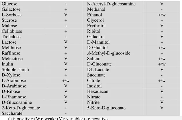
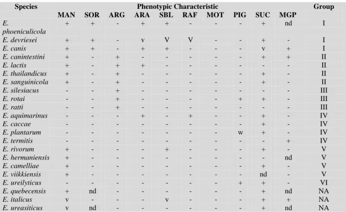
(12) Thèse de Alaa Abdulhussain Al-Seraih, Lille 1, 2016. List of tables Table 1. Some beneficial effects of probiotics on human health. Table 2. Assimilation of different substrates by D. hansenii. Table 3. Phenotypic properties of recently identified Enterococcus species. Table 4. Species of Enterococcus and genotyping methods used for their identification. Table 5. Physiological properties of validly described Enterococcus species. Table 6. Effect of enzymes on antilisterial activity of neutralized cell free supernatant (CFS) and semi-purified enterocin B3A-B3B (SP). Table 7. Spectrum registered with the cell-free supernatant (CFS) before its neutralization (BN) and after its neutralization (AN) as well as that obtained with the semi-purified bacteriocin (SP). Table 8. Determination of minimum inhibitory concentrations (MIC) of Enterocin B3A-B3B. Table 9. Minimum inhibitory concentrations (MICs) of Nisin and Enterocin B3. Table 10. Numbers of LAB isolates in the fecal samples of Iraqi infants. Table 11. Virulence genes present in E. faecalis B3. Table 12. Antibiotic susceptibility of E. faecalis B3A-B3B strain in comparison with a pathogenic strain E. faecalis ATCC29212.. XII. © 2016 Tous droits réservés.. lilliad.univ-lille.fr.
(13) Thèse de Alaa Abdulhussain Al-Seraih, Lille 1, 2016. List of frequently used abbreviations. AAD ABC AEDS AFLP AID AMPs AN ATP ATCC aw BN CDC CFS CFU Cls CM cpn60 CW DBPC DC DMDS DMTS DNA ECDC ED EFSA EMP EPS FAO FIC gad GdpD GF GIT GNB GPB GRAS h HIV IBD IBS IgE. Antibiotic-Associated Diarrhea ATP-Binding Cassette Atopic Eczema/Dermatitis Syndrome Amplified Fragment Length Polymorphism Acute Infections Diarrhea Antimicrobial Peptides Antimicrobial Activity Before Neutralization Of Supernatant With 1M NaOH Adenosine Triphosphate American Type Culture Collection Water Activity Antimicrobial Activity Before Neutralization Of Supernatant Centre For Disease Control And Prevention Cell Free Supernatant Colony Forming Unit Cardiolipin Synthase Cytoplasmic Membrane 60-Kilodalton Chaperonin Cell Wall Double-Blind, Randomized, And Placebo-Controlled Dendritic Cell Dimethylsulphide Dimethyltrisulphide Deoxyribonucleic Acid European Centre For Disease Prevention And Control Entner-Doudoroff European Food Safety Authority Embden-Meyerhof-Parnas Extracellular Polymeric Substances Food And Agriculture Organisation Fractional Inhibitory Concentration Glutamate Decarboxylase Glycerophosphoryl Diester Phosphodiesterase Germfree Gastro Intestinal Tract Gram Negative Bacteria Gram Positive Bacteria Generally Recognized As Safe Hour Human Immunodeficiency Virus Inflammatory Bowel Disease Irritable Bowel Syndrome Immunoglobulin E XIII. © 2016 Tous droits réservés.. lilliad.univ-lille.fr.
(14) Thèse de Alaa Abdulhussain Al-Seraih, Lille 1, 2016. IGS IGSAF IL imm ITS kDa LAB LAPase LC LTA M MAI MALDI-TOF-MS Man-PTS MBI MIC MLN MLSA MRS MRSA MsrC MTA MTL NEC OVA PBP PCR PFGE pI PP PRRs PYR Q–D QPS RAPD rDNA RegIIIγ rep-PCR RP-HPLC rRNA RTE SDS-PAGE sig SOD SP SPF. Intergenic Spacer Intergenic Spacer rDNA Amplification and AluI Fingerprinting Interleukin Immunity Intergenic Transcribed Spacer Kilodalton Lactic Acid Bacteria Leucine Aminopeptidase Laboratory Collection Lipoteichoic Acid Molar Concentration Medium After Incubation Matrix-Assisted Laser Desorption/Ionization-Time of Flight-Mass Spectrometry Mannose Phosphotransferase System Medium Before Incubation Minimum Inhibitory Concentration Mesenteric Lymph Node Multilocus Sequence Analysis de Man, Rogosa And Sharpe Methicillin Resistant Staphylococcus Aureus Macrolide–Streptogramin Resistance Protein Methyl Thioacetate Methanethiol Necrotizing Enterocolitis Ovalbumin Penicillin-Binding Protein Polymerase Chain Reaction Pulsed-Field Gel Electrophoresis Iso-Electric Point Pentose Phosphate Pattern Recognition Receptors Pyrrolidonyl-ß- Naphtylamide Quinupristin-Dalfopristin Qualified Presumption Of Safety Of Microorganisms In Food And Feed Randomly Amplified Polymorphic DNA Ribosomal DNA Regenerating Islet-Derived 3 Gamma Repetitive Element Palindromic Polymerase Chain Reaction Reversed-Phase High-Performance Liquid Chromatography Ribosomal Ribonucleic Acid Ready To Eat Foods Sodium Dodecyl Sulfate Polyacrylamide Gel Electrophoresis Sigma Factor Superoxide Dismutase Semi-Purified Bacteriocin Pathogen-Free Facility XIV. © 2016 Tous droits réservés.. lilliad.univ-lille.fr.
(15) Thèse de Alaa Abdulhussain Al-Seraih, Lille 1, 2016. TA tDNA Th2 TIV tRNA Vat Vgb VRE WHO YPD 2/3CS °C. Teichoic Acid Transfer DNA T Helper2 Trivalent Inactivated Influenza Vaccine Transfer RNA Virginiamycin Acetyltransferase Virginiamycin B Lysase Vancomycin Resistant Enterococcus Faecalis World Health Organization Yeast Extract Peptone Dextrose Two/Three-Component Signal Transduction System Degree Celsius. XV. © 2016 Tous droits réservés.. lilliad.univ-lille.fr.
(16) Thèse de Alaa Abdulhussain Al-Seraih, Lille 1, 2016. General Introduction Human and animal gastrointestinal tracts (GIT) contain trillions of microorganisms which play an important role in the host’s physiology, metabolism, nutrition, and immune function. For these reasons, these microorganisms are considered to be a fully functional virtual organ within the body [Guinane and Cotter, 2013]. The microbial diversity in the GIT evolves with the host’s age until reaching stability [Pan and Yu, 2014]. The estimated weight of total biomass of these microorganisms exceeds 1kg [Scarpellini et al. 2010]. They are mainly present in the large intestine, which contains about 1012 bacteria per gram of colonic tissue [Rial et al. 2016]. The GIT shows variations which depend on its length, oxygen levels, and the flow rates of digesta that move through the stomach to the large intestine [Karasov and Douglas, 2013]. These factors, in addition to plenty of substrates, make the gut microbiota one of the most complex ecosystems on the planet [Round and Mazmanian, 2009; Roeselers et al. 2013]. It has been reported that gut microbiome has a clear impact on a large number of diseases such as dementia, obesity, cancer, irritable bowel syndrome (IBS), inflammatory bowel disease (IBD), rheumatoid arthritis, and ankylosing spondylitis [Myers, 2004; Putignani et al. 2016; Cheema et al. 2016]. Thus, gut microbiota has been proven to be a determining factor in energy metabolism, lipids -oxidation, bile acid, amino acids and glutathione metabolism, in addition to oxidative stress and immune response metabolites [Mardinoglu et al. 2015; Sommer et al. 2016; Rial et al. 2016]. The symbiotic relationship between the resident microbes and GIT is necessary in order to preserve the health and wellbeing of the host, whereas the alterations attributed to environmental changes such as infections or diets, could alter this stability and trigger disease [Gagnière et al. 2016]. Fecal samples are an easy and informative way to investigate and explore the gut microbiota, which contribute to 60% of the fecal mass [O’Hara and Shanahan, 2006]. However, it still necessary to study the fecal microbial contents and estimate to what degree they differ from the mucosal microbiota in composition and function [Eckburg et al. 2005]. Fecal materials harbour wide varieties of beneficial microbes such as yeasts and bacteria, which might fulfil criteria defined in the probiotic guideline [Psomas et al. 1. © 2016 Tous droits réservés.. lilliad.univ-lille.fr.
(17) Thèse de Alaa Abdulhussain Al-Seraih, Lille 1, 2016. 2003; Gareau et al. 2010; Ait Seddik et al. 2016]. Probiotics are “live microorganisms, which when administrated in adequate amounts, confer a health benefit on the host” [FAO/WHO, 2002]. Probiotics have been used for treating diarrhea, anti-pathogen colonization, reducing inflammation, and improving normal colonic flora. [McFarland, 2015; Cruchet et al. 2015]. With the discovery and production of antibiotics, the added-value of probiotics has been neglected. [Bengmark, 2001; Meier and Steuerwald, 2005]. Because of the antibiotic resistance concern around the world and the resurgence of some infectious diseases, probiotics are considered by the World Health Organization (WHO) and the medical community to be a sustainable approach to treating certain diseases such as acute infectious diarrhea (AID), nosocomial diarrhea, antibiotic-associated diarrhea (AAD), necrotizing enterocolitis (NEC), irritable bowel syndrome (IBS), and allergies, among others. [Gareau et al. 2010; Sanders et al. 2014; Cruchet et al, 2015, Riddle et al. 2016; Urbanska et al. 2016; Szajewska et al. 2016]. Studies undertaken during the recent years on probiotics have led to rapid commercial interest [Scarpellini et al. 2008; McFarland, 2015]. In order to be "candidates" for probiotics applications, microorganisms are expected to be safe, to survive in the digestive tract, to produce antimicrobial compounds, to possess good adhesive properties in the intestinal epithelial cells, to modulate immune response, and to tolerate technological processes [Heyman and Ménard, 2002; Wedajo, 2015]. Importantly, antagonism is considered a key criterion for the selection of probiotics; this function enables the killing or inhibition of pathogens [Georgieva et al. 2015]. Lactic acid bacteria (LAB) are an important part of the intestinal microbiota [Derrien and van Hylckama Vlieg, 2015]. This group is a good provider of antimicrobial agents such as the antimicrobial peptides (AMPs), which include bacteriocins, in addition to LAB’s role in the production of lactic acid and H2O2 [Berstad et al. 2016]. LAB group has been classified into many genera, and those with relevance to food and probiotics include Lactobacillus, Lactococcus, Enterococcus, Pediococcus, Leuconostoc and Streptococcus [Holzapfel, 2012]. Enterococcus species have several phenotypic properties. Indeed, they are able to grow under moderately restrictive conditions such as temperatures ranging from 10 to 2. © 2016 Tous droits réservés.. lilliad.univ-lille.fr.
(18) Thèse de Alaa Abdulhussain Al-Seraih, Lille 1, 2016. 45°C, NaCl up to 6.5%, and high pH (pH 9.6) [Sherman, 1937, Teixeira and Facklam, 2003]. Many species belonging to Enterococcus have been reported as commensal bacteria, and have also been proven to be associated with beneficial effects in the intestines of human and animals [Khan et al. 2010]. They also exist as natural microbiota in food such as milk, cheese, and fermented meat, as well as in vegetables and plant materials because of their potential to defy various environmental conditions [Gomes et al., 2010; Henning et al. 2015]. Moreover, Enterococcus spp are commonly found in manufactured food products where they contribute to the development of aroma and ripening of different cheeses and meat products [Gomes et al. 2010; Santos et al. 2015]. They also produce bacteriocins that inhibit the growth of some pathogens and spoilage microorganisms [Yang et al. 2014]. Bacteriocins are ribosomally synthesized antimicrobial peptides produced by both Gram negative and Gram positive bacteria [Drider and Rebuffat, 2011]. They are able to inhibit or kill other competing bacteria by pore-forming in the cell membranes. Bacteriocins can also act as anti-viral, anti-cancer, plant protective, and microbiota regulatory agents [Drider et al. 2016]. The bacteriocins produced by LAB include: (I) Lantibiotics, small (<5 kDa) peptides containing lanthionine and -methyllanthionine, (II) Small (<10 kDa), heat-stable, non-lanthionine-containing peptides, and (III) Large molecules heat sensitive [Perez et al. 2014]. Enterocins, which are bacteriocins produced by enterococci, are remarkable for their spectra, mode of action, molecular weight, and chemical structures [Cintas et al. 2001; Foulquié- Moreno et al. 2003]. They are mainly included the class II bacteriocins. Interest in enterocins has recently increased because of their activity against important food pathogens, including Staphylococcus spp., Clostridium spp., Bacillus spp., Escherichia coli, Campylobacter Pseudomonas spp. and Listeria monocytogenes [Franz et al. 1996; Galvez et al. 1998; Giraffa, 1995; Jennes et al. 2000; Caly et al. 2015; Drider et al. 2016; Liu et al. 2016]. Listeria monocytogenes is a ubiquitous and facultative Gram-positive bacteria causing listeriosis in both humans and animals [Leong et al. 2016]. Listeriosis is a serious foodborne disease frequently incriminated in food poisoning outbreaks around the world [Miyamoto et al. 2015]. In Europe, about 1,642 cases (0.41 cases per 100,000 population) 3. © 2016 Tous droits réservés.. lilliad.univ-lille.fr.
(19) Thèse de Alaa Abdulhussain Al-Seraih, Lille 1, 2016. of listeriosis were confirmed in 2012 with an average case-fatality rate of 17.8 % [EFSA and ECDC, 2014]. However, of over 600 cases of listeriosis confirmed per year in the United States, 100 deaths have been registered by the Centers for Disease Control and Prevention [CDC, 2011]. This infection is particularly hazardous to specific high-risk groups including neonates, older adults, pregnant women, and those with weak immune systems (e.g. cancer, leukemia, HIV, IBD, etc...) [Liu et al. 2010; Miranda-Bautista et al. 2014]. The abilities of L. monocytogenes to grow in up to 10% NaCl, pH 4.7, temperatures ranging from -1.5o to 45o C, and to colonize biotic devices leading to biofilm formation, make this bacterium one of the most important contaminants hitting the food processing industry [Liu et al. 2010; Leong et al. 2016]. A biofilm is an aggregate of microorganisms that adheres to a biotic or abiotic surface, and the adherent cells are embedded in a self-produced matrix of extracellular polymeric substances (EPS), encompassing nucleic acids, proteins, polysaccharides, and lipids [Jung et al. 2015]. The biofilm formation has been involved in many outbreaks recorded around the world, and therefore is considered a serious problem in food industries such as dairy, fish processing, poultry, meat, and ready-to-eat foods (RTE)[Srey et al., 2013]. The mechanism of biofilm formation on the contaminated surfaces includes (1) the microbial adsorption or accumulation on the surface; (2) the attachment and formation of polymer bridges between the microbes and the surface; (3) the colonization or microbial growth and division on the surface [Garrett et al. 2008]. Biofilm helps microbe persistence and resilience to physical and chemical stress by acting as a protective layer from the hostile environment in addition to its nutritional role by trapping the necessary elements [Poulsen, 1999]. L. monocytogenes’ ability to form biofilms is strain-dependent [Milanov et al. 2009]. Even if many physical and chemical techniques (non-bacteriocin methods) such as irradiation, high pressure, ultrasound, ultraviolet light, acids, and other chemicals have been exploited to control L. monocytogenes in foods, they are generally associated with certain undesirable effects in the final product [Liu et al. 2010]. Bacteriocins produced by food-grade bacteria have received great attention, especially with the increasing requests for more natural foods [Chen and Hoover, 2003]. Currently, the lantibiotic nisin is the only bacteriocin that is used as a food additive in more than 50 countries under the designation E234 [Ramu et al. 2015]. Nonetheless, the 4. © 2016 Tous droits réservés.. lilliad.univ-lille.fr.
(20) Thèse de Alaa Abdulhussain Al-Seraih, Lille 1, 2016. bactericidal efficiency of nisin could be affected by environmental conditions such as pH, temperature, food composition, structure, and food microbiota [Zhou et al. 2014]. Moreover, the efficacy of nisin could face the emergence of L. monocytogenes nisinresistant strains [Crandall and Montville, 1998; Draper et al. 2015]. To overcome this problem, different studies suggested the use of bacteriocins combinations [Vignolo et al. 2000; Naghmouchi et al. 2007]. This study provides meaningful insights on this topic by showing that amount of nisin used to master L. monocytogenes could drastically be reduced in combination with enterocin B3A-B3B. The main purpose of this project is the exploration of animal and human microbiota, targeting the isolation of yeasts (non-Saccharomyces) and LAB with anti-Listeria properties and overall probiotic applications. To this end, poultry feces were used for isolation of yeasts with probiotics properties, while the feces of Iraqi children were used as sources of novel LAB.. 5. © 2016 Tous droits réservés.. lilliad.univ-lille.fr.
(21) Thèse de Alaa Abdulhussain Al-Seraih, Lille 1, 2016. Chapter 1. Literature Review 1. Microbial community in gastrointestinal tract and feces The GIT from human or animal origins consists of various anatomical sections extending from the mouth to the anus [Spainhour, 2007]. The microbiological studies of GIT are usually limited to the microbiota of the stomach, small intestine, large intestine, and fecal materials [Mackie and Gaskins, 1999]. As indicated, this organ is composed of trillions of microbes including bacteria, archaea, and eukaryotes, which play a key role in the physiology and health of the host [Roeselers et al. 2013]. Different factors including length, oxygen levels and flow rates of ingested materials, and substrates render the gut one of the most complex ecosystems [Roeselers et al. 2013; Donaldson and Toskes, 1989; He et al. 1999]. The exchanges among gut microorganisms, as well as their interaction with the host immune system, influence the development of health and disease [Clemente et al. 2014]. Humans and animals hold and maintain a diverse but host-specific gut microbial community [Tannock, 1995; Donaldson et al. 2016].The distribution of intestinal microorganisms is varied depending on the anatomic sites (Figure 1). Fecal sampling permits a preliminary insight into the cecal microbial content, but it does not reflect the real composition of microbiota or activities in the proximal large intestine [Eckburg et al. 2005; Mai et al. 2010]. Gut microbial populations have been described in humans, animals and in a wide range of zoological classes, where they contribute to the nutrition, physiology, immunology and protection of their hosts [Eckburg et al. 2005; Mackie and White, 1997; Mackie et al. 1997].. 6. © 2016 Tous droits réservés.. lilliad.univ-lille.fr.
(22) Thèse de Alaa Abdulhussain Al-Seraih, Lille 1, 2016. Fig. 1. Distribution and variations in microbial numbers and composition across the length of the GIT. The human microbiota includes about 1014 bacterial cells; this number is 10 folds higher than the number of human cells present in the body. The bacterial cell numbers goes from 10 2 to 103 bacteria per gram of contents in the stomach and duodenum, progressing to 10 4 to 107 bacteria per gram in the jejunum and ileum, then ends with 109 to 1012cells per gram in the colon [Konturek et al. 2015].. 1.1. Development of gut microbiota Immediately and rapidly after birth, microorganisms, from the mother and the surrounding environment get access to the GIT of neonates [Mead and Adams, 1975; Park et al. 2005]. Development of this microbiota is influenced by several factors during the different steps of life; however, the first three years of life are considered the most critical period to establish the intestinal microbiota of newborns [Rodríguez et al. 2015].. 7. © 2016 Tous droits réservés.. lilliad.univ-lille.fr.
(23) Thèse de Alaa Abdulhussain Al-Seraih, Lille 1, 2016. 1.1.1. Neonate and infancy In spite of serious studies considering the fetus' GIT as "microbiologically sterile,” it has been established that the first bacterial exposure begins when the neonate is in contact with the intestinal, vaginal and surrounding environment microbiota [Jimenez et al. 2005; Onderdonk et al. 2008; Satokari et al. 2009]. The neonate’s GIT is then gradually and constantly colonized by diverse microorganisms. In the GIT of a typical infant, facultative anaerobes such as streptococci, enterobacteria, coliforms and lactobacilli are the first microbes that colonize the host’s intestine, normally in the second to third days of life, while anaerobes including Bifidiobacteria, Clostridia, Bacteriodes and Eubacteria become the dominant microorganisms in the infant’s feces at 1 to 2 weeks of age [Mitsuoka, 1992; Cong et al. 2016]. Three phases of microbe acquisition have been described in infants. The first one occurs during the initial hours of life when the microbial content in feces is zero. The second one takes place between the 10th and 12th hours of life and does include variant microbiota, and the third one occurs when the maternal milk comes through the intestinal tract of the infant. At that point, the microbiota is predominated by Bifidobacteria [McCartney and Gibson, 2006]. Remarkably, a fourth phase related to the introduction of solid food (weaning) to the intestinal tract, which modulates its microbiota as adult type and makes it more complex and diverse, was also described [Benno and Mitsuoka, 1986; Edwards and Parrett, 2002; Bourlioux et al. 2003; Park et al. 2005; Rodríguez et al. 2015].. 1.1.2. Early During the weaning period and when solid foods are introduced to the baby’s intestinal tract, the gut microbiota becomes more diverse and complex [Flint et al. 2012; Rodríguez et al. 2015]. The levels of Bifidobacteria are decreased by about 1 log and became more stable in this period of life. Meanwhile, a remarkable increase of Bacteroides, anaerobic cocci and Clostridia occurs. Additional colonization starts during this period until the adult profile microbiota is shaped [Morelli, 2008; Fallani et al. 2011].. 8. © 2016 Tous droits réservés.. lilliad.univ-lille.fr.
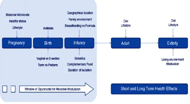
(24) Thèse de Alaa Abdulhussain Al-Seraih, Lille 1, 2016. 1.1.3. Adult The intestinal microbiota of adults is relatively stable over time, and the microbiota of the large intestine becomes more complex conversely to that of children [Zoetendal et al. 1998; Claesson et al. 2011]. The percentage of beneficial bacteria encountered in the gut microbiota such as Bifidobacteria decreases following the weaning and the introduction of solid foods [Salminen and Wright, 2004]. Nevertheless, in adults, Bifidobacteria represent about 1-5% of the total fecal bacterial content [Salminen and Wright, 2004]. Remarkably, the dominant species in the GIT changes with age. For example, B. adolescentis, B. catenulatum/pseudocatenulatum, B. bifidum, and B. longum are the most dominant species displaying considerable variation between individuals [Lahtinen et al. 2011].. 1.1.4. Elderly Limited studies report that structural changes occur in the GIT microbiota of the elderly. The number of beneficial species such as Bifidobacteria diminishes, while the number of species from Clostridia and Enterobacteria populations viewed as detrimental ones for health increase [Gorbach et al. 1967; Mitsuoka, 1982; Rodríguez et al. 2015]. Hopkins and Macfarlane [2002] revealed that Bacteroides species diversity was slightly increased in the feces of elderly subjects, whereas that of Bifidobacteria was decreased. On the other hand, Woodmansey et al. [2004] and Woodmansey et al. [2007] suggested that both Bacteroides numbers and species diversity decreased in the elderly. Overall, the shape of microbiota in the elderly displayed a variation from that established for adults, with the highest levels of Bacteroides spp. and evident abundance of Clostridium spp. [Claesson et al. 2011; Rodríguez et al. 2015].. 1.2. Factors affecting microbiota development and composition Prenatal factors such as the mother’s microbiota, mode of delivery, diet, environment, use of antibiotics and others are associated with establishment and development of GIT microbiota (Figure 2) [Moschen et al. 2012; Rodríguez et al. 2015]. 9. © 2016 Tous droits réservés.. lilliad.univ-lille.fr.
(25) Thèse de Alaa Abdulhussain Al-Seraih, Lille 1, 2016. Fig. 2. Factors influencing the development of infant, adult, and elderly gut microbiota [Rodriguez et al. 2015]. 1.2.1 Maternal microbiota Uterine microbiota is always correlated with intrauterine infection, which is the main cause of infant mortality [Blencowe et al. 2013]. However, recent studies on uterine microbiota with healthy-term pregnancies underpinned a possible transfer of bacteria, or their DNA, from mothers to the placenta tissue [Satokari et al. 2009; Aagaard et al. 2014; Romano-Keeler and Weitkamp, 2014], fetal membranes [Steel et al. 2005; Rautava et al. 2012], amniotic fluid [Bearfield et al. 2002], and umbilical cord blood [Jiménez et al. 2005] of healthy neonates without any indicator of inflammation or infection. Moreover, significant changes in the establishment of neonates’ gut microbiota are supposed as a result of probiotic consumption by mothers during pregnancy [Gueimonde et al. 2006].. 10. © 2016 Tous droits réservés.. lilliad.univ-lille.fr.
(26) Thèse de Alaa Abdulhussain Al-Seraih, Lille 1, 2016. 1.2.2 Mode of delivery It was reported that microbial colonization of the neonate gut is correlated with the mode of delivery, either vaginal delivery or cesarean section [Dominguez-Bello et al. 2016]. Studies point out a strong correlation between the first gut microbiota and the microbial communities of the mother’s vagina (Lactobacillus, Prevotella, or Sneathia) upon vaginal delivery, or of the mother’s skin (Staphylococcus, Corynebacterium, and Propionibacterium) upon cesarean section, when the meconium of newborns has been analyzed [Mueller et al. 2015]. Thus, the vaginal delivery of neonates exposes them to the mother’s vaginal and fecal microbiota, which is a very important source of Bifidobacteria, Bacteroides, and Escherichia coli. Different studies showed that infants born by cesarean section (CS) contain more Bifidobacteria and Bacteroides than those vaginally born [Huurre et al. 2008; Biasucci et al. 2010; Dominguez-Bello et al. 2010].. 1.2.3 Mode of feeding The mode of feeding plays a major role in the development of the infant’s intestinal microbiota. In human cases as a typical model Bifidobacteria and Lactobacilli are frequently detected in the plated samples of breast milk that make it an important source of probiotic bacteria [Fernández et al. 2013]. The comparative studies between breast-fed and formula-fed infants showed a significantly higher number of Bifidobacteria and Lactobacilli and lower counts of Clostridium sp., Bacteroides, Enterobacteriaceae and Staphylococci in the breast-fed infants’ samples [Harmsen et al. 2000; Rinne et al. 2005; Fallani et al. 2010]. Identical Bifidobacterium, Lactobacillus, and Staphylococcus strains were detected in breast milk of mothers and fecal samples of their infants, thus confirming a strong relationship between the mother’s milk and early colonization of the infant’s digestive tract [Martín et al. 2012]. The predominant microbes in the intestinal tract of formula-fed infants were the facultative anaerobes Bacteroides, Clostridium Enterobacteriaceae, Streptococcus, and Staphylococcus, whereas the Bifidobacteria colonization was decreased, leading to a complex microbiota similar to that of adults in these cases [Harmsen et al. 2000; Marques et al. 2010; Fernández et al. 2013].. 11. © 2016 Tous droits réservés.. lilliad.univ-lille.fr.
(27) Thèse de Alaa Abdulhussain Al-Seraih, Lille 1, 2016. 1.2.4. Geographical location and environment Different studies have pointed out a relationship between the intestinal microbiota of the host and the geographical location. Related to this, Hill et al [1971] studied the geographical variations in the incidence of breast cancer and assumed that gut bacteria can produce oestrogens from the biliary steroids present in the colon which could play a direct role in the ætiology of breast cancer. Mueller et al. [2006] conducted a crosssectional study on intestinal microbiota composition on 230 healthy subjects located in Germany, Italy, France, and Sweden. Significant country-age interactions were detected for the German and Italian groups. Notably, the variations between the European intestinal microbiota were only noticed for the Bifidobacterium group. The Bifidobacteria proportion was 2-3 fold higher in the Italian intestinal microbiota than in any other group, and the effect was independent of age. Lee et al. [2011] conducted a comparative study of the composition of fecal microbiota of Korean and American adult twins. They concluded that every geographical area has its own unique microbial “fingerprint,” or identity. Grzeskowiak et al. [2012] studied the gut microbiota of Malawian and Finish infants; they mentioned that Bifidobacteria were dominant in six month old infants, with a higher percentage in Malawian than in Finnish infants. According to these authors, Bifidobacterium adolescentis, Clostridium perfringens, and Staphylococcus aureus were absent in the Malawian infants but present in the Finnish ones. Furthermore, variations in the fecal microbiota content of infants from five European countries with different lifestyle characteristics (Sweden, Scotland, Germany, Italy and Spain) were studied. Bifidobacterium was found to be the predominant species, with about a 40% average of total detectable bacteria, followed by Bacteroides with 11.4% and the Enterobacteriaceae with 7.5%. Infants from the northern European countries appeared to contain high proportions of Bifidobacteria in their feces, in contrast to infants from southern European countries, whose feces contained a diversified microbiota. A higher proportion of fecal microbiota of Bifidobacteria has been associated with breastfed babies, while formula-fed infants have a significant concentration of Lactobacilli, Bacteroides and Clostridium [Fallani et al. 2010].. 12. © 2016 Tous droits réservés.. lilliad.univ-lille.fr.
(28) Thèse de Alaa Abdulhussain Al-Seraih, Lille 1, 2016. The composition of the colon microbiota was also studied for northern Europeans. To this end, fecal samples were collected from 91 healthy humans between age 7 and 52 in France, Germany, Denmark, The Netherlands, and the United Kingdom. The results revealed large inter-individual differences, with Clostridium coccoides and Clostridium leptum as the dominant group followed by Bacteroides. Nonetheless, no significant variation related to geographic origin, age, or gender was noticed [Lay et al. 2005].. 1.2.5 Antibiotics, probiotics and prebiotics The use of antibiotics for bacterial infections has led to the preservation and extension of both human and animal lives, but the development of antibiotic-resistant bacteria became worrisome worldwide. Clearly, resistant bacteria can be found in the intestinal tract [Jernberg et al. 2010; Andersson et al. 2012]. The antibiotic therapies target the pathogenic bacteria, but their effects can afflict the normal microbial communities of the host, mainly those in the gastrointestinal tract [Lode et al. 2001; Bartosch et al. 2004]. The content of the GIT-beneficial anaerobic Bifidobacteria, Lactobacilli, or Bacteroides species could be reduced or even eradicated upon antibiotic treatment, leading to the development of pathogenic species [Sullivan et al. 2001]. A recent study of the shortterm effects of antibiotics on human gut microbiota showed that fluoroquinolones and lactams reduced about 25% of the intestinal microbial diversity and decreased the core phylogenetic microbiota from 29 to 12 taxa [Panda et al, 2014]. Hospitalized patients subjected to a broad spectrum of antibiotic treatment can acquire the symptoms of antibiotic-associated diarrhea resulting from the modification of the composition of their gut microbiota and the proliferation of pathogens such as Clostridium difficile [Vollaard, 1994; Sun et al. 2011; Vincent and Manges, 2015]. The composition of intestinal microbiota could be restored and modulated by probiotics (see page 17) and prebiotics, which play a beneficial role in the gut microbial communities, preventing gut inflammation and other intestinal disease [Hemarajata and Versalovic, 2012; Pourabedin and Zhao, 2015].. 13. © 2016 Tous droits réservés.. lilliad.univ-lille.fr.
(29) Thèse de Alaa Abdulhussain Al-Seraih, Lille 1, 2016. 1.2.6. Other factors Dietary lifestyle and environmental factors have an important impact on the modulation of the composition and metabolic activities of the intestinal microbiota [Gonzaga et al. 2016]. The influence of short-term and long-term dietary changes on the intestinal microbial profile may lead to life-long consequences for the host’s health resulting from the microbial modulation of the immune system [Conlon and Bird, 2014]. Non-dietary lifestyle factors such as smoking, lack of physical exercise, stress, and air-borne toxic particles are tightly linked to intestinal microbiota and host health. Indeed, smoking can significantly affect the intestinal microbiota community and increase the genus Prevotella, mainly in subjects with Crohn’s disease and in healthy subjects that have an increased risk of Crohn’s disease [Benjamin et al. 2012]. Air pollutants can pass to the large intestine via mucociliary clearance from the lungs, in addition to food and water, which affect directly the epithelial cells, immune system and modulation of the intestinal microbiota that linked to increasing of inflammatory bowel diseases (IBD) cases [Beamish et al. 2011]. Stress is another lifestyle factor influencing the bowel activities through the gut-brain axis. Intestinal microbiota of the stressed hosts present a decreasing number of the beneficial intestinal microbiota such as lactobacilli, while pathogenic bacteria such as Escherichia coli and Pseudomonas show high growth ability and epithelial adhesion [Lutgendorff et al. 2008].. 2. Host microbe interactions The microbiota of GIT provides essential health benefits to its host, especially the regulation of immune homeostasis [Honda and Littman, 2016]. The host’s innate immune system consists of the intestinal epithelium and immune system cells such as neutrophils, dendritic cells, monocytes/macrophages, and innate lymphoid cells [Haag and Siegmund, 2015]. The largest interface between host and environment is the luminal surface of the GIT, where the diverse microbiota are in close contact with the immune system of the intestinal mucosa and underlying tissue [Wu and Wu, 2012; Tomasello and Bedoui, 2013]. The intestinal homeostasis is maintained by different protective mechanisms. 14. © 2016 Tous droits réservés.. lilliad.univ-lille.fr.
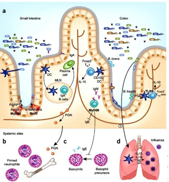
(30) Thèse de Alaa Abdulhussain Al-Seraih, Lille 1, 2016. Several mechanisms depend on the pattern recognition receptors (PRRs) signaling which are triggered by commensal microbes [Kamada et al. 2013]. In the small intestine, AMPs such as peptidoglycan recognition proteins (PGRPs), regenerating islet-derived 3 gamma (RegIIIγ) and defensins are induced as a result of PRR’s stimulation by commensals. However, in the colon, microbes such as Bacteroides fragilis and Bifidobacterium breve induce Treg cells by TLR2 signal (Figure 3) [Chu and Mazmanian, 2013].. 15. © 2016 Tous droits réservés.. lilliad.univ-lille.fr.
(31) Thèse de Alaa Abdulhussain Al-Seraih, Lille 1, 2016. Fig. 3. Pattern recognition receptors (PRRs) signaling promotes immune homeostasis. (a) The small intestine and colon comprise a single layer of intestinal epithelial cells separating the abundant microbiota from host tissues. (b) Peptidoglycan derived from the gut microbiota is necessary to prime neutrophils in bone marrow stores in a Nod1-dependent manner. (c) MyD88 signaling in B cells suppresses serum IgE and inhibits the differentiation of basophils in systemic sites. (d) Commensal gut microbiota induces the production of pro-IL-1β and pro-IL-18 during steady state (signal (1)). During an influenza infection in the lungs, activation of IL-1β and IL-18 mediated by caspase-1 (signal (2)) is critical for clearance of influenza. DC, dendritic cell; MLN, mesenteric lymph node [Chu and Mazmanian, 2013].. 16. © 2016 Tous droits réservés.. lilliad.univ-lille.fr.
(32) Thèse de Alaa Abdulhussain Al-Seraih, Lille 1, 2016. 2.1. Host–microbiota cross-talk After birth, the development of the newborn intestinal microbiota is governed by the interaction between the gut microbial community and the host’s immune system [Critz and Bhandari, 2015]. The neonatal immune system rapidly matures as a result of the influence of microbiota and other important factors [Maynard et al. 2012]. Germfree (GF) mice models showed a defect in intestinal barrier functions and a decrease in inflammatory responses as a consequence of the absence of early microbial stimuli [Sudo et al. 1997]. Oh et al. [2014] studied the responsibility of the gut microbiota to restore and promote the immunity to vaccination of orally inoculated germfree mice models, which previously showed no immune response to the trivalent inactivated influenza vaccine (TIV). Another study conducted on mice models, housed separately in two rooms of the same specific pathogen-free facility (SPF) with two different microbiota, resulted in different mucus barrier properties depending on the influence of intestinal bacteria and their community structure [Jakobsson et al. 2015]. Probiotics were capable of modulating the intestinal immune response by “talking” with the immune cells, implying recognition receptors sensitive to probiotic-derived products such as cell wall components, metabolic products, and DNA [Corthésy et al. 2007].. 2.2. Microbial adhesion and interaction with pathogens The ability of bacteria to adhere to the intestinal surface is a prerequisite for colonization of the squamous and epithelium in the host digestive tract by both beneficial and pathogenic microbes [Pedersen and Tannock, 1989]. The mechanism of adhesion is based on the interaction between the microbe and the targeted surfaces. Hydrophobicity plays a key role in this process owing to the strong correlation between the electrostatic balance, van der Waals interactions, surface hydrophobic character, and the cells’ adhesion behavior [Polak-Berecka et al. 2014]. Various structures such as flagella, fimbriae, and cell wall components are associated with the targeted cell wall to set up the adhesion ability of microorganisms to the intestinal surfaces [Kline et al. 2009; Haiko and Westerlund-Wikström, 2013].. 17. © 2016 Tous droits réservés.. lilliad.univ-lille.fr.
(33) Thèse de Alaa Abdulhussain Al-Seraih, Lille 1, 2016. Different factors govern the bacterial attachment to the host intestinal surfaces. These include digestive enzymes, bile salts, presence of zinc, calcium, magnesium and mucin concentration as well as the pH of the intestine [Ouwehand and Salminen, 2001; Sanchez et al. 2010]. The adhesion ability of beneficial microbes seems to be necessary for the competitive exclusion and displacement of pathogenic microbes and immune system modulation [Castagliuolo et al. 2005; Haiko and Westerlund-Wikström, 2013]. Adhesion of pathogens to intestinal surfaces is the first step of the intestinal infection [Beachey, 1981; Finlay and Falkow, 1997]. Probiotics could develop different mechanisms aimed at inhibiting pathogens colonization of the host intestine, including competition with the pathogens for colonization of the host GIT and therefore occupy the binding sites on the mucus [Vesterlund et al. 2006; Collado et al. 2007a]. Probiotics’ ability to inhibit the adhesion of pathogens showed a high specificity to pathogenic strains [Gueimonde et al. 2006; Collado et al. 2007b]. However, many studies reported that exclusion of pathogenic bacteria by probiotic strains is not related to the adhesion ability of these strains, but could result from different mechanisms involved in the inhibition of pathogens installation [Lee and Salminen, 2009].. 3. Probiotics 3.1. What are probiotics? The term “probiotic” is derived from a combination of the Latin preposition “pro” which means “for” and the Greek noun “bios” meaning “biotic” or “life”. Therefore, this term represents the opposite of “antibiotic” which means “against life” [Guarner et al. 2005; Watson and Preedy, 2015; Nami et al. 2015]. According to a report of the Food and Agriculture Organization (FAO) and World Health Organization (WHO), a probiotic is defined as a “live microorganism which when administrated in adequate amounts confers a health benefit on the host” [FAO/WHO, 2002]. Microorganisms have to fulfil several characteristics to be a candidate for probiotic status. They have to be safe for health, be resistant to digestive tract conditions with as. 18. © 2016 Tous droits réservés.. lilliad.univ-lille.fr.
(34) Thèse de Alaa Abdulhussain Al-Seraih, Lille 1, 2016. much viability as possible, produce antagonistic molecules, adhere to the intestinal epithelial cells, modulate the host immune responses, and be tolerant to technological processes [Heyman and Ménard, 2002]. The bacteria most commonly used as probiotics are the lactic acid bacteria (LAB) group, especially Lactobacillus spp., as well as Bifidobacteria, which received Generally Recognized as Safe (GRAS) status. Nevertheless, different species from other bacteria groups were also tested for their probiotic properties (Figure 4) [Salminen and von Wright, 1998, Watson and Preedy, 2015]. At the same time, the fungal genera Saccharomyces, Debaryomyces, Candida, Kluyveromyces, Pichia, Yarrowia, Metschnikowia, Isaatchenkia and Aspergillus were also commonly proposed as eukaryotes for possible probiotic applications [Nayak, 2011].. Fig. 4. Some examples of prokaryotic and eukaryotic genera commonly used as probiotics [Preedy, 2015].. 3.2. Probiotic criteria and safety assessment To be a candidate for probiotic use, a microorganism has to fulfil the criteria approved by the Food and Agriculture Organization of the United Nations (FAO) and the World Health Organization (WHO) as outlined in the “Guidelines for the Evaluation and 19. © 2016 Tous droits réservés.. lilliad.univ-lille.fr.
(35) Thèse de Alaa Abdulhussain Al-Seraih, Lille 1, 2016. Selection of Probiotics for Food Use” in 2002 (Figure 5) [FAO/WHO, 2002]. These criteria indicate that the identification of a probiotic strain has to be restricted to the strain level because the probiotic effects are strain-specific. Strain identification has to link it to a specific health effect and permit precise observation and epidemiological studies. It was also recommended that updated identification methods be used, and the probiotic strains have to be deposited in an internationally recognized culture collection [FAO/WHO, 2002]. The probiotic selection criteria can be summarized into five major categories [Dunne et al. 2001, Salminen et al. 2004, Holzapfel, 2006, Diez-Gonzalez and Shamberger, 2006, Kailasapathy, 2010]. - ecological, genetic and biochemical properties such as origin, identity, biochemical characteristics, and genetic and metabolic stability. - safety properties; probiotics have to be GRAS (Generally Recognized as Safe), with no invasive potential; non-transferable antibiotic resistance traits, have to be devoid of virulence factors. - physiological properties including (i) resistance to the harsh environmental conditions of the GIT like the low pH of gastric juice, concentration of bile salts and adverse effects of gastric enzymes, and (ii) adhesion ability and viability in the GIT as long as possible. - functional properties related to host health claims such as (i) the ability to colonize the epithelial cells or intestinal mucus and exert competitive exclusion or displacement of target pathogens, (ii) specific antimicrobial activity against pathogens, (iii) stimulation of the immune responses, (iv) selective stimulation of beneficial indigenous bacteria, and (v) restoration of the normal population. - technological properties and performance of the probiotics during manufacturing including growth characteristics in vitro, and survival during the shelf life of the product.. In vitro studies are widely used to gain information about the functional characteristics and safety of probiotics. However, it has been recognized that available tests are not always accurate in predicting the performance and efficacy of probiotics in vivo [Siró, 2011]. The selection of strains and their validation as probiotics should be 20. © 2016 Tous droits réservés.. lilliad.univ-lille.fr.
(36) Thèse de Alaa Abdulhussain Al-Seraih, Lille 1, 2016. based on both in vitro and in vivo demonstrated activities [Siró, 2011]. Probiotics have to pass through various levels of clinical trials that are described by Charalampopoulos [2009]:. 1. Phase I trials: Clinical pharmacology and toxicity, (safety). 2. Phase II trials: Initial clinical investigation effect, (efficacy). 3. Phase III trials: Evaluation of intervention, (effectiveness). 4. Phase IV trials: Post marketing surveillance, (surveillance). Phase II human trials should be designed in the form of double-blind, randomized, and placebo-controlled (DBPC) [FAO/WHO, 2002]. The evaluation of probiotic efficacy in clinical trials is more challenging than the other potential functional foods because the probiotic effects are dependent on the microorganism status [Siró, 2011]. The use of probiotic has significantly increased around the world, and novel probiotic strains are continuously appearing in the markets. Thus, it is recommended that consumers of these probiotics be advised which strains have been used, the minimum concentration of viable cells, their shelf-life, storage conditions, and producer contact details [FAO/WHO, 2002].. 21. © 2016 Tous droits réservés.. lilliad.univ-lille.fr.
(37) Thèse de Alaa Abdulhussain Al-Seraih, Lille 1, 2016. Strain identification and genotypic methods Genus, Species, Strain Deposit strain in international culture collection. Functional characterization In vitro tests animal studies. Safety assessment In vitro and /or animal phase 1 human study. Double blind, randomized, placebo-controlled (DBPC) phase2 human trial or other appropriate design with sample size and primary outcome appropriate to determine if strain /product is efficacious. Phase 3 effectiveness trail is appropriate to compare probiotics with standard treatment of a specific condition. Preferably second independent DBPC study to confirm results. Probiotic Food. Labeling of contents, genus, species, strain designation, minimum numbers of viable bacteria at end of shelflife, proper storage conditions, and corporate contact details for consumers. Fig. 5. Guidelines for the Evaluation of Probiotics for Food Use [FAO/WHO, 2002]. 22. © 2016 Tous droits réservés.. lilliad.univ-lille.fr.
(38) Thèse de Alaa Abdulhussain Al-Seraih, Lille 1, 2016. 3.3. Probiotics and their health effects Beneficial microorganisms can act on their hosts’ health by modulating the composition of the intestinal microbiota or the innate immune system [Frei et al. 2015]. Many studies have highlighted the beneficial effects of probiotic strains by decreasing the risks and treatments of human diseases based on well conducted trials (Table 1).. Table 1. Some beneficial effects of probiotics on human health Probiotic effect stimulation of immune system, gut immune responses and intestinal homeostasis prevention and treatment of diarrhea treatment of irritable bowel syndrome (IBS) enhancement of fecal properties and microbiota treatment of inflammatory bowel disease and constipation protection and cure of Clostridium difficileassociated diarrhea mitigation of lactose intolerance symptoms and food allergies prevention of necrotizing enterocolitis cholesterol-lowering effect therapeutic influence on human immunodeficiency virus (HIV) patients by supporting their immune function reduction of pulmonary damage which resulted from viral infection by development of immune coagulative responses alleviation of the atopic eczema/dermatitis syndrome (AEDS) in specific cases of foodallergic infants. References [Savard et al. 2011] [Guarino et al. 2015] [Moayyedi et al. 2010 Saez-Lara et al. 2015] [Lau et al. 2016] [Pakdaman et al. 2016] [Lambæk et al. 2016] Gilliland et al. 1985; Ooi and Liong, 2010] [Hemsworth et al. 2011; Hemsworth et al., 2012] [Zelaya et al. 2014]. [Viljanen et al. 2005].. Probiotics are used in animal production including poultry, pigs, and ruminants, as well as in aquaculture to improve health, and welfare [Bouchard et al. 2015]. Probiotics permit protection of these animals from severe pathogens including E. coli, Salmonella, Campylobacter, and Clostridium [Papadimitriou et al. 2015]. The relevant beneficial claims of probiotics on farm animals are listed in figure 6 [Cooper et al. 2007].. 23. © 2016 Tous droits réservés.. lilliad.univ-lille.fr.
Figure
![Fig. 4. Some examples of prokaryotic and eukaryotic genera commonly used as probiotics [Preedy, 2015].](https://thumb-eu.123doks.com/thumbv2/123doknet/3718545.110978/34.918.157.770.458.803/fig-examples-prokaryotic-eukaryotic-genera-commonly-probiotics-preedy.webp)
![Fig. 5. Guidelines for the Evaluation of Probiotics for Food Use [FAO/WHO, 2002]](https://thumb-eu.123doks.com/thumbv2/123doknet/3718545.110978/37.918.176.780.93.744/fig-guidelines-evaluation-probiotics-food-use-fao.webp)
+7


Documents relatifs
We consider the lowest order term in the corresponding asymptotic expansion of the scattered field, which can be written as an integral over the center curve of the thin
À travers les cas de la sinusoïde et du dirac, tous deux avec une TFCT de rang 1 dans une convention différente, on re- marque que le choix d’une convention pour obtenir une TFCT
Both tertiary alcohols and protected amines react to form new heterospirocy- cles in good to excellent yields under microwave irradiation in only a few minutes with low
EAAP – 58th Annual Meeting, Dublin 2007 79 Association between a leptin gene polymorphism with milk production traits in dairy cattle L. Moioli, Istituto Sperimentale per la
In this paper, we study whether transition-metal-based ma- terials with perpendicular magnetic anisotropy (PMA) are ad- equate for forward volume spin-wave based information
Editorial: Mathematical Modeling of the Immune System in Homeostasis, Infection and Disease Vitaly Ganusov, Vitaly Volpert, Burkhard Ludewig, Andreas Meyerhans.. To cite
The immune effects of SCFAs are further detailed in Section “Impact of Low Dietary MACs on Immune Cells and Disease Development.” SCFAs play a key role in health and disease and
Based on our previous study, 8 the present study design aimed at testing the hypothesis that birth mode elicits longer-term functional microbiome changes which may impact
