AVIS
Ce document a été numérisé par la Division de la gestion des documents et des archives de l’Université de Montréal.
L’auteur a autorisé l’Université de Montréal à reproduire et diffuser, en totalité ou en partie, par quelque moyen que ce soit et sur quelque support que ce soit, et exclusivement à des fins non lucratives d’enseignement et de recherche, des copies de ce mémoire ou de cette thèse.
L’auteur et les coauteurs le cas échéant conservent la propriété du droit d’auteur et des droits moraux qui protègent ce document. Ni la thèse ou le mémoire, ni des extraits substantiels de ce document, ne doivent être imprimés ou autrement reproduits sans l’autorisation de l’auteur.
Afin de se conformer à la Loi canadienne sur la protection des renseignements personnels, quelques formulaires secondaires, coordonnées ou signatures intégrées au texte ont pu être enlevés de ce document. Bien que cela ait pu affecter la pagination, il n’y a aucun contenu manquant.
NOTICE
This document was digitized by the Records Management & Archives Division of Université de Montréal.
The author of this thesis or dissertation has granted a nonexclusive license allowing Université de Montréal to reproduce and publish the document, in part or in whole, and in any format, solely for noncommercial educational and research purposes.
The author and co-authors if applicable retain copyright ownership and moral rights in this document. Neither the whole thesis or dissertation, nor substantial extracts from it, may be printed or otherwise reproduced without the author’s permission.
In compliance with the Canadian Privacy Act some supporting forms, contact information or signatures may have been removed from the document. While this may affect the document page count, it does not represent any loss of content from the document.
A Novel Role of Cannabinoids in Synaptogenesis
par Sara Hamzeh
F àculté de Pharmacie
Thèse présentée à la Faculté des études supérieures en vue de l'obtention du grade de M.Sc.
en Sciences Pharmaceutiques option Pharmacologie
Août 2008
Cette thèse intitulée:
A Novel Role of Cannabinoids in Synaptogenesis
présentée par : Sara Hamzeh
a été évaluée par un jury composé des personnes suivantes:
Albert Adam, président-rapporteur Jean-François Bouchard, directeur de recherche
Daniel Lamontagne, co-directeur Maurice Ptito, membre du jury
Résumé
Les synapses jouent un rôle important dans la transmission de l'information au sein du système nerveux. Elles montrent une certaine plasticité tout au long de la vie et jouent un rôle essentiel lors du développement, de l'apprentissage et de la mémoire.
Dans cette étude, nous avons exploré un nouveau rôle des cannabinoides endogènes ainsi que leur récepteur CBl dans la formation, le remodellage, et le maintien des synapses. Les cannabinoides endogènes et leurs récepteurs CB 1 sont surtout connus
pour l'effet modulateur qu'ils excercent sur la libération de neurotransmetteurs au niveau de la synapse. Mais, ils sont également impliqués dans plusieurs événements lors du développement. Récemment, il a été rapporté que les cannabinoïdes endogènes diminuaient le nombre de synapses fonctionnelles entre des neurones pyramidaux en culture. Ici, nous démontrons que les cannabïnoides endogènes et les récepteurs CBl régulent la formation de filopodes axonaux et dendritiques (précurseurs de synapse) et sont impliqués lors de la synaptogénèse. La stimulation de neurones corticaux in vitro à l'aide d'un agoniste du récepteur CBl, l'arachidonyl-2'-chloroethylamide (ACEA), produit une diminution significative du nombre de filopodes (précurseurs synaptiques) à DIV8, et par la suite une réduction de la densité synaptique à DIVIO. D'autre part, antagoniser l'action des endocannabinoides et de leur récepteurs CBl à l'aide d'un agoniste inverse, le AM251, ou d'un antagoniste pur, le 02050, augmente la densité des filopodes à DIV8 et éleve le nombre de contacts synaptiques à DIVIO. Aussi, nous avons constaté que ces augmentations étaient bloquées lorsque les cultures étaient préalablement traitées à l'aide du H89 ou du KT5720 (deux inhibiteurs de protéine kinase A(PKA)) ou d'un anticorps blocant lafonction du récepteur DCC (Deleted in Colorectal Cancer). Fait à noter, nous avons observé une diminution significative de la présence des récepteurs DCC à la surface membranaire des cellules traitées avec l'ACEA. D'autre part, nous avons remarqué une augmentation de la présence de DCC à la surface quand les récepteurs CBl étaient antagonisés à l'aide de l'AM251
ou de 1'02050. Cette augmentation a été bloquée lorsque les cellules étaient prétraitées avec le H89 ou le KT5720.Ceci a validé les observations précédentes démontrant que l'activation de la voie de l'adénylate cyclase et de la PKA produisait une augmentation de la formation de contacts synaptiques dépendant de la voie de la nétrine-l et de son récepteur DCC. Afin de confirm,er que l'effet produit par les endocannabinoides et leur récepteur CB] est médié par la nétrine-l et son récepteur DCC, nous avons traité des neurones corticaux provenant d'embryons de souris où le gène codant pour le récepteur DCC a été délété à l'aide d'un agoniste inverse ou d'un antagoniste pur du récepteur CB]. En l'absence du récepteur DCC, l'agoniste inverse AM251 et l'antagoniste 02050 n'ont montré aucune augmentation du nombre de filopodes axonaux et dendritiquse. Le nombre de contact synaptique demeura également inchangé, confirmant ainsi le lien existant entre les endocannabinoïdes, leur récepteur CB] et le récepteur DCC lors de la synaptogenèse. Nous proposons donc que les endocannabinoïdes agissant sur les récepteurs CB], diminuent la concentration cytosolique de l'AMPc, ce qui diminue l'activité de la PKA, bloquant ainsi le recrutement du récepteur de DCC à la surface membranaire, empêchant l'action de la nétrine-l, ce qui diminue la synaptogenèse. Dans cette étude, nous prouvons qu'une interrelation existe entre le système des endocannabinoïdes et celui de la nétrine-l, molécule habituellement impliquée dans le guidage axonal. Ce nouveau mécanisme participe à la formation de synapse au cours du développement neural. Ces résultats démontrent un rôle nouveau majeur des cannabinoides endogènes et représentent une percée importante dans l'identification des mécanismes impliqués dans la synaptogenèse.
Mots-clés: Synaptogenèse, récepteur aux cannabinoïdes CBI, Deleted in Colorectal
Abstract
Synapses play a major role in signalling transduction in the nervous system. They display extensive activity-driven plasticity during development, learning and memory. Here we have explored a new role of endogenous cannabinoids and their CHI receptors in synapse formation, remodelling, and maintenance.
Endogenous cannabinoids and their CHI receptors have been known to regulate neurotransmitter release at the level of the synapse and have also been implicated in several developmental events. Recent/y, if was reported that endogeneous cannabinoids decrease functional synapses in pyramidal neurons. We show here that endogenous cannabinoids and their CHI receptors regulate the dendritic and axonal filopodia formation (synapse precursors) and synaptogenesis obtained from embryonic mice cortical cultures. Stimulating the cortical cultures with the synthetic CHI receptor agonist, arachidonyl-2'-chloroethylamide (ACEA), produces a significant decrease in filopodia number at DIV8, and subsequently a lower synaptic contact density at DIV10 compared with the control group. On the other hand, inhibiting the action of endogenous cannabinoids and their CHI receptors by the inverse agonist AM251 or by the pure antagonist 02050 increases filopodia density at DIV8, and elevates synaptic density formation at DIV10. Furthermore, we found that this increase was reversed when cultures were pre-treated with H89, KT5720 (both inhibitors of Prote in Kinase A (PKA)) or DCCjb antibody, (an antibody which blocks the function of Deleted in Colorectal Cancer Receptor). Interestingly, a decrease of DCC receptors present at the surface of the neurons was observed when treated with ACEA. Conversely, an externalisation of DCC was observed when CHI receptors were antagonised by AM251 or 02050 and this effect was prevented when neurons were pretreated using H89, KT5720. This confirms the previous observations showing that the activation of adenylate cyclase and P KA pathway produces a netrin-l-DCC dependent increase in synaptogenesis.
In order to verify the putative link between cannabinoid and netrin-l systems, we performed in vitro experiments on primary cortical neurons obtained from dcc knockout mouse embryos. In the absence of the DCC receptor, the inverse agonist AM251 and the antagonist 02050 show no increase in axonal and dendritic filopodia, or synapse density confirming a connection between the two systems in the underlying mechanisms of synapse formation. We propose that endocannabinoids acting on their CE, receptors, decrease cytosolic cAMP concentration and inhibit PKA. This blocks the recruitment of the DCC receptor to the membrane surface and therefore, inhibits the action of netrin-l regulating synaptogenesis.
In this study, we show that an interplay between the endogeneous cannabinoids and the DCC / netrin-l pathways regulates synapse formation during neural development. These findings indicate a profound raIe of endogenous cannabinoids and a breakthrough in understanding the mechanisms implicated in synaptogenesis.
Keywords: Synaptogenesis, Cannabinoid Receptor 1, Deleted in Colateral Cancer Receptor, netrin-l, filopodia, Protein Kinase A.
Table of Contents
Résumé ... 111
Abstract ... v
List of Figures.... . .. . .. . .. . .. . .. . ... .. . .. . .. . . .. . .. . .. . .. . .. . .. . ... . .. . . . .. . .. .... ... .. vii
List of Abbreviations ... viii
Dedication. . . .. . .. . . ... . .. . . .. . .. . .. . .. . .. .. .. . . .. . .. . .. . ... ix
Acknowledgements... x
Introduction ... 1
Chapter 1. Literature Review ... 3
1. Synaptogenesis... 3
1.1 Filopodia and Spine formations.. .. .. .. .. .. . .. . .. ... .... .. . .. ... . .. . .. . ... . ... 3
1.2 Axo-Dendritic Contact.. ... ... 5
1.3 Synapse formation ... ... 6
2. Synaptic Plasticity... 8
2.1 Elements that induce Synaptic Plasticity ... 10
2. 2 Signal Transduction Mechanisms ... ... 11
3. PKA and cAMP cycle role in CNS Synapse Fonnation and Plasticity ... 12
3.1 Implication of cAMP in synaptogenesis and plasticity ... ... 13
3.2 Dual role of PKA and cAMP in neuritogenesis and synaptogenesis ... 14
4. Netrins ... 15
4.1 Netrin Structure and Function ... 15
4.2 Netrin Receptors ... 16
4.3 DCC as a Dependence Receptor ... 18
4.4 Developmental and neurological role.. . .. . . .. . .. . .. . .. . .. . .. .. . .. . . .. . .. . .. . . . 19
4.5 Novel Functionsfor Netrins ... 23
5. Cannabinoids ... 23
5.2 CB] receptor: Agonist, Antagonist and Inverse Agonist. . . .... . .... ... 28
5.3 CB] cannabinoid receptor and their behavioural effects ... ... ... ... ... 30
5.4 DSI and DSE: a role of endocannabinoids in short-term plasticity... ... .... ... 31
5.5 Long-Term Potentiation (LTP) and Long-Term Depression (LTD): a role of endocannabinoids in long-term plasticity... 32
5.6 Putative roles of endocannabinoids and their CB] receptors during development and early postnatal period ... 33
Chapter 2. A Novel Role for Cannabinoid Receptor 1 in Synaptogenesis... 37
Introduction ... 37
Materials and Methods... ... ... ... ... ... ... ... 40
Results... ... ... 45
Discussion... 52
Conclusions... ... 58
Chapter 3. General Discussion... ... ... .. ... ... ... 60
Implications of our finding... ... 61
Future Investigations... ... 67
Figures and Legends... ... 71
Bibliography. ... ... ... ... ... ... ... 84
List of figures
Figure 1. Filopodia and Spines: Actin network in neurons
Figure 2. Structure of netrin-l and netrin-l dependence receptors
Figure 3. Natural cannabinoids
Figure 4. CB) and CB2 receptors structures
Figure 5. Inverse agonist, agonist, and antagonist of CB) receptors
Figure 6. CB) receptors are expressed in cortical neuron cultures. They are distributed along dendrites and axons in polarized pyramidal cortical neurons in vitro.
Figure 7. CB) receptors modulate functional synapses in vitro.
Figure 8. Cannabinoids modulate the number of synaptic punctae in vitro: Link between Cannabinoids, PKA, and synaptogenesis
Figure 9. CB) receptors modulate synapse formation in vitro and this effect is dependent of DCC receptor and PKA
Figure 10. Cannabinoids regulate axonal and dendritic filopodia number.
Figure Il. Putative mechanism by which endogenous cannabinoids decrease synaptic density
List of Abbreviations
A AC ACEA 2-AG AM251 AMPA ANOVA Arp2/3 AZ BDNF Ca2+ CaClz CAM CaMK cAMP CapZ CASK CBl CB2 cc Cdc42 cGMP Adult Adenylate Cyclase Arachidonyl-2' -chloroethylamide 2-Arachidonylglycerol N-(piperidin-l-yl)-5-( 4-iodophenyl)-1-(2,4-dichlorophenyl)-4-methyl-lH-pyrazole-3-carboxamide, a CBl receptor Inverse-Agonista-Amino-3-hydroxy-5-Methyl-isoxasole Propionic Acid Analysis of Variance
Actin-Related Protein complex 2/3 Active Zone
Brain-Derived Neurotrophic Factor Calcium
Calcium Chloride Cell Adhesion Molecule
Calciumlcalmodulin-dependent Kinase Adenosine 3', 5'-monophosphate Capping Protein
CaMK-,SH3- and guanylate-Kinase-domain containing protein Cannabinoid Receptor 1
Cannabinoid Receptor 2 Corpus callosum
Cell Division Cycle 42
CNS CNR CRE CREB DAGL DB DCC DCCfb DCCko DIC DIV DSI E ECL ECM EDTA FAAH F-actin FAK FBS FMl-43 FNIII FSK GABA GAP-43 GFP GluRl GRIP
Central Nervous system
Cadherin-related Neuronal Receptor cAMP Response Element
cAMP Response Element Binding prote in
Diacylglycerollipase, the 2-AG synthesizing enzyme DCC-Binding domain
Deleted in Colorectal Cancer
Deleted in Colorectal Cancer function-blocking antibody Deleted in Colorectal Cancer knockout
DifferentiaI Interference Contrast Days In Vitro
Depolarization-induced Suppression of Inhibition Embryonic
Enhanced Chemiluminescence Extracellular Matrix
Ethylenediaminetetraacetic acid Fatty acid amide hydrolase Filamentous actin
Focal Adhesion Kinase Fetal Bovine Serum Fluorescent dye Fibronectin-type III Forskolin
Gamma aminobutyric acid
Growth Associated Prote in of 43 kDa Green Fluorescent Protein
Glutamate Receptor 1
GGP-HRP GMP-HRP GRP-HRP GRP55 GTP GTPase H89 HB-GAM HEK Hoe ICAM Ig Ki KT5720 LTD LTM LTP MAG MAP2 MAPK mEPSC MGL Mintl mRNA Munc N-Cadherin NCAM NFM
Peroxidase AffiniPure Donkey Anti-Goat IgG (H+L) Peroxidase AffiniPure Donkey Anti-Mouse IgG (H+L) Peroxidase AffiniPure Donkey Anti-Rabbit IgG (H+L) G protein-coupled receptor 55
Guanosine Triphosphate Guanosine Triphosphatase
Isoquinoline H89, inhibitor ofPKA
Heparin-Binding Growth-Associated Molecule Human Embryonic Kidney cell .
Hoechst 33258 staining used to detect DNA content Intercellular Adhesion Molecule
Immunoglobulin
In vitro inhibition constant
Hexylester derivative ofK-252a and selective inhibitor ofPKA Long-Term Depression
Long-Term Memory Long-Term Potentiation
Myelin-Associated Glycoprotein Microtubule Associated Protein 2 Mitogen Activated Protein Kinase
miniature Excitatory Post-Synaptic CUITent monoglyceride lipase, a 2-AG degrading enzyme Munc18-1 Interacting protein 1
messenger Ribonucleic Acid Mouse Unc homologs Neural Cadherin
Neural Cell Adhesion Molecule Medium-sized Neurofilament
NGF N-GFP NMDA NMJ NP-40 NT N-WASP 02050 P Pakl PBS PDZ PFA PI3K PKA PKC PLC PSD RET Rac RCM RFP RIM Rho rNet SDS SDS-PAGE SNAP-25
Nerve Growth Factor
Netrin-l: Green Fluorescent Protein chimera N-Methyl-D-Aspartate
Neuromuscular Junction
Tergitol-type NP-40, nonyl phenoxylpolyethoxylethanol Neurotrophin
Neuronal Wiskott - Aldrich syndrome Prote in Pure.antagonist ofCB1 Receptor
Postnatal
P2l-activated kinase Phosphate Buffered Saline PSD-95, Dig and ZO-l Paraformaldehyde
Phosphatidylinositol3-Kinase cAMP-dependent Protein Kinase Calcium-dependent Protein Kinase Phospholipase C
Post-Synaptic Density
Rearranged During Transfection Protooncogene Ras-related C3 botulinum toxin substrate Repulsive Guidance Molecule
Red Fluorescent Protein Rab3-Interacting Molecule Ras Homolog
Recombinant Netrin-l prote in Sodium dodecyl sulphate
Sodium dodecyl sulfate polyacrylamide gel electrophoresis Synaptosomal-Associated Prote in 25
SRl41716A Selective inverse agonist of the CBI receptor
SEM Standard error of the mean STM Short-Term Memory STP SV SV2 SVP-38 SynCAM A9-THC Trk Tsp-1 UNC VAMP2 ZO-1 ZU5 Short-Term Potentiation Synaptic Vesicle
Synaptic Vesicle prote in 2
Synaptic Vesicle Prote in of38 kDa, synaptophysin Synaptic Cell-Adhesion Molecule
Delta-9-tetrahydrocannabinol Tropomyosin-Receptor Kinase Thrombospondin type 1 Uncoordinated
Vesicle-Associated Membrane Protein 2 Zona Occludens prote in 1
1 would fike to dedicate this Thesis to my family in appreciation oJtheir continuous support oJ my dreams and their faith in me at ail times.
Acknowledgements
1 am deeply indebted to my director Dr. Jean-Francois Bouchard and co-director Dr. Daniel Lamontagne, whose help, stimulating suggestions and encouragement helped me in aH the time of research and writing of this thesis. 1 would also like to thank aH the members of Bouchard lab for their friendship and mentorship. In particular, 1 would like to thank Anteneh for aH his help and his great advice on vegetarean restaurants! Marc, for the jokes, and smiles that he brought everyday to the labo Pierre and Nawal for all the coffee breaks and talks about life. 1 also want to thank aIl the hardworking summer students and wish them the best of success: Natalie, Julie, Lynn, Daisy, and Gabriel.
Of course, 1 want to thank aH the members of the Optometry School and the Faculty of Pharmacy for their friendship and help especially Dr. Lamontagne, Dr. Ptito, Dr. Casanova, and Dr. Vaucher and their lab members.
Furthermore, 1 want to thank my family for aIl their help, support, interest and valuable hints. 1 especially dedicate this thesis to my parents, the two most special persons in my life. They, not only gave me life, but also fill it with aIl the love and affection one can wish for. 1 also want to thank my sister and brother, for being a very precious part of my family. FinaIly, 1 would like to thank my husband for his patience, motivation, and love. Thank you for encouraging me to pur sue my dreams.
neurites to form a functional network. While a large amount of information is available on the mechanisms driving neuritogenesis and synaptogenesis, the study of the intricate molecular machinery underlying the intracellular mechanisms has only been recently addressed.
Formation of synapses during development is essential in the wiring of the brain. Synaptogenesis requires molecular recognition cues and guiding interaction cues to regulate the axons and dendrites toward their final target. The morphological development is initiated with the protrusion of filopodia transiently from dendritic shafts. Dendritic filopodia extend and initiate physical contacts with nearby axons or axonal filopodia which results in filopodial stabilization and the formation of functional presynaptic boutons. The increase in the number of boutons coincides with a decrease in the number of transient filopodia and an increase in the number of stable dendritic spines (Ziv NE and Smith SJ, 1996). After the initial contact, a stable synaptic adhesion site is established and the axonal and dendritic compartments differentiate into pre- and postsynaptic specializations respectively.
Synapses play a significant role in plasticity. Mechanisms of synaptic plasticity are regulated by the state of the postsynaptic cell, which is controlled by the interactions of synaptic inputs from multiple sources.
Recent reports on the effects of cannabis on cognitive processes have lead to investigations on their effect on synaptic plasticity. Due to their presence at the fetal and early postnatal periods, endocannabinoids and their CB) receptors have been implicated in playing a significant role in developmental events, such as proliferation and migration of neuronal cells, and synaptogenesis. Although cannabinoids have been reported before to have a retro grade regulatory role on the neurotransmitter release, they have not been clearly implicated in synapse formation. Recently, Kim and Thayer observed that endogenous cannabinoids by activating the CB) receptors decrease the number of synapses, however the underlying mechanisms are still to be uncovered.
Here we show that endogenous cannabinoids via the activation of CB1 receptors with the agoni st ACEA negatively modulate axonal and dendritic filopodia formation. The decrease in axonal and dendritic filopodia number reduces the contact probability between pre- and postsynaptic counterparts and therefore decreases the formation of synapses. This mechanism was found to be dependent of PKA activity and the action of netrin-l, a guidance molecule that acts on DCC receptors. Therefore, a novel role of CB) receptor in regulating synaptogenesis during development has been discovered.
Chapter 1: Literature Review
1. Synaptogenesis
Synaptogenesis is a process involving the formation of neurotransmitter release sites pre-synaptically and receptive fields post-synaptically in neurons. Progress towards understanding the molecular basis of synaptogenesis is still in its early stages.
Neurons acquire the ability to form synapses as part of a deve10pmental maturation process. Synaptic specificity is determined by: (l) neuronal and glial eues that influence competence for synaptogenesis, (2) by long-range and local axon and dendrite guidance eues, (3) by cell-adhesion molecules that mediate contact, (4) and by local presentation of differentiation-inducing molecules. Moreover, the differentiation process is so intricate that neurons develop and maintain molecularly distinct synaptic specializations for excitatory and inhibitory actions, often only microns apart (Linhoff et al., 2006).
1.1 Filopodia and Spine formations
Filopodia formation is a key prerequisite for synaptogenesis in central nervous system neurons. The filopodia are slender cytoplasmic projections which extend from dendritic shafts (Mattila et al., 2008). They contain actin filaments (Figure la) cross-linked into bundles by actin-binding proteins (Hanein et al., 1997). Filopodia extend
to fonn adhesions with the neuronal axons and are attracted by guidance cues that regulate their migratory pathways, and then the y differentiate into stable spines (Mattila and Lappalainen, 2008). A dendritic spine (Figure Ic) IS a small
membranous protrusion from the central stalk of a dendrite that IS typically
electrophysiologically active and synapses with a single axon. TypicaIly, spines have a bulbous head and a thin neck that connects the head of the spine to the stalk of the dendrite. Spiny dendritic stalks host many spines, so because each individual spine typically synapses with a reciprocal axon, a spiny dendrite could receive a multitude of signaIs whereas a traditional dendrite would receive less (Nimchinsky et al., 2002). Dendritic spines, sites of excitatory input in CNS neurons, can be highly dynamic, in later development as weIl as in the mature brain. Spine motility has been proposed to facilitate the fonnation of new synaptic contacts, and they continue to be dynamic even after bearing synaptic connections (Dailey et Smith, 1993).
Figure 1. Filopodia and Spines: Actin network in neurons
•
~---(
'
'-
~",,"
-
;----
---
-, ,Y<?
-
---::;
< '\
\fI
)
Natu,"" Rooview'l Molecul., Celi lIlolosy
1.2 Axo-Dendritic Contact
A synaptic site develops when contact between axons and dendrites is established (Ziv and Smith, 1996; Fiala et al., 1998; Ahmari et al., 2000; Friedman et al.,2000; Alsina et al., 2001 ; Ziv and Garner, 2001). Factors enhancing the axo-dendritic contact therefore are significant role players in synaptogenesis. It was previously thought that axons extend and initiate contact with dendrites or other target cells while dendrites are more stationary. This was best explained by the neuromuscular junction model, which is mainly based on the well-characterized development of the neuromuscular junction (NMJ) (Sanes and Litchman, 1999; Burden, 2002), where target muscles are rather stationary, and axonal growth cones migrate toward the muscle and initiate contact forming a synapse.
Although axonal growth cones play a central role in wiring the nervous system and initiating axo-dendritic contacts, recent numerous studies have also attributed an important role for dendritic filopodia and spines in synaptogenesis (Ziv and Smith, 1996; Jontes et al., 2000; Luscher et al., 2000). The observation that the majority of excitatory synapses in the CNS are formed between spines and varicosities along axons (Gray, 1959; Vaughn, 1989; Harris and Kater, 1994), suggested that dendritic extensions, which are actin filaments, function to efficiently connect axons with a multitude of dendritic shafts, without the need for axons to run convoluted paths (Anderson and Martin, 2001). Dendritic filopodia are often present before the
fonnation of synaptic contacts and spines, and their number decreases as neurons mature, therefore they were believed to initiate axo-dendritic synaptogenesis and to be the precursors for dendritic spines and (Dailey and Smith, 1996; Ziv and Smith, 1996; Smith, 1999) important role players in synaptogenesis.
1.3 Synapse formation
Synapses must remain dynamic during development, allowing neurons to remodel their connections. Such fonns of synaptic plasticity are influenced by the activity of synapses in neurons, and their variations are thought to underlie the adaptive responses of neural circuits, including the adaptive characteristics of leaming and memory (Kandel, 2000). The mechanisms underlying plasticity stem from both the modulation of channel and receptor activity, and the physical movement of channels and receptors into and out of the synapse. The mechanisms that govem synapse fonnation and elimination are fundamental to our understanding of neural development and plasticity.
The wiring of neural circuitry requires that vast numbers of synapses be fonned and disassembled. The subsequent refinement of neural circuitry involves the fonnation of additional synapses coincident with the disassembly of previously functional synapses. There is increasing evidence that activity-dependent plasticity also involves the fonnation and disassembly of synapses (Goda and Davis, 2003).
Complexity of synapse differentiation development arises from the presence of multiple neuron types in the CNS, each fonning molecularly and functionally distinct synaptic specializations.
Differentiation of neurons into glutamatergic or GABAergic subtypes occurs early, before neurons extend axonal processes (Tozuka. and Hisatsune., 2005). Thus, the type of neurotransmitter released from a given presynaptic active zone is predetennined weIl before synapse fonnation (Hampson and Deadwyler, 1999). Consequently, the question arises of how a dendrite can specifically cluster glutamatergic and GABAergic postsynaptic specializations opposite glutamatergic and GABAergic presynaptic contacts, respectively. A likely possibility is that local signaIs from the axon at nascent contact sites direct the aggregation of appropriate postsynaptic proteins at these sites. Recent evidence suggests that b-neurexins could be good candidates for such signaIs. Exogenous focal application of neurexin-l b induces the clustering of glutamatergic and GABAergic postsynaptic receptors, scaffolding proteins and signalling proteins via neuroligins (Dean and Dresbach, 2005).The localization of neuroligin-1 to glutamatergic synapses and neuroligin-2 to GABAergic synapses, and the apparent linkage of neuroligins-l, -3, and -4 to proteins of glutamatergic postsynapses and of neuroligin-2 to proteins of GABAergic postsynapses, suggests that neurexins can influence postsynaptic differentiation by aggregating the proper neuroligin isofonns at nascent contact sites. Therefore, differential expression of neurexin isofonns by glutamatergic versus GABAergic neurons could contribute to local induction of glutamatergic versus GABAergic postsynaptic specializations. However, such a model of postsynaptic specification based solely on neurexins is probably too simplistic. Additional proteins that interact with neurexins and neuroligins and/or that act independently, such as NARP and ephrins, are probably needed to specify appropriate postsynaptic differentiation
(Craig and Linhoff, 2006). Interestingly, N-cadherin is found at hippocampal glutamatergic and GABAergic synapses early in development and then lost from GABAergic synapses (Benson and Tanaka, 1998). Thus, cadherin isoforms could· also contribute to maintaining aspects of specificity. Further studies of synaptic molecules would give a better understanding of the basis for matching pre- and pro-synaptic compartments.
2. Synaptic Plasticity
Synaptic pl asti city was first proposed as a mechanism for learning and memory on the basis of theoretical analysis (Hebb, 1949). The plasticity mIe proposed by Hebb postulates that when one neuron drives the activity of another neuron, the connection between the se neurons is potentiated. Theoretical analysis indicates that not only Hebbian like synaptic potentiation is necessary but also depression between two neurons that are not sufficiently coactive (Stent, 1973, Sejnowski 1977). Depression is necessary for several reasons, among them to prevent all synapses from saturating to their maximal values and thereby loosing their selectivity. It is also important to prevent a positive feedback loop between network activity and synaptic weights. The experimental correlates of these theoretically proposed forms of synaptic plasticity are called long-term potentiation (L TP) and long-term depression (L TD). LTP and L TD, the long-term potentiation and depression of excitatory synaptic transmission, are widespread phenomena expressed at possibly every excitatory synapse. It is now clear that (L TP) and (L TD) are not unitary phenomena. Their mechanisms vary
deperiding on the synapses and circuits in which they operate (Bear and Malenka, 1994).
Recent evidence suggests that induction of L TP may require, III addition to
postsynaptic Ca2+ entry, activation of glutamate receptors and the generation of diffusible intercellular messengers. On the other hand, long-term depression (LTD) requires Ca2+ entry through the NMDA receptor (Yashiro and Philpot, 2008).
Two broad classes of models of synaptic plasticity can be described: 1) Phenomenological models: these are very simple models that are typically based on an input-output relationship between neuronal activity and synaptic plasticity. Phenomenological models are typically used in simulations to account for higher level phenomena such as the formation of memory, or the development of neuronal . selectivity (Bliss and Lomo, 1973).
2) Biophysical models: These more detailed models incorporate more of the cellular and synaptic biophysics of neurons, and are typically used to account for controlled synaptic plasticity experiments. Substantial evidence indicates that the number and strength of synaptic connections can be changed by neuronal activity (Bailey and Kandel, 1993; Bliss and Collingridge, 1993; Malenka, 2003). These changes must be stabilized or consolidated in order for memory to persist. Temporary reversible changes are referred to as short-term memory (STM) and the persistent changes as long-term memory (L TM).
2.1 Elements inducing Synaptic Plasticity
Since its discovery by Bliss and Lomo (1973), LTP has been the object of intense investigation as it is believed to provide an understanding of the cellular and molecular mechanisms by which memories are formed and stored (Bliss and Collingridge, 1993). Calcium influx into the postsynaptic spine is crucial for the induction of many forms of bidirectional synaptic plasticity. Much of the calcium entering the postsynaptic spine cornes through NMDA receptors (Garner and Ziv, 2002). Blocking NMDA receptors pharmacologically can eliminate both L TP and LTD, and a partial block ofNMDA receptors can convert an LTP to LTD. Moreover, experimental results show that a strong postsynaptic calcium transient, in the absence of a presynaptic stimulus can pro duce L TP while a prolonged moderate calcium transient results in LTD (Yang et al., 1999). Many intracellular cascades are initiated after NMDA receptor activation leading to the development of synaptic plasticity. One particular signalling cascade is the one of CaMKII (Kawaguchi and Hirano, 2002). This kinase is unique in that after sufficient stimulation, auto phosphorylation occurs and transforms it from a Ca2+-dependent to a Ca2+-independent state. In this hyper phosphorylated state, CaMKII activity will continue independently of the NMDA receptor activation (Lisman et al., 2002). Moreover, acute CaMKII activation has been reported to enhance GABAA receptor-mediated transmission (Chum et al.,
2002 ; Kawaguchi and Hirano, 2002 ). However, a preferential down regulation of GABAA receptors has been reported in the neocortex after withdrawal from periods of
Sustained CaMKII activity could result in a transient elevation of GABAA receptor function and may trigger a subsequent down regulation of GABAA receptor expression. During brain development, transmitter-gated receptors are operative before synapse formation, suggesting that their action is not restricted to synaptic transmission. GABA, which is the principal excitatory transmitter in the developing brai n, acts as an epigenetic factor to control processes including cell proliferation, neuroblast migration and dendritic maturation. Neurons express functional GABA receptors before formation of functional synaptic contacts (Represa.and Ben-Ari, 2005 ).
2.2 Signal Transduction Mechanisms
In neurons, signal transduction mechanisms underlie the action potentials that travel along nerves. The influx of ions that occurs in response to ligand-gated ion channels often induce action potentials by depolarizing the membrane of the post-synaptic cells, which results in the wave-like opening of Na+ voltage-gated ion channels (Huang and Kandel,1996). In addition, calcium ions are also commonly allowed into the cell during ligand-induced ion channel opening. This calcium can act as a classical second messenger, setting in motion signal transduction cascades and altering the cellular physiology of the responding cell. This may result III strengthening of the synapse between the pre- and post-synaptic neurons by remodeling the dendritic spines involved in the synapse (Kandel, 2000). Overwhelming evidence implicates Ca2+ -sensitive kinases such as PKC and calciumlcalmodulin-dependent kinases (CaMK), as weIl as cAMPIPKA signalling, in
the enhancement of synaptic transmission (Lisman et al.,2002; Nguyen and Woo,2003). Conversely, Ca2+-dependent phosphatases such as calcineurin and prote in phosphatas es 1/2A are shown to decrease synaptic transmission (Morishita et al., 2001; Winder and Sweatt, 2001). Ca2+ levels therefore bi-directionally control synaptic efficacy by influencing the balance between the activity of prote in kinases and phosphatases.
3. Role of PKA and cAMP in CNS Synapse Formation and Plasticity
Adenylyl cyc1ases are a critically important family of multiply regulated signalling . proteins. It is a transmembrane protein that passes through the plasma membrane twelve times. Sorne adenylate cyc1ases are stimulated by Gs proteins, and by
forskolin. In neurons, a majority of adenylate cyc1ases are located next to calcium ion channels for faster reaction to Ca2+ influx; they are suspected of playing an important role in the leaming processes. This is supported by the fact that adenylate cyc1ases are coincidence detectors, meaning that they are only activated by several different signaIs occurring together (Cooper, Mons, and Karpen, 1995). Prote in kinase A, refers to a family of enzymes whose activity is dependent on the level of cyc1ic AMP (cAMP) in the cell. PKA is also known as cAMP-dependent protein kinase. Each PKA is a holoenzyme that consists of two regulatory and two catalytic subunits (Deadwyler et al., 2000). Under low levels of cAMP, the holoenzyme remains intact and is catalytically inactive. When the concentration of cAMP rises, cAMP binds to the two binding sites on the regulatory subunits, which then undergo a conformational change that releases the catalytic subunits. The cAMP signalling
cascade is central to certain types of learning and memory. Changing the strength of connections between neurons is thought to underlie memory formation and may result from the recruitment of new sites of synaptic transmission. New functional synapses can be induced between neurons in culture by elevating cAMP levels (Kim and Thayer, 2001).
3.1 Implication of cAMP in synaptogenesis and plasticity
It had long been believed that almost all synaptic effects of cAMP could be attributed to the direct binding of cAMP to cAMP-dependent prote in kinase (PKA) (Evan and Morgan, 2003). PKA is a ubiquitous serine/threonine protein kinase that regulates many cellular functions. Cyclic AMP-GEFs bind cAMP and selectively activate the Ras superfamily guanine nucleotide binding protein (Rap1A) in a cAMP-dependent but PKA-independent manner. B-Raf, a member of the Raffamily ofSerine/threonine kinases, stimulated by Rap1 subsequently activates mitogen-activated protein kinase (MAPK) pathways (Vossler et al., 1997; York et al., 1998). The MAPK pathways (including at least three pathways: extracellular-signal-regulated kinase (ERK), c-Jun amino-terminal kinase (JNK) , and p38 MAPK) are involved in a widespread set of cellular functions including neuronal differentiation in a transcription-dependent manner (Hazzalin and Mahadevan, 2002), such as ion-channel conductivity, and synaptic release of neurotransmitters. Rapid modulation of synaptic transmission efficacy during the early phase of synaptic plasticity is likely mediated by both pre-and postsynaptic events. However, it remains unclear to what extent these changes contribute to the long-term maintenance of facilitated or depressed synaptic
transmission. It is currently thought that use-dependent modulation of gene expression and protein translation confers long-term plastic changes. In fact, modulation of cAMP-response element (CRE)-regulated gene expression by CRE-binding protein (CREB) appears to be a universal requirement for this process (Kandel, 2001; West et al., 2001).
3.2 Dual role of PKA and cAMP in neuritogenesis and synaptogenesis
To create precise neural circuits in the nervous system, neuritogenesis, axon guidance, and synaptogenesis are the critical cellular processes during neuronal differentiation. Recent studies have examined the role of cAMP in signalling pathways for regulating neuritogenesis and synaptogenesis. A rise in intracellular cAMP concentration by a membrane-permeable cAMP analog, dibutyryl cAMP (dbcAMP), is thought to increase the number of neurites and varicosities (Tojima,Kobayashi, and Ito, 2003). On the other hand, inhibition of cAMP-dependent protein kinase (PKA) activity by a PKA inhibitor (H89) although accelerates, neuritogenesis.neurite outgrowth rate; but it, however, decreases the number of varicosities and the frequency of postsynaptic miniature currents, resulting m suppression of synaptogenesis (Cesa, Scelfo, andStrata, 2007). PKA activity mediates phosphorylation of a gene transcription factor, CREB. On the other hand, inhibition of ERK pathway by PD98059 suppresses both neuritogenesis and neurite outgrowth without CREB phosphorylation. This strongly suggests that PKA simultaneously plays two different roles in neuronal differentiation: inhibition of neuritogenesis and
stimulation of synaptogenesis, via CREB-mediated gene expression. (Tojima et al., 2003)
4. Netrins
Netrins are a family of proteins that guide cell and axon migration during development. Three secreted netrins (netrin-l, ':3/2-like and -41B) have been identified in mammals, in addition to the two GPI-anchored membrane proteins, netrin-G 1 and G2 (Mehlen and LIambi, 2005).
4.1 Netrin Structure and Function
The netrin family of guidance molecules is mainly known to be involved in axon guidance. Netrins are chemotropic; a growing axon will either move towards or away from a source of netrin (Stretaven et al., 1999). Though the detailed mechanism of axon guidance is not fully understood, it is known that netrin attraction is mediated through 40IDCC cell surface receptors and repulsion is mediated through UNC-5 receptors. This protein gradient is bifunctional, attracting sorne axons to the midline and repelling others. Receptors for the secreted netrins include DCC (deleted In
colorectal cancer) and the UNC5 homologues: UNC5 -1/ A, 21B, 3/C, 4/D in mammals. DCC mediates chemoattraction, while repulsion reqmres an UNC5 homologue and, in sorne cases, DCC (Stretaven et al., 1999). The netrin-G proteins bind NGLs (netrin G ligands), single pass transmembrane proteins unrelated to either DCC or the UNC5 homologues. Netrin function is not limited to the developing CNS
midline. Various netrins direct cell and axon migration throughout the embryonic CNS, and in sorne cases continue to be expressed in the mature nervous system (de Castro, 2003). Furthermore, although initially identified for their ability to guide axons, functional roles for netrins have now been identified outside the nervous system where they influence tissue morphogenesis by directing cell migration and regulating ceIl-cell and cell-matrix adhesion.
4.2 DCC, a netrin-l receptor
Deleted in Colorectal Cancer, also known as DCC, is a human gene that has long been implicated in colorectal cancer. The protein coded by the dcc gene is a single transmembrane receptor also known as DCC. Since it was first discovered in a colorectal cancer study in 1990, DCC has been the focus of a significant amount of research (Fearon , Cho, Nigro et al, 1990). DCC held a controversial place as a tumour suppressor gene for many years, and is weIl known as an axon guidance receptor that responds to netrin-l(Hahn SA, Schutte M, Hoque AT, et al., 1996). More recently DCC has been characterized as a dependence receptor, and theories have been put forward that have revived interest in DCC's candidacy as a tumour suppressor gene, as it may be a ligand-dependent suppressor that is frequently epigenetically silenced.
Figure 2. Structure of netrin-l and netrin-l dependence receptors Domain V Domain VI (Th, . . EGF ",pooIB, C·terminaJ baBil: rogIon Netrin·\ UNC5H r wo immunoglobulin domaina Two Ihrombospoodin type 1 oomain. Four immul"lOQfobulin domalna Sbtfibrooctc1in
type III c10malns
AdcictIon
dOpOndenœ domsln
Meblen P and Ulmbi F,Roit of nttrin~1 and ntlrin~l dependence recepton in coloreelll cancers. British Journal of Cancer (2005) 93. 1--6.
The dcc gene is located at 18q21.3, and has a total of 57 possible exons and 43
possible introns. This theoretically results in 13 correctly sliced, putatively functional proteins. The typical DCC protein has one signal peptide motif and eleven domains, including multiple irnrnunoglobulin-like domains, a transmembrane domain, and several fibronectin type 3 domains (Michael A. Reale, Fearon et al., 1994).
DCC has extracellular binding sites for both netrin-l and heparin. Heparin sulphate is believed to also be present during neural growth as a type of co-factor for axon
guidance. Intracellularly, DCC has been shown to have a caspase-3 proteolysis site at Asp 1290 (Mehlen et al., 2001).
DCC and neogenin, two of the netrin-l receptors, have recently been shown to have sites for tyrosine phosphorylation (at Yl420 on DCC) and are likely interacting with Src family kinases in regulating responses to netrin-l(Xiu-Rong Ren et al., 2008).
4.3 Signaling Downstream of Netrin Receptors
Historically, cellular receptors have been thought to be activated when bound to their ligand, and are relatively inactive when no ligand is present. A number of receptors have been found that do not fit into this conceptual mould, and DCC is one of them. These receptors are active both with ligand bound and unbound, but the signaIs transmitted are different when the receptors are ligand bound (Mehlen et al., 2004). Collectively, this type of receptor is known as a dependence receptor because the unbound pathway is usually apoptotic, meaning that cell survival depends on ligand presence. Other receptors also show this functional profile, inc1uding p7SNTR, the
androgen receptor, RET, several integrins and Patched (Mehlen et al., 2004).
When DCC is present on the membrane and bound to netrin-l, signaIs are conveyed that can lead to proliferation and cell migration (Zanker et al, 2007). Only in the absence of the DCC receptor is there an absence of downstream signaling. There are therefore three possible signaling states for dependence receptors: on (ligand-bound, migration and proliferation), off (ligand-unbound, apoptosis inducing) and absent (lack of signal).
4.4 Developmental and Neurological Roles
DCC's role in commissural axon guidance is perhaps it's most characterized. In the developing spinal cord, commissural neurons located dorsally extend axons ventrally using a mechanism dependent on a ventral midline structure, the floor plate (Imondi and Kaprielian, 2001). A gradient of netrin-1 is produced from the floor plate, which allows orientation of the extending axons, ai ding the development of the dorsal-ventral axis of the brain and spinal cord. A variety of receptors are present on the axon surface which either repe1 or attract axons to the midline. When membrane DCC is stimulated by netrin-1, it promotes axon progression towards the midline (Kaprielian" Runko., and lmondi, 2001).
Several other molecules are also involved in the guidance of axons to and across the midline. The slit proteins have repulsive functions, as opposed to netrins, and are mediated by the transmembrane protein Robo (Simpson et al., 2000). Axonal growth cones that are attracted to the midline by netrinlDCC signaling eventually cross the floor plate. When this occurs they lose responsiveness to netrin and become repulsed by slitiRobo signaling. This is accompli shed by the formation of a DCC-Robo complex, which inhibits attractive netrinlDCC signaIs while allowing slitiRobo signaIs (Farmer et al., 2008). Netrin also has other receptors, the UNCS family. The UNCS receptors have repellant migratory responses to netrin binding, and have similar effects to the slitlRobo system (Farmer et al., 2008).
The intracellular signaling responses to netrin-1 are not fully understood. Several phosphorylation events have been established, as have the involvement of several src
family kinases and small GTPases, but the sequence of events has not yet been determined (Zheng et al., 2003). DCC is also required to be recruited to lipid rafts for axon outgrowth and apoptotic signaling. Activating PKA increased the distance over which axons tumed toward a source of netrin-l, whereas PKA inhibition reduced this distance (Bartoe et al., 2006). However, in contrast to the cyc1ic nuc1eotide switch model, inhibiting PKA does not cause these axons to be repelled by netrin-l (Bouchard et al., 2004; Moore, and Kennedy, 2006). Thus the mechanisms underlying chemoattraction to netrin-l are independent of mechanisms required for cyclic nucleotide-dependent switching. PKA regulates the sensitivity of spinal commissural axon chemoattraction to netrin-l and mobilizes DCC from an intracellular vesicular pool to the growth cone plasma membrane (Bouchard et al., 2004; Moore and Kennedy, 2006).
Axon growth and pathfinding are crucial events involved in the establishment of the correct neuronal circuitry during the development of the nervous system. This is achieved by the complex integration of intracellular mechanisms mediated by several highly conserved families of guidance cues, including the netrins (Huber et al., 2003). Netrins are bifunctional ligands attracting or repelling different classes of neurons, depending on the expression of the receptors at the cell surface of the growth cone (Round and Stein, 2007).
Remodelling of the actin cytoskeleton within the neuronal growth cone is an important step in the response of an axon to attractive and repulsive cues leading the axon to advance, retract, or tum in the appropriate direction. Rho-family GTPases, in
particular Racl, Cdc42 and RhoA are important regulators of cytoskeletal dynamics in neuronal and non-neuronal cells and act downstream of most guidance cue receptors (Hall, 1998; Luo, 2000; Dickson, 2001; Govek et al., 2005). Rho GTPase activities regulate growth cone extension and axon outgrowth (Mueller, 1999; Dickson, 2001). Rac1 promotes the formation of actin-based lamellipodia, which provides tension necessary for neurite elongation, while Cdc42 is responsible for the formation of filopodia that serves to orient the growth cone by sensing the extracellular environment (Waterman-Storer et al., 1999; Suzukia and Takahashi, 2008).
RhoA activity also affects the growth cone morphology by decreasing actin polymerization, regulating actomyosin contraction and inducing growth cone collapse (Tashiro et al., 2000). A widely accepted model in neuronal morphogenesis suggests that attractive cues promote actin polymerization and neurite outgrowth via activation of Rac1 and Cdc42 whereas repulsive cues induce neurite collapse via RhoA activation (Hall, 1998; Mueller, 1999). However, in sorne cases, their roles in neurite outgrowth seem to be interchanged (Meyer and Feldman, 2002). For instance, Sema3D-induced chick dorsal root ganglia (DRG) growth cone collapse is mediated by Rac1 (Jin and Strittmatter, 1997). Similarly, Rac1 activity is required for Ephrin-A2-mediated growth co ne collapse in chick retinal cells and sensory neurons (Jumey et al., 2002). Conversely, RhoA activity has been implicated in mediating neurite outgrowth (Sebok et al., 1999; Arakawa et al., 2003) and axon guidance (Bashaw et al., 2001; Yuan et al., 2003). lndeed, low concentration of stromal cell-derived factor (SDF-la) promotes axon elongation by activation of the RhoA/mDia pathway in
cultured cerebellar granule neurons (Arakawa et al., 2003). Similarly, a Dbl family RhoGEF was found to promote axon attraction in Drosophila, in a Rho but not Rac or Cdc42-dependent manner (Bashaw et al., 2001). Rac 1 and Cdc42 are important downstream signaling components of netrin-l receptor DCC signaling (Li et al., 2002b; Shekarabi and Kennedy, 2002; Shekarabi et al.,2005). Rho GTPases are implicated in the signalling mechanisms induced by the netrin-l receptor UNC5 (Briançon-Marjollet and Lamarche-Vane, 2008). Rac1, Cdc42 and RhoA have been shown to play a role in UNC5a signalling and are implicated in the regulation of neurite outgrowth (Sebok et al., 1999; Arakawa et al., 2003).AIso recent studies showed that UNC5a highly activate RhoA and to a lower extent Rac1 and Cdc42 in response to netrin-l in fibroblast cells (Cirulli andYebra, 2007). Interestingly, UNC5a has been shown to induce neurite outgrowth but not growth cone collapse in NIE-115 neuroblastoma cells in a netrin-I-dependent manner (Huber et al., 2003). UNC5a was shown to strongly activate RhoA and thus RhoA activity is thus required for UNC5a-mediated neurite outgrowth (Cirulli and Yebra, 2007).
DCC is developmentally regulated, being present in most fetal tissues of the body at higher levels than what is found in adult tissues (Jiang et al., 2003). DCC and netrin have been found to be specifically involved in the secondary migration of neural crest cells into the pancreas and developing gut structures, and may prove to be vital to other areas during fetal growth (Jiang et al., 2003).
4.5 Novel Functions for Netrins
N etrin-l and DCC play a role in the development of the embryonic nervous system, specifically in the formation ofaxonal and dendritic filopodia, and have a possible role in initiating synaptic contacts and modulating synaptic transmission. Recently, Bouchard et al. showed that activation of PKA enhances the presence of DCC at the cell surface and increases axon extension in response to netrin-l. While inhibiting adenylate cyc1ase, PKA, or exocytosis blocks DCC translocation decreasing axon extension (Bouchard et al., 2004; Bouchard et al. 2008).
5. Cannabinoids
The marijuana plant contains more than 60 bioactive ingredients, of which delta9-tetrahydrocannabinol (.A9-THC) is mainly responsible for its psycho active properties.
Cannabinoids are a group of substances that are structurally related to A9 -THC
or that bind to cannabinoid receptors. Currently, there are three general types of cannabinoids: herbaI cannabinoids occur uniquely in the cannabis plant; endogenous cannabinoids are produced in the bodies of humans and other animaIs; and synthetic cannabinoids which are produced in a laboratory (Lambert and Fowler, 2005). Phytocannabinoids also called natural cannabinoids, herbaI cannabinoids, and c1assical cannabinoids, are only known to occur naturally in significant quantity in the cannabis plant, and are concentrated in a viscous resin that is produced in glandular structures known as trichomes (Lambert and Fowler, 2005). In addition to cannabinoids, the resin is ri ch in terpenes, which are largely responsible for the odour
of the cannabis plant (Lambert and Fowler, 2005). Phytocannabinoids are nearly insoluble in water but are soluble in lipids, alcohols, and other non-polar organic solvents (Taura et al., 2007). However, as phenols they fonn more water-soluble phenolate salts under strongly alkaline conditions (Taura et al., 2007).
An
natural cannabinoids are derived from their respective 2-carboxylic acids (2-COOH) by decarboxylation (Thakur et al., 2005). Many cannabinoids have been isolated from the cannabis plant.An
classes derive from cannabigerol-type compounds and differ mainly in the way this precursor is cyclised (Burns and Ineck, 2006).Figure 3. Natural cannabinoids Type Cannabigerol-type CBG Cannabichromene-type CBC Cannabidiol-type CBD Tetrahydrocannabinol-and Cannabinol-type THC,CBN Cannabielsoin-type CBE ÎSO- Tetrahydrocannabinol-type iso-THC Cannabicyclol-type CBL Cannabicitran-type CBT http://eu.wikipedia.org/wiki/Caouabinoids Skeleton Cyclization
5.1 Cannabinoid Structure and Function
In the 1970's, it was believed that ,A9-THC produced its effects by perturbing neuronal membranes due to its lipid-soluble, hydrophobie nature. It was subsequently demonstrated that saturable, stereo selective, high-affinity membrane binding sites for cannabinoids are present in the mammalian brain (Thakur et al., 2005). There are currently two known subtypes, CBI which is expressed mainly in the brain, but also in the lungs, liver and kidneys and CB2 which is mainly expressed in the immune system and in hematopoietic cells (Begg et al., 2005). Mounting evidence suggests that there are novel cannabinoid receptors that is, non-CB 1 and
non-CB2, which are expressed in endothelial cells and CNS. One of the se is the GPR55, which was identified and cloned for the first time in 1999 (Sawzdargo et al., 1999). Later it was identified by an in silico screen as a putative cannabinoid receptor because of a similar amino acid sequence in the binding region (Baker et al., 2006). GPR55 is only 13.5% identical to CBI and 14.4% identical to CB2, and its mRNA is present in the brain and periphery (Ryberg et al., 2007). The physiological role of GPR55 is unclear. Mice with a target deletion of the GPR55 gene show no specifie phenotype (Johns et al., 2007). GPR55 is widely expressed in the brain, especially in the cerebellum. GPR55 is not expressed in the periphery, except for the jejunum and ileum (Ryberg et al., 2007). This profile as a distinct non-CB I/CB2 receptor which responds to a variety of both endogenous and exogenous cannabinoid ligands, has led sorne groups to suggest GPR55 should be categorised as the CB3 receptor, and this
complicated by the fact that another possible CB) receptor has been discovered in the hippocampus, although its gene has not yet been cloned, (De Fonseca and Schneider, 2008) suggesting that there may be at least four cannabinoid receptors which will eventually be characterised.
The currently recognized cannabinoid receptors, CBI and CB2, are about 45% similar in their protein sequences (Munro et al., 1993). The existence of specific membrane receptors for plant-derived substances in mammalian neurons triggered a search for an endogenous ligand. In 1992, this search culminated in the identification of arachidonyl ethanolamide, named anandamide, a brain-derived lipid that binds to cannabinoid receptors and mlmlCS the biological effects of A9-THC (Mechoulam et al., 1998). Three years later, a second endogenous cannabinoid, 2-arachidonoylglycerol (2-AG) was isolated (Mechoulam et al., 1998). In the ensuing years, several other related lipids with endocannabinoid properties were identified, but have been characterized less extensively (Pagotto et al., 2007). CBI receptor is a 7 pass transmembrane super family member. It couples to inhibitory G-proteins (Gi/o) and therefore, cannabinoid agonists inhibit adenylate cyclase and decrease the activity of protein kinase A (Collins et al., 1994). Stimulation of adenylyl cyclase has been reported in pertussis toxin-treated ceIl, suggesting that in the absence of functional Gi/o coupling, the CBI receptor can activate Gs (OHanas and Onali, 2006). Activation of CBI receptors also inhibits synaptic transmission, probably via inhibition of voltage-gated Ca2+ channels and activation of K+ channels (Deadwyler
et al., 1993). The cannabinoid CB 1 receptor is one of the rnost abundant G-protein-coupled receptors in the brain (Begg et al., 2005).
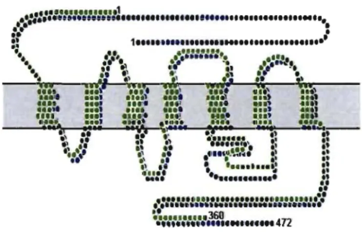
Figure 4. CBI and CB2 receptors Structures
••
::::::::::::::::::l
... ..
::..
:
:: 1 ... ••••••
"
•
:'1'"
•
" .
••
•
::'::ll::~~..
'
.-;-
.. .•.
'l,.
••
.1.
.. ~)i'"
_
••
1
:
.
~"'1 ,...
.
.
,
,ft
'"
III .....••. .1. .... ,., .,
~".
'
••• ,. .' •• 1 . , •• , •••• • . .. ., . t ,:::
:.
:::; :::'
.
:::
.
:::: Sii
:
U!
;
.... • •• ' .. : :.. • .... ! ... .)"" ....'::
..
~:::
!
-::
ri
at::
:.
__ t::
.
·:·c~.::: •
•
Siraiker A. J NeurpbY!liol, 2003; Ho_leu AC. Pharmacol Rev, 2002; Howleu AC. "andb Exp Ph.rm.co~ 2005
5.2 CBI receptor: Agonist, Antagonist and Inverse Agonist
Historically, laboratory synthesis of cannabinoids was often based on the structure of herbaI cannabinoids and a large number of analogs have been produced and tested. Later novel cornpounds were not related to natural cannabinoids but actually based on the structure of the endogenous cannabinoids. Synthetic cannabinoids are particularly use fuI in experirnents to deterrnine the relationship between the structure and activity of cannabinoid cornpounds and for studying cannabinoid receptor rnechanisrns. Sorne ofthese are described below:
Arachidonyl-2'-chloroethlamide (ACEA) is a potent and highly selective CBl receptor agoni st (Ki
=
1.4 nM). It displays a greater than 1400-fold selectivity over CB2 receptor active in vivo (Hillard et al., 1999).AM251 is a CB 1 cannabinoid receptor inverse agoni st. AM251 is structurally very
close to SR141716A (rimonabant), which both are biarylpyrazole cannabinoid receptor inverse agonists. In AM251, the p-chloro group attached to the phenyl substituent at C-5 of the pyrazole ring is replaced with a p-iodo group. The resulting compound exhibits slightly better binding affinity for the CBl receptor with a Ki value of 7.5 nM compared to SR141716A, which has a Ki value of 11.5 nM (Hillard et al, 1999; Pertwee, 2005). However AM251 is about two-fold more selective for the CBl receptor when compared to SR 141716A. AM 251 displays a Ki value at CBl receptors which is 306-fold selective over CB2 receptors. In comparison, SR141716A
displays a Ki value which is 143-fold selective over CB2 receptors (Hillard et al,
1999; Pertwee, 2005).
02050 is a high affinity cannabinoid CB1 receptor pure antagonist (Ki = 2.5 nM);
5.3 CDI cannabinoid receptor and their behavioural effects
Cannabis derivatives are among the most ancient and frequently consumed drugs. Cannabinoid dependence and self-administration have been verified in animal tests (Tsou et al., 1995; de Fonseca et al., 1997; Martellotta et al., 1998; Tanda et al. 2000), further confirming that cannabinoids ho1d a considerable abuse potential (Abood and Martin, 1992). It is generally considered that the recreational use of cannabinoids is related to their positive modulatory effects on brain-rewarding processes along with their ability to positively influence emotional states and remove stress responses to environmental stimuli (De Fonseca et al., 1997). Indeed, recent studies have shown that dopamine release is significantly increased in the nucleus accumbens after cannabinoid treatment presumably because of increased activity of dopaminergic neurons in the ventral tegmental area (Chen et al., 1990; Tanda et al., 1997). In addition, cannabinoid exposure decreases corticotropin-releasing hormone level in the amygdala, which may account for the reduced stress responses (De Fonseca et al., 1997).
The neuronal cannabinoid receptor CB1 has been shown to be responsible for most
behavioral effects of cannabinoids (Ledent et al., 1999; Zimmer et al., 1999). Accordingly, CB) knock-out animaIs do not develop cannabinoid dependence or self-administration (Ledent et al., 1999). CB) receptors are widely distributed in the brain (Tsou et al., 1998), suggesting that several brain areas may be affected by cannabinoids and contribute to their behavioral effects and abuse potential.
Thus, to understand how cannabinoids modulate emotional states, one should consider that other brain regions may also play important roles in different aspects of these phenomena and elucidate the role of CBI receptors at the synaptic, cellular, and network levels in these regions.
5.4 DSI and DSE: a role of endocannabinoids in short-term plasticity.
In the hippocampus, depolarization of pyramidal neurons induces a short-term suppression of GABAergic IPSCs. This phenomenon termed Depolarization-induced Suppression of Inhibition (DSI) is blocked by perfusion of CBI receptor inverse agonists (SR141617A and AM251), and it is absent in enrl-/- mice (enrl: gene co ding for CBI receptors). DSI expressed in the hippocampus is transient «30s) and suppression of the resulting inhibition could not account for long-term plasticity of synapses. However, more recent in vivo studies fail to confirm DSI in response to a variety of behaviourally relevant neuronal activation patterns. Furthermore, recent studies demonstrate that not all neuronal phenotypes in the hippocampus exhibit the capacity for endocannabinoid-mediated DSI. In the cerebellum, researchers obtain similar results. Moreover, Kreitzer and Regehr found that both climbing fiber and parallel fiber excitatory inputs to Purkinje neurons could also be transiently suppressed following Purkinje cell depolarization, the Depolarization-induced Suppression of Excitation (DSE) phenomenon. They showed also that DSE could be blocked by AM251. This suggests that endocannabinoids have a fundamental role in DSI/DSE and synaptic plasticity.


![Figure 5. Inverse agonist, agonist, and antagonist of CD] receptors](https://thumb-eu.123doks.com/thumbv2/123doknet/11585705.298357/53.918.206.509.343.701/figure-inverse-agonist-agonist-antagonist-cd-receptors.webp)
