HAL Id: hal-02461858
https://hal.archives-ouvertes.fr/hal-02461858
Submitted on 30 Jan 2020
HAL is a multi-disciplinary open access
archive for the deposit and dissemination of
sci-entific research documents, whether they are
pub-lished or not. The documents may come from
teaching and research institutions in France or
abroad, or from public or private research centers.
L’archive ouverte pluridisciplinaire HAL, est
destinée au dépôt et à la diffusion de documents
scientifiques de niveau recherche, publiés ou non,
émanant des établissements d’enseignement et de
recherche français ou étrangers, des laboratoires
publics ou privés.
Chloe Topolski, Cédric Dumas, Jérôme Rigaud, Caroline Cao
To cite this version:
Chloe Topolski, Cédric Dumas, Jérôme Rigaud, Caroline Cao. Why is circular suturing so difficult?.
Human Factors and Ergonomics Society Europe Annual Meeting 2019, Oct 2019, Nantes, France.
�hal-02461858�
In D. de Waard et al., [more] (Eds.) (2019). Proceedings of the Human Factors and Ergonomics Society Europe Chapter 2018 Annual Conference. ISSN 2333-4959 (online). Available from http://hfes-europe.org
Why is circular suturing so difficult?
Chloe Topolski1,2, Cédric Dumas2,Jerome Rigaud3, Caroline G.L. Cao11Wright State University,2IMT Atlantique Bretagne-Pays de la Loire, 3Centre Hospitalier Universitaire de Nantes 1United States of America, 2,3France
Abstract
Suturing is a basic surgical skill that is requires much training to achieve competency. Circular suturing is even more challenging, especially in minimally invasive surgery. In a radical prostatectomy procedure, circular suturing is
performed to reconnect the bladder and urethra after the prostate has been removed. Task analysis of linear suturing and circular suturing, each using the laparoscopic and robot-assisted approaches, was performed and validated. Results revealed that circular suturing involves more motoric and perceptual constraints than linear suturing, requiring depth perception for proper alignment of two differently sized circular sutures. Robotic surgical systems such as the da Vinci Surgical System can reduce some of these constraints by providing a stereoscopic view of the circular structures and increasing the manipulability of the needle and tissue. These findings will inform the design of training and assessment of performance, as well as assistive tools to enhance the performance of circular suturing.
Background
In surgery, suturing is performed to close incisions or gaps in the anatomy when diseased tissue has been removed. Suturing is one of the most difficult basic technical skills in surgery. It requires hand-eye coordination, dexterity and precision to place evenly spaced stitches with equal tension to achieve good approximation of tissue. In minimally invasive surgery such as laparoscopic surgery or robot-assisted surgery, intracorporeal suturing is even more difficult due to the limited degrees of freedom in manipulation and constrained space. These suturing tasks may take place along any tissue in any location inside the body. The surfaces and tissues inside the abdomen are all distinct and require different techniques in order to properly manage them during surgery. Additionally, the differences in structure size and shape has a big impact on how the surgeon can perform different suturing tasks.
In urology, after a radical prostatectomy (complete removal of the prostate) is performed to reduce the risk of cancer or to mitigate the spread of cancerous cells, the urethra and bladder neck are joined together with sutures in a process called the urethrovesical anastomosis. This anastomosis involves circular suturing and is considered to be the most difficult part of the entire operation (Ghazi & Joseph, 2018). The urethrovesical anastomosis involves the joining of the ends of two tubular structures –the urethra and the bladder (see Figure 1). This means that the surgeon must suture around the outside circumference of both tubes to ensure the tissues are
securely connected while still allowing fluid to pass through the lumen of the tubes. As this method differs from the more common linear suture where the stitches are made across a straight line, a drastically different technique is needed. The intricacies of these different tasks are outlined in many surgical texts but are not explained in detail. Novice surgeons have to rely on guided training with expert surgeons in order to fully grasp the concepts and methods of circular suturing that make it so challenging. Not only is the task difficult to learn, it is also difficult to teach to novice surgeons, especially in the minimally invasive approach.
Surprisingly, the robotic surgical system da Vinci (Intuitive Surgical, Inc.) that had been struggling to demonstrate value in laparoscopic surgery provided the solution to this difficult urological procedure. In fact, the use of the da Vinci Surgical System in urological procedures increased from 8% in 2004 to 67% in 2010 and is now used in more than 70% of prostatectomy procedures (Voilette et. al, 2015). Nevertheless, the robotic system has not been able to completely nullify the difficulties inherent to the urethrovesical anastomosis, such as bimanual dexterity in instrument manipulation (Chen et al, 2018). While the da VinciÒ has no doubt improved many aspects of minimally invasive surgery (Ballantyne, 2002), the urethrovesical anastomosis still proves to be a challenging task for many surgeons.
This study is the first step towards an understanding of the requirements and constraints in circular suturing for the purpose of surgical skills training, as well as for developing an objective assessment metric for circular suturing performance. Ultimately, an assistive tool may be developed to make explicit the requirements to augment the performance of novice and expert surgeons alike.
what makes circular suturing so difficult 3
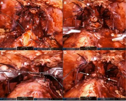
Figure 1: This illustrates 4 different stages of circular suturing in an urethrovesical
anastomosis. As the surgeon progresses, the urethra (indicated by small white circle in A) and bladder neck (indicated by large white circle in A) are brought closer together and joined (part C) and secured (part D), thus completing the anastomosis.
Materials and Methods
Data Collection
To gather initial information about circular suturing tasks, ten surgical texts and manuals were consulted and reviewed to learn the basic steps necessary to complete a urethrovesical anastomosis procedure (Croce & Olmi, 2000, Davis, 2016, Ghazi & Joseph, 2018, Hudgens, 2015, Johnson & Cadeddu, 2019, Joseph, 2008, Lierse, 1987, Secin et. al 2006, Sundaram et. al, 2010, Yuh & Gin, 2018). Observation and recording of five robot-assisted radical prostatectomy surgeries procedures were completed at the Centre Hospitalier Universitaire de Nantes, supplemented by 12 videos of the same surgery found online from other hospitals and training programs. The live procedures ranged from 1.5 hours to 6 hours in duration. The online videos were a mix of laparoscopic or robot-assisted radical prostatectomies; each video averaged around two hours long. Surgeon consent was obtained for the operating room observation portion of the process. Visual recordings of the live observations were taken from the da VinciÒ intraoperative camera; no patient data or audio were included in the recordings.
Four expert surgeons were interviewed. All surgeons consented to being video recorded as they were interviewed. The interview consisted of three main portions: review of a pre-selected video, a structured interview, and reviewing the hierarchical task analysis diagrams. First, the surgeons were asked to observe a video of an expert completing an urethrovesical anastomosis and make comments throughout the video relating to technique and procedure (Mollo & Falzon, 2004). Next, the interviewer asked questions about certain aspects of the procedure and the surgeon’s past experiences with the procedure. Finally, the surgeons were asked to review the four task analyses and verify the content and sequence of steps.
Data Analysis
A task analysis was performed following the procedure in Cao et. al (1996) and four hierarchical task analysis (HTA) diagrams (linear and circular suturing, and laparoscopic and robotic suturing) were constructed to match the techniques observed in the operating rooms. All HTA were validated by four expert surgeons.
A cognitive task analysis was performed by interviewing four expert surgeons at the Centre Hospitalier Universitaire de Nantes in Nantes, France. The transcripts of each of the interviews was synthesized to extract common themes based on the language used. This information was organized and classified to supplement the HTA. By doing this, it became easier to address the specific differences in each of the tasks and which steps of the tasks were more difficult overall.
Results
Hierarchical Task Analysis
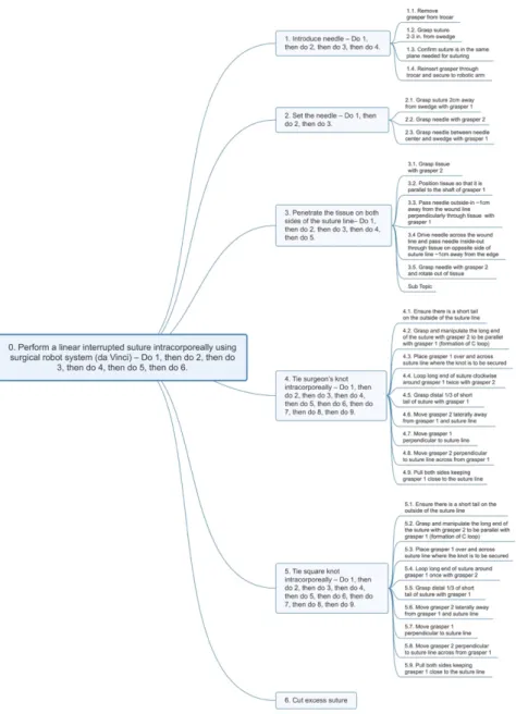
Figures 2-5 show the hierarchical decomposition of the four suturing tasks: laparoscopic linear suturing, laparoscopic circular suturing, robotic linear suturing, and robotic circular suturing. Comparing linear and circular suturing, the first sublevel of the task decomposition was similar; this sublevel contained six to seven steps. The only difference was between circular and linear suturing where two steps were needed to penetrate the tissue since there are two distinct structures to pass the needle through. Distinct differences appeared in the second sublevel of the task decomposition. Circular suturing was more complex than linear suturing, requiring more sub-tasks that were not necessary for the linear suture.
When comparing the robotic approach with the laparoscopic approach, the task decomposition showed that in many of the second-level subtasks, the robotic approach was less constrained than the laparoscopic approach. In the robotic approach, it was not necessary for the needle to be set as meticulously as in laparoscopy since the robot wrist motions can adapt easily to different angles. While there were notable differences in the content of the subtasks, the procedure ultimately remained very much the same.
what makes circular suturing so difficult 5
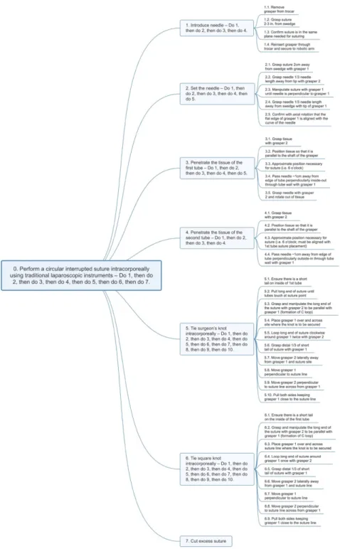
Figure 2: HTA of a circular suturing task using the laparoscopic approach. There are seven first-level subtasks and 37 second-level subtasks included in the diagram, all of which are necessary to perform a circular suture using this approach.
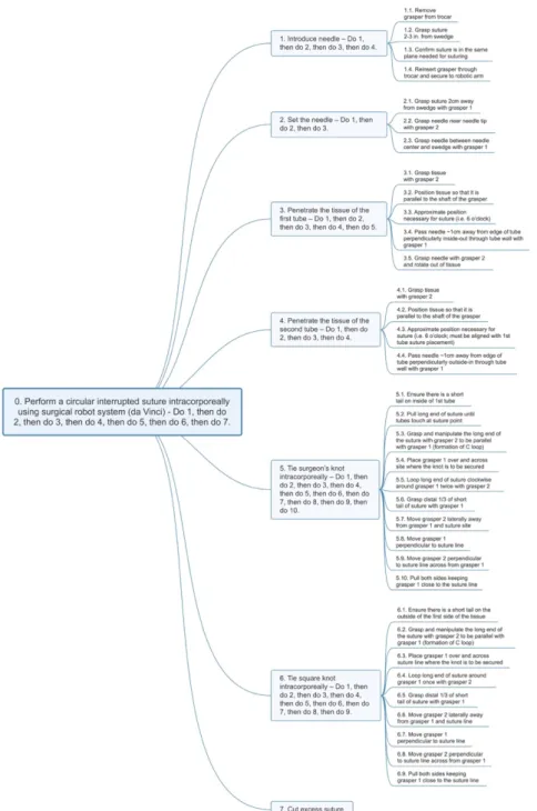
Figure 3: HTA of a circular suturing task using the robot-assisted surgical approach. The second level not only has 12 fewer subtasks than the laparoscopic approach, but the tasks are also simpler and less exigent.
what makes circular suturing so difficult 7
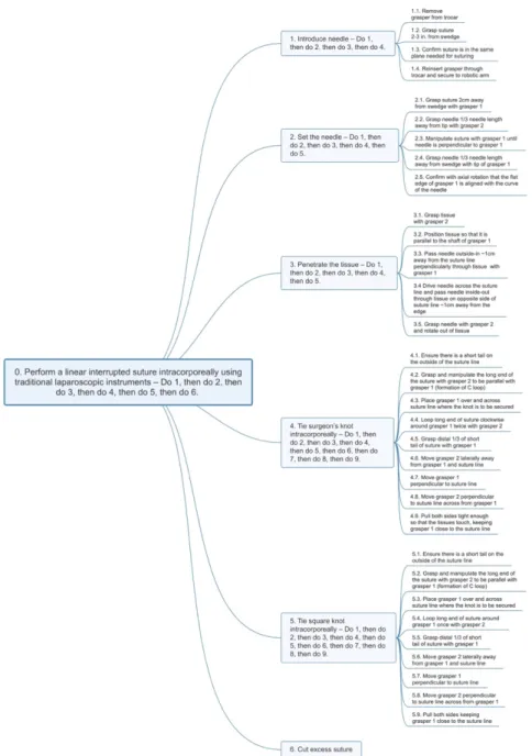
Figure 4: HTA of a linear suturing task using the laparoscopic approach. There are 6 first-level subtasks and 32 second-first-level subtasks necessary in order to complete a linear suture using this approach.
Figure 5: HTA of a linear suturing task using the robot-assisted surgical approach. This approach has 2 fewer subtasks than the laparoscopic approach and is lower in complexity in the “set the needle” task.
Cognitive Task Analysis
Tables 1-3 summarize the results of the cognitive task analysis. Task requirements and constraints were abstracted from the interviews and classified into two levels of abstraction: execution (skills) and planning. The execution or skill of the surgeon was
what makes circular suturing so difficult 9
further broken down into two more levels: motor movements and perception. Table 1 reveals the additional degrees of freedom that the robotic system afforded in manipulating tissue and orienting the needle. Table 2 reveals additional requirements for the circular suturing task, such as the changing orientation of the needle for each stitch, which align with the capabilities of the robotic system in Table 1. Finally, the need to visualize and plan extensively in circular suturing compared to linear suturing is summarized in Table 3. Notably, the placement of the stitches in circular suturing required mental imagery in planning, and constant adjustments during execution.
Table 1: Comparing laparoscopic and robot-assisted suturing techniques. Laparoscopic Robot-assisted
Few degrees of freedom – one axis of rotation More degrees of freedom – wrist motion extremely helpful for needle orientation Better for linear sutures, circular sutures become
more difficult with changing angles of insertion Can easily adapt to linear or circular sutures Orientation of needle in grasping tool critical Orientation of needle in grasping tool not as important 2D view of surgical field lacking depth for circular
suturing High-definition and stereoscopic view of surgical field good for circular suturing
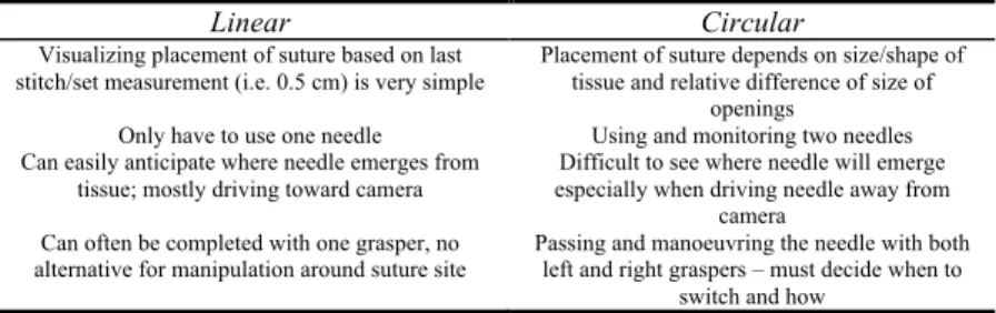
Table 2: Comparing the execution tasks of linear and circular suturing. Linear Circular
Angle of insertion remains consistent Angle of insertion changing Alignment of needle the same for each stitch Alignment and orientation of needle has to be varied precisely
Easy alignment, no concern with twisting or
stretching Different size circumference of openings complicates alignment Can most often use dominant hand to do majority
of suture
Required to use left and right hand with same amount of dexterity
Table 3: Comparing the planning tasks of linear and circular suturing.
Discussion
From the hierarchical task analysis alone, it is not clear why circular suturing is more difficult than linear suturing. Even though there are differences in the number of subtasks at the second level of task decomposition, the differences seem minor as the suturing tasks follow the same technique of needle insertion-needle pull through-suture pull through-repeat needle insertion. Similarly, whether the suturing is performed laparoscopically or with the robotic system, the steps and subtasks are
Linear Circular
Visualizing placement of suture based on last
stitch/set measurement (i.e. 0.5 cm) is very simple Placement of suture depends on size/shape of tissue and relative difference of size of openings
Only have to use one needle Using and monitoring two needles Can easily anticipate where needle emerges from
tissue; mostly driving toward camera especially when driving needle away from Difficult to see where needle will emerge camera
Can often be completed with one grasper, no
alternative for manipulation around suture site Passing and manoeuvring the needle with both left and right graspers – must decide when to switch and how
similar, further confirming that these different approaches follow the same technique in performing a suturing task.
While the execution steps used in linear and circular suturing are essentially the same, the cognitive task analysis revealed marked differences at the execution and planning levels. As linear suturing involves working in one plane, the angle of needle insertion remains consistent for all stitches. In circular suturing, however, the angle of insertion changes with each subsequent stitch. This varying angle of the needle must vary with the tangent of the curve around the circular structure.
Additionally, in urethrovesical anastomosis, the two structures being sutured together have different circumference which complicates the alignment process. Linear sutures which often bring two pieces of tissue together in the same plane are easy to align without any stretching or twisting. In circular suturing, the surgeon must also be able to use both the left and right tools with the same amount of dexterity. A linear suture can often be completed entirely with one hand, while both hands are need to achieve multiple angles of the needle in circular suturing.
Not only is circular suturing more difficult in terms of motor control, but perceptual constraints also play a major role in how a circular suture is completed. In linear suturing, visualizing where the needle should be placed next, based on the position of the previous suture, is relatively easy. However, in the anastomosis task, the positioning of the structures, as well as the difference in size of the structures, makes it more difficult to determine where the next stitch should be placed. Circular suturing most often involves using two needles and keeping track of these needles and sutures can become confusing. Additionally, visualizing these two needles around the circumference of the bladder neck can be difficult. As the surgeon has to drive the needle through the back of the bladder neck, away from the camera, to a point occluded by tissue, where the needle exits the tissue is often a matter of guessing. The planning process throughout all of these steps also changes between linear and circular suturing. For example, the spacing of stitches in a linear suture can be pre-determined based on the length of the suture, such as 5 mm. For a circular suture, the spacing is different on each of the two structures to be joined, due to their size difference. The corresponding stitches on the bladder neck and the urethra must align to ensure an even and tight closure. The passing and manipulating of the needle also require more planning and adjustments in a circular suture. While a linear suture can be conducted simply with one grasper, a circular suture requires the surgeon to decide when to switch directions, when to switch needles, and when to switch hands and grasps to maintain the optimal physical control over the process.
Clearly, many of these requirements are being addressed by the increased degrees of freedom in the surgical robot. Laparoscopic tools are very rigid compared to the robotic end-effectors; the wrist motion of the robotic tool allows for easier needle manipulation that is crucial in circular suturing. Laparoscopic instruments are adequate in linear suturing where the suture is only being applied across a single plane of tissue. However, in circular suturing where the plane of action is constantly changing, the wrist motion of the robotic tools allows the surgeon much more
what makes circular suturing so difficult 11
freedom. The setting of the needle in robot-assisted surgery is not as strict as it is in laparoscopic surgery because the wrist motion allows for rotation in different directions rather than just the one axis of rotation that the laparoscopic tools offer. Another major benefit of the robotic system is the stereoscopic view provided in the operational console. This stereoscopic view is useful in visualizing the circular structures. Laparoscopic screens display the surgical video in 2D only, not allowing the surgeon to have accurate depth perception within the surgical field.
Considerations for Future Work
What is not included in this analysis is the timeline of each approach for the suturing task. A separate timeline analysis, in combination with the task analysis, would more precisely reveal which subtask is time-consuming or which subtask is more difficult. Current teaching materials for minimally invasive linear suturing may be adequate for teaching the order of steps when adapted for circular suturing. However, it is clear that there are additional perceptual and motoric requirements that need to be included in the training instructions. More explicit instructions can be developed for training, as well as for evaluation of performance in circular suturing.
Conclusion
In both laparoscopic and robot-assisted minimally invasive surgery, circular suturing is considered a challenging task to teach and to learn. The joining of the bladder and urethra after a radical prostatectomy procedure is just one example of this type of task. In this study, analysis of four different intracorporeal suturing approaches was conducted through observations of live surgeries, interviews, and video review with expert surgeons. The results of this analysis revealed that circular suturing requires depth perception and proper alignment of two differently sized circular structures, as well as additional motoric manipulations of needle and tissue. Utilizing robotic techniques can mitigate some of these constraints by providing a stereoscopic view of the surgical field as well as increasing the manipulability of both the needle and tissue. The ability to use mental imagery during the planning phase seems to be an important factor in the success of the task. These findings will inform future design of training and assessment methods, and assistive technologies for surgical performance. Acknowledgements
This project was made possible through funding from the FAME Cluster of the NExT Project in Pays de la Loire. The authors are grateful to Drs. Karam, De Vergie, and Nedelec of the CHU Nantes for their domain expertise.
References
Ballantyne, G.H. (2018). Robotic surgery, telerobotic surgery, telepresence, and telementoring. Surgical Endoscopy, 16(10), 1389-1402.
Cao, C.G.L., MacKenzie, C.L., & Payandeh, S. (1996). Task and motion analyses in endoscopic surgery. In ASME Dynamic Systems and Controls Division
proceedings, (Fifth Annual Symposium on Haptic Interfaces for Virtual Environment and Teleoperator Systems),Volume 58, (pp. 583-590). Atlanta,
Georgia USA.
Chen, J., Oh, P.J., Cheng, N., Shah, A., Montez, J., Jarc, A., Guo, L., Gill, I.S., & Hung, A.J. (2018). Use of Automated Performance Metrics to Measure Surgeon Performance during Robotic Vesicourethral Anastomosis and Methodical Development of a Training Tutorial. Journal of Urology 200(4), 895-902.
Croce, E. & Olmi, S. (2000). Intracorporeal Knot-Tying and Suturing Techniques in Laparoscopic Surgery: Technical Details. JSLS : Journal of the Society of
Laparoendoscopic Surgeons 4(1), 17-22.
Davis, J.W. (2016). Robot-assisted radical prostatectomy New York, New York: Springer Science + Business Media.
Gill, I.S. & Hung, A.J. (2018). Use of Automated Performance Metrics to Measure Surgeon Performance during Robotic Vesicourethral Anastomosis and Methodical Development of a Training Tutorial. Journal of Urology, 200(4), 895-902.
Ghazi, A. & Joseph, J.V. (2018). The Urethrovesical Anastomosis in H. John and P. Wiklund Robotic Urology (pp. 375-389) Cham: Springer International Publishing.
Hudgens, J.L. (2015). Systematic Method in J.L. Hudgens and R.P. Pasic
Fundamentals of Geometric Laparoscopy and Suturing (pp. 25-38)
Tuttlingen, Germany: Endo Press.
Johnson, B.A. & Cadeddu, J.A. (2019). Radical Prostatectomy in Robotic-Assisted
Minimally Invasive Surgery (pp. 239-247) Cham: Springer International
Publishing.
Joseph, J. (2008). Vesicourethral Anastomosis in H. John, & P. Wiklund Robotic
Urology (pp. 109-116) Berlin: Springer.
Lierse, W. (1987). Male Urethra in W. Lierse Applied Anatomy of the Pelvis (pp. 171-174) Berlin: Springer Berlin Heidelberg.
Mollo, V., Falzon, P. (2004). Auto- and allo-confrontation as tools for reflective activities. Applied Ergonomics, 35(6) 531-540.
Secin, F.P., Karanikolas, N., Touijer, K.,& Guillonneau, B. (2006). Laparoscopic Radical Prostatectomy: Techniques and Complications in S. Naito, Y. Hirao, and T. Terachi Endourological Management of Urogenital
Carcinoma (pp. 129-145) Tokyo: Springer-Verlag.
Sundaram, C.P., Gjertson, C.K., & Koch, M.O. (2010). Laparoscopic and Robotic-Assisted Laparoscopic Radical Prostatectomy in S. Tsuda and O.Y. Kudsi
Essential Urologic Laparoscopy (pp. 301-331) Totowa, New Jersey USA:
Humana Press.
Violette, P.D., Mikhail, D., Pond, G.R., Paulter, S.E. (2015). Independent predictors of prolonged operative time during robotic-assisted radical prostatectomy.
Journal of Robotic Surgery 9(2), 117-123.
Yuh, B., Gin, G. (2018). Robot-Assisted Radical Prostatectomy in Y. Fong, Y. Woo, W.J. Hyung, C. Lau, and V.E. Strong The SAGES Atlas of Robotic Surgery (pp. 113-125) Cham: Springer International Publishing.