PROTECTION 0F
PRIMARY
CULTURES 0F
MOUSE HEPATOCYTES AGAINST FAS-INDUCED
APOPTOSIS
Role ofEGF receptor
ïntrinsic activity
and
intracellular redox state
par Lina Musallam
Département de Pharmacologie Faculté de Médecine
Thèse présentée à la Faculté des études supérieures en vue de l’obtention du grade de
Philosophiae Doctor (Ph.D.) en Pharmacologie
April, 2003
‘\)
j
24 ‘ J) N —f, \
Direction des bibliothèques
AVIS
L’auteur a autorisé l’Université de Montréal à reproduire et diffuser, en totalité ou en partie, pat quelque moyen que ce soit et sur quelque support que ce soit, et exclusivement à des fins non lucratives d’enseignement et de
recherche, des copies de ce mémoire ou de cette thèse.
L’auteur et les coauteurs le cas échéant conservent la propriété du droit
d’auteur et des droits moraux qui protègent ce document. Ni la thèse ou le
mémoire, ni des extraits substantiels de ce document, ne doivent être imprimés ou autrement reproduits sans l’autorisation de l’auteur.
Afin de se conformer à la Loi canadienne sur la protection des
renseignements personnels, quelques formulaires secondaires, coordonnées
ou signatures intégrées au texte ont pu être enlevés de ce document. Bien
que cela ait pu affecter la pagination, il n’y a aucun contenu manquant.
NOTICE
The author of this thesis or dissertation has granted a nonexclusive license allowing Université de Montréal to reproduce and publish the document, in part or in whole, and in any format, solely for noncommercial educational and
research purposes.
The author and co-authors if applicable retain copyright ownership and moral
rights in this document. Neither the whole thesis or dissertation, nor
substantial extracts from it, may be printed or otherwise reproduced without the author’s permission.
In compliance with the Canadian Privacy Act some supporting forms, contact
information or signatures may have been removed from the document. While this may affect the document page count, it does not represent any loss of
Université de Montréal faculté des études supérieures
Cette thèse intitulée
PROTECTION 0F PRIMARY CULTURES 0F
MOUSE HEPATOCYTES AGAINST FAS-INDUCED
APOPTOSIS: Role of EGF receptor intrïnsic activity
and intracellular redox state
présentée par: Lina Musallam
a été évaluée par un jury composé des personnes suivantes:
Dr. Trang Hoang, président-rapporteur Dr Pierre S. Haddad, directeur de recherche Dr Marc Bilodeau, co-directeur de recherche
Dr Marie-Josée Hébert, membre dujzuy
Résumé
Le foie est très sensible à l’apoptose. De nombreuses études ont rapporté une induction
marquée de l’apoptose des hépatocytes au cours des infections virales (hépatites B, C et
fulminante) et dans les hépatopathies alcooliques. Étant donné que les hépatocytes
expriment le récepteur Fas de façon constitutive, ceci soulève son implication f as dans le
développement des pathologies hépatiques. Ainsi, le but de cette thèse est d’étudier les
mécanismes de protection activés par le facteur de croissance épidermique (EGF) contre
l’apoptose induite par le Fas chez les hépatocytes de souris en culture primaire. En
particulier, ces études ont concentré sur l’implication de l’état rédox et de l’activité tyrosine
kinase (TK) du récepteur EGf. Il est à noter que l’injection du Fas in vivo résulte en
apoptose massive du foie (—100%) alors que seulement 5-10% de hépatocytes en culture
meurent par apoptose suite à leur traitement avec le Fas. Nos résultats démontrent ici que la
résistance relative des hépatocytes en culture à l’apoptose est due à la perte importante du
contenu cellulaire en glutathion (GSH) durant l’isolation et la culture des cellules (totalisant
—75% de perte après seulement 2h en culture). L’augmentation des niveaux de GSH
intracellulaire et surtout du ratio de glutathion réduit (GSH)/total (GSx), en enrichissant le
milieu avec la cysteine et méthionine (deux acides aminés précurseurs de GSH) a amené
une sensibilité accrue à l’apoptose induite par le Fas. En plus, l’EGF, qui est comni pour
son effet anti-apoptotique, a significativement diminué les niveaux de GSH intracellulaires,
i\,
en parallèle à l’effet protecteur de l’EGF contre l’apoptose et ils étaient contrecarrés par PD168393, un inhibiteur de l’activité TK du récepteur EGf. La réplétion de la cellule en GSH a diminué l’effet protecteur de l’EGF contre l’apoptose induite par le Fas. Cette modulation de l’apoptose par l’état rédox semble être due à la régulation de l’activité des caspases. Il est à noter cependant, que malgré le fait que PDI6$393 a aboli l’autophosphorylation du R-EGF, l’activation des voies de MAPK et PI3K ainsi que l’augmentation de l’expression de l’ARNm et de la protéine de BCL-xL, il n’a pas complètement inhibé l’effet anti-apoptotique de l’EGF. Ceci suggère que des mécanismes indépendants de l’activité TK du récepteur EGF participe aussi dans la protéger contre l’apoptose. En conclusion, un état rédox oxydative semble protéger contre l’apoptose induite par le Fas dans les cultures primaires d’hépatocytes de souris. Ce processus est même utilisé par les facteurs de croissance, tel que l’EGF, et il est dépendant sur l’activité TK du récepteur.
Mots-clés apoptose, hepatocytes, Fas, EGF, BCL-x1, caspases, glutathion, état rédox, protection, activité tyrosine kinase
Abstract
The liver is highly sensitive towards apoptosis. Numerous injurious events resuit in the loss of hepatocytes by apoptosis, including hepatitis C, B and alcoholic liver disease. Since hepatocytes constitutively expless fas-R, the fas system lias been involved in the development of liver pathologies. Therefore, the aim of these studies was to investigate the rnechanisms of protection afforded by epidermal growth factor (EGf) against Fas-induced apoptosis in primary mouse hepatocyte cultures. In particular, these studies focused on the involvement of the intracellular redox state and of the tyrosine kinase (TK) activity of the
EGf receptor. 0f note, in vivo injection of anti-fas resuits in massive apoptosis of the liver (--100 %) while exposure of primary hepatocytes cultures to Fas causes 5-10 % apoptotic bodies only. Here, our findings indicate that the relative resistance of mouse hepatocytes in culture toward fas-induced apoptosis is due to the loss of the important stocks oC glutathione (GSH) during the isolation procedure and culture (totaïling 75% loss afier 2h culture). The increase ofintracellular GSH levels and especially ofthe reduced (GSH)/ total (GSx) glutathione ratio, by supplementing the media with cysteine and methionine (precursor amino acids of GSH), enhanced ceil sensitivity toward fas-induced apoptosis. furthermore, EGf, a known anti-apoptotic agent, significantly lowered GSH levels and GSHIGSx ratio in a time- and dose-dependent manner. In fact, EGF treatment decreased GSSG reductase activity, which may account for the decline in GSH/GSx ratio. These changes occurred in parallel to the anti-apoptotic effect of EGf on mouse hepatocytes in culture and were inhibited by PD168393, a specific inhibitor ofthe TK activity ofthe EGF
vi
receptor. Ceil replenishment with GSH diminished the protective effect of EGf against Fas-induced apoptosis. This redox modulation of apoptosis seerns to occur through regulation ofthe activity ofcaspases. 0f note, and in spite ofthe fact that PD16$393 has effectively abolished the autophosphorylation of the EGF-R, the activation of MAPK and PI3K pathways as well as the induction of BCL-xL mRNA and protein expressions, it did not completely inhibit the anti-apoptotic effect of EGF. This indicated that some TK independent pathways are involved in the anti-apoptotic signal of EGf against Fas-induced apoptosis. In conclusion, an intracellular oxidative state seems to protect against f as induced apoptosis in primary mouse hepatocyte cultures. This mechanisrn is used bv growth factors such as EGF to protect against apoptosis and it is dependent on the TK activity of the receptor.
Keywords apoptosis, f as, EGf, BCL-xL, caspases, glutathione, redox state, protection, tyrosine kinase activity.
TABLE 0F CONTENT
TABLE 0F CONTENT vii
LIST 0F TABLES xii
LIST 0F FIGURES xiii
LIST 0f ABBRIVIATIONS xvii
GENERAL INTRODUCTION
A.CELL DEATH BY APOPTOSIS 2
1. CELL DEATH: A POINT 0f NO RETURN 2
2. TYPES 0f CELL DEATH: NECROSIS VS APOPTOSIS 3
2.]. Necrosis 3
2.2. Apoptosis 4
3. ROLE 0f APOPTOSIS IN HEALTH AND DISEASE 8
3.]. Apoptosis in physioïogy N
3.2. Apoptosis in pathology J t)
4. APOPTOSISANDTHELIVER 10
4.]. Theliver 10
viii
B.APOPTOSIS : CELLULAR MECHANISMS 13
1. INDUCTION 0F APOPTOSIS 13
2. EXECUTION 0F APOPTOSIS 1$
2.]. THE FAMILY 0F ICE-PR0TEA SES (CIA SPA SES) 18
2.2. THEPRO-AFOPTOTICFROTEINS 0F THEBC’L-2 fAMILY 21
2.3. Diablo/Srnac prote ins 23
3. DEGRADATION AND ELIMTNATION 0F APOPTOTIC CELLS 23
CJ’AS RECEPTOR / FAS LIGAND SYSTEM 25
1. FAS SYSTEM AND ITS ROLE [N DISEASE 25
2. FAS RECEPTOR SIGNAL TRANSDUCTION 26
2.]. fADDpathway: 30
2.2. Fas signatiing in hepatocytes (type lIceils): 30
2.3. FADD-independent signalling: 31
D.ANTI-APOPTOTIC MECHANISMS 33
1. GROWTH FACTORS AND PROTECTION AGANST APOPTOSIS 33
2. ANTI-APOPTOTIC PROTENS 0F THE BCL-2 FAMILY 34
3. PHOSPHATIDYLNO$ITOL 3-OH KINASE/ Akt PATHWAY 36
5. TNHIBITOR 0f APOPTOSIS PROTEINS (IAP) .38
E.EPIDERMAL GROWTH FACTOR (EGF) 40
1. STRUCTURE 0f THE EGf RECEPTOR 40
2. ACTIVATION 0f EGf-R 41
3. EGf-R SIGNAL TRANSDUCTION PATHWAYS 42
3.1. Mitogen-Activated Protein Kinases Pathway (MAPK): 42 3.2. Phosphatidyl InositoÏ 3-OH Kinase Pathway (PI 3-K). 46
3.3. Phosphohpase Cy Pathway (PLCj: 46
3.4. Reactive oxygen species (ROS): 47
F. INTRACELLULAR REDOX STATE 4$
1. REACTIVE OXYGEN SPECIES (ROS) 49
2. CYTOTOXIC EFFECTS 0f ROS 52
3. INTRACELLULAR ANTI-OXIDANT MECHANISMS AND REDOX STATE 56
3.1. Scavengers 56
3.2. Enzymes 57
3.3. G$Hsystem 5cS
4. REDOX SIGNALLING 62
X
4.2. Oxidative modfications ofproteins. 63
4.3. $ignaÏÏing pathways targeted by ROS 61
4.4. Ligand-induced R OS production 66
5. REDOX REGULATION 0F APOPTOSIS 67
5. 1. Oxidative stress sensitizes cetis towaïd apoptosis 67
5.2. ROSprotect celis against apoptosis 68
CHAPTER 2 OBJECTIVES AND HYPOTHESIS 70
A.RATIONAL 71
B.HYPOTHESES 72
C.OBJECTIVES 73
CHAPTER 3 EXPERIMENTAL RESULTS 74
3.1 ARTICLE NO. 1 RESISTANCE TO FAS INDUCED APOPTOSIS IN HEPATOCYTES: ROLE 0F GSH DEPLETION BY CELL ISOLATION AN!)
CULTURE 75
3.2 ARTICLE NO. 2 ROLE 0F EGF RECEPTOR TYROSINE
ACTIVITY IN ANTI-APOPTOTIC EFFECT 0F EGF ON MOUSE
HEPATOCYTES 122
3.3 ARTICLE NO. 3 EGF MEDIATES PROTECTION AGAINST FAS
INDUCED APOPTOSIS BY DEPLETING AN!) OXIDIZING
CHAPTER 4 DISCUSSION & CONCLUSION. 220
CONCLUSION 230
xii
LIST 0F TABLES
CHAPTER 1 INTRODUCTION
Table 1 Comparative table between the moiphologiccil and biochemical properties
ofapoptosis and necrosis 5
CHAPTER 3 EXPERIMENTAL RESULTS
ARTICLE NO. 2
Table 1 Effect of PD]68393 and Tyrphostin AG]478 on II332F04 incoiporation 11210
mouse hepatocytes treated with EGF 172
ARTICLE NO. 3
Table 1 Efftct of GSHmee on the intracelliilar Ïevels of GSx, GSH and GSH/G& ratio inmouse hepatocytes treated with EGf 207
LIST 0F FIGURES
CHAPTER 1
INTRODUCTION
Figure 1 Morphology ofdying ceils by necrosis and apoptosis 6
Figure 2 Mechanisms of induction and inhibition ofapoptosis 16
figure 3 fas receptor signallingpathways 2$
Figure 4 EGf receptor signalÏingpathways 44
Figure 5 Sources ofreactive oxygen species (RO$) and their nzechanisrns ofeÏimination 50
figure 6 RO$cytotoxiceffects 54
CHAPTER 3
EXPERIMENTAL RESULTS
ARTICLE NO. 1
figure 1 Effect ofccli isolation and culture on total glutathione (GS±,) levels 10$
xiv
figure 3 Effect of medium N and C+M on the expression levels ofprocapsase-8, Bid
andBcL-xLproteins 112
figure 4 Effect ofmedittrn N and C+M on Fas-induced caspase-8 activation 114
Figure 5 Efftct of extended time in culture on G$x and GSH levels as weIÏ av GSH/GSx ratio in ceils cultured in mediztrn C+M 116
Figure 6 Effect of tirne in culture on Fas ability to induce caspase-8 activation ami
apoptosis in medium C+M 11$
figure 7 Ivlodulation of the protease activity of Immun recombinant activated
caspase-8 by GSH/GSx ratio 120
ARTICLE NO. 2
figure] Effect Kinetics ofEGf-R phosphoiylation in mouse hepatocytes treated with
EGF in the absence andpresence ofTy,phostin AG1478 160
Figure 2 Anti-apoptotic effect of EGf on primaly mouse hepatocyte cultures
subjected to fas receptor stimulation 162
figure 3 Effect PD168393 on EGF-R phosphoiylation in mouise hepatocytes treated
figure 4 Effect of FD]68393 on post-receptor events in nzouse hepatocytes treated
withEGf 166
figure 5 Morphotogy ofEGf-treated mouse hepatocytes in the presence ctndabsence
ofFD168393 16$
figttre 6 Effect of PD168393 and Tyiphostin AG1478 on the anti-apoptotic response
ofEGf in mouse hepatocytes 170
ARTICLE NO. 3
Figure J Effect of anti-fcis and/or EGf on intraceib Jar ghttathione leveÏs ctncl
GSH/GSx ratio 20$
figure 2 Effect of anti-fas and/or EGf on the activily ofgÏutathione reductase (G]?,)
210
Figztre 3 Tirne-course and dose-response curves of intraceÏhdar G$x levels and
GSH/G$x ratio foltowing exposure to EGF 212
figure 4 Effect ofEDi 68393 on EGf-induced decrease ofGSx, GSH Ïeveïs as iveÏÏ as
GSH/GSx ratio and G]? activity 214
Figure 5 Effect ofghttathione monotheyl ester (‘G$Hmee on protective effect ofEGf against Fas-induced caspase-3 activation and ceil death by apoptosis.... 216
xvi
figure 6 EJfect of GSHrnee on EGF receptor (EGf-R) autophosphoiyÏation cind
LI$T 0F ABBRIVIATION$
AIF-1 : Apoptosis inducing factor- 1
ALT : Alanine aminotransferase
Apaf-1 : Apoptosis protease-activating factor-1
ASK-1 : Apoptosis signal regulating kinase-1
ASI : Aspartate aminotransferase
ATP Adenosine tris-phosphate
BCL-2 : 3-ceil lyrnphoma
BIR : Baculovirus repeats
CAD : Caspase dependent DNase
CARI : Caspase recruitment domain
xviii
DAG 1 ,2-di-acyl gilycerol
DAXX : Death associated protein
DB : Death domain
DEB : Death effector domain
Diablo Direct IAP binding protein with low pI
DISC : Death inducing signalling complex
EGF : Epidermal growth factor
EGF-R : EGF receptor
ERK : Extracellular signal regulated kinase
FADD : fas associated protein with death domain
Fas-L t Fas ligand
Fas-R : fas receptor
GC$ y-glutamylcysteine synthetase
GDP : Guanyl dis-phosphate
GF : Growth factors
GS : Glutathion synthetase
GSH : Glutathione, reduced form
GSSG : Glutathione, oxidizedform
GSx Total Glutathione
GTP : Guanyl tris-phosphate
HGF : Hepatocyte growth factor
IAP : Inhibitors of apoptosis proteins
ICAD : Inhibitor of caspase dependent DNase
ICE : Interleukin 1 f3 converting enzyme
xx
1P3 Inositol 1,4,5- tris-phosphate
JNK : Jun-arnino terminal kinase
LDH : Lactate dehydrogenase
MAPK : Mitogen-activated protein kinase
MEK-1 : MAPK kinase-1
MPT Mitochondrial permeability transition
NAU(P)H : Nicotimide adenine denucleotide (phosphate), reduced forrn
NGF : Nerve growth factor
02 : Superoxide dismutase
011 Hydroxyl radical
PARP Poly(ADP-ribose)polymerase
PI : Phosphatidylinositol
PIP2 : Phosphatidylinositol 4,5-dis-phosphate
PKC : Protein kinase C
PLCy : Phopholipase Cy
PTH : Phosphotyrosine homology domain
ROS : Reactive oxygen species
RTK : Receptors tyrosine kinase
SH- : Suifflydryl group
SU-2 : Src-homology-2 domain
Smac : Second mitochondrial activator of caspases
SOS : Son of Sevenless
TGF-Π: Tumor growth factor-a
TNfΠ: Turnor necrosis factor c
TRADD : TNF receptor associated protein with death domain
TRX : Thiorodoxin
To nîyparents,
To mj’ brotit ers anct siste,
To alt rnyfainity andfriends,
for their sttpport air dfortire gooct anci tire bact
xxiv
ACKNOWLADGEMENTS
I extend my gratitude and appreciation to many people who made this Ph.D. thesis possible.
Above ail, immense thanks to my mom and dad for their unconditional love, priceless
advice and continuous motivation and support. They were aiways the back that I can lean on during the difficuit times, the first to cheer me up and to make my problems seem 50
trivial. I love you very much. Many many thanks to my sister Iman and brothers Salah and Hisham for their love, support and taking over my share of houseshold shores, a huge incentive for thesis writing. Speciai thanks to $aÏah for his invaluable help and patience during my computer difficulties.
Special thanks to my friends, fatiha Moukdar, Loan Nguyen and Marcia Nagaoka, who helped make my post-graduate adventure fun!
I am deeply indebted to my supervisor Marc Bilodeau for welcoming me in his laboratory, for his guidance and help in shaping my scientific curiosity and for his financial heip when I needed it. I am grateful to my supervisor Pierre Haddad for his moral support, his belief in my capacities and for the chance that he offered me to pursue a career in research. Many thanks are due to Chantai Éthier, who was the first to introduce me to research bench work and for her help throughout my research. Many thanks also go to Christian Demers and Paul Desjardin for their technical help and their patience with my endless questions. Many thanks go to the personnel of Hôpital Saint-Lue as well as the students who participated in various ways to ensure my research succeeded. I would also like to acknowiedge with
much appreciation the financial support provided by fonds de la recherche en santé du
Québec (FRSQ) and fonds pour la formation des chercheurs et l’aide à la recherche
CHAPTER 1
1. CELL DEATH: A POINT 0F NO RETURN
Ceil structure and function are maintained in dynamic stability by cell’s genetic program. Preserving this intracellular dynamic equilibrium in spite of the normal physiological demands, the constrains of neighbouring ceils and the availability of metabolic substrates is called homeostasis. Disturbance of this equilibrium by more excessive physiological stresses or pathological stimuli may bring about a number of physiological and morphological cellular adaptations, in which a new but altered steady state is achieved to preserve the viability of the ccli and to modulate its fiinction as a response to such stimuli.
If the limits of adaptive response to a stimulus are exceeded, or in certain instances when
adaptation is not possible, a sequence of events follows, loosely terrned ceil injury. Celi injury is reversible up to a certain point, but if the stimulus persists, or if the stimulus is severe enough from the beginning, the ccli reaches the point of no return and suffers irreversible ccii injury and ceil death.(1)
Ccli death, the ultimate resuit of celi injury, is one of the most crucial events in pathology. affecting every celi type and being the major consequence of ischemia, infection, toxins and immune reaction. In addition, it is criticai during embryogenesis, lymphoid tissue development and hormonaily induced involution. Finaily, it is the aim of cancer radiotherapy and chemotherapy.( 1)
n
2. TYPES 0F CELL DEATH: NECROSIS VS APOPTOSIS
The causes of ccli injury and ccli death range from externai stimuli (such as physical, chemicai, and infectious agents,) to internai causes (such as hypoxia, immunoiogical
reactions and genetic alterations). There are two major morphoiogicai types of ccii dcath,
necrosis and apoptosis. Ceil death by necrosis is norrnally observed aCter a pathologicai or
injurious event. Apoptosis is a programmed type of ceii death which necessitates the active
participation of the celi and one which, in contrast to necrosis, is associated with normal
physioiogy in addition to pathoiogicai conditions (2-4).
2.1. Necrosis
Morphoiogicaily, necrosis is characterized by organeiie and generai ceiiular sweiling, ioss
of membrane integrity (organeiles and plasmatic) and rupture of lyzosomes ieading to
membrane breakdown and ccii iysis (sec figure 1) (2;3;5). These morphologicai phenomena
are the resuit of important biochemical disturbances of intracelluiar homeostasis including
depletion of adenosine tris-phosphate (ATP), loss of seiective membrane permeabiiity and
impairment of ionic pumps function. These cause the ioss of ionic horneostasis (calcium,
potassium and sodium) and the ieak of cytoplasmic enzymes (e.g. alaninc aminotransferase
(ALT), aspartate aminotransferase (AST), lactate dehydrogenase (LDH)). Necrosis induces
a local inflammatory reaction aimed at eiiminating the necrotic ceiis. However, this
2.2. Apoptosis
The second form of ccli death is apoptosis. The term apoptosis was first used by Keru, Wyliie and Currie in 1972 (6), to distinguish a forrn of ccli death morphologicaily distinct from necrosis. Apoptosis is characterized by cytoplasmic shrinldng, membrane blebbing, dilated endoplasmic reticulum, and nuclear chromatin condensation and fragmentation (sec Figure 1)(7). Mitochondria remain unchanged morphoÏogicaÏly, but its energetic ftrnction is severely compromised since mitochondriai potentiai dissipates. Activation of caspases (Cysteinyl aspartate-specific protease; (8)), endonucleases (9; 10) and transgiutarninases (11) are responsible for these morphoiogical changes (3;12;13). In final stages ofapoptosis, the ccii separates into intact, membrane-bound apoptotic boches (14) (sec figure 1). This type of ccii death is ofien hard to observe in vivo under physiologicai conditions. Indeed,
apoptosis does flot norrnaiiy induce an inflammatory reaction and the dying celis are rapidiy phagocytosed by non-activated tissue macrophages. (3 ;6; 15)
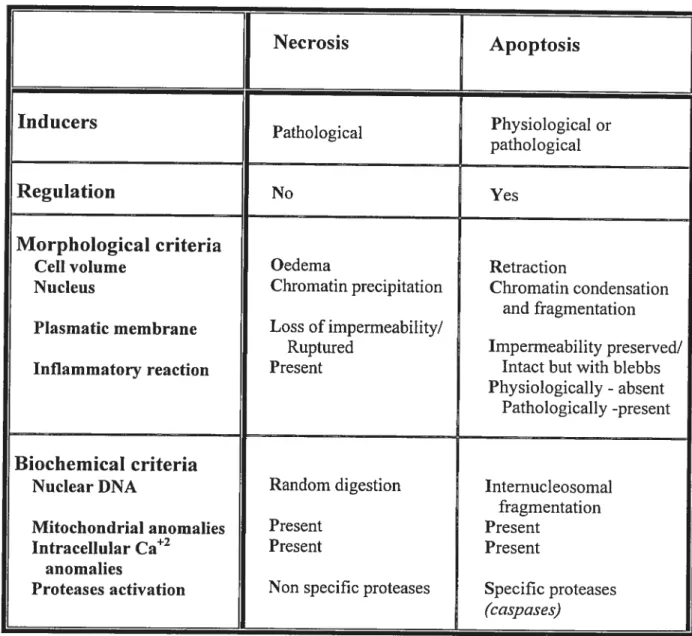
Table 1 Comparative table behveen the morphotogical and biochemicat properties of apoptosis (md necrosis Necrosis Apoptosis Inducers Pathological Physiological or pathological Regulation No Yes Morphological criteria
Celi volume Oedema Retraction
Nucleus Chromatin precipitation Chromatin condensation and fragmentation Plasmatic membrane Loss ofimpermeability/
Ruptured Imperrneability preserved/
Inflammatory reaction Present Intact but with blebbs
Physiologically - absent
Pathologically -present
Biochemical criteria
Nuclear DNA Random digestion Internucleosomal
fragmentation
Mitochondrial anomalies Present Present
Intracellular Ca2 Present Present
anomalies
Proteases activation Non specffic proteases Specific proteases
Figure 1 Morphology of dying celis by necrosis rnd apoptosis
Morphology of normal (A), necrotic (B) and apoptotic (C) mouse hepatocytes, where
arrows indicate apoptotic bodies. Hepatocyte nuclei can be visualized under a microscope
equipped with ultraviolet epifluorescence following staining with Hoechst 33258, a specific
DNA fluorochome. Normal nuclei appear homogenous and intact (D), as opposed to
apoptotic nuclei which are condensed (E), fragmented and very bright (F). In panel (G), a
T-ce!! undergoing apoptosis show the characteristic blebbing of the cytoplasmic membrane
(photo from Zimmermann, KC. et al. Pharmacology & Therapeutics (2001) 92: 57-70).
Non-activated tissue macrophages (panel H), stained with fluorescein, ingest apoptotic
bodies (stained with rodamine) to prevent inflammation (photo from $avill J et aï. Nature
A
B
C
)
I-D
3. ROLE 0F APOPTOSIS IN HEALTH AND DISEASE
3.1. Apoptosis in physiotogy
The concept of spontaneous ce!! death as a physiologica! event was first reported by
Walther f!emming in 1885 who noticed that many of the epithelial cel!s !ining the
regressing ovarian follicu!es showed fragmented nuc!ei typica! of what is now known as
apoptosis that he termed “chromatolysis”. (2) Later on, embryologists also understood the
importance of “chromato!ysis” as a morphogenetic mechanism during embryogenesis.
Indeed, the formation of digits, the deve!opment of the immune (through the elimination of
seif-reactive lymphocytes) and of the nervous systems were ai! shown to necessitate this
specia! kind of ceil death. However, these finding were thought to be limited to embryonic
tissue. (2)
WIty tue explosion in stttdies ht articles related to apoptosis? In 1972, Kerr et a!. (6) have
documented the diversity of the occurrence of apoptosis in different tissues and in a wide
range of physiologica! and patho!ogica! conditions. Later, Wyllie (10) has demonstrated
that nuclear apoptotic events inc!uded the activation of an endonuclease that resu!ted in the
degradation of chromatin into nuc!eosome-sized DNA !adders that have becorne a
biochemica! hal!mark of apoptosis. However, these findings remained without significant
recognition for a!most a decade. The discovery that the adult hermaphrodite
9
apoptosis, hurtied apoptosis into hot topic status in the 1990s. (16) The study of mutants of C. eÏegans in the apoptotic processes identified 3 genes which are involved in the regulation of apoptosis in ail somatic ceils (ced-3, ced-4 and ced-9). In addition, these genes have been shown to have marnrnaiian homologues (caspases (17), apoptosis protease-activating factor-1 tApaf-l] (1$) and B-cell iyrnphoma [BCL-2] (19) famiiies of genes respectiveiy)(20). With this regain of interest in apoptosis, researchers have focused on the physiological aspects of apoptosis as a widespread phenomena. Indeed, apoptosis
has been shown to be involved in severai physioiogical processes, such as:
• Acquisition of immunologicai self tolerance in T celis (21),
• Elimination of viraiiy-infected ceils by cytotoxic T cells (21),
• Down-reguiation of the immune reaction afler inflammatory reaction (Activated
induced ceii death) (21;22),
• Organ atrophy foiiowing hormonal retrievai (23),
• Establishment of the immune priviieged sites (such as testes, ovaries, uterus and
retina) (22;24)
• Maintenance of tissue homeostasis (i.e. appropriate number of ceils) and is therefore
3.2. Apoptosis inpathotogy
Given the widespread and critical role of apoptosis in biology, it is not surprising that
deregulation of apoptosis occurs frequently during pathological conditions. Indeed, the
aberrant activation of apoptosis may contribute to a number of diseases such as
neurodegenerative disorders (e.g. Alzheimer and Parkinson) (26) and viral infection.
Several viruses were shown capable of inducing (27-29) or inhibiting (30-32) apoptosis
directly in infected ceils or to induce their elimination by cytotoxic T cells through
activation of the fas receptor (f as-R) and/or Granzyme B systems (33-35). In contrast,
impaired apoptosis may be a significant factor in the etiology of diseases such as cancer
(36;37) and autoimmune disorders (systemic lupus erythematosis (34) ancl
lymphoproliferative disorders (38; 39 ;40)).
4.
APOPTOSIS AND THE LIVER
4.1. TIte tiver
The liver, which constitutes approximately 2% of body weight in adult humans, resides at
the crossroads between the digestive tract and the rest of the body. Hence, it plays a major
role in maintaining the body’s metabolic homeostasis. This includes 1) the uptake of
dietary amino acids, carbohydrates, lipids and vitamins, their subsequent storage.
rnetabolism and release into the blood and the bile; 2) the biotransformation of hydrophobic
11
water-soluble derivatives that can be excreted into the bile or the urine; 3) the synthesis of
semm proteins such as albumin, vitamin transporters and blood clofting factors. In addition.
the liver is integrated into the reticuloendothelial system of defence of the body against
foreign macromolecules and microorganisms. (41)
Hepatic disorders therefore have severe consequences on overali health. The liver is
vuinerable to a wide variety of metabolic, toxic, microbial, circulatory and neoplastic
insults that cause liver injury both by necrosis and apoptosis (1;41;42).
4.2. Physiotogicat andpatltotogicat retevance of tiver ceit apoptosis
In normal liver, apoptosis is a rare event. Indeed, only about 1-5 apoptotic celis are found
out of 10 000 hepatocytes in the rodent liver. (43) This very low rate could be explained by
the low rate of turnover of hepatocytes under physiological conditions. However, this rate
is increased during liver involution to remove excessive hepatocytes. This can occu
following withdrawal of hypertrophic or hyperpiasic stimuli (e.g. increased functional
demand, over feeding, pregnancy or during severe protein loss) (42) or during liver atrophy
following left portal vein ligation (14;44). furthermore, aberrant activation of apoptosis has
been noted in metabolic liver diseases such as alcoholic liver disease (where high levels of
Fas ligand were detected, see below). (45) In addition, several studies have reported a
marked increase in the levels of apoptosis in fulminant hepatitis (46) and viral infections
such as hepatitis B(42;47) and hepatitis C(4$), as well as in immune-mediated liver
failure of apoptosis to delete genetically altered celis appears to contribute to the development ofhepatocellular cancer and cholangiocarcinorna. (37)
Based on this evidence, new therapeutic approaches could be developed to treat theses diseases. Indeed, pharmacological inhibition of apoptosis may arneliorate liver injury and/or promote liver regeneration. On the other hand, purposeftil induction of apoptosis in malignant ceils may be usefiul in treating hepatobiliary cancers. Thus, understanding the mechanisms of apoptotic induction and inhibition is highly relevant for the cÏinical diagnosis and treatment of liver diseases. (49)
13
B. APOPTOSIS : CELLULAR MECHANISMS
The apoptotic process is divided into three phases: induction, execution, and finaily
degradation and elimination. During the induction phase, celis receive signais to initiate the
apoptotic process, decode and interpret them. When the ceii takes the decision to commit
suicide, it activates the executioner caspases: this is the point of no return. These caspases
then cleave multiple intraceliuiar targets (such as endonucieases, transglutaminases, other
caspases) to produce the biochemical (DNA iadder, protein precipitation and degradation of
cytoskeieton components) and morphoiogicai (chromatin fragmentation, membrane
blebbing, cytoplasm disintegration) aiterations characteristic of apoptosis. Finally.
apoptotic bodies are phagocytosed by macrophages and neighbouring ceiis. (7;15) (see
f igure 1)
1. INDUCTION 0F APOPTOSIS
Apoptosis can be initiated by different signais from outside as weil as inside the ceil
1. Ceil-surface death receptors of the Tumour Necrosis factor Π(TNfa) family, such
as f as and TNf receptors, are among the best characterized extracelluiar inducers
of apoptosis. Binding of iigands to their receptors initiates a fairiy weii-defined
caspase signaliing cascade that activates, in an amplification ioop, other executioner
BCL-2 family. The latter then transmit the signal through activation of die
mitochondrial apoptotic pathway (type II cells, see below). (50-53)
2. Granule-mediated apoptosis: granules released by cytotoxic T celis contain perforin
ta pore-forming protein which facilitates the entry of granules components into the
target cell) and Granzyme B (a serine protease which cleaves afier an aspartate
residue) (54). It has been suggested that Granzyme B can directly cleave anci
therefore activate caspase-3, which then activates caspase-7. (55)
3. Genotoxic agents, such as topoisomerase inhibitors (e.g. etoposide), anti
metabolites (e.g. 5-fluorouracil), DNA damaging agents (like cisplatin) and
y—irradiation, are among the best-studied initiators of apoptosis. Damage to DNA is
sensed by p53, which upregulates Bax and therefore activates the mitochondriaÏ
apoptotic cascade. (53;56-5$)
4. Loss of survival signais, such as growth factors (e.g. epidermal growth factor
[EGF], hepatocyte growth factor [HGF], nerve growth factor [NGF], etc) and
contact with the extraceilular matrix, which are normally perceived by cells.
activates the apoptotic program by default. (59-6 1) This has been shown to occur
through the association of Bad with Bax, two pro-apoptotic proteins of the BCL-2
famiiy (see below), in the cytoplasm thus favouring the oligornerization and the
transiocation of the complex to the mitochondria. The sequence culminates with the
15
5. Mitochondrial-elicited death pathway: numerous pro-apoptotic pathways converge on the mitochondria to induce mitochondrial permeability transition (MPT): the outer membrane becomes protein-permeable while the iimer membrane continues to retain matrix proteins but dissipates the mitochondrial transmembrane permeability
(A’J’). MPT triggers the activation of caspases and nucleases through the release of
pro-apoptotic proteins normally confined to the mitochondrial interniembrane space, such as cytochrome e and Apoptosis Inducing Factor-Ï (AIF-1). (13). The release of cytochrome e stimulates the assernbly of the apoptosome (procaspase-9, cytochrome e and Apaf- 1) in the cytoplasrn and therefore the activation of the downstream caspase-3. On the other hand, AIF-1 transiocates to the nucleus and activates a nuclear DNase (reviewed in (67;68)).
Figure 2 Mech anisms of iudttction and inhibitioit of apoptosis
Pro-apoptotic rncchanisrns: (1) Ceil surface death receptors (e.g. Fas, TNFŒ) activate initiator caspases (e.g. caspas-$) to cleave Bid and generate tBid. (2) Growth factors (GF) deprivation induces the dephosphorylation of Bad and its dissociation of 14-3-3 proteins. (3) Genotoxic agents increase the expression of Bax in a p53-dependent manner. Ail these apoptotic stimuli induce the transiocation of Bax from the cytosol to the mitochondria. This culminates in the release of cytochrome e and Diablo/Smac. Diablo/Smac relieves the inhibitory effect of IAP. Cytochrome c forms the apoptosome with Apaf- 1, procaspase-9 and ATP. This activates caspase-9, which then activates caspase-3. The latter caspase is responsible for the degradation of cytoskeleton components (e.g. fodrin, gelsolin) as well as ICAD (inhibitor of CAD), thus releasing CAD (Caspase Activated DNase). CAD is the DNase responsible for the fragmentation of DNA into 180-200 bp, which are the haflmark of apoptosis.
Anti-apoptotic rnechanisrns (in Black boxes). There are several checkpoints for where apoptotic signal can be inhibited. (4) fLIP is a natural inhibitor of the formation ofthe DISC complex. (5) lAPs (Inhibitors of apoptosis proteins) are natural inhibitors of active caspases. (6) Anti-apoptotic members of the BCL-2 (e.g. BCL-2) act directly on Bax activity in the mitochondria. (7) Akt, a serine/threonine kinase, phosphorylates Bad on serÏ36, thus favouring its sequestration with 14-3-3 proteins, and procaspase-9, thus preventing its activation.
o
J
.. — e.1.
•. . •.. O4;
O 00 0 O •0 OI
o
t
O -. le _..1 o ee fl • o • • •tl E C C —e
00
‘t NC
C
2. EXECUTION 0F APOPTOSIS
During this phase, the fate of the celi is set. Indeed, the celiular response to the above rnentioned apoptotic signais is reversible and depends on the intraceilular context. Irreversible activation of the apoptotic process can 5e faciiitated (pro-apoptotic) or abrogated (anti-apoptotic) by specific proteins whose expression and activity are under tight control. In this section, the pro-apoptotic proteins wiil be discussed. The anti apoptotic proteins will be detailed in section D.
There are three key pro-apoptotic families of genes:
1. ICE (interleukin-1 converting enzyme) farniiy, which are now best known as
caspases;
2. Pro-apoptotic members ofthe BCL-2 family (e.g. Bid, Bax);
3. Inhibitors oflnhibitor Apoptosis Proteins (e.g. DiabÏo/Smac)
2.1. THE FAMIL
Y
0F ICE-PROTEASES (CASPASES)Proteases: Proteases participate in protein destruction and in the reguiation of protein activities in different contexts. They are involved in protein maturation (e.g. caspase-1 in the context of pro-IL-1 processing), regulate and arnplify specific reactions (e.g. the ciotting and complement cascades) and allow celis to migrate (e.g. proteases of the
19
extracellular matrix). In addition, they are responsible for the degradation of proteins outside the celi (digestive proteases) and in a similar aibeit more controlÏed and discriminating manner inside the ceil (jroteosome). Finally, in the context of celi death, they act as triggers ofthe apoptotic process, as regulatory elements within it, and ultimately as a subset ofthe effector elements ofthe machinery itseif. (54)
Caspases: Ced-3 encodes a protein which resemblcs the mammalian interleukin 1
1-converting enzyme (ICE), commonly called caspase-1.(69) They are proteases with a reduced cysteine residue in their active site (QAÇXG) which confers them the specificity of cleavage afier an aspartate residue.(70) This specificity of cleavage is shared with another protease, Granzyme B (see section B.1).To date, 14 caspases have been identifled in mammals. Caspases are constitutively expressed and are norrnally present as inactive precursors in celis. Caspases have a ver)’ conserved structure composed of a N-terminal prodomain of varying length, and a domain that, afier processing, will give rise to two subunits: a large subunit (17-21 KDa) which contains the active site, and a smali subunit (10-14 KDa). (70-72)
Upon receiving an apoptotic signal, the pro-forms (zymogens) of caspases undergo sequential proteolytic processing at internai aspartate residues (releasing the srnall subunit then the prodornain) to generate the active enzyme. Caspases then assemble into two large subunits and two small subunits thus forming the active caspases.(12;73;74) In addition, the cysteine in the active site must be reduced in order for the caspases to function. Indeed,
several in vitro as well as in vivo studies have reported that oxidants inhibit the proteolytic activity of caspases while anti-oxidants restore or maintain their activity. (75-77)
Based on the length of their prodomain, caspases can be divided into two groups: class 1. which contains a relatively long prodomain (e.g. caspases 1, 2, 4, 5, 8, 9, 10, 12, and 13) and ctass II containing a short prodomain (e.g. caspases 3, 6, 7).(54;70) The long prodomains in many class I caspases contain protein-protein interaction domains that play a crucial rote in the recruitment of caspases to specific death complexes. This triggers oligomerization of procaspase molecules, thus facilitating their activation by autocleavage. For example, the prodomain of caspases-8 and -10 contain a Death Effector Domain (DED), which mediates their binding to adaptor molecules FADD (Fas Associated protein with Death Domain) (7$;79) or TRADD (TNF Receptor Associated protein with Death Domain) (80). Other caspases (such as 1, 2, 4, and 9) contain a Caspase Recruitment Domain (CARD, (81), which plays an important role in the interaction between different caspases as well as with various adaptor proteins. Class II caspases, which lack a long prodomain, also lack the ability to auto-activate and require cleavage by class I caspases to get activated.(54;70;74) For this reason, class I caspases are referred to as initiators caspases whule class II as executioner ones. The fact that caspases can auto-activate and/or activate others constitutes a caspase cascade that not only amplifies the process but also transmits the signal from one compartment to another within the cett. Caspases are activated or inactivated through a series of intracellular steps, or pathways, in response to death or survival signals, which are subject to multiple regulations (see Section D).
21
2.2. THE PRO-APOPTO TIC PROTEINS 0f THE BCL-2 FAMIL V
As mentioned above, mitochondria play an important role in apoptosis. In particular, they release AIF-1 and cytochromec (64;6$;$2), which are responsible for the mediation ofthe apoptotic signal to downstream effectors. BCL-2 farnily members (mammalian homologue ofced-9) have been identified as key regulators of cytochrome e release.(13;6$;83;84)
The BCL-2 famiiy is characterized by conserved motifs known as the BCL-2 homology domain (3H). Members of this family have at least one of the four 11H identified. According to their fiinction and structure, these proteins have been subdivided into three subfamilies (68;$5):
1. Anti-apoptotic proteins, such as BCL-2, BCL-x1, BCL-w and MCL-1. BCL-2 and
BCL-xL contain ail four BH (BH1-BH4), but other members contain at least BHI and BH2. (discussed in detail in Section D.2)
2. BH3-only containing pro-apoptotic proteins which include Bid, Bad, Blk.
3. Classical pro-apoptotic proteins such as Bax and Bak, which share sequence homology in ail BH domains except BH4.
These proteins canform homo- and heterodimers that invoives the 3H dornains (84;$6;87). The BH3 domain of pro-apoptotic proteins appears to be required for the interaction between anti- and pro-apoptotic proteins ($8).
BH3-onty coittaining pro-apoptotic proteins: Members of the BH3-only subset of the
BCL-2 family coimect proximal signais to the core apoptotic machinery. These proteins, of which Bid is a prototype, are capable of heterodimerizing with members of other subsets of the BCL-2 family.(73) They are found in the cytosol under non-apoptotic conditions. Upon receiving the apoptotic signai, they become activated. For example, Bid activation by cleavage (following Fas-R stimulation) favours the interaction of truncated Bid with Bax. This interaction induces a conformational change of Bax leading to its oligornerization and insertion into the outer mitochondriai membrane (65;89;90) and finally culminating in the activation ofthe downstream caspase cascade.
Classicaipro-apoptotic proteins: The expression of Bax and Bak has been associated with
high sensitivity toward apoptosis. In fact, genotoxic agents induce apoptosis by increasing the expression Ïevels of Bax. Bax has been shown to form pores in synthetic lipid bilayers that resemble the channel-forming properties of the bacterial toxins coÏicins and dtphteria.(91) 0f note, the channel forming activity of Bax (and Bak) has not been yet dernonstrated in vivo. Bax transiocation to the mitochondria has been shown to cause cytochrome c release and activation ofcaspases, leading to celi death by apoptosis. (64-66) However, the exact mechanism responsible for these proteins’ function are flot yet elucidated.(64-66;89;90)
, -, z_J
2.3. Diabto/Srnacproteins
Diablo (direct IAP binding protein with low pI) /Smac (second mitochondrial activator of caspases) is a mammalian protein analogous to the pro-apoptotic Drosophila molecules. Grim, Reaper, and HID. (92;93) It is released from the mitochondria following apoptotic stimulation in a Bid-dependent manner to relieve inhibition of caspases by the inhibitor of apoptosis proteins (lAPs; see section D.5; (94)). Once released from the mitochondria. Diablo/$mac binds to lAPs and antagonizes their anti-apoptotic effect. It therefore assists the initiator caspase-9 and effector caspases (caspase-3, caspase-6, and caspase-7) in
becoming active, ultirnately leading to celi death (95;96). Extra-mitochondrial transiocation of Diablo/Smac along with cytochrome c and other pro-apoptotic proteins represent important regulatory checkpoints for mitochondria-mediated apoptosis.
3. DEGRADATION AND ELIMINATION 0F APOPTOTIC CELLS
When the full complement of caspases that are necessary for the proper execution of the cell death program have become activated, the final disintegration phase begins. In this phase, the biochemical bases for the morphological changes take place.
Nuctetts: Active caspase-3 is responsible for the cleavage of the Inhibitor of Caspase Dependant DNase (CAD), an inhibitory protein which retains Caspase Dependent DNase (CAD) in the cytoplasm (97). This degradation releases CAD, which transiocates to the nucleus. This nuclease cuts the DNA between the nucleosomes thus producing 180-200 bp DNA fragments. The activity of CAD is responsible for the DNA laddering that is a
hallmark of apoptosis.(98) Lamin A (99), Lamin B (100) and NuMA (101) are also
degraded by caspases which transiocates to the nucleus (e.g. caspase-6). This leads to the
disassembly and the destruction of the nuclear cytoskeleton thus further contributing to
nuclear fragmentation. In order to ensure that the damage done by the apoptotic process is
flot repaired, PARP (poly (ADP-ribose) polyrnerase), which is involved in the repair of
damaged DNA, is also degraded by caspase-3. (102) The mitochondrial protein AIF
released in parallel to cytochrome c following apoptotic stimulation induces high molecular
weight DNA fragments in a caspase-independent manner. (103)
Ptasnta membrane: Structural breakdown of the celi includes cleavage of fodrin (104).
membrane-associated cytoskeletal protein, as well as direct cleavage of gelsolin (105), an
actin regulatory protein, resulting the disruption of actin filaments. Taken together these
two events can be held responsible for dissociation of the submembrane cytoskeleton and
the destabilization of the plasma membrane (membrane blebbing). In addition, the
asymmetrical distribution of phosphatidylserine (PS) is lost resulting in the presentation of
PS on the external surface of apoptotic cells. This has been shown to facilitate the
phagocytosis of apoptotic cells by macrophages and neighbouring ceils thus reducing the
25
C.
FAS RECEPTOR / FAS LIGAND SYSTEM
1. FAS SYSTEM AND ITS ROLE IN DISEASE
In 1989, two groups independently isolated mouse-derived antibodies that were cytotoxic for various human ceil unes (108). The celi surface proteins recognized by these antibodies
were named f as and APO- 1. The fas system is composed of Fas receptor (Fas-R) and Fas ligand (Fas-L) (109). Fas-R is highly expressed in activated mature lymphocytes (110) and human lymphocytes transfected with Epstein-Ban virus or human immunodeficiency virus (108;1 11). In addition, many nonlymphoid tissues express high levels of Fas-R, including
liver, heart, lung, kidney and ovary (112). Fas-L is primarily expressed by activated T celis
and plays a role in peripheral T- and B-cell homeostasis. (14;34;47; 113)
The apoptotic potential of Fas-R / Fas-L system has been mostly studied in the immune
system. Indeed, it was observed the Ïpr (lymphoproliferative) and gld (generalized
lymphoproliferative disorder) phenotypes (39) were associated with progressive
lymphadenopathy and immune complex syndrome that resemble the human autoimmune
disease systemic lupus erythematosus (38;40). A major role of Fas system in lymphoid
ceils lies in the control the immune response. It acts as a seif-limiting feedback in activated
lymphocytes through a process called activation-induced celi death. Indeed, afier antigen
driven expansion of lymphocytes, there is upregulation of f as-L on activated T celis that is
cytotoxic T ceils eliminate virally infected and genetically altered celis by activating the
f as apoptotic death signal. (33;34)
One of the early indications that the fas system plays an important role in hepatic homeostasis and therefore in the development of hepatic disorders is the observation that injection of anti-fas antibody to mice caused massive hepatic apoptosis and animal demise shortly thereafier (115). Hepatocytes constitutively express Fas-R and may upregulate its expression in a variety of liver diseases (14;42;47). Numerous studies have iinplicated f as-R and Fas-L in both hepatitis B (42;47) and C (4$), where infected celis appear to be extremely sensitive to f as-mediated apoptosis (52). In alcoholic liver disease (116) as weB as Wilson’s disease (117), Fas-L mRNA was found to be expressed in hepatocytes. This expression is hypothesized to mediate hepatocyte loss observed in these disorders. Therefore, the mechanisms of apoptotic induction of the Fas system as well as protection against it in the liver should be of great therapeutic interest.
2. FAS RECEPTOR SIGNAL TRANSDUCTION
The structure of fas-R indicated that it is a type I transmembrane protein (46 KDa), which
belongs to the TNf and NGF receptor family. It possesses 3 cysteine-rich extracellular
domains involved in the recognition and binding of the fas-L. The cytoplasmic domain
contains a conserved region (about 70 amino acids) that is necessary and sufficient to
27
involved in the recniitment of and the protein-protein interaction with FADD (20; 10$; 113). an adaptor protein that also contains a DD region.
It is weIl documented that ceil death induced by Fas-R stimulation is dependent on caspase activation.(20;51;1 19) However, there some reports describing a caspase-independent killing.(5 1 ;120-122) This led to the hypothesis of two independent signal transduction pathways activated at the level of Fas-R: one is mediated by the adaptor protein DAXX (death associated protein; (123)) which can enhance fas-induced apoptosis through the activation of jun-amino terminal kinase (JNK) cascade; the other involves the adaptor protein FADD and the subsequent recruitrnent of caspases to the Death Inducing Signalling Complex (DISC) and therefore the activation ofthe caspase cascade (51;78;79;124).
Figure 5 fas receptor signattiitgpathways
Binding of Fas ligand (Fas-L) to Fas receptor (Fas-R) induces the trimerization of the latter.
This activates Daxx- and FADD- dependent signalling pathways. (1) In the former, Daxx
associates with the death domain (DD) of the Fas-R. This activates Apoptosis Signal
regulating Kinase-1 (ASK-Ï), which resuits in the activation of the jun-arnino terminal
kinase (JNK) pathway leading to ceil death by apoptosis. (2) The fADD pathway is
initiated by its recruitment to the Fas-R. Procaspase-8 molecules are then recruited to form
the Death Inducing Signalling Complex (DI$C) and therefore get activated. (2a) In type II
ceils (e.g. hepatocytes), caspase-$ cleaves Bid to generate tnincated Bid (tBid). tBid
associates with Bax protein in the cytoplasm and induce its transiocation to the
mitochondria resulting in the release of cytochrome e and Diablo/Smac proteins into the
cytoplasm. Diablo/Smac relieve the inhibitory effect of IAP (Inhibitors of Apoptosis
Proteins) thus facilitating the activation of caspases. Cytochrome c along with Apaf- I
(apoptosis protease activating factor- 1), procaspase-9 and ATP form the apoptosome,
which culminate in the activation of caspase-9. Caspase-9 then cleaves and activates
caspase-3. (2b) In type I cells (e.g. lymphocytes), caspase-8 directly cleaves and activates
caspase-3. Active caspase-3 degrades several cellular components leading to celi death by
30
2.1. fADD patit way:
fas-induced activation of the caspase signalling cascade also involves two major pathways termed type I and type II. Basically, they differ in the involvement of the mitochondria in
fas signal transduction. In type I ceÏÏs, such as lymphocytes, there ïs activation of a
sufficient amount of initiator caspases at the receptor level to directly transmit the signal to effector caspases. (20;125) Hepatocytes are type II celis, where the signal generated at the receptor ïs not strong enough to transduce the signal and needs to be amplified. Therefore, the weak apoptotic signal is transmitted to the mitochondria where it induces MPT and
activates downstream caspases (see Section 3.1 and beÏow).(5 1 ;1 19)
2.2. fas sigitatting in hepatocytes (type II ce!ts):
Binding of the f as-L or of anti-Fas antibodies to fas-R in hepatocytes resuits in receptor trimerization. The clustering of the DD in the intracellular portion of the receptors recruits the adapter molecule called FADD via the DD ofthe latter (50;79). Procaspase-8 molecules
(fLICE) are recruited to the Fas-RJFADD complex through an interaction mediated by the
DED present on FADD along with the prodornain of caspase-2, thus forming the DISC complex. The close proximity of procaspase molecules favours their oligomerization, and stirnulates their autocleavage resulting in their activation.(79; 119) Active caspase-$ then cleaves Bid, a 3H3 only pro-apoptotic protein of the BCL-2 family. This generates a p15-Bid fragment (tp15-Bid) which transiocates to the outer membrane of the mitochondria.(73) tBid can interact, through its BH3 domain, with the anti-apoptotic BCL-xL which
destabilizes the BCL-xL/BAX heterodimers.(126) This favours the homo-oligomerization
of BAX. In addition, this association prevents the formation of the anti-apoptotic complex
between BCL-XL and Apaf-1/procaspase-9, leading to the release of the latter. (73;127)
Consequently, cytochrorne c ($2)as well as Diablo/Srnac (92;93) are released from the
mitochondria, although the mechanisms of this liberation are flot yet ftilly elucidated.
(51;82;1 19;128)
Once released from the mitochondria, cytochrome e interacts with Apaf- 1 and procaspase-9
(in an ATP-dependent manner) to form a complex known as the apoptosorne ($3;127).
Formation of the apoptosorne resuits in the cleavage and activation of caspase-9, which in
turn leads to the cleavage and activation of caspase-3, a central executioner caspase.(73)
Caspase-3 then cleaves and activates downstream caspases and other important proteins.
which are directly or indirectly responsible for the systernatic dismantiing of the apoptotic
cell and finally culminating in ceil death by apoptosis. (see Section B.3)
2.3. fADD-independent signatting:
Several pieces of evidence demonstrated the involvement of caspase-independent pathways
in the modulation of the apoptotic signal.(120;122;124) One of these pathways is the JNK
pathway.(129) The involvement of this pathway in apoptosis was initially characterized in
UV irradiation and TNf-Πtreatment. (129-131) Fas-mediated apoptosis was shown to
require JNK activation in 293 and L920, but not in HeLa cells (123). On the other hand, a
32
fas-induced apoptosis (132). This suggested that the involvement of the JNK pathway in Fas-induced apoptosis was celi specific, which signifies that the biological relevance of its implication requires further investigation.
The JNK pathway was linked to the Fas-R through the discovery of the DAXX protein in 1997 using an interaction trap system in yeast (123). DAXX associates with the DD offas R, although it lacks a DD of its own. In addition, Yang et al. have shown that the simultaneous overexpression of f as-R and DA)O( activated the INK pathway and enhanced Fas-induced ceil death by apoptosis. This effect of DAXX was shown to be mediated by the MAP kinase kinase kinase ASK- 1 (poptosis Signal regulating Kinase-l) (121;133).
D. ANTI-APOPTOTIC MECHANISMS
1. GROWTH FACTORS AND PROTECTION AGAINST APOPTOSIS
Not ail fas-bearing ceils are susceptible to f as-induced apoptosis. Indeed, depending on the extracellular environment and the intracellular context in which the apoptotic signal is received, the celi may or may flot undergo apoptosis. Growth factors are known for their ability to antagonize apoptosis. Growth factors such as NGf, HGf, EGf, and IL-3 have been shown to protect various types of celis (e.g. astiocytes, PC 12, hepatocytes, HepG2, lymphocytes, fibroblasts, etc.) against a variety of apoptotic inducers including physical (UV, y-radiations), chemical ones (cisplatinum, 5-fluoracil), and ceil-surface death receptors (fas, TNf) (46;134-136). This protection has been mainly associated with the increase in the expression of the anti-apoptotic members of the BCL-2 family (e.g. BCL-2 and BCL-xL) (137;13$), as well as the inactivation of pro-apoptotic proteins (e.g. Bad, caspase-9), the latter foÏÏowing their phosphorylation by the serine/threonine kinase Akt (see below). (62;139;140)
Celis possess anti-apoptotic mechanisms to prevent the unintentional activation of the apoptotic program. Several of these mechanisms were discovered due to their abnormal expression (lack of or over expression) in diseases such as cancers and viral infections. Many types of cancer have developed mechanisms to avoid apoptosis. These include overexpression of anti-apoptotic proteins of the BCL-2 family, downregulation and
j
prevention of the recruitment of initiator caspases, overexpression of IAP or expression of
decoy receptors (intact extracellular domain but truncated intracellular domain) (36).
In parallel, several viruses have adapted anti-apoptotic mechanisms to delay and even
prevent the onset of the apoptotic program in infected ceils in order to give them the time to
replicate. Herpes virus (141), Baculovirus (30;142), and Polyomavirus Middle T antigen
(143) are some examples ofthese viruses.
Here will be discussed some of the key inhibitory factors of apoptotic signalling, including
the BCL-2 family, IAP, FLIP and Akt. (see Figure 2)
2. ANTI-APOPTOTIC PROTEINS 0F THE BCL-2 FAMILY
The original protein BCL-2 was discovered in 1985 by Tsujimoto et al. (144) in follicular B
ceYl lymphoma, hence its name B-cell lymphoma-2 (BCL-2). In this type of cancer, the
BCL-2 gene is translocated from chromosome 1$ (where it normally resides) to
chromosome 14 where it becomes juxtaposed with the immunoglobulin heavy-chain (IgH)
locus. Such transiocation brings this anti-apoptotic gene under the influence of a strong
promoter whose normal job is to drive high levels of 1g gene expression in B-cells. This
results in the loss of normal regulation of the BCL-2 gene, and in the production of
inappropriately high levels of B CL-2 protein in germinal center B-cells. This contributes to
the neoplastic B-cell expansion by preventing celi turnover (by inhibiting apoptosis) rather
Furtherrnore, its expression increases the survival of celis deprived of trophic factors such
as growth factors (GF) and cytokines. In addition, the anti-apoptotic effect of several GF
has been associated with the increase in the expression levels of B CL-2 or BCL-xL proteins
($6;137).
These proteins have been localized in the membranes of mitochondria, nucleus and
endoplasmic reticulum. (67) BCL-xL was the first member of this family to be crystallized.
This revealed a structure similar to the pore-forming bacterial toxins (colicins and
diphteria) (91;145). As mentioned above, proteins of the BCL-2 family can forrn homo
and heterodimers.($5;146;147) The balance between cell survival and death seems to be
regulated by the proportion of anti- or pro-apoptotic dimers formed (146;147) where by
overexpression of BCL-2 would favours the formation of BCL-2/BCL-2 dimers. The high
proportion of anti-apoptotic dimers prevents the release of cytochrome c and the
subsequent transmission of the apoptotic signal (7 ;68 ; 85; 86).
Several hypotheses regarding the anti-apoptotic action of BCL-2 and BCL-xL proteins have
been put forth, including regulation of the mitochondrial megachannel (148;149)and anti
oxidant function (67;86;150;151). However, their exact mechanism of action is flot yet
36
3. PHOSPHATIDYLINOSITOL 3-OH KINASE! Akt PATHWAY
The PI 3-K is one of the phosphorylation pathways activated by GF (see Section E.3.2).
Several studies have recently irnplicated this pathway, and especially the downstream
serine/threonine kinase Akt, in the protection against apoptosis. Indeed, inhibition of the PI
3-K pathway by wortmannin or LY294002 sensitizes ceils to apoptosis (152). In parallel.
wortmannin treatment was found to enhance f as- and TNf-a-induced caspase-3
activation.(153) This effect was shown to resuit from two phosphorylation events upstream
of casapse-3 activation: 1) phosphorylation of the pro-apoptotic protein Bad (62;1 54-1 56)
and 2) phosphorylation ofprocaspase-9.
As mentioned above, Bad can heterodimerize with BCL-2 and BCL-xL to antagonize their
anti-apoptotic effect. Gf, such as IL-3 and NGf, specifically induce the phosphorylation of
Bad on serine 136 (pBad-136) through Akt (62;156;157). This favours the association of
pBad-136 with 14-3-3 proteins instead of BCL-2 or BCL-xL therefore sequestering Bad in
the cytoplasm and preventing it from transiocating to the rnitochondria.(139) Thereby, this
phosphorylation promotes cell survival by allowing the unhindered action of anti-apoptotic
proteins ofthe BCL-2 family (139;155;156).
In parallel, it was demonstrated that serine phosphorylation of pro-caspase-9 (sen 96)
prevents its autocleavage and therefore its activation (15$). In fact, cytochrome c-induced
proteolytic processing of procaspase-9 was defective in cytosolic extracts from ceils
PI 3-KJAkt-dependent phosphorylation inactivates the important apoptotic mitochondrial
pathway. These two combined phosphorylation events contribute to the protective effect of
PI 3-K/Akt activation against apoptosis.
4. FLICE-INHIBITORY PROTEIN (FLIP)
The formation of the DISC complex in ceil-surface death receptor signalling, such as Fas
and TNF-a, can be regulated by a family of proteins called FLIP (fLICE-inhibitory
protein). This family of inhibitors was originally identified in oncogenic viruses, including
Kaposi’s sarcoma-associated human Herpes virus-8 (HHV-$) (160) and molluscipox virus
(141;161). Furthermore, high levels of FLIP were detected in melanomas where it promotes
escape from T-cell induced apoptosis (160). In parallel, some studies have suggested that
T-cell sensitivity toward Fas-induced apoptosis was conelated with the decrease in FLIP
rnRNA expression levels (162). Therefore, FLIP seems to inhibit apoptosis, and especially
the activation of procaspase-8. FLIP contains two DED that mediate FLIP binding to the
prodomain of caspase-8 and -10. This prevents the recruitment of procaspase molecules to
these receptors and their subsequent activation(162;163). There are different forms of FLIP.
The longest form (FLIPL) bas, in addition to the two DED, the equivalent of an inactive
n j
5.
INHIBITOR 0F APOPTOSIS PROTEINS (IAP)
Recently, key intrinsic inhibitors of caspases called IAP have been identified and they
represent important regulatory factors in apoptotic signalling. They were originally
identified as viral products that can inhibit the defensive apoptotic response of the host celi
to allow more time for the virus to replicate (164). Proteins in this family (such as XIAP.
HIAP1, cIAP2, NIAP and survivin) possess one or more baculovirus repeats (BIR), a
characteristic cysteine-rich domain of about 70 amino acids. They also contain a carboxy
terminal RING zinc-fingerthat can act as ubiquitin ligase (165;166).
In general, lAPs are thought to function at the caspase activation step in the ceil death
pathway. They bind to the inactive, prodomain-containing caspase (zymogen) and prevent
it from being processed into the active enzyme.(1 67; 168) The BIR domains of XIAP and
HIAPI have been shown to bind and inhibit caspase-3, -7, and -9 (167;169;170). Even the
smallest member of the IAP family, survivin, which contains only a single BIR, is also
capable of inhibiting caspase-3 (171). Despite the sequence similarities between the BIR
dornains of XIAP, they exhibit different affinities in term of binding and inhibiting
caspases. Indeed, BIR2 specifically inhibits caspase-3 and -7 while 31R3 inhibits caspase-9
only (170;172-174). Furthermore, it has been shown that XIAP can be cleaved into BIR1-2
and BIR3-RING zinc-finger fragments by activated caspases in vitro and in Fas-treated
celis (172). Exogenous expression of a BIR3-RTNG fragment potently suppresses Bax
the mitochondria, and the formation of the apoptosome complex, culminates in the
recruitment and activation of caspase-9 (172).
As for the ubiquitin ligase ability of XIAP and clAPi, it appears to be responsible for auto
ubiquitination and self-degradation of these lAPs through the proteosome, in response to
certain apoptotic stimuli (166). The RING finger may also function as a negative regulator
ofother apoptotic components, since the RING finger of clAPi mediates the ubiquitination
of caspase-3 and -7 (165). This suggests that lAPs initially bind and inhibit caspases, then
subsequently ubiquitinate and trigger the degradation of the IAP-caspase complex.
Mammalian lAPs could be important in diseases since the cIAP2 gene is ofien translocated
in mucosa-associated lymphoid tissue lymphomas (175) and ML-IAP/Livin is ofien
abnormally expressed in melanoma ceil unes. (176). In addition, elevated expression of
40
E.
EPIDERMAL GROWTH FACTOR (EGF)
1. STRUCTURE 0F THE EGF RECEPTOR
The EGF receptor (EGF-R) is a 170 kDa transmembrane glycoprotein with intrinsic
enzymatic activity. It is constituted of three distinct structural elements (178-183):
a) Extracethdar N-terminus domabt: It possess two cysteine-rich regions that form
the binding site for specific ligands such as EGf and Tumour growth factor-Œ
(TGfa);
b) Transinembrane domain: it is responsible for anchoring the receptor in the
membrane;
c) Cytoptasmic C-terminus doniain: it is comprised ofthree regions
i) Juxtamembrane region. It contains negative-feedback phosphorylation sites
for Protein Kinase C (PKC; Thr 654) and Mitogen activated Protein Kinases
(MAPK; Thr 669).
ii) Tyrosine kinase region (TK). This region, which contains the ATP binding
site (Lysine 721), catalyses the transfer of the y-phosphate of ATP to a
proteins. The TK region is highly conserved between the different receptors Tyrosine Kinase (RTK).
iii) Carboxy-terminat region: it comprises 5 tyrosine residues (Tyr 1173, 1 148, 1086, 1068, 992) that are the autophosphorylation sites of the EGF-R. These phosphorylated residues serve as docking sites for Src-homolgy-2 (SH2)- anci
phosphotyrosine homology (PTH)- containing proteins (see below) (181; 184).
2. ACTIVATION 0F EGF-R:
The monomeric EGF-R is inactive: it lias a weak affinity toward its ligand and a low rate of
TK activity. The binding of EGF to EGF-R induces a conformational change of the
extracellular domain of EGF-R, which favours the dimerization of two ligand-monomers
complexes. This dimerization stabilizes the interactions between the cytoplasmic domains.
increases EGf-R affinity toward the ligand and amplifies its TK activity. (185) The exact
mechanism by which EGf-R dimerization induces the activation of the TK activity is not
yet elucidated. However, the current conceptual model postulates that the carboxy-terminal
region of the receptor, in the basal non-phosphorylated state, functions as an auto-inhibitory
substrate of the TK activity of the EGF-R. The conformational change induced by EGF
binding places the C-terminus in proximity of the TK domain of the other monomer,
leading to its phosphoiylation on specific tyrosine residues (Tyr 1173, 1148, 1086, 1068.
992). This, in tum. relieves the inhibitory effect that the C-terminus exerts on the TK