Published in: Journal of Alzheimer's Disease (2010), vol. 10, pp 1119-1132 DOI:10.3233/JAD-2010-091633
Statut: Postprint (author’s version)
Peroxisome Proliferator-Activated Receptor Gamma
Enhances the Activity of an Insulin Degrading
Enzyme-Like Metalloprotease for Amyloid-β Clearance
Ira Espuny-Camachoa,b, Diana Domingueza,b, Pascal Merchiersc, Luc Van Rompaeyc,Dennis
Selkoed and Bart De Stroopera,b
aCenter for Human Genetics, KU Leuven, Leuven, Belgium
b Department of Developmental and Molecular Genetics, VIB, Leuven, Belgium cGalapagos Genomics NV, Mechelen, Belgium
d Department of Neurology, Harvard Medical School and Center for Neurologic Diseases,
Brigham and Women's Hospital, Boston, MA, USA
Abstract.
Peroxisome proliferator-activated receptor gamma (PPARγ) activation results in an increased rate of amyloid-β (Aβ) clearance from the media of diverse cells in culture, including primary neurons and glial cells. Here, we further investigate the mechanism for Aβ clearance and found that PPARγ activation modulates a cell surface metalloprotease that can be inhibited by metalloprotease inhibitors, like EDTA and phenanthroline, and also by the peptide hormones insulin and glucagon. The metalloprotease profile of the Aβ-degrading mechanism is surprisingly similar to insulin-degrading enzyme (IDE). This mechanism is maintained in hippocampal and glia primary cultures from IDE loss-of-function mice. We conclude that PPARγ activates an IDE-like Aβ degrading activity. Our work suggests a drugable pathway that can clear Aβ peptide from the brain.
Keywords: Amyloid-β (Aβ) clearance, insulin-degrading enzyme (IDE), metalloprotease,
peroxisome proliferator-activated receptor gamma (PPARγ)
Supplementary data available online (http://www.j-alz.com/issues/20/vol20-4.html#supplementarydata)
INTRODUCTION
Over the last few years, several alternative approaches to treat Alzheimer’s disease (AD) patients have been proposed. Most of them tackle either directly or indirectly the amyloid peptide generating pathway, as it is thought to be the driving mechanism behind the patho-genesis. It remains an issue of debate to what extent amyloid is directly responsible for the disease or to what extent additional factors are essential in the neurodegenerative process. Thus, abnormal phosphorylation of tau and tangle formation, an inflammatory component, and oxidative stress are all factors that likely contribute to the neurodegenerative process.
Recently, it has become clear that some antiinflammatory compounds, like certain NSAIDs or PPARγ agonists appear to modulate not only inflammation, but might also have a more direct effect on amyloid generation [1-7]. Lately, several clinical trials in AD patients have been performed using different NSAID compounds and duration of the treatments. Whereas most of them resulted in no or small benefits, it was proposed that NSAIDs action could be more effective for prevention instead of treatment of the disease [8]. Previous studies have also demonstrated that a subset of the NSAID family can act as PPARγ agonists [7]. On the other hand, first results from clinical trials using the PPARγ agonist, rosigli- tazone, showed a preserved cognition in AD and mild cognitive impairment (MCI) patients [9,10], although more recent studies, including preliminary results of a Phase III study were recently reported [11]. This study failed to demonstrate any significant effect in the treated patients (http://www.alzforum.org/drg/drc/default.asp? type=keyword&keyword=PPAR). This does not rule out the possibility that other drugs targeting PPARγ with, for instance, a better exposure in the brain, could have potential in the future. Thus, there is a huge interest in understanding how these compounds affect the amyloid burden in AD, and whether both type of agonists activate similar targets or whether they affect different mechanisms important for Aβ down-regulation.
PPARγ is a member of the Peroxisome Prolifera- tor Activated Receptor family (PPARs), a group of nuclear transcription factors. PPARγ activation plays important physiological roles in the regulation of lipid metabolism, adipocyte differentiation, insulin action, cell proliferation, and regulation of inflammation [12].
Recent studies, using either NSAIDS or PPARγ agonists, have related the activation of PPARγ under inflammatory conditions with a transcriptional repression of the BACE gene, ultimately resulting in reduced Aβ production [4,5]. We have previously found that PPARγ activation, even in the absence of inflammatory- mediated Aβ production, also affected clearance of Aβ in different types of cells in culture [13].
Concerning mechanisms for Aβ catabolism in the brain, several Aβ-degrading enzymes have been identified to date. Metalloproteases including insulindegrading enzyme (IDE or insulysin), the vasopep- tidases neprilysin (NEP or CD10), endothelin converting enzyme (ECE) and angiotensin converting enzyme (ACE), and the matrix metalloproteinases 2 and 9 (MMPs); the serine protease tissue plasminogen activator (tPA)-plasmin and the cysteine protease cathepsin B have been implicated in Aβ peptide turnover using in vitro assays [14-20]. Loss-of-function genetic mouse models have demonstrated the in vivo relevance of some of these candidate enzymes. Deficiencies in NEP, IDE, ECE, MMP2, MMP9, plasmin, and cathepsin B can each result in increased Aβ levels in the brain [19,2125]. Conversely, overexpressing IDE or NEP markedly reduced Aβ deposits and associated neuropathology in AβPP transgenic mice [26]. In humans, low ECE and plasmin levels and reduced IDE activity have been observed in AD patients [27-29], suggesting that lower activity of these enzymes could contribute to the generation of AD pathology. Indeed, several studies have shown that aging decreases the activity of some of these Aβ-degrading proteases in vivo [30,31].
Therefore, the search for modulators of Aβ degradation as a possible target for intervention in AD has gained considerable therapeutic relevance over the last few years.
Here, we further investigate the PPARγ-enhanced Aβ clearance mechanism and the putative role of the known Aβ degrading enzymes described to date. We propose that PPARγ expression activates an IDE-like metalloprotease for Aβ degradation.
MATERIALS AND METHODS
GENERATION OF ADENOVIRUSES AND PLASMIDS
The Ad5/GFP, PPARγ recombinant adenoviruses were generated by Galapagos Genomics NV as described previously [32].
The pcDNA3.1 (-) HA-Dyn2 K44A (Dynamin II dominant negative mutant) construct was purchased from ATCC, USA.
DRUGS AND PEPTIDES USED IN CULTURE
Lactacystin (10 µM), ammonium chloride NH4Cl (30 mM), and bafilomycin (10 µM) were
purchased from Calbiochem, Vel/Merck Eurolab, and Mikrobielle chemie, respectively. Insulin (1, 10 µM) and glucagon (0.1, 1 µM) were purchased from Sigma. Aβ1-40 synthetic
peptide was used at 0.1 µM concentration, and batches were obtained from Sigma and Bachem. The protease inhibitors: phenylmethylsulfonyl fluoride (PMSF) (30, 100, 300 µM in ethanol), pepstatin A (1, 3, 10 µM in DMSO), N-[N-(L-3-transcarboxyirane- 2-carbonyl)-L-Leucyl]-agmatine E64 (10, 100 µM in H2O), diaminoethanetetraacetic acid EDTA (3,10 mM in
50 mM Tris-Base, 0.9% NaCl pH 8), 1, 10- phenathroline (500 µM in DMSO), Phosphoramidon disodium salt (10, 30,100 µM inH2O), DL-Thiorphan (10, 30, 100 µM in
DMSO), TNF-α protease inhibitor- 1 TAPI-1 (10,30,100 µMinDMSO), leupeptinand antipain dihydrochloride (10,30, 100 µMinDMSO) were obtained from Sigma. 76307 Pefabloc SC (100 µM in H2O) was obtained from Fluka. Galardin, ilomastat GM6001 (10, 30 µM in DMSO)
was purchased from Biomol.
ANTIBODIES
The mouse monoclonal WO2 (epitope Aβ4-10) used for the immunodetection of Aβ and
sAβPPα was purchased from Abeta GmbH (Germany). The rabbit polyclonal B7/8 antibody (Aβ peptide), generated in our laboratory as previously described [33], was used for Aβ immunoprecipitation. Full length AβPP was detected using the rabbit polyclonal B63.1 antibody generated against the 15 C-terminal amino acids of AβPP, as described [34]. The HA.11 clone 16B12 antibody, used to detect HA tag from dynamin II (K44A) mutant, was purchased from Covance. Transferrin Receptor (H68.4) was detected with the monoclonal antibody obtained from Zymed Laboratories (San Francisco, USA). IDE levels were detected using either a polyclonal antibody (IDE-1) [35], or the monoclonal antibody (9B12) purchased from Covance (USA). Actin levels were analyzed with the monoclonal antibody (A 5441) obtained from Sigma.
CELL CULTURE
Primary murine hippocampal or mixed glia cultures were established from brains of embryonic E17.5 or newborn (P0) pups from wild-type, NEP-/-, IDE -/-, or NEP-IDE double knockout mice as described before [36]. Briefly, the dissected brain cortices were suspended in HBSS medium supplemented with trypsin (0.25% v/v) and incubated at 37°C for 15 min.
Single cell suspensions obtained from hippocampi or total brain were plated on poly-L-lysine-coated plastic dishes in minimal essential medium (MEM) supplemented with 10% horse serum. For hippocampal cultures the medium was changed 3-4 h after plating to serum-free neurobasal medium with B27 supplement (GIBCO) and treated with cytosine arabinoside (3
× 10-7 M) to prevent non-neuronal (glia) cell proliferation. The glial cultures were allowed to grow for 1 week before starting the experiments. Cells were infected with recombinant adenovirus defined as 1.000 multiplicity of infection (MOI) for 16 h and kept in culture for another 48-72 h.
HEK293 wild-type cells were kept in culture using DMEM/F12 medium (GIBCO) with 10% FBS. Forvi- ral infection, cultures (60% confluence) were infected with recombinant adenovirus with 100 MOI. After 24 h, medium was discarded, and a minimum volume of DMEM supplemented with 10% FBS was added to the cells for different time periods. When indicated, HEK293 cells were also transfected with pcDNA3.1 (-) HA-Dyn2 K44A (to block endocytosis), using lipo- fectamin 2000 reagent (Invitrogen) for 3 h. Cells were transfected either 3 h prior to adenoviral infection or 24 h after infection (48 h after plating), and therefore the reporter construct was expressed for 72 or 48 h, respectively.
GEL ELECTROPHORESIS AND IMMUNOBLOTTING
For the detection of IDE, HA-dyn II, and actin levels cells were lysed in 1% Triton buffer, and post-nuclear fractions were isolated by centrifugation at 10,000 X g at 4° C for 15 min. Protein concentrations were determined by Bradford assay [37]. Proteins were resolved in 10% Bis-Tris SDS gels (Invitrogen) and transferred to nitrocellulose membranes for western blot detection using the antibodies described above. For intracellular Aβ, cells were lysed in 200 µl of ice-cold RIPA buffer, and post-nuclear fractions were isolated by centrifugation at 10,000 X g at 4°C for 10 min. All the material was used for immunoprecipitation using B7/8 antibody. Immunoprecipitated samples were extensively washed, loaded on 12% Bis-Tris SDS gels (Invitro- gen) and transferred to nitrocellulose membranes for immunoblotting using WO2 antibody. Samples of conditioned medium were resolved in 12% Bis-Tris gels for Aβ and sAβPPα detection and transferred to nitrocellulose membranes. Immunoblot analysis was performed using WO2. Detection of signal was performed using the Chemiluminescence kit (PerkinElmer Life Science).
AΒ STABILITY ASSAY
Murine primary cultures and HEK293 cells were transduced with adenovirus and incubated for 16 h in DMEM and 10% FBS before adding the Aβ1-40 synthetic peptide (0.1
µM) for 3 or 24 h. When protease inhibitors or peptides were used in culture they were
incu-bated 30 minbefore Aβ addition. Conditioned medium was collected and subjected to immunoblotting analysis for Aβ stability.
SMALL
INTERFERENCE
RNA
(SIRNA)
DOWN-REGULATION
TECHNOLOGY
siRNA oligos already desalted, and converted to the 2'hydroxyl form to increase stability were obtained from Dharmacon, USA. Oligos were designed against murine Sphingomyelinase (nSMase) as control or human IDE. The sense and antisense sequences for murine nSMase were 5,-GCGCUUGGGAGACUUUCUGdT- dT-3' and
5'-CAGAAAGUCUCCCAAGCGCdTdT- 3’, respectively. The sense and antisense sequences for human IDE were 5'-UCAAAGGGCUGGGUUAAUA- UU-3' and 5'-PUAUUAACCCAGCCCUUUGAUU- 3’,respectively. dsRNA oligos at 100nM concentration were delivered into the cells by liposomal transfection (lipofectamine 2000, Invitrogen) for 3 h. 48 or 72 h after transfection, the medium was refreshed and 16 h later Aβ1--40 synthetic
peptide was added to the culture medium (as described above) and incubated for 3 h. Levels of Aβ in the medium were analyzed by western blot (WO2 antibody). IDE levels were detected in cell extracts by immunoblotting (polyclonal IDE-1 and monoclonal 9B12 antibodies).
CELL SURFACE PROTEIN LOCALIZATION BY BIOTINYLATION ASSAYS
We tested how effective the mutant HA-Dyn2 K44A (dyn II) was in blocking endocytosis in HEK293 cells. Cells in 10 cm dishes were grown to 70-80% confluence before the assay. Cells kept at 4°C were washed 3 times in Dulbecco’s PBS (pH 8), followed by incubation of the cells with 0.8 mM Sulfo-NHS-SS-Biotin reagent (in DMSO) or just the solvent DMSO in PBS pH 8 for 30 min. Cells were washed again 3 times with PBS pH 8 followed by another incubation with PBS pH 7 supplemented with 100 mM glycine and 0.5% bovine serum albumin (BSA) for 15 min. Cells were rinsed 3 times with PBS pH 7 supplemented with 100 mM glycine. Cell extracts were prepared in 1% Triton buffer with protease inhibitors and equal amount of protein was incubated with streptavidin beads overnight at 4° C to precipitate biotinylated proteins. Samples were resolved by SDS PAGE and immunoblotted with antibodies specific for transferrin receptor (Tf-R) and AβPP(B63.1).
QUANTITATIVE PCR
Total mRNA was extracted from HEK293 cells GFP- or PPARγ-transduced (in quadruplicates) with Trizol (Invitrogen) as described elsewhere [38]. mRNA samples were treated with DNAse I Turbo DNA-freeTM kit (Ambion Inc. Austin, TX) for 30 min at 37 °C. First strand cDNA was synthesized from 4 µg of DNAse I-treated total RNA with random primers (Invitrogen) using SuperScriptTM II RNAse H-RT (Invitrogen). Primers for qPCR reaction were
designed with PrimerExpress software (Applied Biosystems). The efficiency of qPCR amplifications was validated in four different cDNA dilutions from human kidney tissue with actin and beta-glucuronidase precursor (GUSB) primer sets as normalizers. The quantitative PCR reaction mixtures consisted of LightCycler 480 SYBR Green I Master (Roche), 250 nM of primers and 10 ng cDNA. Samples were run in triplicates on LightCycler 480 (Roche). The reaction steps were an initial denaturation step of 10 min at 95°C, thermal cycling conditions were 15 s at 95°C and 1 min at 60°C for 46 cycles. Relative expression levels of the different genes are given as a percentage of actin and GUSB (normalizer housekeeping genes): 2-∆∆Ct
whereby ∆Ct = Cttarget-Ctnormalizer, ∆∆Ct = ∆CtPPARγ-∆CGFP. For primer sequences see
supplementary data (experimental procedures).
RESULTS
PPARΓ-INDUCED Aβ DEGRADATION IS NOT DEPENDENT ON
CLATHRIN- OR CAVEOLIN-MEDIATED ENDOCYTOSIS
We recently showed that PPARγ activation causes decreased Aβ levels in a variety of cell culture systems, including primary neurons and glial cells. Moreover, when exogenous synthetic Aβ was added to the culture medium of HEK293 cells, PPARγ activation resulted in its enhanced clearance. The mechanism induced by PPARγ does not seem to involve a secreted enzyme as shown previously [13] by a cell-free in vitro assay where conditioned medium from GFP-transduced HEK293 AβPPsw cells incubated in a 50:50 ratio with fresh or with medium from hPPARγ-transduced cells showed no faster degradation. In addition, we have performed cell-based experiments where either fresh medium or conditioned medium from hPPARγ-transduced cells were added to GFP- transduced HEK293 cells coinfected
with AβPPsw adenovirus. The level of Aβ in the conditioned medium was not different among the different conditions [13]. We have now further investigated the mechanism for degradation and tested the hypothesis that an intracellular or plasma membrane-bound protease could mediate the reported Aβ-degradation. We analyzed whether major endocytic pathways play a role in the Aβ clearance induced by PPARγ activation. GFP- or PPARγ- transduced HEK293 cells were transfected with empty vector (Figs 1A-C lanes 5-6) or with a dominantnegative form of Dynamin II (dyn II carrying the K44A mutation) that has an HA-tag at the N-terminus (Figs 1A-C lanes 1-4). The K44A mutation in dyn II is known to block clathrin- and caveolin-mediated en- docytosis [39,40]. Cells were subsequently incubated with synthetic Aβ1-40 peptide, and the levels of sAβPPα and Aβ in the conditioned media were
detected as above. The dyn II transgene was expressed for either 48 or 72 h, resulting in different levels of expression as detected by western blot using an antibody against the HA-tag (Fig. 1C). Levels of sAβPPα in the conditioned medium increased as a consequence of endocytosis inhibition, especially after 72 h of dyn II expression (Fig. 1A), as previously reported [41]. The clearance of Aβ was, however, not affected in the PPARγ-expressing cells upon endocytosis inhibition (Fig. 1A), suggesting that intracellular uptake of the peptide from the medium is not necessary for its degradation. It is relevant to mention that the levels of synthetic intracellular Aβ were reduced in GFP cells transfected with dyn II (K44A) (Fig. 1B compare lanes 1, 3, with 5), indicating that Aβ peptide added to the medium is taken up by the cell via a dyn II-mediated pathway. The intracellular Aβ band detected corresponds mainly to the synthetic Aβ peptide as shown in Supplementary Fig. 1 (available online, http://wwwj-alz. com/issues/20/vol20-4.html#supplementarydata). As a positive control for dyn II activity we assayed cell surface levels of the Transferrin Receptor (Tf-R) (Fig. 1D) and AβPP (Fig. 1E). Both proteins accumulated at the cell surface when mutant dyn II (K44A) is expressed (compare lanes 6 and 4), in agreement with previous reports [41]. Similarly, non-specific treatments aimed at blocking receptor-mediated endocytosis, i.e., Na- Cl (100 mM) or sucrose (400 mM) treatment [42,43], did not affect Aβ clearance in PPARγ-expressing cells (data not shown). In addition, we specifically tested whether known Aβ-uptake mechanisms, low density lipoprotein receptor related protein (LRP) or scavenger receptor-mediated [44-50], were playing a role in the PPARγ-induced Aβ clearance. GFP- or PPARγ- transduced HEK293 cells pre-incubated either with RAP, a general inhibitor of LRP mediated endocyto- sis [47], or with the anionic polyssacharides fucoidan and dextran sulfate, known to bind and block scavenger receptor-mediated Aβ uptake [48,51], and incubated with synthetic Aβ peptide showed no difference in Aβ levels in the media compared with control non-treated cells (data not shown).
In conclusion, since PPARγ-activated Aβ degradation depends neither on a secreted protease [13] nor on mechanisms for Aβ peptide uptake, we hypothesized that a membrane-bound enzyme at the cell surface was responsible for Aβ clearance in this system.
PPARΓ-ENHANCED
Aβ
CLEARANCE
IS
BLOCKED
BY
PHENANTHROLINE, INSULIN, OR GLUCAGON TREATMENT
To characterize the protease responsible for the down-regulation of Aβ levels, we performed a series of protease inhibition profiling studies. We have previously tested the role of the proteasome and the lysoso- mal/endosomal proteolytic system by the use of lacta- cystin and the alkalinizing agent NH4Cl or the proton pump ATPase inhibitor Bafilomycin,
respectively [13] (see Table 1). Blocking the proteasome and the lysoso- mal/endosomal systems did not rescue Aβ stability in PPARγ expressing cells [13]. Thus, these results con-firm that the Aβ-mediated degradation does not depend on uptake of the peptide followed by internal degradation.
On the other hand, we sought to identify the protease profile of the Aβ-degradation. Therefore, we performed pre-incubation with aspartic, cysteine, or serine protease inhibitors followed by synthetic Aβ peptide incubation. None of these inhibitors had an effect on the
PPARγ-induced Aβ clearance (final concentrations indicated in Table 1). However, EDTA (3 and 10 mM), partially blocked the PPARγ-dependent Aβ clearance (Table 1), compatible with (a) metallo- protease(s) in the Aβ-degradation pathway. We next used a panel of specific metalloprotease inhibitors to discriminate among the known Aβ-degrading metalloproteases (see Table 1): i) 1,10-phenanthroline blocks IDE, NEP, MMPs, ACE, and ECE [34,52]; ii) phospho- ramidonblocks NEP, ECE, and ACE [53,54]; iii) thior- phan blocks NEP and ACE [53,54]; and iv) GM6001 and TAPI-1 blocks ADAMs and MMPs [25,55,56], as does a specific MMP2, MMP9 inhibitor [25]. Among these various inhibitors, only phenanthroline effectively blocked Aβ clearance (from 45.6 ± 7.9% inPPARγ non-treated to 109.6 ± 28.4% in phenanthroline treated cells, p < 0.05 N = 3) (Fig. 2A and Table 1). Therefore, we ruled out the contribution of the metalloproteases NEP, ECE1, ACE, and MMPs and identified IDE as a possible candidate. IDE is the only known Aβ-degrading protease with this profile. IDE is a zinc- metalloendopeptidase that belongs to the M16 pitrilysin family presenting a Zn-binding motif (HXXEH) which is inverted from the canonical one present in the va- sopeptidases and MMPs. IDE is known to cleave a variety of small peptides, including insulin, glucagon, and Aβ [55-58]. Increasing the concentrations of these alternative IDE substrates have been shown to compete with Aβ degradation in vitro [35,52,61]. To evaluate a possible role for IDE in PPARy-induced Aβ degradation, we performed competition studies between synthetic Aβ peptide and increasing concentrations of other IDE substrates, i.e., the hormone peptides insulin and glucagon. HEK293 cells transduced with GFP or PPARy were pre-incubated with insulin (Table 1 and Fig. 2B) or glucagon (Table 1 and Fig. 2C) and assayed for synthetic Aβ1-40 degradation. Increasing concentrations of insulin and glucagon effectively blocked the
PPARγ-dependent Aβdegradation (from 10.7 ± 4.3% to 22.1 ± 10.5% in 1 µM and 67.7 ± 12.3% in 10 µM insulin treated cells, p < 0.05 N = 3; and from 10.8 ± 6.0% to 35.6 ± 11.5% in 0.1 µM and 85.6 ± 7.4% in 1 µM glucagon treated cells, p < 0.005, N = 3). These data are consistent with the role of an IDE-like protease in the reported degradation [35,52,61].
Fig. 1. Expression of a dominant negative Dynamin II mutant form (dyn II) does not rescue
Aβ stability in PPARγ-expressing cells. A — C, GFP- or PPARγ-transduced HEK293 cells transfected with a dominant negative mutant form of HA-tagged Dynamin II for 48 (lanes 3-4), or 72 h (lanes 1-2), or with empty vector for 72 h (lanes 5-6) were incubated with a synthetic Aβ1-40 peptide (0.1 µM) for 3 h. A) Levels of sAβPPα and Aβ were detected by
western blot from media samples using WO2 antibody. B) intracellular synthetic Aβ was detected by immunoprecipitation (B7/8 antibody) and immunoblotting (WO2 antibody) from total cell extracts (RIPA buffer). C) Total cell extracts were immunoblotted for the levels of expression of dyn II using an antibody against the HA tag. D,E) HEK293 cells transfected with empty vector (lanes 1, 3-4) or with dyn II (lanes 2, 5-6) were subjected to cell surface biotinylation labeling (lanes 4, 6) or DMSO control labeling (lanes 3, 5). Levels of Transferrin Receptor (D) and AβPP (E) were detected in total fractions (lanes 1-2) and in streptavidin precipitated extracts of cell surface biotin-labeled proteins (lanes 3-6).
Fig. 2. Phenanthroline, insulin and glucagon block the PPARγ-enhanced mechanism for Aβ
clearance. HEK293 cells were infected with either GFP or PPARγ adenovirus. Cells were treated for 30 min with phenanthroline, insulin, glucagon, or control treatment (solvent) prior to the addition of Aβ1-40 synthetic peptide (0.1 µM) to the medium. A-C) Samples of
conditioned medium from cells non-treated (A-B lanes 1-2 and C lanes 5-6), or treated with 500 µM phenanthroline (A, lanes 3-4), 1 and 10 µM insulin (B, lanes 3-6) or 0.1 and 1 µM glucagon (C, lanes 1-4) were subjected to immunoblotting for sAβPPα and Aβ levels (WO2 antibody). A-C) Aβ levels were quantified from 3 independent experiments and values were normalized to the levels of sAβPPα (Aβ sAβPPα) and referred to GFP set at 100%. Asterisks represent significant differences between non-treated and treated PPARγ-transduced cells determined by Student’s test: *p < 0.05, **p < 0.005.
PPARΓ INDUCES A NOVEL Aβ DEGRADATION MECHANISM
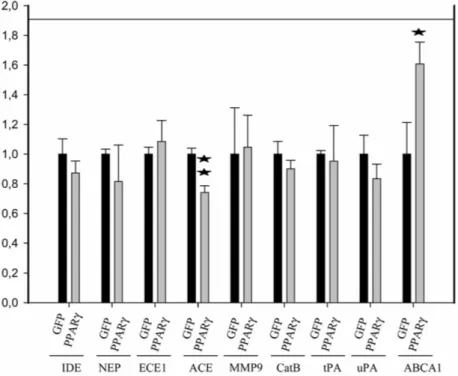
Next, we checked whether the mRNA levels of any of the known Aβ-degrading proteases and, in particular, the IDE mRNA levels were altered by PPARy transcriptional activity, as an IDE upregulation would effectively increase Aβ catabolism [26]. Quantitative PCR experiments demonstrated that mRNA levels of insulin degrading enzyme (IDE), neprilysin (NEP), endothelin converting enzyme1 (ECE1), angiotensin converting enzyme (ACE), matrix metalloproteinase 9 (MMP9), cathepsin B (CATPB) (the known Aβ-degrading en-zymes [14-20]) or of tissue plasminogen activator (tPA) and urokinase plasminogen activator (uPA) (the known activators of plasmin mediated Aβ-degradation [62]) did not increase under PPARγ-overexpression conditions in HEK293 cells (Fig. 3). Indeed, ACE mRNA levels were decreased in cells overexpressing PPARγ (Fig. 3). As a positive control, the mRNA levels of ABCA1, a member of the ATP-binding cassette transporter family and a known PPARγ downstream target, were increased upon PPARγ overexpression [63-66] (Fig. 3). mRNA levels of the Aβ-degrading enzyme, plasmin in HEK293 cells were below detection limits of qPCR and were therefore not further analyzed.
We conclude that activation of PPARγ does not increase the transcriptional activity of the IDE gene, neither of any of the other genes encoding identified Aβ- degrading enzymes.
Table 1 : Summary of protease inhibitor effects on Aβ degradation
Inhibitor Concentration Target peptidases Effect on clearance
Lactacystin 10 µM proteasome - NH4 Cl 10, 30 mM lysosome/endosome - Bafilomycin 10 µM lysosome/endosome - E64 10, 100 µM cysteine - Pepstation A 1,3, 10 µM aspartic - Leupeptin 10, 30, 100 µM cysteine/serine - Antipain 10, 30, 100 µM cysteine/serine - PMSF 30, 100, 300 µM serine -
Pefabloc, AEBSF 100 µM serine -
EDTA 1,3, 10 µM metallo +
GM6001 10, 30, 100 µM metallo -
Phosporamidon 10, 30, 100 µM MMPs/ADAMs metallo - DL-Thiorphan 10, 30, 100 µM
NEP > ECE > ACE
metallo - 1, 10-Phenanthroline 250, 500 µM NEP > ACE metallo + TAPI-1 10, 30, 100 µM metallo - MMPs/ADAM 17 MMP-2/9 5, 10, 50 µM metallo - MMP-2/9 Insulin 1, 10 µM IDE + Glucagon 0.1, 1 µM IDE +
-, no or little inhibition; +, potent inhibition. NH4Cl, ammonium chloride; PMSF, phenylmethylsulfonyl fluoride; E64, N-[N-(L-3-transcarboxyirance-2-carbonyl)-L- Leucyl]-agmatine; EDTA, Diaminoethanetetraacetic acid; GM6001, galardin, ilomastat; TAPI-1, TNF-π protease inhibitor-1; MMPS, matrix metalloproteases; ADAMs, a desinte- grin and metalloprotease; NEP, neutral endopeptidase 24.11 (neprilysin); ECE, endothelin- converting enzyme; ACE, angiotensin-converting enzyme; IDE, insulin degrading enzyme.
THE Aβ CLEARANCE MECHANISM IS CONSERVED IN WILD TYPE AND
IDE KNOCK OUT PRIMARY GLIA AND HIPPOCAMPAL-DERIVED CELLS
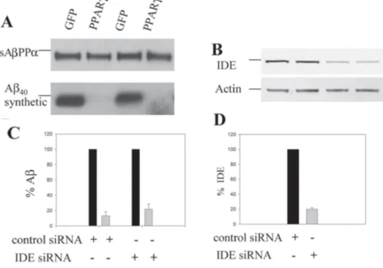
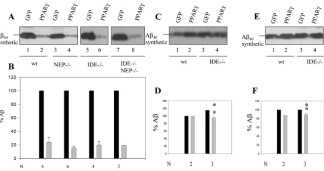
We reasoned that even if PPARγ does not seem to affect total mRNA levels of IDE (Fig. 3), it might be possible that PPARγ enhances its enzymatic activity indirectly by affecting the expression levels of an activity modulator or changing the localization of the enzyme vis-a-vis major Aβ pools. We used two different approaches in an attempt to validate the hypothesis that IDE is responsible for the PPARγ-induced Aβ degradation. We first performed siRNA experiments to down-regulate IDE levels in GFP- or PPARγ- transduced HEK293 cells and analyzed the stability of synthetic Aβ in the culture medium. As a control, we used dsRNA oligos directed against a murine mR- NA sequence that should not affect any human mR- NA target (Fig. 4). Down-regulation of IDE protein by about 79.8 ± 1.9% (N = 3) (Figs 4B, D) did not restore Aβ stability in the medium of PPARγ-treated cells (Figs 4A, C). These experiments suggest that IDE does not mediate the Aβ-degradation mechanism activated by PPARγ . However, siRNA experiments do not completely deplete IDE from the cell, and the residual IDE levels might be sufficient for activation by PPARγ. Therefore, we examined in a second experiment primary hippocampal neurons and glial cells from IDE knock-out mice. We also tested primary neurons and glial cells from NEP knock-out and IDE/NEP double knock-out mice, as NEP is also a well documented Aβ- degrading protease (Genotyping data for IDE and NEP is available in Supplementary Fig. 2). Cells were incubated with Aβ for 24 h. PPARy expression decreased Aβ levels in glial and hippocampal cells derived from wild-type animals but also in NEP—/— cells (Figs 5A- B lanes 1-2 and 3-4 and Figs 6A-B lanes 1-2), as expected.
Unexpectedly, the Aβ-lowering effect ofPPARy was still present in IDE-deficient cells, whether they were single IDE knock out (IDE—/—, Figs 5A-B lanes 56), or also lacking one (IDE—/— NEP+/—, Figs 6A-B lanes 3-4), or two alleles of the NEP gene (IDE—/— NEP— /—, Figs 5A-B lanes 7-8 and Figs 6A-B lanes 5-6). The absence of IDE expression was confirmed by western blot analysis (see Supplementary Fig. 3).
These data rule out IDE (and NEP) as the principal players in the PPARγ-enhanced clearance mechanism. It could still be argued, however, that the activation of PPARy in the cell could affect the activity of more than one enzyme, one of them being IDE. Because Aβ degradation was very efficient after the 24 h period, we tested shorter and less efficient incubation times (10 and 3 h) to try to detect a partial contribution of IDE to Aβ clearance. However, also at these shorter incubation intervals, no change in Aβ clearance between wild type and IDE knock outs could be observed (Supplementary Fig. 4).
We conclude that neither IDE nor NEP contribute to the PPARγ-enhanced clearance of Aβ.
Fig. 3. PPARγ expression does not up-regulate the mRNA levels of any of the known-Aβ
degrading enzymes. Graphic shows the result of quantitative PCR experiments to measure relative mRNA levels of insulin degrading enzyme (IDE), neprilysin (NEP), endothelin converting enzyme 1 (ECE1), angiotensin converting enzyme (ACE), matrix metalloproteinase 9 (MMP9), cathepsin B (CATPB), tissue plasminogen activator (tPA), urokinase plasminogen activator (uPA), and the ABCA1 member of the ABC transporter protein family in PPARγ compared to GFP-transduced cells. Experiments were performed in 3-4 different samples per group, and all Ct values were measured in triplicates. Ct values were normalized to 2 internal housekeeping genes following the formula: ∆Ct = Ctarget
-Ctnormalizer, ∆∆Ct = ∆CtPPARγ — ∆CtGFP and represented 2-ΔΔCt values. Asterisks represent
significant differences between GFP- and PPARγ-transduced cells determined by Student’s test: *p < 0.05, **p < 0.005.
Fig. 4. Down-regulation of IDE levels does not rescue Aβ stability in PPARγ-transduced
HEK293 cells. GFP- or PPARy-transduced HEK293 cells were transfected with control siRNA oligos (single) or siRNA oligos against IDE for 72 h, and incubated with Aβ1-4o (0.1
µM) for 3 h. A) sAβPPα and Aβ were detected from samples of conditioned medium (WO2).
B) IDE and actin levels in cell extracts were detected by immunoblotting using specific antibodies. C) Aβ levels were quantified from 3 independent experiments and values were normalized to the levels of sAβPPα (AβZsAβPPα) and referred to GFP set at 100%. D) Levels of IDE were quantified and normalized to actin levels. Graphic shows representation of IDE levels relative to 100% from cells transfected with control oligos.
PHENANTHROLINE AND GLUCAGON RESCUE Aβ STABILITY IN WILD
TYPE AND IDE KNOCK OUT PRIMARY GLIA AND
HIPPOCAMPAL-DERIVED CELLS
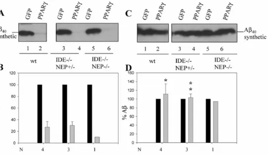
Importantly, the metalloprotease inhibitor phenanthroline and the hormone peptide glucagon could still block the PPARγ-induced Aβ clearance in wild type (Figs 5C-F lanes 1-2 and Figs 6C-D lanes 1-2) (hippocampal cells from 27.3 ± 9.6% to 111.4 ± 23.5%, p < 0.05 N = 4), in IDE—/— (Figs 5C-F lanes 3-4) (glial cells from 19.8 ± 5.5% to 89 ± 3.4%, p < 0.005 N = 3 in phenanthroline-treatedandto 96 ± 4.6%, p < 0.005 N = 3 in glucagon-treated cells), in IDE— /— NEP+/- (Figs 6C-D lanes 3-4) (hippocampal cells from 30.2 ± 6.4 to 103.3 ± 8.3 p < 0.005
N = 3) and in IDE-/- NEP-/- (Figs 6C-D lanes 5-6).
These results demonstrate that the PPARγ-induced Aβ clearance mechanism is indeed present also inbrain cells such as glia and hippocampal cultures and that this pathway identified in HEK293 cells is mechanistically conserved in cell types that are directly relevant
to the disease process, further underlining its physiological relevance. Moreover, although the PPARγ- activated mechanism is independent from IDE activity, it presents a similar inhibitor profile as this well- characterized Aβ degrading enzyme.
Fig. 5. Phenanthroline and glucagon treatment rescue Aβ stability in wild type and in IDE—
/— glia cultures. GFP- or PPARγ-transduced murine primary glial cultures from wild-type (A-F, lanes 1-2), NEP—/— (A-B, lanes 3-4), IDE—/— (A-B, lanes 5-6 and C-(A-F, lanes 3-4), and IDE—/—NEP—/— (A-B, lanes 7-8) were either non-treated (A-B), phenanthroline-treated (C-D) or glucagon-treated (E-F) prior to incubation with Aβ1-40 synthetic peptide (0.1 µM) for 24
h. Aβ stability was analysed by western blot using the WO2 antibody. B,D,F) Aβ levels were quantified from N = 2-6 independent experiments and values were normalized to the levels of sAβPPα (Aβ/sAβPPα) and referred to GFP set at 100%. Asterisks represent significant differences between non-treated and treated PPARγ-transduced cells (N = 3-4) determined by Student’s test: *p < 0.05, **p < 0.005.
Fig. 6. Glucagon treatment rescues Aβ stability in wild type,IDE—/—NEP+/— and IDE—/—
NEP—/— hippocampal cultures. GFP- or PPARγ-transduced murine primary hippocampal cultures from wild-type (lanes 1-2), IDE—/—NEP+/— (lanes 3-4) and IDE—/—NEP—/— (lanes 5-6) were either non-treated (A-B), or glucagon-treated (C-D) prior to incubation with
Aβ1-40 synthetic peptide (0.1 µM) for 24 h. Aβ stability was analyzed by western blot using the
WO2 antibody. B,D) Aβ levels were quantified from N = 1-4 independent experiments and values were normalized to the levels of sAβPPα (Aβ/sAβPPα) and referred to GFP set at 100%. Asterisks represent significant differences between non-treated and treated PPARγ-transduced cells (N = 3-4) determined by Student’s test: *p < 0.05, **p < 0.005.
DISCUSSION
We demonstrate here that PPARγ activation enhances a mechanism for Aβ clearance that is not dependent on known general mechanisms for Aβ uptake, clathrin or caveolin-mediated, as well as specific LRP or scavenger-mediated endocytosis [44-50]. Besides, we exclude intracellular mechanisms for protein degradation such as proteasomal-, endosomal-, or lysosomal- dependent pathways in the PPARγ -induced Aβ degradation. Thus, as PPARγ activation does not enhance the activity of any secreted protease [13] and internalization of Aβ peptide is not required for efficient degradation, we inferred that the process occurs at the plasma membrane. The mechanism is conserved in neurons and glia cells and canbe activatedby known PPARγ agonists like pioglitazone, troglitazone, and rosiglitazone from the TZD family, as previously shown [13].
Recent in vivo data have shown that endogenous PPARy activation causes Aβ reduction in hippocampus and cortex of a transgenic mouse model for AD [1]. These authors have shown that PPARγ can suppress BACE transcription, providing a link towards inhibition of Aβ generation, especially under inflammatory conditions. Moreover, their work proves the presence and activation of the endogenous PPARγ transcription factor within the brain.
In addition, a small human clinical trial using the PPARγ agonist rosiglitazone showed a better cognitive performance of treated versus placebo patients [10], although other study showed no effects [11]. Therefore, the treatment with PPARγ agonists in preclinical and clinical stages could have beneficial effects, although little is known about the working mechanisms behind these effects.
We presently report that PPARy activation induces an Aβ-clearance mechanism with a metalloprotease profile very similar to the known Aβ-degrading enzyme IDE. This was further confirmed by the findings that Aβ clearance was efficiently blocked in a dose-dependent manner by glucagon and insulin hormones, similarly to IDE activity [35,52,61]. However, PPARγ activation does not result in a transcriptional increase of the IDE gene. Moreover, PPARγ induced Aβ-degradation is still active in cell lines knocked-down for IDE expression and in primary cultures isolated from IDE loss-of- function mice. These data indicates that PPARγ regulates the activity of a metalloprotease similar, but not identical to IDE.
Overall, further work is required to unravel the identity of the sought protease whose activity can be modulated by the use of the well-known PPARγ agonists towards Aβ degradation in hippocampal and glia cells.
In conclusion, our work presents a novel mechanism for the clearance of Aβ that can be targeted by drugs of known pharmaceutical families. Our data add to the growing literature that PPARy activation might decrease Aβ burden in AD patients by enhancing its clearance besides other PPARγ reported functions like modulation of inflammation and down-regulation of BACE transcriptional activity. Overall, this novel mechanism could provide a very attractive drug target for further exploration in the treatment or prevention of AD.
ACKNOWLEDGMENTS
The authors thank Dr. Lola Ledesma and Dr. Wim Annaert for critical discussion and suggestions, and Dr. Guy Froyen and Dr. Idoya Lahortiga for the help with the preparation of the qPCR data.
The co-authors Pascal Merchiers and Luc Van Rompaey have been, or are currently, employees from Galapagos Genomics NV which uses adenoviral technology for commercial purposes.
For experiments involving animals, written approval from the local ethics and animal experimentation committees were obtained. Our animal facilities are under regular supervision by government-certified veterinarians and housing of and experimentation with animals conforms to common animal welfare and protection guidelines and adheres to the highest possible standards and have been approved.
This research was supported by a Pioneer award from the Alzheimer’s Association (to BDS); the Fund for Scientific Research, Flanders; the K.U. Leuven (GOA and Methusalem); the EuropeanUnion; and the Federal Office for Scientific affairs, Belgium (IUAP P6/43/).
Authors’ disclosures available online (http://www.j-
alz.com/disclosures/view.php?id=319).
REFERENCES
[1] Heneka MT, Sastre M, Dumitrescu-Ozimek L, Hanke A, Dewachter I, Kuiperi C,
O’Banion K, Klockgether T, Van Leuven F, Landreth GE (2005) Acute treatment with the PPARgamma agonist pioglitazone and ibuprofen reduces glial inflammation and
Abeta1-42 levels in APPV717I transgenic mice. Brain 128, 14Abeta1-42-1453.
[2] Jantzen PT, Connor KE, DiCarlo G, Wenk GL, Wallace JL, Rojiani AM, Coppola D,
Morgan D, Gordon MN (2002) Microglial activation and beta -amyloid deposit reduction caused by a nitric oxide-releasing nonsteroidal anti-inflammatory drug in amyloid precursor protein plus presenilin-1 transgenic mice. J Neurosci 22, 2246-2254.
[3] Lim GP, Yang F, Chu T, Chen P, Beech W, Teter B, Tran T, Ubeda O, Ashe KH,
Frautschy SA, Cole GM (2000) Ibuprofen suppresses plaque pathology and inflammation in a mouse model for Alzheimer’s disease. J Neurosci 20, 5709-5714.
[4] Sastre M, Dewachter I, Landreth GE, Willson TM, Klock- gether T, van Leuven F,
Heneka MT (2003) Nonsteroidal antiinflammatory drugs and peroxisome proliferator-activated receptor-gamma agonists modulate immunostimulated processing of amyloid precursor protein through regulation of beta-secretase. J Neurosci 23, 9796-9804.
[5] Sastre M, Dewachter I, Rossner S, Bogdanovic N, Rosen E, Borghgraef P, Evert BO,
Dumitrescu-Ozimek L, Thal DR, Landreth G, Walter J, Klockgether T, van Leuven F, Hene- ka MT (2006) Nonsteroidal anti-inflammatory drugs repress beta-secretase gene promoter activity by the activation of PPARgamma. ProcNatl Acad Sci U SA 103, 443-448.
[6] Weggen S, Eriksen JL, Das P, Sagi SA, Wang R, Pietrzik CU, Findlay KA, Smith TE,
Murphy MP, Bulter T, Kang DE, Marquez-Sterling N, Golde TE, Koo EH (2001) A subset of NSAIDs lower amyloidogenic Abeta42 independently of cyclooxygenase activity.
Nature 414, 212-216.
[7] Yan Q, Zhang J, Liu H, Babu-Khan S, Vassar R, Biere AL, Citron M, Landreth G (2003)
Anti-inflammatory drug therapy alters beta-amyloid processing and deposition in an animal model of Alzheimer’s disease. J Neurosci 23, 7504-7509.
[8] Lleo A, Galea E, Sastre M (2007) Molecular targets of nonsteroidal anti-inflammatory
drugs in neurodegenerative diseases. Cell Mol Life Sci 64, 1403-1418.
[9] Risner ME, Saunders AM, Altman JF, Ormandy GC, Craft S, Foley IM, Zvartau-Hind
ME, Hosford DA, Roses AD (2006) Efficacy of rosiglitazone in a genetically defined pop-ulation with mild-to-moderate Alzheimer’s disease. Pharma- cogenomics J 6, 246-254.
[10] Watson GS, Cholerton BA, Reger MA, Baker LD, Plymate SR, Asthana S, Fishel MA,
Kulstad JJ, Green PS, Cook DG, Kahn SE, Keeling ML, Craft S (2005) Preserved cognition in patients with early Alzheimer disease and amnestic mild cognitive impairment during treatment with rosiglitazone: a preliminary study. Am J Geriatr Psychiatry 13, 950-958.
[11] Rabiner EA, Tzimopoulou S, Cunningham VJ, Jeter B, Zvartau-Hind M, Castiglia M,
Mistry P, Bird NP, Matthews J, Whitcher B, Nichols TE, Lai R, Lotay N, Saunders A, Reiman E, Chen K, Gold M, Matthews PM (2009) Effects of 1 months of treatment with the PPARγ agonist rosiglitazone on brain glucose metabolism in Alzheimer’s Disease: A18F-FDGPET study. Alzheimers Dementia 5, P39-40.
[12] Berger J, Moller DE (2002) The mechanisms of action of PPARs. Annu Rev Med 53,
409-435.
[13] Camacho IE, Serneels L, Spittaels K, Merchiers P, Dominguez D, De Strooper B (2004)
Peroxisome-proliferator-activated receptor gamma induces a clearance mechanism for the amyloidbeta peptide. J Neurosci 24, 10908-10917.
[14] Backstrom JR, Lim GP, Cullen MJ, Tokes ZA (1996) Matrix metalloproteinase-9 (MMP-9)
is synthesized in neurons of the human hippocampus and is capable of degrading the amyloidbeta peptide (1-40). J Neurosci 16, 7910-7919.
peptide by endothelin-converting enzyme. J Biol Chem 276, 24540-24548.
[16] Howell S, Nalbantoglu J, Crine P (1995) Neutral endopeptidase can hydrolyze
beta-amyloid(1-40) but shows no effect on beta-amyloid precursor protein metabolism.
Peptides 16, 647-652.
[17] Hu J, Igarashi A, Kamata M, Nakagawa H (2001) Angiotensinconverting enzyme
degrades Alzheimer amyloid beta-peptide (A beta); retards A beta aggregation, deposition, fibril formation; and inhibits cytotoxicity. J Biol Chem 276, 47863-47868.
[18] Kurochkin IV, Goto S (1994) Alzheimer’s beta-amyloid peptide specifically interacts with
and is degraded by insulin degrading enzyme. FEBSLett 345, 33-37.
[19] Mueller-Steiner S, Zhou Y, Arai H, Roberson ED, Sun B, Chen J, Wang X, Yu G,
Esposito L, Mucke L, Gan L (2006) Antiamyloidogenic and neuroprotective functions of cathepsin B: implications for Alzheimer’s disease. Neuron 51, 703-714.
[20] Tucker HM, Kihiko M, Caldwell JN, Wright S, Kawarabayashi T, Price D, Walker D,
Scheff S, McGillis JP, Rydel RE, Estus S (2000) The plasmin system is induced by and degrades amyloid-beta aggregates. J Neurosci 20, 3937-3946.
[21] Eckman EA, Watson M, Marlow L, Sambamurti K, Eckman CB (2003) Alzheimer’s
disease beta-amyloid peptide is increased in mice deficient in endothelin-converting enzyme. J Biol Chem 278, 2081-2084.
[22] Farris W, Mansourian S, Chang Y, Lindsley L, Eckman EA, Frosch MP, Eckman CB,
Tanzi RE, Selkoe DJ, Guenette S (2003) Insulin-degrading enzyme regulates the levels of insulin, amyloid beta-protein, and the beta-amyloid precursor protein intracellular domain in vivo. Proc Natl Acad Sci U S A 100, 4162-4167.
[23] Iwata N, Tsubuki S, Takaki Y, Shirotani K, Lu B, Gerard NP, Gerard C, Hama E, Lee HJ,
Saido TC (2001) Metabolic regulation of brain Abeta by neprilysin. Science 292, 1550-1552.
[24] Melchor JP, Pawlak R, Strickland S (2003) The tissue plasminogen
activator-plasminogen proteolytic cascade accelerates amyloid-beta (Abeta) degradation and inhibits Abeta- induced neurodegeneration. J Neurosci 23, 8867-8871.
[25] Yin KJ, Cirrito JR, Yan P, Hu X, Xiao Q, Pan X, Bateman R, Song H, Hsu FF, Turk J, Xu
J, Hsu CY, Mills JC, Holtzman DM, Lee JM (2006) Matrix metalloproteinases expressed by astrocytes mediate extracellular amyloid-beta peptide catabolism. J Neurosci 26, 10939-10948.
[26] Leissring MA, Farris W, Chang AY, Walsh DM, Wu X, Sun X, Frosch MP, Selkoe DJ
(2003) Enhanced proteolysis of betaamyloid in APP transgenic mice prevents plaque formation, secondary pathology, and premature death. Neuron 40, 10871093.
[27] Kim M, Hersh LB, Leissring MA, Ingelsson M, Matsui T, Farris W, Lu A, Hyman BT,
Selkoe DJ, Bertram L, Tanzi RE (2007) Decreased catalytic activity of the insulin-degrading enzyme in chromosome 10-linked Alzheimer disease families. JBiol Chem
282, 7825-7832.
[28] Ledesma MD, Da Silva JS, Crassaerts K, Delacourte A, De Strooper B, Dotti CG (2000)
Brain plasmin enhances APP alpha-cleavage and Abeta degradation and is reduced in Alzheimer’s disease brains. EMBO Rep 1, 530-535.
[29] Yoshizawa T, Iwamoto H, Mizusawa H, Suzuki N, Matsumoto H, Kanazawa I (1992)
Cerebrospinal fluid endothelin-1 in Alzheimer’s disease and senile dementia of Alzheimer type. Neuropeptides 22, 85-88.
[30] Saido TC, Iwata N (2006) Metabolism of amyloid beta peptide and pathogenesis of
Alzheimer’s disease. Towards presymp- tomatic diagnosis, prevention and therapy.
[31] Saito T, Takaki Y, Iwata N, Trojanowski J, Saido TC (2003) Alzheimer’s disease,
neuropeptides, neuropeptidase, and amyloid-beta peptide metabolism. Sci Aging
Knowledge Environ 2003, PE1.
[32] Michiels F, van Es H, van Rompaey L, Merchiers P, Franck- en B, Pittois K, van der
Schueren J, Brys R, Vandersmissen J, Beirinckx F, Herman S, Dokic K, Klaassen H, Narinx E, Hagers A, Laenen W, Piest I, Pavliska H, Rombout Y, Lange- meijer E, Ma L, Schipper C, Raeymaeker MD, Schweicher S, Jans M, van Beeck K, Tsang IR, van de Stolpe O, Tomme P (2002) Arrayed adenoviral expression libraries for functional screening. NatBiotechnol 20, 1154-1157.
[33] De Strooper B, Simons M, Multhaup G, Van Leuven F, Beyreuther K, Dotti CG (1995)
Production of intracellular amyloid-containing fragments in hippocampal neurons ex-pressing human amyloid precursor protein and protection against amyloidogenesis by subtle amino acid substitutions in the rodent sequence. EMBOJ 14, 4932-4938.
[34] Esselens C, Oorschot V, Baert V, Raemaekers T, Spittaels K, Serneels L, Zheng H,
Saftig P, De Strooper B, Klumperman J, Annaert W (2004) Presenilin 1 mediates the turnover of telen- cephalin in hippocampal neurons via an autophagic degrada- tive pathway. J CellBiol 166, 1041-1054.
[35] Vekrellis K, Ye Z, Qiu WQ, Walsh D, Hartley D, Chesneau V, Rosner MR, Selkoe DJ
(2000) Neurons regulate extracellular levels of amyloid beta-protein via proteolysis by insulindegrading enzyme. J Neurosci 20, 1657-1665.
[36] De Strooper B, Saftig P, Craessaerts K, Vanderstichele H, Guhde G, Annaert W, Von
Figura K, Van Leuven F (1998) Deficiency of presenilin-1 inhibits the normal cleavage of amyloid precursor protein. Nature 391, 387-390.
[37] Bradford MM (1976) A rapid and sensitive method for quantitation of microgram
quantities of protein utilizing the principle of protein-dye-binding. Anal Biochem 72, 248-254.
[38] Chadderton T, Wilson C, Bewick M, Gluck S (1997) Evaluation of three rapid RNA
extraction reagents: relevance for use in RT-PCR’s and measurement of low level gene expression in clinical samples. Cell Mol Biol (Noisy-le-grand) 43, 1227-1234.
[39] Altschuler Y, Barbas SM, Terlecky LJ, Tang K, Hardy S, Mostov KE, Schmid SL (1998)
Redundant and distinct functions for dynamin-1 and dynamin-2 isoforms. J Cell Biol 143, 1871-1881.
[40] Nabi IR, Le PU (2003) Caveolae/raft-dependent endocytosis. J Cell Biol 161, 673-677. [41] Chyung JH, Selkoe DJ (2003) Inhibition of receptor-mediated endocytosis demonstrates
generation of amyloid beta-protein at the cell surface. J Biol Chem 278, 51035-51043.
[42] Daukas G, Zigmond SH (1985) Inhibition of receptor- mediated but not fluid-phase
endocytosis in polymorphonuclear leukocytes. J Cell Biol 101, 1673-1679.
[43] Park RD, Sullivan PC, Storrie B (1988) Hypertonic sucrose inhibition of endocytic
transport suggests multiple early en- docytic compartments. J Cell Physiol 135, 443-450.
[44] Coraci IS, Husemann J, Berman JW, Hulette C, Dufour JH, Campanella GK, Luster AD,
Silverstein SC, El-Khoury JB (2002) CD36, a class B scavenger receptor, is expressed on microglia in Alzheimer’s disease brains and can mediate production of reactive oxygen species in response to beta-amyloid fibrils. Am J Pathol 160, 101-112.
[45] Deane R, Wu Z, Sagare A, Davis J, Du Yan S, Hamm K, Xu F, Parisi M, LaRue B, Hu
HW, Spijkers P, Guo H, Song X, Lent- ing PJ, Van Nostrand WE, Zlokovic BV (2004) LRP/amyloid beta-peptide interaction mediates differential brain efflux of Abeta isoforms.
Neuron 43, 333-344.
Scavenger receptor-mediated adhesion of microglia to beta-amyloid fibrils. Nature 382, 716-719.
[47] Kounnas MZ, Moir RD, Rebeck GW, Bush AI, Argraves WS, Tanzi RE, Hyman BT,
Strickland DK (1995) LDL receptor- related protein, a multifunctional ApoE receptor, binds secreted beta-amyloid precursor protein and mediates its degradation. Cell 82, 331-340.
[48] Paresce DM, Ghosh RN, Maxfield FR (1996) Microglial cells internalize aggregates of
the Alzheimer’s disease amyloid beta-protein via a scavenger receptor. Neuron 17, 553-565.
[49] Sagare A, Deane R, Bell RD, Johnson B, Hamm K, Pendu R, Marky A, Lenting PJ, Wu
Z, Zarcone T, Goate A, Mayo K, Perlmutter D, Coma M, Zhong Z, Zlokovic BV (2007) Clearance of amyloid-beta by circulating lipoprotein receptors. Nat Med 13, 1029-1031.
[50] Santiago-Garcia J, Mas-Oliva J, Innerarity TL, Pitas RE (2001) Secreted forms of the
amyloid-beta precursor protein are ligands for the class A scavenger receptor. J Biol
Chem 276, 30655-30661.
[51] Husemann J, Loike JD, Kodama T, Silverstein SC (2001) Scavenger receptor class B
type I (SR-BI) mediates adhesion of neonatal murine microglia to fibrillar beta-amyloid. J
Neu- roimmunol 114, 142-150.
[52] Qiu WQ, Walsh DM, Ye Z, Vekrellis K, Zhang J, Podlisny MB, Rosner MR, Safavi A,
Hersh LB, Selkoe DJ (1998) Insulindegrading enzyme regulates extracellular levels of amyloid beta-protein by degradation. J Biol Chem 273, 32730-32738.
[53] Iwata N, Tsubuki S, Takaki Y, Watanabe K, Sekiguchi M, Hosoki E,
Kawashima-Morishima M, Lee HJ, Hama E, Sekine-Aizawa Y, Saido TC (2000) Identification of the major Abeta1-42-degrading catabolic pathway in brain parenchyma: suppression leads to biochemical and pathological deposition. Nat Med 6, 143-150.
[54] Shirotani K, Tsubuki S, Iwata N, Takaki Y, Harigaya W, Maruyama K, Kiryu-Seo S,
Kiyama H, Iwata H, Tomita T, Iwatsubo T, Saido TC (2001) Neprilysin degrades both amyloid beta peptides 1-40 and 1-42 most rapidly and efficiently among thiorphan- and phosphoramidon-sensitive endopeptidases. JBiol Chem 276, 21895-21901.
[55] Koon HW, Zhao D, Na X, Moyer MP, Pothoulakis C (2004) Metalloproteinases and
transforming growth factor-alpha mediate substance P-induced mitogen-activated protein kinase activation and proliferation in human colonocytes. JBiol Chem 279, 45519-45527.
[56] Yoshisue H, Hasegawa K (2004) Effect of MMP/ADAM inhibitors on goblet cell
hyperplasia in cultured human bronchial epithelial cells. Biosci Biotechnol Biochem 68, 2024-2031.
[57] Authier F, Posner BI, Bergeron JJ (1996) Insulin-degrading enzyme. Clin Invest Med 19,
149-160.
[58] Becker AB, Roth RA (1995) Insulysin and pitrilysin: insulindegrading enzymes of
mammals and bacteria. Methods Enzy- mol 248, 693-703.
[59] Duckworth WC, Bennett RG, Hamel FG (1998) Insulin degradation: progress and
potential. Endocr Rev 19, 608-624.
[60] Kurochkin IV (2001) Insulin-degrading enzyme: embarking on amyloid destruction.
Trends Biochem Sci 26, 421-425.
[61] Bennett RG, Duckworth WC, Hamel FG (2000) Degradation of amylin by
insulin-degrading enzyme. J Biol Chem 275, 36621-36625.
[62] Myohanen H, Vaheri A (2004) Regulation and interactions in the activation of
[63] Chawla A, Boisvert WA, Lee CH, Laffitte BA, Barak Y, Joseph SB, Liao D, Nagy L,
Edwards PA, Curtiss LK, Evans RM, Tontonoz P (2001) A PPAR gamma-LXR-ABCA1 pathway in macrophages is involved in cholesterol efflux and atherogen- esis. Mol Cell 7, 161-171.
[64] Costet P, Luo Y, Wang N, Tall AR (2000) Sterol-dependent transactivation of the ABC1
promoter by the liver X recep- tor/retinoid X receptor. J Biol Chem 275, 28240-28245.
[65] Repa JJ, Turley SD, Lobaccaro JA, Medina J, Li L, Lustig K, Shan B, Heyman RA,
Dietschy JM, Mangelsdorf DJ (2000) Regulation of absorption and ABC 1-mediated efflux of cholesterol by RXR heterodimers. Science 289, 1524-1529.
[66] Venkateswaran A, Laffitte BA, Joseph SB, Mak PA, Wil- pitz DC, Edwards PA, Tontonoz
P (2000) Control of cellular cholesterol efflux by the nuclear oxysterol receptor LXR alpha. ProcNatlAcadSci USA 97, 12097-12102.