© FEBS 1987
The pH dependence of the active-site serine DD-peptidase of Streptomyces R61
Louis VARETTO', Jean-Marie F R 6 R E ' , Martine NGUYEN-DISTECHE', Jean-Marie GHUYSEN' and Claude HOUSSIER^ ' Service de Microbiologie and ^ Laboratoire de Chimie macromolectilaire et Chimie physique, Institut de Chimie, Universite de Liege (Received June 16/September 18,1986) - EJB 86 0609Titration of the active-site serine DD-peptidase of Streptomyces R61 shows that formation of acyl enzyme during hydrolysis of the substrate Ac2-L-Lys-D-Ala-D-Ala and enzyme inactivation by the /?-lactam compounds benzylpenicillin, iV-acetylampicillin and ampicillin relies on the acidic form of an enzyme's group of p/T K 9.5. It is proposed that protonation of a lysine e-amino group facilitates initial binding by charge pairing with the free carboxylate of the substrate and the ^-lactam molecules. Lowering the pH from 7 to 5 has no effect on the second-order rate constant of enzyme acylation by benzylpenicillin and A^-acetylampicillin but results in a decreased rate constant of acylation by ampicillin and Ac2-L-Lys-D-Ala-D-Ala. Protonation of the side-chain amino group of ampicillin and a decreased efficacy of the initial binding of the j)eptide to the enzyme seem to be responsible for the observed effects. Whatever the molecule bound to the enzyme, there is no sign for the active involvement of an enzyme's histidine residue of pK 6.5 — 7.0 in the hydrolysis pathway.
Two families of bacterial enzymes specifically interact with ^-lactam compounds. The ^-lactamases, which hydrolyse the ^-lactam ring, can play an important role in resistance. The DD-peptidases, which participate in the last stages of wall peptidoglycan metabolism, are the primary targets of these antibiotics [1, 2]. Many of the ^-lactamases (classes A and C) and DD-peptidases characterized so far are serine enzymes and react with /?-lactam compounds according to reaction (1).
E-D*- E + P(s) (1)
where E = enzyme, D = ^-lactam, ^.^2and^.^3 = first-order rate constants, K = dissociation constant of E • D (Michaelis complex) and E —D* = serine ester-linked acyl enzyme [3]. Most often, the value of /t:+3 is high or very high with the )S-lactamases, and so low with the DD-peptidases that a slight stoichiometric excess of /?-lactam compound causes long-lasting immobilization of the protein at the level of the acyl enzyme and these latter enzymes behave as penicillin-binding proteins [2—5]. Another major difference is that breakdown of the acyl enzyme formed by reaction between penicillins and DD-peptidases involves either the direct release of the penicilloyl moiety or fragmentation of this moiety, with the formation of A^-formyl-D-penicillamine and a novel, unstable acylglycylenzyme [6]. The extracellular active-site serine DD-peptidase of Streptomyces R61 has been crystallized [7] and its primary structure established [8, 9]. Although this primary structure shows little homology with that of class A i?-lactamases. X-ray diffraction studies [10, 11] show striking similarities in the relative positions of secondary structure elements in the R61 DD-peptidase and the Bacillus ceretts and Bacillus licheniformis ^-lactamases. Apart from the fact that in all studied DD-peptidases and /J-lactamases of classes A
Correspondence to J.-M. Frere, Service de Microbiologie, Institut de Chimie B6, Universite de Liege au Sart-Tilman, B-4000 Liege, Belgium
Abbreviation. Ac, acetyl.
Enzymes. D-Alanyl-D-alanine carboxypepUdase or DD-peptidase (EC 3.4.16.-); ^-lacUmase (EC 3.5.2.6).
and C, the active-site serine is followed by an Xaa-Xaa-Lys sequence (which suggests that this lysine probably plays an important role), little is known concerning the enzymes' functional groups involved in substrate binding and catalysis. Studies of the pH dependence of the kinetic parameters of class A and C ^-lactamases [12, 13] indicate the possible in-volvement of a carboxylate and of a Lys or Tyr residue in the enzymatic mechanisms.
Much less is known about the interaction between the DD-peptidases and their peptide substrates. However, all avail-able data [2, 14] indicate that these enzymes catalyse acyl-transfer reactions from R-D-Ala-D-Ala-terminated carbonyl donors (D) according to a pathway which closely resembles reaction (1)
HY
where HY is H2O or a suitable R-NH2 peptide or amino acid. The goal of the present work was to obtain further infor-mation on the possible functional groups involved in catalysis and penicillin binding by the Streptomyces R61 DD-peptidase. When this study was initiated, it was known [2] that at pH 7.0 and 37°C (unless otherwise stated) (a) the K. k+2 and k + 3 values for the interaction with benzylpenicillin were 13 mM (25°C), 180s-* (25°C) and 1.4xlO-*s-' ; (b) the cor-responding values for the interaction with ampiciUin were 7.2 mM, 0.77 s "' and 1.4 x lO""* s " ' ; (c) the K^ and k^t values for the best substrate Ac2-L-Lys-D-Ala-D-Ala were about 10 mM and 50 s ~ \ respectively and (d) the activity of the enzyme decreased with increasing ionic strength [15].
MATERIALS AND METHODS Enzymes
The Streptomyces R61 DD-peptidase and the Bacillus licheniformis ^-lactamase (2000 units/mg protein) were purified as described [16, 17]. Peroxidase and D-amino acid oxidase were from Boehringer, Mannheim, FRG.
Peptide substrate and inhibitor
Ac2-L-Lys-D-Ala-D-Ala was from UCB Bioproducts (Braine-rAUeud, Belgium). ["*C]Ac2-L-Lys-D-Ala-D-Ala and [^*C]Ac2-L-Lys-D-Ala-D-lactate (with the label in the acetyl groups; specific radioactivity 50 Ci/mol) were prepared by reacting L-Lys-D-Ala-D-Ala or L-Lys-D-Ala-D-laetate (from UCB Bioproduets) with [**C]aeetic anhydride (from Amersham International, UK) as desedbed [18].
P-Lactam compounds
Benzylpenieillin was from Rhone Poulenc, France, and ampieillin (Pentrexyl®) from Bristol Benelux, Brussels. ['*C]Benzylpenicillin (50 Ci/mol) was from Amersham In-ternational, UK. Benzylpenieillin amide was a gift from Prof. H. Vanderhaeghe (Rega Instituut, KUL, Leuven, Belgium). A^-Aeetyl ampieillin, Af-l'^CJaeetylampieillin and A^-aeetyl a-phenylglyeylglyeine were synthesized as described in the Appendix.
Buffers
The following buffers were used: pH 5, 10 mM sodium caeodylate/HCl; pH 6, 13 mM sodium eaeodylate + HCl or 13 mM sodium phosphate; pH 7,10 mM sodium phosphate; pH 8, 7 mM sodium phosphate or 25 mM Tris/HCl; pH 9, 6 mM potassium borate or 12 mM glyeine + NaOH; pH 10, 2.5 mM borate or 25 mM glyeine + NaOH. The ionie strength of all the reaction mixtures was adjusted to a conductance of 0.1 mS using a Methrohm E527 conduc-tometer either by modifying the buffer concentrations or, in the case of the glyeine buffers, by adding NaCl. Moreover, in the experiments involving varying substrate concentrations, the ionie strength was adjusted by NaCl addition to that obtained at the highest substrate concentration.
Thin-layer chromatography
Polygram Sil-G plates (Macherey Nagel, Duren, FRG) were used. Solvent A was CHCls/methanol/acetie acid (88/ 10/2; v/v/v).
Paper electrophoreses
These were performed at pH 6.5 on Whatman 3MM paper using a Gilson high-voltage electrophorator model DW (60 V/ em). The buffer was collidine/acetic acid/water (9/2.7/1000; v/v/v). Mobilities (in em/h towards the anode) of the various standard compounds were as follows: benzylpenicilhn, 16; benzylpenicilloate, 25; phenylacetylglycine, 22; phenyl-acetylglyeylglyeine, 18; A^-acetylampicillin, 14; penicilioate of A^-acetylampiciUin, 24; A^-acetyl a-phenylglycylglycine, 17. Radioactive measurements
Radioactive areas on paper strips were located with a Packard 2000 Radiochromatogram scanner. The amount of radioactivity was measured with a Packard Tri-carb liquid scintillation spectrometer.
DD-Peptidase activity
The hydrolysis of Ac2-L-Lys-D-Ala-D-Ala was stopped by heating the solutions in boiling water for 1 min and the
amount of released D-Ala measured enzymatically (D-amino acid oxidase procedure) as described [19]. Alternatively, the amount of [*'*C]Ac2-L-Lys-D-Ala released from [^*C]Ac2-L-Lys-D-Ala-D-Ala was separated from the residual substrate by paper electrophoresis and estimated as described [15].
Estimation ofacyl ({^*CJAc2-L-Lys-D-alanyl) enzyme [^'*C]AC2-L-Lys-D-Ala-D-Ala (5.3 mM) or [''*C]Ac2-L-Lys-D-Ala-D-Lac (4 mM) was incubated wih the R61 DD-peptidase (25 nM) for 15 s at 0°C, in buffers of varying pH (final volumes 15^1). The reaction mixtures were supplemented with 15 ^l\ denaturing buffer (0.125 M Tris/HCl pH 6.8 con-taining 20% glyeerol, 2% sodium dodecyl sulphate, 10% 2-mercaptoethanol and 0.001% bromophenol blue), main-tained in boiling water for 30 s and submitted to gel electro-phoresis at pH 8.3 in the presence of sodium dodecyl sulphate. Estimation of the amounts of radioactive acyl enzyme was performed by fluorography [20, 21] of the gels using pre-flashed Kodak X-Omat XR films, and scanning. Controls were samples of the R61 DD-peptidase treated with [''*C]benzylpenicillin under conditions where all the enzyme was converted into ["*C]benzylpenicilloyl enzyme. For more details, see [14].
Kinetic parameters for the interaction with P-lactam compounds
Value o/k + 3. After complete inactivation of the enzyme by ampieillin or benzylpenieillin and elimination of the free jS-lactam compound by addition of the B. licheniformis /?-lactamase, recovery of enzyme activity was measured as described [22]. With [*'*C]benzylpenicillin, the radioactive acyl enzyme was isolated by filtration on Sephadex G-25 in water and incubated at 37 °C in buffers of varying pH. Samples were removed at inereasing intervals, submitted to high-voltage electrophoresis and the released compounds were estimated by radioactivity measurements. With A^-acetylampicillin, acyl enzyme was not very stable and the value of fc + 3 was measured by adding /J-lactamase and tripeptide substrate to mixtures containing the enzyme and a large excess of antibiotic, and monitoring the release of D-alanine with time [23].
Values of the inactivation rate constant. With ampieillin, k^ {i.e. fc+2[D]/([D] + K)ifk+i <^ A:,} was computed by measur-ing the degree of enzyme inactivation as a function of time. With A^-acetylampicillin, k^ was not much larger than k+3 and it was computed by measuring the residual activity at the steady-state. (For more details, see [5].) The inactivation of the enzyme by benzylpenieillin was monitored by measuring the decrease of protein fluorescence [4] at 320 nm. Fluores-cence measurements were performed using a Sigma ZWS-11 stopped-flow mixing imit (Biochem, Munieh, FRG) adapted to a Dia-Log optical and detection set-up (Garching Instru-ment, Dusseldorf, FRG). The signals were analysed by a data acquisition and treatment system for fast, transient optical signals as described [24]. Excitation was at 280 nm. The ex-ponential decrease of fluorescence was analysed on the basis of the linearized equation In [(F, - F^)/(Fo-F^)] = -/c,/ where Fa, F, and Fa, were the fluorescence intensities at times 0, t and after stabilization of the system was reached. Equal volumes (80 |il) of enzyme solution (4.4 jiM) and ben-zylpenieillin solution (0.67 mM) made in the same buffers (of varying pH) were mixed rapidly. The k^ values were computed by linear regression using 200—400 points at 1-ms intervals.
Curve-fitting
A general multiparametric curve-fitting program was used [25].
RESULTS
Kinetic parameters for the hydrolysis of Ac2-L-Lys-D-Ala-D-Ala
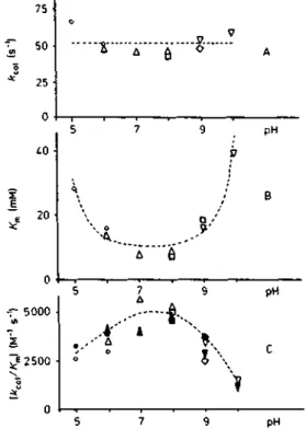
The effects of pH on K^ and /Cd were first determined at substrate concentrations ranging over 2 —lOmM, by following the release of D-Ala (i.e. the reaction product of the k+2 step) and using Hanes' linearization of the steady-state equation [D]/v = (K^ + [D])/F. The ratio k^JK^ was also determined at a 1 mM (i.e. 4, K^) substrate concentration under which conditions the velocity of the reaction was pro-portional to [D]. The linearity of product formation with time (5, 10 and 15 min) was verified in all cases. Note that on the basis of reaction (2), k^JK^ is equivalent to the second-order rate constant k+2/K of enzyme acylation. The experimental values of/Teat, ^m and k^JK^ (i.e. A:+2/^0 as a function of pH are shown in Fig. 1 A, B and C.
Using ['*C]Ac2-L-Lys-D-Ala-D-lactate at a concentration (4 mM) equivalent to 0.1 A:^ [26] and at pH 7, about 1.8% of the enzyme occurred as acyl enzyme at the steady state of the reaction. At this depsipeptide concentration, and if A:+2 was much larger than k+3, a maximum of 10% of total enzyme would be trapped as acyl enzyme. Using [''*C]Ac2-L-Lys-D-Ala-D-Ala as substrate at a 5.3 mM concentration, no acyl enzyme was detected at the steady state of the reaction whether thepH was 5, 7 or 10.
Inactivation by benzylpenicillin, ampicillin and 'N-acetylampicillin
Preliminary experiments showed that at pH 7, the interac-tion with A^-acetylampicillin was characterized by a small k+2/ K value (about 8 M"* s"') and a relatively high k+3 value of 5x10"'* s"* (half-life 23 min). Four types of experiments were carried out which led to the following observations.
a) The pH dependence of the pseudo-first-order rate constant k^ of acyl-enzyme formation was determined at benzylpenicillin and ampicillin concentrations well below the value of K. Hence kJ[D] was equivalent to k + 2IK. It was assumed that the same condition applied to the interaction with Af-acetylampicillin. Since with this latter compound the values of k+3 was rather high, the rate of formation of the acyl enzyme was estimated by measuring the residual activity after the interaction between the enzyme and the ^-lactam had reached the steady state (see [5] and legend of Fig. 2B). The experimental values of A;,/[D] (i.e. k+2/K) as a function of pH are shown in Fig. 2 A (for benzylpenicillin). Fig. 2 B (for A^-acetylampicillin) and Fig. 2C (for ampicillin). One should note that benzylpenicillinamide (0.1 mM) did not cause any enzyme inactivation after a 10-min incubation at 37 °C, in-dicating a kJ[D] value of less than 2 M"^ s"^
b) The pH dependence of the first-order rate constant k+3 of acyl enzyme breakdown was examined by following the recovery of the enzyme activity. Fig. 3A, C and D depict the results obtained with benzylpenicillin, A^-acetylampicillin and ampicillin, respectively.
c) The effects of pH on the nature of the reaction products arising through breakdown of benzylpenicilloyl enzyme are shown in Fig. 3B. Phenylacetylglycine (filled symbols) was
75 50 25 0 20 D -A " " 6 pH
.A'
i\
pH - 5000 2500Fig. 1. pH dependence of^a, (A), K„ (B) and \i.a«jKn (C) for the hydrolysis of Ac2-L-Lys-D-Ala-D-Ala by the active-site serine DD-peptidase o/Streptomyces R6L *;„, and K^ were obtained from initial velocity measurements using 2, 4, 6, 8 and 10 mM substrate. Filled symbols in (C): the enzyme was incubated with 1 mM substrate for 5, 10 and 15 min. For other conditions, see text. Buffers: (O, • ) cacodylate; (A, A) phosphate; (D, • ) Tris/HCl; (O. • ) borate; (V. • ) glycine/NaOH. Dashed curves were obtained by computer-aided fitting
produced at a constant rate at pH 5 - 1 0 while ben-zylpenicilloate (open symbols) was produced only at pH > 8. At pH 9 and 10, the experiments were carried out in borate buffers. In glycine-containing buffers, an additional degrada-tion product, presumably, phenylacetylglycylglycine [27], was also formed. The sum of phenylacetylglycine and phenylace-tylglycylglycine was equivalent to the amount of phenylacetyl-glycine alone obtained in borate buffers.
d) The compounds originating by breakdown of the acyl enzyme formed with A^-['*C]acetylampicillin were examined by thin-layer chromatography in solvent A (in which system, Af-acetylampicillin had an Rp value of 0.44). The main com-pound obtained at pH 10 had an Rp value of 0.05, which is that of the product formed by /?-lactamase hydrolysis of 7V-acetylampicillin. The product observed at pH 7.0 had the same R^ (0.22) in solvent A and the same electrophoretic mobility (17 cm/h towards the anode) at pH 6.5 as authentic A^-acetyl-a-phenylglycylglycine.
DISCUSSION
Benzylpenicillin, ampicillin, A^-acetylampicillin and Ac2-L-Lys-D-Ala-D-Ala possess a free carboxylate ofpK ^ 3 at one end of the molecule. Ampicillin contains in addition an amino group with a pK value of 7.25 at 25 °C [28] or 6.7 at 37 °C (as redetermined by titration of the sodium salt of ampiciUin).
No acyl enzyme accumulates detectably at the steady state of the reaction with Ac2-L-Lys-D-Ala-D-Ala used at
concentra-528 10.10' o A--..,A
V
pH 10's
O\ 300 100 • 5 7 9 pH 5 7 9 pHFig. 2. pH dependence of the pseudo-first-order rate constant k^ of acyl enzyme formation during interaction between the active-site serine DD-peptidase and 0.67mM benzylpenicillin (A; pH 5 — 9.5), 2 fiM benzylpenicillin (A; pH 10), 200 pM fi-acetylampicillin (B) and 10 nM ampicillin (C). Dashed curves were obtained by computer-aided fitting. For buffers, see Fig. 1. (A) The values at pH 5—9.5 were obtained at 25 °C by fluorescence stopped-flow as explained in the text. The value at pH 10 was obtained by incubating the enzyme (0.6 nM) and benzylpenicillin (2 jtM) at the same temperature. Samples (10 |il) were removed at 2-min intervals, and diluted threefold in 5 mM sodium phosphate pH 7.0 containing 2 ng ^-lactamase and 60 nmol substrate. The amount of D-alanine produced was estimated after 10 min at 37 °C. (B) The enzyme (1.3 nM) was incubated at 37 °C with 0.2 mM N-acetylampicillin. Samples (10 jil) were removed after 5,10, 20 and 30 min and supplemented with 20 nl of 2 mM substrate prepared in the corresponding buffer. After 5 min at 37 "C, the amount of free D-alanine was estimated. The results indicated that, in all cases, the steady state was reached after less than 20 min of interaction between the enzyme and A^-acetylampicillin. The value of k^ was estimated using the following equation
IED*],» k.
where k+3 was obtained from the data shown on Fig. 3C. (C) The enzyme (0.6 jiM) was incubated at 37 °C with 10 nM ampicillin, 10-^1 samples were removed at increasing intervals and diluted threefold in 5 mM sodium phosphate pH 7.0 containing 20 ng /?-lactamase and 60 nmol substrate. The amount of D-alanine was estimated after 10 min
tions equivalent to 0.2 x K^ at pH 5, 0.65 x K^ at pH 7 and 0.13 X Km at pH 10. Note also that little variation of the K^ at pH 7.0 was observed between 37° and 0°C (unpublished data). Since 1.8% of total enzyme can be trapped as acyl enzyme during reaetion with Ac2-L-Lys-D-Ala-D-lactate (at 0°C, pH 7 and a concentration equivalent to 0.1 K^), it is concluded that certainly less than 1 % of total enzyme occurs as acyl enzyme during reaction with the peptide. Since K^
= k+3K/(k+2 + k+3) and as shown elsewhere [5], these levels of enzyme acylation yield k+ilk+i values of 4 (depsipeptide, pH 7) and >22 (peptide, pH 5), > 190 (peptide, pH 7) and > 14 (peptide, pH 10). Although these data must be taken
2-7 0- 2-7 9 pH - 20-10 2 • pH pH
Fig. 3. pH dependence of the first-order rate constant of enzyme re-covery through breakdown of the acyl enzyme formed by reaction be-tween the active-site serine DD-peptidase and benzylpenicillin (A), H-acetylampicillin (C) and ampicillin (D). pH dependence of the rate of release of henzylpeniciltoate (B; open symbols) and phenylacetylglycine (B; filled symbols) from benzylpenicilloyl enzyme. For buffers, see Fig. 1. (A, D) The enzyme (51 nM) was completely inactivated by a 10-min incubation at 37°C with 10 (iM benzylpenicillin (A) or 100 (lM ampicillin (D) in buffers of varying pH. The mixtures were supplemented with 2 jxg (A) or 20 ng (D) ^-lactamase and maintained at 37 °C. After 0,30,60 and 90 min, samples (30 nl) were removed and supplemented with 60 nmol substrate. The amount of D-alanine produced was estimated after a 10-min incuba-tion at 37 °C. (B) The enzyme (26 nmol) was inactivated by reacincuba-tion with 100 nmol ['"CJbenzylpenicillin for 5 min at 37°C in 300 M1 of 10 mM sodium phosphate pH 7.0. The buffer salts and the excess of free ['*C]benzylpenicillin were eliminated by filtration on Sephadex G-25 in water and the radioactive acyl-enzyme preparation was divided into lOO-jtl fractions each containing 20 nCi. These fractions were freeze-dried, the residues dissolved in 100 \>\ buffer of varying pH and the resulting solutions incubated at 37 °C. Samples (20 jxl) were removed at varying intervals (0, 30, 60 and 90 min) and sub-mitted to high-voltage paper electrophoresis at pH 6.5 for 60 min. The ['*C]benzylpenicilloyl enzyme remained at the origin. ['''ClPhe-nylacetylglycine (filled symbols) and [''*C]benzylpenidlloate (open symbols) migrated as mentioned in Materials and Methods. (C) The enzyme (200 nM at pH 10, 11 nM in the other cases) was incubated at 37°C with 1.33 mM (at pH 10) or 0.33 mM (in the other cases) N-acetylampicillin. After 30 min, inactivation was complete and the solutions were simultaneously supplemented with 20 |ig of /3-lactamase and 1.2 mM (fmal concentration) substrate. At varying intervals, 50-nl samples were removed and analysed for their content of D-alanine. The filled symbols in (A) refer to the sum benzylpenicilloate -I- phenylacetyl glycine whose individual values were measured in B
with caution (since only one substrate concentration has been used), it is evident that the ^+3/^+ 2 ratio for the reaction with the peptide is > 10 at all pH values and that, consequently, ^cat = ^+2 and Km — K. In agreement with these results, peptide analogues that inhibit the enzyme competitively have ^i values comparable to the K,n for the peptide substrate [29].
^ -EHy D .-EHj-D- • E H j . Table 1. Values ofpK.2
EH-D* »-EH-Pls)
II
.11.
F.n »-E-D •E-D
Fig. 4. General model for the interaction between the Streptomyces R61 DD-peptidase and carbonyl-donor substrate or inactivators. Ki, K2, Ko, K'D and K'D are dissociation constants. Our discussions assumes that the lower branch is inoperative (K'D i> KD) for both substrate and /?-lactam, and that the upper branch is only operative for j?-lactams (P-lactums: K'D = KD,k'+2 = ^ +2;substrate:/Tb P ^D) Thedeacyla-tion step (k + 3) on which no data can be obtained with the substrate, can be studied independently with the ^-lactams. In that case, an additional step is found ( E H - D * * ' - ' ° " ' ' , EH -I- P'. Curve-fitting, when performed with the complete models (i.e. assuming that Ab and KD are not much larger than KD), does not significantly improve the agreement between the calculated and measured values
The variations of the kinetic parameters shown in Fig. 1 thus refiect the infiuence of pH on the dissociation constant of the Michaelis complex (K) and of the first-order rate constant (k + 2) for the transformation of that complex to acyl enzyme. The effects of pH on the rate of hydrolysis of the acyl enzyme remain unknown.
The value of A^^ increases (Fig. 1B) at pH values lower than 7 and higher than 8, expressing a decreased affinity of the free enzyme for the substrate. Assuming that EH 2 • D and E • D (see Fig. 4) are not detectably formed (i. e. K'i and K'D
P KD), the dashed line and curve shown on Fig. 1A and B are obtained and fitting of the data to Eqn (3) gives k + 2
= 51.5 s"', pKi = 5.3, pK2 = 9.48 and KD = 10.4 mM.
(3) A:,
Fitting the data shown on Fig. 1C (dashed curve) on the basis of Eqn (4) (where 4950 M " ' s~Ms the value ofk+2/KD computed from the results obtained above) yields pKy and pA^2 values of 4.92 and 9.47, respectively.
Fitting without prior fixation of the k+2/Kv values has also been attempted but does not significantly decrease the total relative standard deviation and the agreement with the values obtained from analysis of the K^ curve is not so good {k+2/ = 4250 M and = 4.7). This suggests that formation of the Michaelis complex with the substrate is pos-sible only with the central form EH and that the observed variations of the k^JK^ ratio can be accounted for by the sole variation of K^, i.e. of A^.
Eqns (3) and (4) can easily be derived from Eqns (8) and (9) of Kaplan and Laidler [30]; our data correspond to case 4 in Table 1 (k2 < ^3) of that article \ when (A-1 + k2)/k2 is replaced by
A^D-' It should how^ei^e noted that a printing mistake occurred in [30]. Eqn (9}^giving kc/K^ is not obtained as printed by dividing Eqn (7). giving kc, by Eqn (8), giving K^. Instead, Eqn (9) should read
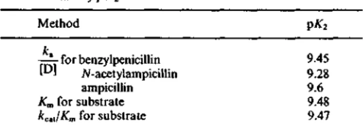
kc [H] 1 + Method 1^ —— for benzylpenicillin P J Af-acetylampicillin ampicillin K^ for substrate kcu/Km for substrate
9.45 9.28 9.6 9.48 9.47
The rate of enzyme acylation by benzylpenicillin and A^-acetylampicillin remains unchanged between pH 5 and 8 (fig. 2 A and B) and, in that case, /k'+2/A^b = k+2/KD. Curve fitting, performed on the basis of Eqn (5) (dashed curves in Fig. 2 A and B) yields pA:2 values of 9.45 (Fig. 2 A) and 9.28 (Fig. 2B). The values of k+2/KD are 9800 M " ' s " ' for benzylpenicillin (at 25 °C)
Af-acetylampicillin (at 37 °C). and 7.6 M" s , - 1 [D]
1 +;
JC2 for (5) [H]With ampicillin (Fig. 2C) the observed decrease of A:,/[D] at pH 5 and 6 can be attributed to the protonation of ampicillin itself (D -hH* *± DH"^; pK = 6.7). Indeed, the dashed curve shown on Fig. 2C is obtained with values of
265M"»s''forA:+2/A:D, 1 7 7 M " ' s " ' for ^ + 2, DH-'/ATDH*
and 9.6 for
pA^2-Unfortunately, with any of the )S-lactam compounds tested, it is not possible to decide whether the observed decrease of k + 2/K at high pH is due to a decrease of/:+2 or to an increase of K. However, since the values of pAr2 derived from the different experiments are strikingly similar (Table 1) and since, as shown above, protonation of an enzyme group of pA" X 9.5 greatly increases the efficacy of the initial binding step of the tripeptide substrate, it is proposed by extension that the same mechanism applies to the interaction with the ^-lactam compounds. This more efficient binding might be due to charge pairing between the free carboxylate of the substrate/inactivator and a postively charged group near the enzyme's active site. In support of this view, amidation of the carboxylate group causes, at least, a 5000-fold decrease in the inactivating potency of benzylpenicillin.
Titration of the active-site serine ^-lactamases of class A and C [which react with the /?-lactam compounds according to reaction (1) but with a k+3 of very high value] also reveals an enzyme's group of pA" 8.5-10 [12, 13]. Remarkably, the R61 DD-peptidase and these /?-lactamases possess a highly conserved lysine residue located at the third position on the carbonyl side of the active-site serine residue (Ser-Xaa-Xaa-Lys). It may thus be hypothesized that the group of pA^ 8.5 — 10 detected in all these enzymes is the £-amino group of that particular lysine residue and that this amino group plays an important role in the formation and/or productiveness of enzyme-ligand associations.
While deprotonation of the group of pK « 9.5 similarly decreases the affinity of the enzyme for the peptide substrate and penicillins, a major difference between the two classes of compounds is observed at low pH. Whereas interaction with the substrate appears to rely on a group of pA' w 5, in contrast, binding of penicillins remains stable when the pH is lowered from 7 to 5. A group of pA^ » 5.0 seems to occur in the active site of serine ^-lactamases [12, 13] but the stability of the
DD-peptidase does not allow measurements to be performed at a pH lower than 5. Further experiments are needed to explain coherently the results obtained at pH < 7.
Independent study of the deacylation (fc+3) step is only possible with the ^-lactam compounds. The value of k+3 is low at pH between 5 and 8 but it increases at higher pH. As shown in Fig. 3 B, this increase is due to attack of the acyl (benzylpenicilloyl) enzyme by OH" ions with formation of benzylpenicilloate. The second-order rate constant computed
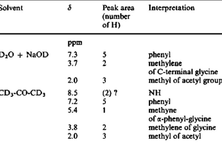
Table 2. NMR characteristics ofN-acetyl-a-phenylglycyigiycine The reference for chemical shifts was trimethylsilylpropionate in D2O and hexamethyl disiloxane in CD3COCD3
for this reaction is about 4 M " * s higher than that observed (0.7 M
\ a value which is not much s"') for the hydrolysis of a-methylpenicilloate by OH" ions [31]. In contrast, the rate of release of phenylacetylglycine is pH-independent between pH 5 and 10 (Fig. 3B). Hence, irrespective of the pH, rupture of the C5 — C6 bond of the acyl (benzylpenicilloyl) enzyme and generation of phenylacetylglycyl-enzyme is rate-determining [32]. In some respects, these results are at variance with those reported for the interaction between benzylpenieillin and the membrane-bound DD-peptidase o{Bacillus stearothermophilus [33]. In this latter case, k+3 was maximal at pH 5 - 6 but experiments were not performed at pH 8. At pH 6.5, phenylacetylglycine was also the only product released.
Finally, it should be stressed that there is no sign of an imidazole group of pK x 7 as it occurs in the active site of chymotrypsin and related peptidases. Yet, earher studies suggested that a group of pK x 7.0 might be involved in the transp)eptidation reactions catalysed by the R61 DD-peptidase [15]. Such an enzyme group would play a role neither in the hydrolysis of the peptide substrate nor in the interaction with ^-laetam compounds.
APPENDIX
Synthesis ofN-acetyl and 'N-[^'*CJacetylampicillin
A^-AcetylampiciUin was prepared as follows. Ampieillin (100 mg) was dissolved in 0.3 ml acetonitrile by dropwise addition of 1 M NH4HCO3 and the solution was sup-plemented with 0.1 ml acetic anhydride and maintained at 20 °C. The disappearance of the free amino group was monitored by spotting 5-nl samples on a filter paper and spraying with a ninhydrin solution. After 20 min, the blue-brown color characteristic of ampieillin failed to appear and the solution was evaporated to dryness. Analysis of the reac-tion nMxture by thin-layer chromatography using solvent A (see below) and detection with bromocresol green (after drying the plates at 120°C for 60 min; acidic compounds appear yellow) revealed one single compound of Rf 0.44. This com-pound (A^-acetylampicillin) completely disappeared upon treatment with the ^-lactamase and gave rise to one single compound (the corresponding penicilioate) of Rp 0.05.
Af-[^*C]Acetylampidlhn (specific radioactivity 0.82 Ci/ mol) was prepared as follows. Ampieillin (4 mg) was dissolved in 55 nl of a mixture of acetonitrile and 1 M NH4HCO3 solution (9/1; v/v). The solution was supplemented with 1 nl of 0.5% [**C]acetic anhydride in toluene (Amersham; 120 Ci/ mol) and maintained for 2 h at 20 °C. Non-radioactive acetic anhydride (20 |il) and acetonitrile (10 nl) were added and after 16 h at 20 °C, the reaction mixture was evaporated to dryness and the residue dissolved in 2 ml 50 mM sodium phosphate pH 7.0. The A^-acetylampicillin concentration (55 mM) was estimated by measuring both the absorbance of the solution at 230 nm (e = 2400 M " * cm"') and the rate with which the preparation inactivated the R61 DD-peptidase (using the non-radioactive A^-acetylampicillin as control).
Solvent D2O + NaOD CD3-CO-CD3 5 ppm 7.3 3.7 2.0 8.5 7.2 5.4 3.8 2.0 Peak area (number ofH) 5 2 3 (2)? 5 1 2 3 Interpretation phenyl methylene of C-terminal glyeine methyl of acetyl group NH phenyl methyne of a-phenyl-glycine methylene of glyeine methyl of acetyl
Synthesis ofH-acetyl a-phenylglycylglycine
a) a-Phenylglycine (5 g, 33 nmol) was dissolved in 100 ml of water by adjusting the pH to 9 with NaOH. Acetic anhydride (10 ml) was added and after 10 min at 20 "C, the solution was cooled to 0°C and acidified with HCl. The white crystals were collected, washed with cold water and dried (melting point 190_93°C, yield4g).
b) A^-Acetyl a-phenylglycine was coupled to glyeine ethyl ester using dicycloliexylcarbodiimide as described [34], but the procedure for the isolation of the adduct was modified as follows. Urea was eliminated by filtration, the solvent dry-evaporated and the residue redissolved in hot ethylacetate. After cooling and eUmination of residual urea, the organic solution was washed successively with a 10% solution of sodium bicarbonate and water, dried over MgS04 and eon-centrated until it became slightly cloudy.
c) The ester was crystalhzed by addition of petroleum ether (40 - 60 °C b.p.) (melting point 222 - 224 "C; yield 0.4 g), dissolved in 30 ml dioxane containing 0.2 g NaOH and saponified during 2 h at 20 °C. The solution was cooled to 0°C, acidified with HCl, dry-evaporated, and the residue dis-solved in hot acetone. After elimination of the sohd (NaCl) and evaporation of the solvent, the residue was dissolved in hot ethylacetate, the solution washed successively with a 7% solution of potassium bicarbonate and water, dried over MgSO4 and concentrated until it became cloudy. Yellowish crystals (melting point 160°C) were obtained by addition of petroleum ether (40—60°C b.p.). They were dissolved in a minimum volume of water containing a stoichiometric amount of NaOH and the solution acidified with HCL White crystals appeared after several days at 4°C (melting point 182—185 °C). They were analysed by thin-layer chromatog-raphy in solvent A (one single spot of/?F 0.22—0.24) and by NMR in D2O + NaOD and CD3-CO-CD3 (Table 2). Mass spectra of positive and negative secondary ions were recorded using a quadrupolar extranuclear (model 7-162-8) mass spectrometer [35]. The compound was dissolved in glyeerol using camphorsulfonic acid as a promoting agent, m/z for positive ions: 251 [MH+], 233 (H20)], 223 [MH+-(CO)], 176 [MH+-(NH2-CH2-COOH)], 146 [176-[MH+-(CO)], 106 [C6H5-CH=NH^]; m/z for negative ions: 249 [(M-H+)"], 204 [(M-H+)--(COOH)], 174 [(?) possibly (M-H+)"-(CH3-CO-NH-OH)], 119 [C6H5-CH2-CO"].
531 This work was supported in part by the Fonds de la Recherche
Scientifique Medicate, Bnissels, Belgium (contract 3.4507.83), an Ac-tion concertee with the Belgian Government (convenAc-tion 79/84-11) and a Convention tripartite between the Region wallonne. Continental
Pharma and the University of Liege. We wish to thank Dr E. De Pauw for obtaining the mass spectra, and Dr L. Christiaens and Mr R. Bertin for their help in the synthesis of A^-acetyl-a-phenylglycylglycine and the curve-fitting analysis, respectively.
REFERENCES
1. Ghuysen, J. M., Frere, J. M., Leyh-Bouille, M., Nguyen-Disteche, M., Coyette, J., Dusart, J., Joris, B., Duez, C , Dideberg, O., Charlier, P., Dive, G. & Lamotte-Brasseur, J. (1984) Scand. J.
Infect. Dis. Suppl. 42, 17-37.
2. Frere, J. M. & Joris, B. (1985) CRC Crit. Rev. Microbiol. 11, 299-396.
3. Frere, J. M., Duez, C , Ghuysen, J. M. & Vandekerckhove, J. (1976) FEBS Lett. 70, 257-260.
4. Frere, J. M., Ghuysen, J. M. & Iwatsubo, M. (1975) Eur. J.
Biochem. 57,343-351.
5. Ghuysen, J. M., Frere,J. M., Leyh-Bouille, M., Nguyen-Disteche, M. & Coyette, J. (1986) Biochem. J. 235,159-165.
6. Frere, J. M., Ghuysen, J. M., Degelaen, J., Loffet, A. & Perkins, H. R. (1975) Nature (Lond.) 258, 168-170.
7. Kelly, J. A., Knox, J. R., Moews, P. C , Hite, G. J., Bartolone, J. B., Zhao, H., Joris, B., Frere, J. M. & Ghuysen, J. M. (1985)
J. Biot. Chem. 260, 6449-6458.
8. Joris, B., Jacques, Ph., Frere, J. M., Ghuysen, J. M. & Van Beeumen, J. (1987) Eur. J. Biochem. 162, 519-524.
9. Duez, C , Piron-Fraipont, C , Joris, B., Dusart, J., Ureda, M. S., Martial, J., Frere, J. M. & Ghuysen, J. M. (1987) Eur. J.
Biochem. 162,509-51%.
10. Kelly, J. A., Dideberg, 0., Charlier, P., Wery, J. P., Libert, M., Moews, P. C , Knox, J. R., Duez, C , Fraipont, C , Joris, B., Dusart, J., Frere, J. M. & Ghuysen, J. M. (1986) Science ( Wash.
DC) 231,1429-1431.
11. Samraoui, B., Sutton, B. J., Todd, R. J., Artimyuk, P. J., Waley, S. G. & Phillips, D. C. (1986) Nature (Lond) 320, 378-380. 12. Waley, S. G. (1975) Biochem. J. 149, 547-551.
13. Bicknell, R., Knott-Hunziker, V. & Waley, S. G. (1983) Biochem.
J. 213,61-66.
14. Nguyen-Disteche, M., Leyh-Bouille, M., Piriot, S., Frere, J. M. & Ghuysen, J. M. (1986) Biochem. J. 235,167-176.
15. Frere, J. M., Ghuysen, J. M., Perkins, H. R. & Nieto, M. (1973)
Biochem. J. 135, 483-492.
16. Fossati, P., Saint-Ghislain, M., Sicard, P. J., Frere, J. M., Dusart, J., Klein, D. & Ghuysen, J. M. (1978) Biotechnol. Bioeng. 20, 577-587.
17. Thatcher, D. R. (1975) Methods Enzymol. 43, 653-664. 18. Perkins, H. R., Nieto, M., Frere, J. M., Leyh-Bouille, M. &
Ghuysen, J. M. (1973) Biochem. J. 131, 707-718.
19. Frere, J. M., Leyh-BouiUe, M., Ghuysen, J. M., Nieto, M. & Perkins, H. R. (1976) Methods Enzymol. 45, 610-636. 20. Bonner, W. M. & Laskey, R. A. (1974) Eur. J. Biochem. 46, 8 3
-88.
21. Spratt, B. G. (1977) Eur. J. Biochem. 72, 341 -352.
22. Frere, J. M., Leyh-Bouille, M., Ghuysen, J. M. & Perkins, H. R. (1974) Eur. J. Biochem. 50, 203-214.
23. Kelly, J. A., Frere, J. M., Klein, D. & Ghuysen, J. M. (1981)
Biochem. J. 199,129-136.
24. Houssier, C. & O'Kinski, C. T. (1981) NATO Adr. Study Inst. Ser.
B64, 309-339.
25. Meites, L. & Meites, L. (1972) Talanta 19, 1131-1139.
26. Pratt, R. F., Faraci, U. S. & Govardhan, C. P. (1985) Anal.
Biochem. 144, 204-206.
27. Marquet, A., Frere, J. M., Ghuysen, J. M. & Loffet, A. (1979) Biochem. J. 777,909-916.
28. Hou, J. P. & Poole, J. W. (1969) J. Pharm. Sci. 58,1510-1515. 29. Perkins, H. R., Nieto, M., Frere, J. M., Leyh-Bouille, M. &
Ghuysen, J. M. (1973) Biochem. J. 131, 707-718.
30. Kaplan, H. & Uidler, K. J. (1967) Can. J. Biochem. 45, 5 3 9 -546.
31. Frere, J. M. (1981) Biochem. Pharmacol. 30, 549-552. 32. Frere, J. M., Ghuysen, J. M. & De Graeve, J. (1978) FEBS Lett.
88,141-150.
33. Blumberg, P. E., Yocum, R. R., Willoughby, E. & Strominger, J. L. (1974) J. Biol. Chem. 249, 6828-6835.
34. Sheehan, J. C. & Hess, G. P. (1955) J. Am. Chem. Soc. 77,1067-1070.
35. Pelzer, G., De Pauw, E. & Marien, J. (1985) Analysis 13, 3 6 8 -372.


