African Journal of Biotechnology Vol. 4 (5), pp. 409-421, May 2005 Available online at http://www.academicjournals.org/AJB
ISSN 1684–5315 © 2005 Academic Journals
Full Length Research Paper
Polyphasic identification of a new thermotolerant
species of lactic acid bacteria isolated from
chicken faeces
N’Dèye Maïmouna SOW
1,2, Robin Dubois
DAUPHIN
1, Dominique
ROBLAIN
1, Amadou
Tidiane GUIRO
2and Philippes THONART
11Centre Wallon de Biologie Industrielle (CWBI) -Unité de Bio-Industries, 2, Passage des Déportés, 5030 Gembloux, Belgique.
2Institut de Technologie Alimentaire de Dakar, Routes des Pères Maristes Dakar, Sénégal.
Accepted 14 September, 2004
Two thermotolerant and desiccation tolerant lactic acid bacteria (TDLAB) were pointed out from twenty isolated strains from soils and dried chicken faeces. Samples were collected in poultry farms in the vicinity of Dakar, Senegal (West Africa). The two new isolates were called Sp.4 (Sp.4=CWBI-B534=LMG7278) and Sp.20 (Sp.20=CWBI-B545=LMG7279). They are Gram-positive, catalase-negative, facultatively anaerobic, non-motile, and non-spore-forming rods. Both produce D/L lactic acid via homofermentative pathway. Growth of the strains occurred between 15°C and 44°C. The optimum temperature for growth was in 30°C-37°C temperature and pH 3-8 range. Desiccation treatment in glycerol showed 30% survival rates. Complex total fatty acid pattern of the strains showed the presence of C14:0, C16:0, C16:1, C18:0, C18:1, C18:2, C18:3.
SDS-PAGE of total protein of both strains placed them in L. plantarum group. AFLP analysis showed a phylogenetic proximity of the two strains with L. plantarum stricto sensu species. Specific amplified 16s rDNA restriction analysis (ARDRA) of the 16S rDNA gene, however, showed that these thermotolerant strains were not L. plantarum. ITS sequencing revealed that Sp.20 (LMG 7279) could be classed into Lactobacillus paraplantarum species since the short sequence of ITS showed 95% of similarity with reference species. Polyphasic identification shows that Sp.4, (the type strain is LMG 7278T) represent a new species within the genus Lactobacillus with only 88%+/-1 ITS sequence similarity with reference species. For which the name Lactobacillus aminata sp. nov. is proposed.
Key words: Lactobacillus, thermotolerant and desiccation tolerant lactic acid bacteria, intergenic transcribed sequence, 16s rDNA restriction analysis, AFLP, SDS-PAGE.
INTRODUCTION
Lactic acid bacteria (LAB) comprise a diverse group of Gram-positive, non-spore forming bacteria and widely involved in the production of fermented foods (Kandler
*Corresponding author. E-Mail: sow.n@fsagx.ac.be. Tel: 00 32 81 62 23 05 Fax: 00 32 8161 42 22.
and Weiss, 1986). They are responsible for lactic fermentation in several products such as dairy products, ensilage, ham and sausage. They also play the obviously important role in human and animal health with regard to their use as probiotics (Herrero et al., 1996, Tannock et al., 1999). Among promising probiotic species are members of genera Lactobacillus, Bifidobacterium and Enterococcus (Indu Pal Kaur et al., 2002).
In addition to the requirements for good safety and functional properties, LAB and particularly probiotic formulations should be able to withstand food processing and storage conditions (Svensson et al., 1999). A big challenge associated with LAB production is therefore the loss of viability during processing (Desmond, et al., 2002). Two inactivation mechanisms are known for LAB: thermal and dehydration inactivation (Lievense et al., 1994b). Thermal inactivation is caused by denaturation of critical cell components, mainly DNA and RNA. Cytoplasmic membrane damage is generally considered as the main mechanism of dehydration inactivation (Lievense and van’Riet, 1994a). Despite the improvement that occur the culture, the low survival rate during desiccation remains a major problem (Hodzic et al., 1995).
LAB belong to the genus Lactobacillus have been isolated from a variety of habitats, including plant, dairy products, meat products, sewage, manure, humans and animals (Kandler and Weiss, 1986). Some new Lactobacillus sp. have been isolated from chicken faeces and intestine, including: L. aviarius from intestine of chicken (Fujisawa et al., 1984), L. gallinarum and L. Johnsonii from chicken faeces (Fujisawa et al., 1991), L .fermentum subsp.cellobiosus and L. animalis from gastro-intestinal tracts of chicken (Gusils et al., 1999). Most of these strains so far have been mesophiles. Some LAB used in diary industry, for example L. delbrueckii, L. helveticus and S. thermophilus are already known as thermotolerant starter species from their temperature range of growth (Delcour, et al., 2000). We still have little knowledge about the biodiversity of thermotolerant lactic acid bacteria (TLAB) in nature, because very few studies have been done that focus on LAB from the standpoint of their thermotolerance (Niamsup et al., 2003). The isolation of Lactobacillus sp. from chicken faeces under high temperatures has been reported for the first time by Niamsup et al., (2003). But the isolation of Lactobacillus sp. from chicken faeces under simultaneously high temperature and dehydrated conditions (very low water activity) has not been reported. The screening of microorganisms from drastic environments appears, as a good strategy to obtain useful strains with simultaneously thermotolerance and high desiccation tolerance. There may have been a strong evolutionary pressure that has favoured some mechanisms increasing the tolerance to desiccation (Pichereau et al., 2000). However, to decipher desiccation tolerance mechanism, the topic is not the resistance of particular known structures like spores and akinetes, but rather the tolerance to extreme desiccation of vegetative cells like many Cyanobacteria. Understanding the mechanism of desiccation tolerance holds promise not only because it may resolve a cell biology enigma, but also, because, it may find biotechnological applications.
The achievement of the stabilization of dried cells will make possible the production and the storage of an increasing variety of bacterial strains in manufacture of food or pharmaceutical preparations. In this present study, two new isolates selected on the basis of heat and desiccation tolerance is described.
MATERIALS AND METHOD Samples and cultivation
Soils and faeces samples were collected from different poultry farms in Dakar (Senegal, West Africa). Strains isolation was carried out according to Nakayama et al. (1967). Five grams of sample were mixed with 100 ml of GYP medium contained 1% glucose (w/v), 1% yeast and 1% peptone. The sample suspension was heated for 10 min at 80°C and incubated anaerobically at 30°C. After 48 h of incubation, 100 µl of the mixture were spread onto the
surface of GYP agar containing 1% CaCO3. The culture was
incubated anaerobically at 30°C. Acid producing bacteria were recognised by the clear zones around the colonies. Bacteria were purified by several isolations and fresh cultures of these isolates were conserved at –80°C with glycerol (30%) as cryoprotective agents.
Phenotypic characterisation
Cells morphology was studied with a light microscope by phase-contrast (Zeiss Axiocam, x 100) and Gram determination was performed. Catalase production was determined by transferring fresh colonies from MRS (De Man et al., 1960) agar to a slide glass
and adding H2O2 (30%). Pseudo-catalase production was
determined by benzidine test according to Gerhardt et al. (1981). Growth conditions were studied in MRS broth from 15°C to 55°C, and in 3 to 8 pH range values. Lactic acid configuration was determinated by enzymatic method, according to manufacter’s instructions (Boehringer Mannheim). Carbohydrate fermentation patterns were determined using API 50 CHL test (Bio Mérieux) in duplicate at 30°C.
Thermotolerance of each isolate was estimated after heating bacterial suspension at 80, 90 and 100°C for 10, 10 and 5 min, respectively. Viability assays were performed after dilution on GYP
agar (containing 1% of CaCO3) count. The plates were incubated
anaerobically at 30°C. Time of decimal reduction at 80°C and 90°C
(DT) was calculated by following equation:
DT= - t/[Log (N/No)]
t: time (minutes), N: survival cell count, No: initial cell count. Thermoresistant isolates were subjected to a hyperosmotic stress to determine their tolerance to desiccation. The bacterial cells were suspended in glycerol solution with Aw (water activity) = 0.17 for 7 days (Sonan, 1997). Viability was estimated epifluorescence microscope AxioCam HRc type (Zeiss) with FITC excitation filter according to Boulos et al. (1999) modified method with two fluorescent dead/live markers: rhodamine 123 and the
propydium iodide. Rhodococcus erythropolis T902 (Weekers et al.,
1997) and L. plantarum G100-CWBI-B076 were used as
references. Pictures were analysed using Axiovision (Zeiss) software package.
SDS-PAGE
Cultures were grown anaerobically on MRS agar for 24 h at 28°C. The preparation of the cell extracts and the gel electrophoresis (SDS-PAGE) were carried out as earlier described (Pot et al., 1994). The normalised and digitised protein patterns were numerically analysed and clustered with the reference profiles (culture collection strains and industrial isolates) in the 'LAB' database as currently available (GelCompar™ 4.2 software, Applied Maths, Belgium).
FAME analysis
Total lipids extraction was performed according to the adapted method of Ito et al., (1969). Fatty acid methyl esters were prepared
by incubating the lipid extracts at 80°C for 4 h in 2 ml of CH3OH,
containing 15% (v/v) of KOH. The fatty acids were extracted with chloroform and analysed by gas chromatography on a HP 6890 (Hewlett Packard, Waldbronn, Allemagne) equipped with a
flame-ionization detector and a SPTM–2560, 100 m*0.25 mm*0,2 µm
fused silica capillary column. The conditions were as follows: injector temperature, 260°C; detector temperature, 260°C; carrier gas (helium) flow rate, 3 ml/min. The oven temperature was programmed from 140°C during 5 min to 240°C at 4°C/min. For peak identification, standard solution (Sigma) was used.
The results were relative percentages of fatty acids, determined from peak areas of methyl esters. They were means of three independent experiments. The ratio between the standard deviations and the means values was between 2 and 5%.
Amplification of ITS and 16S rDNA
For DNA preparation, cells were cultivated in MRS broth at 30°C. They were harvested in the late exponential phase and DNA was extracted essentially as described by Marmur (1961).
Universal primers were used to amplify the 16S rDNA and intergenic transcribed sequence (ITS) genes. For ITS amplification, forward R16: 5’-GGGTGAAGTCGTAACAAGGTA-3’ and reverse R23: 5’-KASTGCCAGGGCATCCAGCGT-3’ were used. These primers are derived from conserved regions of 16S and 23S rDNA genes, respectively, and can be used to amplify the ITS sequence of all prokaryotic DNA tested so far. For 16S rDNA amplification,
forward1 (PO): 5’-GAAGAGTTTGATCCTGGCTCAG-3’ and
reverse 1 (P6): 5’-CTACGGCTACC TTGTTACGA-3’ were used. The oligonucleotides were purchased from Eurogentec (Liège, Belgium). Each PCR mixture (50 µl) contained a reaction mix of 20mM Tris-HCL, 20mM of each deoxynucleoside triphosphate, 0.1
mM of each primer, 1 U of the Taq polymerase (Eppendorf) and 50
ng of DNA template. All amplifications were carried out in a Master Cycler Personal (Eppendorf) and the following programme was used: initial denaturation for 5 min at 95°C, 30 cycles of denaturation (1 min at 95°C), annealing (1 min at 55°C), and extension (2 min at 72°C). The amplified products were separated in a 1.5% (w/v) agarose gel electrophoresis in TAE buffer, at 120 mA followed by ethidium bromide staining. Gels were photographed with Polaroid Type 667 positive film using a 260 nm UV source.
AFLP
Total genomic DNA is digested by two restriction enzymes (EcoRI
and TaqI). Small DNA molecules (15-20 bp) containing one
compatible end are ligated to the appropriate ‘stricky end’ of the restriction fragments. Both adaptors are restriction halfsite-specific,
Sow et al. 411
and have different sequences. These adaptors serve as binding sites for PCR primers.
For this study, adaptor A (EcoRI, hexacutter):
5’-CTCGTAGACTGCGTACC-3’ 3’-CTGACGCATGGTTAA-5’
and adaptor B (TaqI, tetracutter):
5’-GACGATGAGTCCTGAC-3’ 3’-TACTCAGGACTGGC-5’
were used.
Some restriction fragments were amplified selectively: PCR primers are specifically hybridised with the adaptor ends of the restriction fragments. Since the primers contain at their 3’-end one or more so-called ’selective bases’ that extend beyond the restriction site into the fragment, only those restriction fragments that have the appropriate complementary sequence adjacent to the restriction site will be amplified. Following primer combination was used:
E01: 5’-GACTGCGTACCAATTCA-3 T01: 5’-CGATGAGTCCTGACCGAA-3’
PCR products are separated according to their length on a resolution polyacrilamide gel using DNA sequencer (ABI 377). Fragments that contain an adaptor specific for the restriction halfsite created by the 6-bp cutter are visualised due to the 5’-and labelling of the corresponding primer with the fluorescent dye FAM. The resulting electrophoretic patterns were tracked and normalised using GeneScan3.1 software (Applera, USA). Normalised tables of peaks, containing fragments of 50 to 536 bp, were transferred into
the BioNumericsTM 2.5 software (Applied Maths, Belgium). For
numerical analysis, data intervals were delineated between the 50-and 500 bp b50-ands of the internal size st50-andard. Clustering of the pattern was done using the Dice coefficient and the UPGMA algorithm. The profiles were compared with the reference profiles of the lactic acid bacteria taxa as currently available in LMG database (Gent, Belgium).
Amplified 16S rDNA Restriction Analysis (ARDRA)
Aliquots of 10 µl of 16S rDNA PCR products were digested in a 20 µl final volume with restriction endonucleases as specified by the
manufacturer. The following enzymes were used: AccII, Taq I
(Pharmacia). According to Ingrassia et al. (2001), LAB 16S rDNA
can be characterised by digestion with Taq I and AccII.
Restricted DNA fragments were analysed by horizontal electrophoreses in 2 % agarose gel. Gels were stained and photographed as described previously.
16S rDNA and ITS sequencing
The amplified 16S rDNA was purified by using the Microcon YM-100 (Millipore) system according to the manufacturer’s instructions. Each amplified ITS was purified after cutting corresponding fragment from agarose gel using Qiaex II gel extraction system (Qiagen). After a second amplification, ITS fragment was purified a second way using the Microcon YM-100 (Millipore).
The determination of nucleotide sequences was performed by Sanger (1977) method using an applied Biosystems 3100 DNA sequencer, the protocols of the manufacturer (Applied Biosystems, USA) and the BigDye ABI Prism terminator v3.1 cycle
Table 1. Oligonucleotides used as primers for 16S rDNA sequencing.
Primer Position Length (bp) Sequence
F1 (forward) 21-39 18 5'CTGGCTCAGGAYGAACG-3' R1(reverse) 533-551 18 5'CTGCTGGCACGTAGTTAG-3' F2 (forward) 374-392 18 5'GAGGCAGCAGTMGGGAAT-3' R2 (reverse) 778-796 18 5'AATCCTGTTYGCTMCCCA-3' F3 (forward) 742-759 17 5'ACACCAMTGGCGAAGGC-3' R3 (reverse) 1100-1118 18 5'CCAACATCTCACGACACG-3' F4 (forward) 961-979 18 5'GCACAAGCGGYGGAGCAT -3' R4 (reverse) 1241-1262 21 5'TGTGTAGCCCRGGTCRTAAG-3'
M=G, A; Y=T, G, C; R=A, T: on L. plantarum
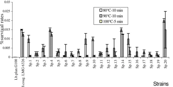
Figure 1. Representative survival rates of TLAB (Sp.) and control strains after heat treatment at 80°C, and 90°C for 10 min. No resistance was observed when cells were heated at 100°C for 5 min.
sequencing ready reaction kit. The sequencing primers used for ITS sequencing are the same used for PCR amplification; those for
16S rDNA are listed in Table 1. They were designed by Oligoplayer
software, on the basis of comparison of the 16S rRNA gene
sequences of several species of Lactobacilli. These sequences
come from the data banks of the EMBL and were available via service SRS of BEN (Belgian EMBnet-http://www.embnet.org). The
position indicates the annealing of the primer 3’ end to E. coli 16S
rDNA in forward (F) or reverse (R) orientation. The electrophoretic profiles were analysed by Genescan Analysis software (Applied Biosystems). Forward and reversed sequences were aligned and compared using the Revseq and Clustal software available on BEN (http://www.be.embnet.org) site.
Sequence assembly was performed by using Vector NTI software. The sequence were compared to sequences available in
Genbank database by using FASTA 3
(http://www.ebi.ac.uk/fasta33/) and the results obtained were used to identify isolates to genus or species level. Phylogenetic trees based on the neighbour-joining method were constructed by using Systat 3.0 software. The 16S rDNA nucleotide sequences obtained were aligned within the most similar ones of the Ribosomal Database Project.
Nucleotide sequence accession numbers
Bacterial ITS and 16S rDNA sequences obtained in this study were
deposited in Genbank nucleotide database and are available under
following accessions numbers (Genbank via Bankit:
http://www.ncbi.nlm.nih.gov/BanKIt).
Sp4=CWBI-B534= LMG 7278, 16S rDNA complete sequence: AY594275
Sp4=CWBI-B534= LMG 7278, short ITS complete sequence: AY594277;
Sp20=CWBI-B545= LMG 7279, 16S rDNA complete sequence: AY594276;
Sp20=CWBI-B545= LMG 7279, short ITS complete sequence: AY594278.
RESULTS
Isolation of thermotolerant lactic acid bacteria Twenty thermotolerant lactic acid bacteria (TLAB) were isolated from soils and chicken faeces samples, by heating them at 80°C for 10 min. All of them are Gram-positive rods, catalase and pseudo-catalase-negative, homofermentative, and non-spore-forming. These new isolates are called Sp.1 to Sp.20.
The results of thermotolerance (survival rates at various temperature values, Figure 1) showed that after
0 0.005 0.01 0.015 0.02 0.025 0.03 L b. pl an .G 10 0 B .c oa g. L M G 6 32 6 Sp .1 Sp .2 Sp .3 Sp .4 Sp .5 Sp .6 Sp .7 Sp .8 Sp .9 Sp .1 0 Sp .1 1 Sp .1 2 Sp .1 3 Sp .1 4 Sp .1 5 Sp .1 6 Sp .1 7 Sp .1 8 Sp .1 9 Sp .2 0
Strains
% su rv iv al r at es80°C-10 min 90°C-10 min 100°C-5 min
Sow et al. 413
Table 2. Dt values determination for the best heat resistant strains:
Sp.4, Sp.14, Sp.20 and B. coagulans LMG 6326. D80°C (min) D90°C (min) Sp.4 2.62 2.50 Sp.14 2.62 2.56 Sp.20 2.70 2.62 B. coagulans LMG 6326 2.64 2.63
0
10
20
30
40
50
60
R.ery thopo lis T 902 Lb.pl antar um G 100Sp.1 Sp.2 Sp.4 Sp.7 Sp.9Sp.10 Sp.13Sp.14Sp.15Sp.17Sp.18Sp.19Sp.20Strains
%
s
ur
vi
va
l r
at
es
a
ft
er
7
j
at
A
w
=
0.
17
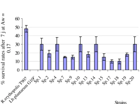
Figure 2. Residual viability (bacterial suspension had a week in glycerol solution, Aw =0.17). Cells were
treated with fluorescent markers (rhodamine 123 and propidium iodide using modified method of Boulos
et al., 1999). Sp.3, 5, 6,8, 11, 12 and 16 did not survived after osmotic treatment like L. plantarum G100,
a sensitive strain.
treatment, survival cell counts of Sp.4, Sp.14, Sp.20 and B. coagulans LMG6326 (a control strain) were higher.
There was no survival for any strains after heating at 100°C for 5 min. DT value is the time recurred at fixed temperature to reduce 90% of lived cells. For vegetative forms, generally, D70°C are between 10-15 s and for spore forming bacteria, D84°C are between 2 min. Table 2 shows that D80°C and D90°C values for vegetative heat tolerant cells of Sp.4, Sp.14 and Sp.20 were in the same values range as B. coagulans LMG6326, a spore-forming strain.
Determination of TLAB desiccation tolerance
Cells were maintained at low water activity (Aw=0.17) in all glycerol solution. After hyperosmotic treatment (for 7 days) at Aw = 0.17, the loss of viability was different from one strain to another (Figure 2). This reference strain (Rhodococcus erythropolis T902) has been previously
described by Weekers et al. (1997) to have good dehydration resistance.
In reference to R. erythopolis T902, the control stain, only Sp.1, Sp.4, Sp.10, Sp.14 and Sp.20 show better resistance with 30% of viability after desiccation treatment. The variation of water activity in the environment of the bacterial cells has a significant effect on the cellular viability (Lievense etal., 1994b).
Physiological analysis
Physiological patterns of selected strains were compared to two control strains; L. plantarum G100-CWBI B76 and B. coagulans LMG 6326. All strains produce D/L lactic acid from glucose (Table 3). The optimum temperature of growth for all strains is between 30°C and 37°C except Sp.4 and Sp.20, where growth occurred between 15 to 44°C. The control strains were inactive at 15°C. The pH
Table 3. Physiological characteristics (lactic acid production, pH and growth temperature) of the
Sp.4, Sp.20, L. plantarum G100 and B. coagulans LMG 6326.
Growth Temp (°C) Growth pH
Strains Acid lactic
15 30 37 44 55 2 3 4 5 6 7 8
Sp.4 D/L + ++ ++ ++ - - + + ++ ++ ++ ++
Sp.20 D/L + ++ ++ ++ - - + + ++ ++ ++ ++
L. plantarum G100 D/L - ++ ++ - - - - ++ ++ ++ ++ ++
B. coagulans LMG 6326 D/L - ++ ++ + - - - - ++ ++ ++ ++
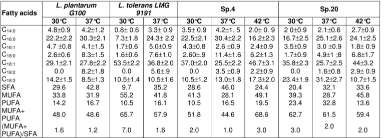
Table 4. Total fatty acid composition and modifications induced by growth temperature.
L. plantarum G100 L. tolerans LMG 9191 Sp.4 Sp.20 Fatty acids 30°C 37°C 30°C 37°C 30°C 37°C 42°C 30°C 37°C 42°C C14:0 4.8±0.9 4.2±1.2 0.8± 0.6 3.3± 0.9 3.5± 0.9 4.2±1. 5 2.0± 0. 9 2 0±0.9 2.1±0.6 2.7±0.9 C16:0 22.2±2.2 30.3±2.1 7.3±1.8 24.3± 2.2 22.5±2.1 30.4±2.2 16.2±2.3 16.7±2.5 25.1±2.6 24.1±2.5 C16:1 4.7 ±0.8 4.1±1.5 1.7±0.6 5.0±0.9 4.3±0.8 2.6 ±0.9 2.4±0.9 3.5±0.9 3.0 ±0.9 1.8± 0.9 C18:0 2.6±0.6 8.3±1.5 1.6±0.6 7.6±1.0 2.60±.9 11.4±1.6 6.2±1.3 1.7±0.9 4.9±1 ;8 6.8±1.7 C18:1 29.1±2.1 27.8±2.2 53.5±2.2 36.8±2.0 37.0±2.0 25.5±2.2 46.7±3.1 35.8±2.3 25.7±2.5 44±3.2 C18:2 0.0 8.2±1.8 0.0 5.6±.9 0.0 3.5 ±0.9 2.2±0.9 0.0 1.6±0.8 2.9± 0.9 C18:3 14.2±1.5 8.5±1.3 10.5±1.4 10.5±1.6 10.5±1.2 13.0±1.8 17.3±2.0 23.4±1.9 31.2±2.7 10.7±1.5 SFA 29.6 42.8 9.7 35.2 28.6 46.0 24.4 20.4 32.1 33.6 MUFA 33.8 31.9 55.2 41.8 41.3 28.1 49.1 39.3 28.7 45.8 PUFA 14.2 16.7 10.5 16.1 10.5 16.5 19.5 23.4 32.8 13.6 MUFA+ PUFA 48.0 48.6 65.7 57.9 51.8 44.6 68.6 62.7 61.5 59.4 (MUFA+ PUFA)/SFA 1.6 1.2 7.0 1.6 2.0 1.0 3.0 3.0 2.0 2.0
SFA: saturated fatty acids, MUFA: mono-unsaturated fatty acids, PUFA: poly-unsaturated fatty acids.
0% 20% 40% 60% 80% 100% L.plan tarum G10 0-30° C L.plan tarum G10 0-37° C L.tole rans L MG9 191-3 0°C L.tole rans L MG9 191-3 7°C Sp .4-30°C Sp .4-37°C Sp .4-42°C Sp.20 -30°C Sp.20 -37°C Sp.20 -42°C Strains/Temperature of culture
%
o
f
pi
cs
a
re
as
C18:3 C18:2 C18:1 C18 C16:1 C16:0 C14Figure 3. Total cellular fatty acid composition analysis in SP.4, SP.20 and control strains, in Reletion with
at which growth occurs for Sp.4 and Sp.20 was found in pH 3-8 range values. L plantarum G100 did not growth under pH 4 and B. coagulans LMG 6326 did not growth under pH 5. The pattern of fermented carbohydrates on API 50 CHL galleries showed a diversity of fermentative metabolism in comparisonb with the control strain and with two thermotolerant strains (G12 and G22) isolated by Niamsup et al. (2003). All strains produce lactic acid from glucose, ribose, L-arabinose, D-fructose, melibiose, raffinose and from gluconate. G12 and G22 ferment D-xylose, but not galactose and lactose, while Sp.4 and Sp.20 ferment galactose and lactose but not D-xylose.
Fatty acid analysis
Study fatty acid modification in relation with temperature is a suitable method to discriminate TLAB strains (Niamsup et al., 2003). The changes in lipid composition enable the microorganisms to maintain membrane functions in the face of environmental fluctuations. In particular, the temperature-induced variations in lipid composition of bacteria are generally thought to be associated with the regulation of liquid-crystalline-to gel-phase transition temperature for the maintenance of an ideal ‘functional’ physiological state of cell membrane. The fatty acid composition analysis, in relation with growth temperature is given in Tables 3 and 4.
Thermotolerant strains, including L. tolerans LMG 9191, Sp.4 and Sp.20, have ~20% of unsaturated fatty acids (mono + poly) more then L .plantarum G100. L. plantarum G100 and L. tolerans LMG 9191 are not be able to grow at 42°C. In order to maintain membrane fluidity when temperature of culture increase from 30°C to 37°C (normal LAB growth temperature), all strains reduced synthesis of unsaturated fatty acid (C18:1, C18:3) and increase synthesis of saturated fatty acid (C16:0, C18:0-Table 4-figure 3). Such behaviour was already observed by Russell et al. (1984, 1989, 1990, 1999, 2002) and Grahay et al. (1987).
The oleic acid rate increases when temperature increase from 37°C to 42°C, out of normal LAB growth temperature range (Table 4). Such modifications seemed to be common to eucaryotes and procaryotes. They were already notified by Steel et al. (1994) on S. cerevisiae during heat ant oxidative stresses. The increase of the proportion of oleic and linoleic acids with temperature has also been observed in the thermotolerant Hansenula polymorpha (Wijeyaratne et al., 1986). In this yeast species, the relative percentage of UFA proved to be higher at 50°C than at 20°C. Guerzoni et al. (2001) also reported the same modifications in L. helveticus in the presence of multiple stress factors, such as low pH and high NaCl concentration. These authors show that oleic acid level increased when L. helveticus was submitted to the combinations of acid, osmotic and heat stress.
Sow et al. 415
SDS-PAGE of total proteins
The dendrogram of the cluster analysis with a subset of the reference strains (culture collection strains and industrial isolates from the BCCM/LMG LAB database, Gent, Belgium, BCCM.LMG@RUG.ac.be) is given below (Figure 4). The type strains are indicated by T. Sp.4 (7278) and Sp.20 (7279) are in a cluster within plantarum group SDS-PAGE cannot separated them from the three species which constituted this group. The protein patterns in Figure 4 show three clusters: L. pentosus cluster, L. plantarum/L. paraplantarum cluster and L. alimentarius/L. farciminis cluster. Sp.4 (7278) and Sp.20 (7279) seemed closely related to L. pentosus.
16S rDNA sequences comparison
The 16S rDNA sequences of Sp.4 and Sp.20 were determined and compared with available 16S rDNA sequences in the Genbank database. The sequences were similar to each other (>99% similarity). This approach is not suitable because of high identity value shared by L. plantarum and L. pentosus (Collins et al., 1991; Quere et al., 1997).
AFLP results
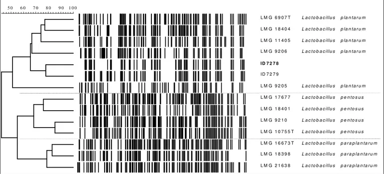
The plantarum group has a genetic heterogeneity, which was demonstrated by Dellaglio et al. (1975), on the basis of DNA/DNA hybridisation data. Three groups have been identified which were later classified as L. plantarum stricto sensu, L. pentosus (Zanoni et al., 1987) and L. paraplantarum (Curk et al., 1996). These three species are closely related genotypically and show highly similar phenotypes. So, a correct identification of some new isolates as one of these strains is complicated by the ambiguous response of traditional physiological tests and molecular methods (Torriani et al., 2001). The dendrogram of the AFLP clusters analysis with reference strains (L. plantarum, L. pentosus and L. paraplantarum, Figure 5) shows that Sp.4 and Sp.20 constituted separated subgroups within plantarum species, in contrast with SDS-PAGE results.
PCR-ARDRA of 16S rDNA
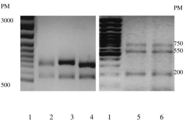
16S rDNA was amplified from three L. plantarum strains and from Sp.4 and Sp.20. Fragments of 1500 bp were obtained. The Figure 6 presents the patterns of the 16S rDNA polymorphism after Acc II restriction digestion. All L. plantarum patterns (Figure 6, lanes 2, 3 and 4) after digestion with AccII, show the same restriction profile characterised by the presence of three fragments (~175, 300 and 450 bp). Sp.4 and Sp.20 pattern (lanes 5
Lactobacillus plantarum LMG 17675 Lactobacillus pentosus LMG 10755T Lactobacillus pentosus R-4699 Lactobacillus pentosus LMG 18401 Lactobacillus pentosus LAB 160 ID 7278 ID 7279 Lactobacillus plantarum LMG 9205 Lactobacillus plantarum LMG 8413 Lactobacillus plantarum LMG 6907T Lactobacillus paraplantarum LMG 16673T Lactobacillus paraplantarum LMG 14775 Lactobacillus plantarum LMG 8403 Lactobacillus plantarum LMG 12167 Lactobacillus pentosus LAB 159 Lactobacillus plantarum LMG 18053 Lactobacillus plantarum LMG 14773 Lactobacillus alimentarius LMG 9188t2 Lactobacillus alimentarius LMG 9187T Lactobacillus paralimentarius LMG 19152T Lactobacillus kimchii LMG 19822T Lactobacillus farciminis LMG 9189 Lactobacillus farciminis LMG 9200T Lactobacillus farciminis LMG 17703 Lactobacillus arizonensis LMG 19807Tt2
Figure 4. Dendrogram showing the relationship between electrophoresis protein patterns of Sp.4 (7278), Sp.20 (7279)and LMG LAB
database strains. . . . . . . . . . . . . . . . . . . . . . . . . . . . . . . . . . . . . L a c to b a c illu s L a c to b a c illu s L a c to b a c illu s L a c to b a c illu s L a c to b a c illu s L a c to b a c illu s L a c to b a c illu s L a c to b a c illu s L a c to b a c illu s L a c to b a c illu s L a c to b a c illu s L a c to b a c illu s p la n ta r u m p la n ta r u m p la n ta r u m p la n ta r u m p la n ta r u m p e n to s u s p e n to s u s p e n to s u s p e n to s u s p a ra p la n ta ru m p a ra p la n ta ru m p a ra p la n ta ru m L M G 6 9 0 7 T L M G 1 8 4 0 4 L M G 1 1 4 0 5 L M G 9 2 0 6 ID 7 2 7 8 ID 7 2 7 9 L M G 9 2 0 5 L M G 1 7 6 7 7 L M G 1 8 4 0 1 L M G 9 2 1 0 L M G 1 0 7 5 5 T L M G 1 6 6 7 3 T L M G 1 8 3 9 8 L M G 2 1 6 3 8 1 0 0 9 0 8 0 7 0 5 0 6 0
Figure 5. Dendrogram showing the relationship between AFLP profiles of Sp.4 (7278), Sp.20 (7279)and reference strains (LMG LAB database).
Figure 6. Photo-negative ethidium bromide stained agarose gel
electrophoresis of amplified 16S rDNA of strains digested with
AccII. 1: 100 bp DNA ladder used as molecular size marker
(PM); 2: L. plantarum G100; 3: L. plantarum L115; 4: L
.plantarum ’Ma’; 5: Sp.4; 6: Sp.20.
Figure 7. Photo-negative ethidium-bromide stained agarose gel
electrophoresis of amplified 16SrDNA of strains digested with Taq I.
Lane 1: 100 bp DNA ladder used as molecular size marker (PM);
Lane 2: L. plantarum G100;Lane 3: L. plantarum L115; Lane 4: L.
plantarum ’Ma’;Lane 5: Sp.4; Lane 6: Sp.20.
Table 5. Restriction fragment length polymorphism obtained
after 16S rDNA digestion with Acc II and Taq I. Sizes were
expressed in bp.
and 6)) show a new pattern with ~300, 450 and 700 bp fragment. According to this result, it may be affirmed that Sp.4 and Sp.20 are not L. plantarum.
All L. plantarum patterns (lanes 2, 3 and 4) show after digestion with TaqI the same restriction profile with two fragments (~550, 750bp). Sp.4 and Sp.20 both show (figure 7) a different profile from the one of L. plantarum,
Sow et al. 417
with three fragments (~200, 550 and 750 bp). These results, confirm that Sp.4 and Sp.20 are genetically different from L .plantarum. Restriction fingerprinting profiles are given in Table5 with different fragment sizes.
Table 6. Homology values calculated (Vector NTI, Maryland, USA)
for short ITS sequences.
G100=L. plantarum G100-CWBIB076,
DSM10667=L. paraplantarum DSM10667
U97138=L. paraplantarum U97138,
U97134=L. pentosus U97134.
Intergenic transcribed spacer (short ITS) sequencing and sequence alignment results
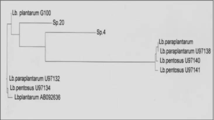
ITS of Sp.4, Sp20 and L .plantarum G100 were sequenced and these sequences were compared with those of L. plantarum stricto sensu, L. pentosus and L. paraplantarum, found in NCBI Genbank. Sequences alignment did not introduce any gaps, and there were no ambiguous positions that could lead to bias in the inference of phylogeny. This was not surprising, since the sequences were derived from closely related species. Table 6 shows homology percentages calculated for different ITS sequences (Vector NTI) Comparing short ITS homology (Table 6), Sp.20 could be classified into L. paraplantarum cluster with 95% of ITS similarity with L. paraplantarum U97138 and 96% of ITS similarity with L. paraplantarum DSM10667. Moreover, the percentage of homology between reference species and thermotolerant strains are way out the permissive value (97.5%), proposed by Tannock et al. (1999) to classify two strains in the same species. Therefore, Sp.4 isolate could not be classified into any existing taxon. Sp.4 represent a new species (Figure 8) within Lactobacillus genus with only 88%+/-1 ITS sequence similarity with reference species. Figure 8 shows phylogenetic relationship between L. plantarum, L. paraplantarum, L. pentosus, Sp.4 and Sp. 20 based on short ITS alignment.
Sp. 20 and Sp.4 as inferred by the neighbour-joining method with short ITS sequence alignment. *Sequences were obtained from NCBI Genbank. Alignments were realised on Vector NTI Software.
Strains Restriction enzymes
L.plantarum reference AccII TaqI L. plantarum G100 450-300-175 750-550 L. plantarum “Ma“ 450-300-175 750-550 L. plantarum L115 450-300-175 750-550 New TDLAB Sp.4 700-450-300 750-550-200 Sp.20 700-450-300 750-550-200 Homology (%) G100 Sp.4 Sp. 20 DSM 10667 U97138 U97134 G100 100 86 94 92 91 92 Sp.4 86 100 95 89 88 87 Sp.20 94 95 100 96 95 89 DSM 10667 92 89 96 100 95 92 U97138 91 88 95 95 100 92 U97134 92 87 89 92 92 100 PM 3000 500 1 2 3 4 1 5 6 PM 3000 500 1 2 3 4 1 5 6 PM 750 550 200
Figure 8. Phylogenetic tree showing the relative positions of L. plantarum, L.paraplantarum*, L. pentosus*,
DISCUSSION
Twenty strains were isolated from soils and dried chicken faeces. Two strains (Sp.4 and Sp.20) are resistant to both heat and desiccation stresses. These strains produce massive amount of lactic acid; they are catalase negative, Gram positive and non-spore-forming rods. A polyphasic approach was employed for the taxonomic identification of these two strains, including techniques with a wide discriminative power (growth conditions, auxanography, fatty acids analysis, SDS-PAGE of proteins, genomic AFLP, 16S rDNA gene restriction analysis, and ITS sequencing). Culture studies showed large range of growth temperatures (15°C to 44°C) with an optimum at 30-37°C and pH values (3 to 8). Sugar fermentation profiles obtained by API 50 CHL indicated that Sp.4 and Sp.20 have heterogeneous carbon metabolism. They ferment lactose and galactose but not D-xylose, in contrast with thermotolerant strains recently isolated by Niamsup et al. (2003).
The cellular fatty acid composition is the result of a sum of complex phenoma maintaining optimal viability of cell under stress conditions (Russell et al., 1995). Linoleic acid probably results from oleic acid after conversion by desaturase activity (Guillot et al., 2000). It seemed that survival and growth of Sp.4 and Sp.20 at 42°C can be related to the high proportion of MUFA and to an overall equilibrium in unsaturated/saturated fatty acid ratio. Some thermotolerant strains, (Niamsup et al., 2003) present similar oleic acid rates when cultures were incubated at 42°C. Guillot et al. (2000) reported that in Lactococcus lactis, at high temperature, the UFA/SFA increases from 1.7 to 2.7. An oxygen-dependent desaturase induction was postulated in thermotolerant yeast strains with the roles of preventing increased oxygen and reactive oxygen species (ROS) accumulation in the membrane at superoptimal temperatures and protecting the cells from damage generated by oxidative and thermal stresses (Guerzoni
et al., 1997). It seemed that TLAB thermal responses are similar to those reported by Guerzoni et al. (1997). These results clearly identify an increase in unsaturation level of fatty acids as a response to exposure to superoptimal temperatures. This behaviour suggests that desaturase activation or hyperinduction plays an important role in the response to heat stress at least in thermotolerant strains. In addition, FTIR spectroscopy provided evidence for changes in membrane fluidity as result of the unsaturation of fatty acids (Szalontai et al., 2000). According to these authors, the frequency of symmetric CH2 stretching is higher in unsaturated fatty acid, because of loading. The decrease of this frequency enhances rigidification of membranes (Szalontai et al., 2000). In Lactobacilli, temperature is reported to induce chances in fatty acid in relation to the regulation of the degree of fatty acid unsaturation, cyclization and proportions of long-chain fatty acids containing (Dionisi et al., 1999). However, the range of temperatures generally studied has not exceeded the optimal ones (except in Niamsup et al., 2003). Moreover, fatty acid or precursor supplementation in culture media strongly affects cellular fatty acid profiles. In particular, oleic acid is known to be incorporated into LAB membrane.
SDS-PAGE protein profile analysis indicated that these isolates constitute a homogeneous phenon/subgroup within plantarum group, even if they cannot be separate from L. paraplantarum, L. pentosus and L. plantarum. It is possible that environmental adaptation (to heat and dehydration) performs suitable physiological modifications and induces typical metabolism profiles. Some of regulatory mechanisms responding to an environmental stress condition are common to those found in Gram-positive bacteria, and many stress responses are conserved at the protein level. It was demonstrated by Prasad et al. (2003) by enhancing protective effect of prestressing L. rhamnosus HN 001 with heat or osmotic shock on the viability of dried preparations during storage.
These results confirm the insufficiency of biochemical and physiological analysis to characterise new wild isolates, because the metabolism can be modified with evolution and environment. We further complete this taxonomic study using genotypic techniques with diverse taxonomic discriminative powers (Torriani et al., 2001). AFLP analyses confirm that Sp.4 and Sp.20 cannot be separated to plantarum group, since the three constituted species are phylogenetiquely closely related. The 16S rDNA sequences of Sp.4 and Sp.20 were determined and compared with available 16S rDNA. sequences in the Genbank database. % of 16S rDNA homology to each other is up to 99% similarity. This approach is not suitable because of high identity value shared by L. plantarum and L. pentosus (Collins et al., 1991; Dellaglio et al., 1975; Quere et al., 1997). PCR-ARDRA, which has been successfully used for differentiation of a variety of microorganisms (Moschetti et al., 1997; Ventura et al.,
2001) is rather preferred. The 16Sr DNA was amplified and, after restriction with AccII and TaqI, a polymorphism was found with each restriction enzyme between L. plantarum control strains Sp.4 and Sp.20. ARDRA technique described here can be considered as a reliable and a reproducible molecular tool to discriminate new isolated from plantarum stricto sensu species (Garcia-Martinez et al., 1999).
ITS (short fragment) of L. plantarum G100, Sp.4 and Sp.20 were sequenced and compared with those of L. plantarum stricto sensu, L. paraplantarum and L. pentosus. Sequence analysis revealed that Sp.20 belonged to paraplantarum species with 95-96 % of ITS homology. Sp.4 appears like a new species within the genus Lactobacillus with only 88% ITS sequence similarity with reference species and the name Lactobacillus aminata sp. nov is proposed for it.
Lactobacillus aminata sp. nov (Sp.4) cells are Gram-positive, non-motile, non-spore-forming, catalase-negative rods of 1x2-3 µm in size, which occur singly, in pair or as, short chain. After growth at 30°C for 48 h, colonies on GYP (glucose yeast peptone) agar are ~1-2 mm in diameter, white, convex, circular and smooth, obligatory homofermentative and produce large among of D/L lactic acid. It grows at 15 to 44°C and from pH 3 to 8. It does not hydrolyse aesculin, does not produce hydrogen sulphide, does not reduce nitrate, does not liquefy gelatin and does not produce dextran from sucrose. Acid is produced from glucose, ribose, L-arabinose and D-fructose, melibiose, saccharose, galactose, methyl -D-mannoside, mannose, mannitol sorbitol, thehalose, maltose, lactose, arbutine, melezitose, D-turanose and D-raffinose. Glycerol, erythritol, D-arabinose, L-xylose, methyl- -xyloside, dulcitol, inositol, inulin, starch, glycogen, xylitol, D-lyxose, D-tagatose, D-fucose, D-arabitol, L-arabitol, gluconate, 2-ketogluconate and 5-ketogluconate are not fermented. The major cellular fatty acid produced when culture was incubated at 42°C is a straight chain unsaturated acid, C18:1. The GenBank accession number for Sp.4 16S rDNA complete sequence is AY594275 and Sp.4 short ITS complete sequence is AY594277. L. aminata has been deposited in the BCCM/LMG Bacteria Collection (University of Gent, Gent, Belgium) as LMG ID 7278 and in the Centre Wallon de Biologie Industrielle- Unité de Bio-industries (CWBI) Culture Collection (Faculté de Sciences Agronomiques de Gembloux, Belgium) as CWBI-B534. Isolated from faeces of chickens, coming from Senegal, the type strain is Sp.4 (=LMG ID 7278T=CWBI-534).
This study demonstrates the usefulness of polyphasic criteria for identification of natural isolates, which often show wide microbial heterogeneity (Dykes et al., 1994). The ability to discriminate between closely related strains and the capacity to identify unusual or atypical isolates can be improved by combining different techniques, such as physiological analysis, SDS-PAGE of proteins, AFLP,
Sow et al. 419
ARDRA and ITS sequencing.
Use of lactic acid starter is more and more important since potential health benefits were showed (Daly et al., 1998). So, freezing is widely used for long-term preservation of their viability, easy transport and technological properties (acidification activity, aroma compounds production, probiotic potentialities). However, freezing causes a loss of viability acidification activity of LAB (Fonseca et al., 2003). Moreover, bacterial resistance to stress including freezing, frozen storage and desiccation depends on the strain and on production conditions, stabilisation (concentration, cryoprotection, freezing) and storage (Fonseca et al., 2001).
The aim of this work was to isolate and identify thermotolerant and desiccation tolerant strains. The next step will to estimate potential properties like probiotic and behaviour of Sp.4 and Sp.20 during storage at room temperature. Such strains have been prepared by their background to support drastic environment (heat, low Aw). In nature, they are exposed to variable factors, such as temperature, and the availability of nutrients and water. Survival in this changing environment requires a wide range of fast, adaptive responses (Van de Guchte et al., 2002). In the majority of cases, the bacterial response leads to transcriptional activation of genes whose products cope with a given physico-chemical stress. Gene regulators respond to specific signals (such as environmental and cellular signals) by stimulating or inhibiting transcription, translation, or some other event in gene expression, so that the rate of synthesis of gene products is appropriately modified. Microorganisms that are able to offer a successful physiological/biochemical adaptation are better suited to colonising the changing niche (Ramos et al., 2001). In this study, new isolates are able to give suitable response to desiccation and heat stresses. They are be able to support storage at room temperature (data not published), where cytoplasmic membrane damages are generally considered as the main mechanism of dehydration (Lievense et al., 1994b). Economically and practically, it will be a real progress in dairy industry to screen rustic LAB strains particularly in developing hot countries even in all over the world.
ACKNOWLEDGEMENTS
This work was supported by the Commissariat Général aux Relations Internationales de la Région Wallonne. The authors are grateful to the Unité de Phytopathologie de la Faculté Universitaire des Sciences Agronomiques de Gembloux for ARDRA analyses. We would also like to thank the Laboratory of Microbiology of Gent for technical assistance, and Progenus (Gembloux, Belgium) for sequencing analyses.
REFERENCES
Boulos L, Prévost M, Barbeau B, Coallier J, Desjardins R (1999). J. Microbiol. Methods. 37: 77-86.
Collins MD, Rodrigues UM, Ash C, Aguirre M, Farrow JAE, Martinez-Murcia A, Phillips BA, Williams AM, Wallbanks S (1991).
Phylogenetic analysis of the genus Lactobacillus and related
lactic acid bacteria as determined by reverse transcriptase sequencing of 16S rRNA. FEMS Microbiol. Letl. 7 7 : 5 - 1 2 .
Curk F, Bringel MC, JC, Hubert (1996). Characterization of Lactobacilli
by Southern-type hybridization with a Lactobacillus plantarum
pyrDFE probe. Int. J. Syst. Bacteriol. 46: 588-594.
Daly C, Davis R (1998). The biotechnology of lactic acid bacteria with emphasis on applications in food safety and human health. Agric. food Sci. Finland 7: (2) 251-264.
Delcour J, Ferain T, Hols P (2000). Advances in the genetics of
thermophilic lactic acid bacteria. Curr. Opinion Biotechnol. 11: 497-504.
Dellaglio F, Bottazzi V, Vescovo M (1975). Deoxyribonucleic acid
homology among Lactobacillus species of the subgenus
Streptobacterium Orla –Jensen. Int. J. Syst. Bacteriol. 25: 160:172.
De Man JC, Rogosa M, Sharpe ME (1960). A medium for cultivation of Lactobacilli. J. Appl. Bacteriol. 23: 133-135.
*Colette D, Catherine S, Fitzgerald GF, Kevin C, Ross R, Paul (2001).
Environmental adaptation of probiotic Lactobacilli towards
improvement of performance during spray drying. Inter. Dairy J. 12: 183- 190.
Desmons S, Zgouli S, Destain J, Thonart P (1997). Freeze-drying ofa
lactic acid bacteria. Lactobacillus acidophilus. Mededelingen
Faculteit Landbouwwkundige en Toegepaste Biologische
Wetenschappen. Universiteit Gent. 62:No 4b, 1713-1716.
Devereux J, Haeberli P,Smithies O (1984). A comprehensive set of
sequence analysis programs for the VAX. Nucleic Acids Res. 12: 387-395.
Dionisi F, Golay PA, Elli M, Fay LB (1999). Stability of cyclopropane and conjugated linoleic acids during fatty acid quantification in lactic acid bacteria. Lipids 34: 1107-1115.
Dykes GA, Von Holy A (1994). Strain typing in the genus Lactobacillus.
Letl. Appl. Microbiol. 19: 63-66.
Eyssen H, De Pauw G, Stragieragier J, Verhulst A (1983). Cooperative formation of -muricholic acid by intestinal microorganisms. Appl. Environ. Microbiol. 45: 141-147.
Fonseca F, Béal C, Corrieu G (2001). State diagrams and sorption isotherm of bacterial suspensions and fermented medium. THERMOCHIMICA ACTA. 366: 167-182, XP002198084.
Fonseca F, Béal C, Mihoub F, Marin M, Corrieu G (2003). Improvement
of cryopreservation of Lactobacillus delbrueckii ssp. bulgaricus CFL1
with addtitives displaying different protective effects. Int. Dairy J. (sous presse).
Fujisawa T, Shirasaka S, Watabe J, Mitsuoka T (1984).
Lactobacillus aviarius sp. nov: a new species isolated from the intestine of chiken. Syst Appl. Microbiol. 5: 414-420.
Fujisawa T, Benno Y, Yaeshima T, Mitsuoka T (1991). Taxonomic
study of the Lactobacillus acidophilus group, with recognition of
Lactobacillus gallinarum sp. nov. and Lactobacillus johnsonii sp.
nov. and synonymy of Lactobacillus acidophilus group A3
(Johnson et al. 1980) with the type strain of Lactobacillus
amylovorus (Nakamura 1981). Int. J. Syst. Bacteriol. 42: 487-491.
Garcia –Martinez J, Acinas SG, Anton AI, Rodriguez-Valera F (1999). Use of 16S-23S ribosomal genes spacer region in studies of procaryotic diversity. J. Microbiol. Methods. 36: 55-64.
Gerhardt P.(Eds.), 1981. Methods for general bacterial. American Soc.
Microbiol. Press, Washington.
Gevers DG, Huys, Swings (2001). Applicability of rep-PCR
fingerprinting for identification of Lactobacillus species. FEMS
Microbiol. Letl. 205 : 31-36.
*Gounot AM, Russell NJ (1999). Physiology of cold-adapted micro-organisms. In: Margesin R, Schinner F (Eds.), Cold-adapted Organisms. Ecol. Physiol. Enzymol., Mol. Biol. Springer, Berlin, 33-55.
Graham PH, Sadowsky MJ, Keyser HH, Barnet YM, bradley RS, Cooper JE, Delay DJ, Jarvis BDW, Roslycky EB, Strijdom BW, Young JPW (1991). Proposed mininal standards for the description of new genera and species of rood and stem-nodulating bacteria. Int. J. Syst. Bacteriol. 41: 582-587.
Grahay F, Thonart PH, Lognay G, Mampuys JP (1987). Effet of culture conditions of the yeast Candida curvata on lipids yield and
composition. Proceeding 4th Europeen Congres on Biotechnology,
ECBH, 3.O.M.Neijssel, R.P. Van der Meer, K. Ch. A. M Luyben (Eds) Gram Christian (1884).Ueber die isolirte Farbung der Schizomyceten in SchnittÄund Trockenpraparaten. Fortschritte der Medicin, 2: 185-189.
GuerzoniME, Ferruzi M, Sinigaglia M, Criscuoli GC (1997). Increased
cellular fatfy acid desaturation as s possible key factor in
thermotolerance in Saccharomyces cerevisiae. Can. J. Microbiol.
43: 569-576.
GuerzoniME, Lanciotti R, Cocconcelli P, Sandro (2001). Alteration in
cellular fatty acid composition as a response to salt, acid, oxidative
and thermal stresses in Lactobacillus helveticus. Microbiol. 147:
2255-2264.
Guillot A, Obis D, Mistou MY (2000). Fatty acid composition and
activation of glycine-betaine transport in Lactococcus lactis subjected
to osmotic stress. Int J Food Microbiol. 55: 47-51.
Gusils C, Palacios J, Gonzales S, Olivier G (1999). Lectin-like protein fractions in lactic acid bacteria isolated from chickens. Biol. Phar. Bull. 22: 11-15.
Herrero MB, Mayo B, González, Zuárez JE (1996). Evaluation of technologically important traits in lactic acid bacteria isolated from spontaneous fermentations. J. Appl. Bacteriol. 81: 565-570. Hodzic D, Llabrés G, Zgoulli S, Gilsoul JJ, Thonart PH (1995). Study of
Residual Water Types Study in freeze-dried Lactobacillus
acidophilus by Nuclear Magnetic Resonance. Lebensm.Wiss. U. Technol. 28: 21-24.
Kaur IP, Chopra K, Saini A (2002). Probiotics:potential pharmaceutical
applications. Eur. J. Pharm. Sci. Pharm. Appl. 15 (1): 1-9.
Ingrassia I, Roques C, Prevots F (2001). Preliminary experiments for
ARDRA validation on flora associated with intestinal mucosa. Lait,
81: 263-280.
Ishibashi N, Shimamura S (1993). Bifidobacteria: Research and
Development in Japan. Food Tech. June, pp.126-135.
Ito M, Connor WE, Blanchette EJ, Treadwell CR, Vahouny George V (1969). Inhibition of lymphatic absorption of cholesterol by cholestane-3ßbeta, 5 , 6ßbeta-triol. J. Lipid Res. 10: 694-702.
Kandler O, Weiss N (1986). Genus Lactobacillus Beijerinck 1901,
212 AL. In Bergey's Manual of Systematic Bacteriology: Edited by PHA Sneath, NS, ME Sharpe, JG Holt. Baltimore: Williams and Wilkins. 2: 1208-1234.
Lievense LC, van’t Riet K (1994a). Convective drying of bacteria. II. Factors influencing survival. Adv. Biochem. Eng./Biotechnol. 51: 71-89.
Lievense LC, Verbeek MAM, Tackeme T, Meerdink G, van’t Riet K
(1994b). Machanism of dehydration inactivation of Lactobacillus
plantarum. Appl. Microbiol. Biotechnol. 41: 90-94.
Lilly DM, Stillwell RH (1965). Probiotics: growth promoting factors
produced by micro-organisms. Science 147: 747-748.
Marmur J (1961). A procedure for the isolation of DNA from microorganisms. J. Mol. Biol. 3: 208-218.
Moschetti Giancarlo, Blaiotta Giuseppe, Villani Francesco, Coppola
Salvatore (2000). Specific Detection of Leuconostoc mesenteroides
subsp. mesenteroides with DNA Primers Identified by Randomly
Amplified Polymorphic DNA Analysis. App. Environ. Microbiol. 66: 422-424.
Nakayama O, Yanoshi M (1967). Spore-bearing lactic acid bacteria
isolated from rhizosphere. I. Taxonomic studies on Bacillus
laevolacticusnov. sp. and Bacillus racemilacticus nov. sp. J. Gen. Appl. Microbiol. 13: 139-153.
Niamsup P, Sujaya N, Tanaka M, Sone T, Hanada S, Kamagata Y, Lumyong S, Assavanig A, Asano K, Tomita F, Yokota A (2003).
Lactobacillus thermotolerans sp.nov., a novel thermotolerant species isolated from chicken faeces. Inter. J. Systematic Evol. Microbiol. 53: 263-268.
stress induced multiresistances: influence of extracellular coumpounds. Int. J. food Microbiol. 55: 19-25.
Pot B, Vandamme P, Kersters K (1994). Analysis of electrophoretic whole-organism protein fingerprints. Chemical Methods in
Prokaryotic Systematics. M. Goodfellow and A.G. O'Donnell (Eds.). J. Wiley and sons, Chichester.
Prasad J, Mc Jarrow P, Gopal P (2003). Heat and Osmotic Responses
of Probiotic Lactobacillus rhamnosus HN001 (DR20) in Relation to
Viability after Drying. Appl. Environ. Microbiol. 69: 917-925.
Quere F, Deschamps A, Urdaci MC (1997). DNA probe and
PCR-specific reaction for Lactobacillus plantarum. J. Appl. Microbiol.
82: 783-790.
Ramos JL, Gallegos MT, Marque S, Ramos-Gonzales MI, Espinoza-Urgel M, Segura A (2001). Responses of gram-negative bacteria to certain environmental stressors. Curr. Opinion Microbiol. 4: 166-171. Russell NJ (1984). Mechanisms of thermal adaptation in bacteria:
blueprints for survival. Trends Biochem. Sci. 3: 108-112.
Russell NJ (1989). Functions of lipids: structural roles and membranes
functions. In: Ratledge, C. Wilkinson S.C. (Eds.), Microbial lipids. 2:
279-365. Academic press Lond.
Russell NJ, Fukunga N (1990). A comparison of thermal adaptation of membrane lipids in psychrophilic and thermophilic bacteria. FEMS Microbiol. Rev. 75: 171-182.
Russell NJ, Nichols DS (1999). Polyunsaturated fatty acids in marine bacteria - A dogma rewritten. Microbiology 145: 767-779.
Russell NJ (2002). Bacterial membranes: the effects of chill storage and food processing. An overview. Int. Food Microbiol. 79: 27-34. Sanger F, Nicklen S, Coulson AR (1977). Chain Sequencing with
Chain-Terminating Inhibitors, Proc. Nat. Acad. Sci. USA. 74: 5463. Sonan Henri Guillaume (1997). Viabilité au traitement de
déshydratation d’une série de bactéries lactiques. DES en gestion des biotechnologies. CWBI/FSAGX, Belgique.
Steels EL, Learmonth RP, Watson K (1994). Stress tolerance and
membrane lipid unsaturation in S. cerevisiae grown aerobically or
anaerobically. Microbiol. 140: 569-576.
Sow et al. 421
Svensson U, Saxelin M, Grenov B (1999). The technology of probiotics. Trends Food Sci Technol. 10: 387-392.
Szalontai B, Nishiyaina Y, Gombo Z, Murata N (2000). Biochim Biophys Acta. 1509: 409-419.
Tannock GW, Tilsala-Timisjarvi A, Rodtong S, Munro J, Ng K,
Alatossava T (1999). Identification of Lactobacillus Isolates from the
Gastro-intestinal Tract, Silage, and Yoghurt by 16S-23S rRNA Gene Intergenic Spacer Region Sequence Comparisons. Appl. Environ.
Microbiol., 65, No9:4264-4267.
Torriani S, Giovanna EF, Dellaglio F (2001). Differentiation of
Lactobacillus plantarum, L. pentosus and L.paraplantarum by recA
Gene Sequence Analysis and Multoplex PCR Assay with recA
Gene-Derived Primers. Appl. Environ. Microbiol. 67: 3450-3454.
Van de Guchte M, Serror P, Chervaux C, Smokvina T, Erlich SD,
Maguin E (2002). Stress reponse in lactic acid bacteria. Antonic
Leewen-hock. 82: 187-216.
Ventura M, Elli M, Reniero R, Zink Ralf (2001). Molecular microbial
analysis of Bifidibacterium isolated from different environments by
the species-specific amplified ribosomal DNA restriction analysis (ARDRA). FEMS Microbiology Ecology. 36: 113-121.
Vos P, Hogers R, Bleeker M, Reijans M, Van de Lee T, Hornes M Frifiters A, Pot J, Peleman J, Kuiper M, and Xabeau M. 1995. AFLP:
A new technique for DNA fingerprinting. Nucleic Acids Res. 23:
4407-4414.
Weekers F, Jacques Ph, Springael D, Mergeay M, Dielsl, Thonart Ph (1997). Med. Fac. Land Bauw. Univ. Gent. 62/4B : 1999-1902. Wijeyaratne SC, Ohta K, Chavanichi S, Mahamontri V, Nilubol N,
Hayashida S (1986). Lipid composition of a thermotolerant yeast
Hansenula polymorpha. Agric. Biol. Chem. 50: 827-832.
Zanoni P, Farrow JA, Farrow E, Philips BA, Collins MD (1987).
Lactobacillus pentosus (Fred Peterson and Anderson) sp. nov., nom.
