Université de Montréal
Neogenin function and modulation in spinal motor neuron
development
Par
Louis-Philippe Croteau
Programmes de biologie moléculaire Faculté de Médecine
Thèse présentée à la Faculté de médecine en vue de l’obtention du grade de PhD
en biologie moléculaire
Septembre 2017
i
Résumé
Le système nerveux est la structure fonctionnelle la plus complexe que l’on connaisse. Cette structure permet aux organismes multicellulaires de percevoir leur existence en tant qu’entités individuelles. Le fonctionnement du système nerveux repose sur l’assemblage précis des circuits neuronaux. Pour ce faire, pendant le développement, les projections axonales des neurones sont guidées par des signaux de guidages. Ceux-ci se trouvent dans l’environnement extracellulaire et permettent aux neurones de trouver leurs cibles respectives. Pour détecter et pouvoir répondre aux signaux de guidage, les cônes de croissance à la pointe des axones expriment des récepteurs de signaux de guidage. Ceux-ci, lorsqu’ils se lient à leurs ligands, induisent des cascades de signalisation en aval, menant à des modulations dans la poussée et la direction des axones. Bien que nous ayons fait de grands progrès dans la compréhension des effets qu’ont les signaux de guidage sur les axones, on en sait beaucoup moins sur la façon dont ces signaux agissent en concert.
Les neurones moteurs qui résident dans la colonne moteur latérale (LMC) de la moelle épinière projettent leurs axones dans les membres, s'appuient sur une multitude de signaux de guidage. Cette thèse démontre que le récepteur
transmembranaire Neogenin est impliqué dans plusieurs aspects du développement des neurones LMC, y compris la différenciation et la ségrégation des sous-types de neurones LMC ainsi que le guidage de leurs axones.
Notre laboratoire a précédemment démontré que les axones LMC sont guidés de manière synergique par Netrin-1 et les ephrins. Je vous ferai part de mes
ii
résultats explorant le mécanisme sous-jacent à l'activité synergique de Netrin-1 et d’ephrin-A5 dans le guidage des axones LMC. Je démontre que ephrin-A5 sensibilise un sous-ensemble d'axones LMC à Netrin-1. Je propose que cela est la conséquence d'une augmentation induite par ephrin-A5 dans l'abondance de Neogenin conduisant à une liaison améliorée de Netrin-1 dans les cônes de croissance LMC. En outre, je montre que la modulation de ephrin-A5 des niveaux de Neogenin dépend de
l'interaction entre ephrin-A5 et son récepteur EphA4. Je montre également que contrairement à la répulsion des axones de ephrin-A5, la sensibilisation à Netrin-1 se produit indépendamment de la queue cytoplasmique d'EphA4. Ces résultats
suggèrent que la répulsion des axones induite par ephrin-A5 et la sensibilisation à Netrin-1 se produisent dans des voies moléculairement distinctes.
Netrin-1, Neogenin et les ephrins sont vastement impliqués lors du développement du système nerveux et au-delà. Notre démonstration que les
interactions ephrin-A5 / EphA4 modulent l'abondance de Neogenin et la sensibilité à Netrin-1 ouvre la possibilité a ce que les ephrins puissent avoir un impact profond sur les processus cellulaires dépendants de la signalisation Netrin-1 et Neogenin.
iii
Abstract
The nervous system is the most exquisitely complex functional structure in the known universe. The nervous system is what enables multicellular organisms to experience life as individual entities. The functionality of the nervous system relies upon the precise assembly of neuronal circuits. To achieve this, during development, the extending axons of neurons are guided by cues in the extracellular environment that enable neurons to find their respective targets. To sense and respond to
extracellular cues, growth cones at the tip of axons express guidance cue receptors. The interaction between guidance cues and their receptors induce downstream signaling cascades which lead to modulations in axon outgrowth and directionality. Although we have made great progress in understanding how individual cues guide axons, much less is known about how these cues act in concert.
Motor neurons that reside within the lateral motor column (LMC) of the spinal cord extend axons that innervate the limbs, rely on a multitude of guidance cues. The evidence presented in this thesis shows that the transmembrane receptor Neogenin is implicated in several aspects of LMC neuron development including LMC subtype differentiation and segregation as well as the guidance of their axons.
Our lab has previously shown that LMC axons are guided synergistically by Netrin-1 and ephrins. I will be presenting results exploring the mechanism underlying the synergistic activity of Netrin-1 and ephrin-A5 in LMC axon guidance. I provide evidence that ephrin-A5 sensitizes a subset of LMC axons to Netrin-1. I propose that this is a consequence of an ephrin-A5 induced increase in the abundance of
iv
Neogenin leading to enhanced Netrin-1 binding in LMC growth cones. Furthermore, I show that the ephrin-A5 modulation of Neogenin levels is dependent on the
interaction between ephrin-A5 and its receptor EphA4. I also show that contrarily to the repulsion from ephrin-A5, sensitization to Netrin-1 occurs independently from the cytoplasmic tail of EphA4. These results suggest that the ephrin-A5 induced axon repulsion and sensitization to Netrin-1 occur in molecularly distinct pathways.
Netrin-1, Neogenin and ephrins are vastly implicated during the development of the nervous system and beyond. Our demonstration that ephrin-A5/EphA4
interactions modulate the abundance of Neogenin and sensitivity to Netrin-1 brings forth the possibility that ephrins may have a profound impact on cellular processes implicating Netrin-1 and Neogenin.
v
Table of contents
Résumé --- i
Abstract --- iii
List of abbreviations --- viii
List of figures --- x
Acknowledgements --- xii
CHAPTER I - INTRODUCTION --- 15
1 An introduction to axon guidance --- 16
2 The Growth Cone --- 18
3 Guidance cues --- 20
4 Netrin-1 --- 21
4.1 Netrin-1 in commissural axon guidance --- 21
4.2 Netrin-1 receptors --- 23
4.3 DsCAM --- 23
4.4 Dcc --- 24
4.5 Neogenin --- 25
4.6 Neogenin as a receptor for RGMs --- 25
4.7 Neogenin as a receptor for Netrin-1 --- 27
4.8 Chick Neogenin --- 28
4.9 Neogenin/Dcc structure --- 30
5 Netrin-1 signaling --- 32
6 Modulators of Netrin-1 signaling --- 34
6.1 Heparin sulfate proteoglycans --- 34
6.2 Slit/Robo --- 35
6.3 Draxin --- 37
6.4 Dcc/Neogenin cleavage --- 38
7 An introduction to Eph/ephrin signaling --- 40
7.1 Eph/ephrin signaling in axon guidance --- 42
8. Spinal motor neuron development --- 44
8.1 LMC neuron differentiation --- 48
9 LMC axon guidance --- 49
vi
10.1 Eph/ephrin forward signaling--- 52
10.2 Eph/ephrin reverse signaling --- 53
10.3 Eph/ephrin cis attenuation --- 55
10.4 GDNF / GFRa1/RET --- 56
10.5 Npn-2/Sema3F --- 57
11 Netrin-1 in LMC guidance --- 58
11.1 In vivo requirement for Netrin-1 in LMC axon guidance --- 59
11.2 LMCm and LMCl axons respond to Netrin-1 in vitro --- 60
12 Synergy between Netrin-1 and ephrins in LMC guidance --- 61
CHAPTER II - MATERIALS AND METHODS --- 65
CHAPTER III - RESULTS --- 74
1. Neogenin in the responsiveness of LMCl axons to Netrin-1 --- 75
Rationale --- 75
Neogenin is required for the responsiveness of LMCl axons towards Netrin-1--- 75
Dcc rescues the loss of LMCl responsiveness to Netrin-1 --- 76
2. Netrin-1 and ephrin-B2 collapse LMCm growth cones synergistically --- 78
Rationale --- 78
3. Neogenin in LMC differentiation --- 80
Rationale --- 81
Expression of Neogenin, RGMa and RGMb during LMC neuron development --- 81
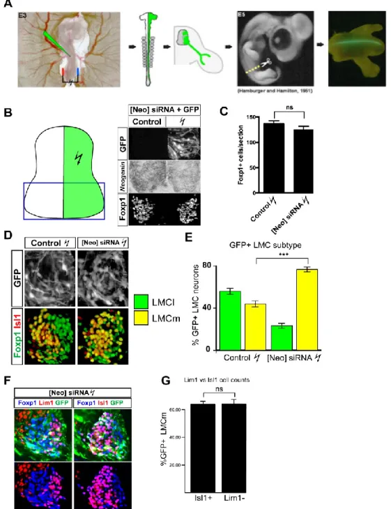
Knockdown of Neogenin alters LMC neuron subtype differentiation --- 83
LMC neuron subtype distribution within the LMC is altered in Neogenin KD embryos - 88 Investigating the requirement for RGMa and RGMb in LMC subtype differentiation --- 90
Knockdown of Neogenin alters lumbar LMC axon projections --- 92
Neogenin may be dispensable for the subtype differentiation of LMC neurons in mice - 94 4. Sensitization of spinal motor neurons to Netrin-1 by ephrin-A5 --- 96
Rational --- 96
Netrin-1 induces a protein kinase A-dependent re-localization of Neogenin in LMC growth cones --- 97
Ephrin-A5 increases Neogenin protein levels in LMC growth cones --- 104
Ephrin-A5 induces an increase in the co-localization of Neogenin and EphA4 --- 107
vii
Ephrin-A5 increases Neogenin growth cone surface levels --- 107
Ephrin-A5 enhances Netrin-1 binding in growth cones --- 110
Binding of ephrin-A5 to EphA4 is necessary for the ephrin-A5 induction of Neogenin in growth cones --- 112
Overexpression of EphA4 potentiates LMC growth cones to the ephrin-A5-dependent increase in Neogenin abundance --- 114
The ephrin-A5-induced Neogenin upregulation is independent of PKA, Src family kinase, protein synthesis, proteosomal or γ-secretase degradation --- 115
The intracellular domain of EphA4 is dispensable for potentiating the ephrin-A5 induced increase in Neogenin abundance --- 117
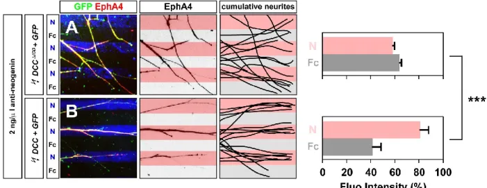
Ephrin-A5 sensitization of lateral LMC axons to Netrin-1 --- 119
The cytoplasmic tail of EphA4 is dispensable for the sensitization of LMCl axons to Netrin-1 by ephrin-A5 --- 122
CHAPTER IV - DISCUSSION --- 124
Neogenin in LMC neuron differentiation --- 125
Neogenin in LMC subtype segregation --- 125
Neogenin in LMC subtype differentiation --- 127
Sensitization of LMCl axons to Netrin-1 by ephrin-A5 --- 129
Netrin-1 increases and redistributes Neogenin to the growth cone periphery --- 129
Ephrin-A5 on receptor dynamics in LMC growth cones --- 130
Ephrin-A5/EphA4 attenuation of Neogenin cleavage as a possible mechanism underlining the sensitization to Netrin-1 --- 132
CHAPTER V – CONTRIBUTIONS AND PERSPECTIVES --- 137
Neogenin as a mediator of attractive Netrin-1 signaling in LMC neurons --- 138
Speculations on the requirement for Neogenin dependent transcriptional regulation for the expression of Lim1 by LMC neurons --- 138
Optimization of the chick in ovo electroporation protocol --- 139
The sensitization of LMC neurons to Netrin-1 by ephrin-A5 --- 140
A potential for ephrin-A5 induced upregulation of Neogenin in various cell types --- 140
A potential requirement for ephrin-A/EphA induction of Neogenin for neural tube closure --- 142
Speculations on the requirement for ephrin/EphA4 dependent upregulation of Dcc/Neogenin for LTP --- 143
viii
List of abbreviations
ADAM A disintegrin and metalloproteinase APP Amyloid precursor protein
BC Boundary cap
BMP Bone morphogenic protein CNS Central nervous system Dcc Deleted in colorectal cancer
DsCAM Down syndrome cell adhesion molecule E Chick Embryonic day
F-actin Filamentous actin FAK Focal adhesion kinase FNIII Fibronectin type III
GAP GTPase activating protein
GDNF Glial-cell-line-derived neurotrophic factor GEF Guanine nucleotide exchange factor GPI Glycosylphosphatidylinositol
GFRα1 GDNF receptor alpha-1 HH st. Hamburger-Hamilton stage HMC Hypaxial motor column HRP Horseradish peroxidase HSPG Heparin sulfate proteoglycan IF Immunofluorescence
ICD Intracellular domain Ig Immunoglobulin
KD Knock down
ix
LMCl Lateral LMC LMCm Medial LMC LMO4 Lim-only protein 4 MMC Medial motor column MN Motor neuron
MT Microtubule Np Neuropilin
OE Olfactory epithelium
PGC Preganglionic motor column PKA Protein kinase A
PS1 Presenilin-1 RA Retinoic acid
Raldh2 Retinaldehyde dehydrogenase-2 RGM Repulsive guidance molecule RGC Retinal ganglion cell
RTK Receptor tyrosine kinase Sema Semaphorin
SFK Src family kinase SpMN Spinal motor neuron TCA Thalamocortical axon
WNT Wingless-type MMTV integration site WRC Wave regulatory complex
x
List of figures
Figure 1 The structural and spatial divisions in a typical growth cone ... 19
Figure 2 Chick Neogenin expression in the spinal cord closely reassembles the combined expression of Dcc and Neogenin in the mouse ... 29
Figure 3 Netrin-1 signaling ... 31
Figure 4 Early development of spinal motor neurons of the LMC ... 47
Table 1 LMC dorso-ventral axon guidance summary ... 52
Figure 5 Model for the guidance of LMC axons by Netrin-1 ... 58
Figure 8 Netrin-1 and ephrins are integrated synergistically by LMCm and LMCl axons ... 62
Figure 6 Neogenin is required for LMCl axon growth preference for Netrin-1 ... 77
Figure 7 Dcc can functionally substitute for chick Neogenin in LMCl axon preference for Netrin-1 ... 78
Figure 9 Netrin-1 and ephrin-B2 act synergistically on LMCm growth cone collapse ... 80
Figure 10 Expression of Neogenin, RGMa and RGMb during LMC neuron development ... 83
Figure 11 Knockdown of Neogenin alters LMC subtype differentiation ... 85
Figure 12 Neogenin is required for establishing Lim1 expression in LMC neurons... 87
Figure 13 The Distribution of LMCm and LMCl neurons is altered in [Neo]siRNA embryos 89 Figure 14 A possible implication for RGMb in LMC subtype differentiation ... 92
Figure 15 Knockdown of Neogenin alters LMC axon projections ... 93
Figure 16 LMC neuron subtype differentiation in mice lacking Neogenin appears normal .... 95
Figure 17 Netrin-1 induces a re-localization of Neogenin in LMC growth cones ... 98
Figure 18 Netrin-1 at 10 ng/mL is sufficient in redistributing Neogenin within LMC growth cones ... 99
Figure 19 Netrin-1 induced re-localization of Neogenin is PKA dependent ... 100
Figure 20 Netrin-1 induces specific changes in protein levels and distribution at the growth cone ... 103
Figure 21 The effects of ephrin-A5 on protein levels and distribution at the growth cone .... 105
Figure 22 Ephrin-A5 increases the proportion of fluorescence overlap between Neogenin and EphA4 ... 108
Figure 23 Ephrin-A5 increases surface enriched Neogenin in growth cones ... 109
Figure 24 Ephrin-A5 enhances Netrin-1 binding in growth cones ... 111
Figure 25 Blocking ephrin-A5/EphA4 interactions inhibits the ephrin-A5 induced increase in Neogenin levels and overlap with EphA4 ... 113
Figure 26 Overexpression of EphA4 enhances the ephrin-A5 induced increase in Neogenin abundance ... 115
Figure 27 The ephrin-A5 induced Neogenin upregulation prevails despite inhibiting specific cell functions ... 117
Figure 28 The cytoplasmic tail of EphA4 is dispensable in potentiating the ephrin-A5 induced increase in Neogenin abundance ... 119
xi
Figure 30 The cytoplasmic tail of EphA4 is dispensable in the sensitization of LMC axons to Netrin-1 by ephrin-A5... 123 Figure 31 Tentative model for the sensitization of LMCl axons to Netrin-1 ... 134 Supplementary Figure 1 Growth cone area measurements... 136
xii
Acknowledgements
First and foremost, I would like to thank my PhD supervisor, Dr. Artur Kania, for providing me with the opportunity to pursue graduate studies as a member in his lab. Over the years, he has dedicated an enormous amount of time discussing my experiments with me and I am grateful for all the scientific wisdom he has shared. I am also grateful for his encouragement in having high scientific standards and for participating in conferences. Thank you Artur for seeing the opportunity in turning an unexpected electroporation artifact into my first scientific publication. You have also provided me the opportunity of collaborating with several labs, experiences that have enriched my skill set and scientific career. Above all, thank you Artur for listening to what I had to say and taking my opinions into consideration.
I would also like to acknowledge Meirong Liang, our lab technician, for all the help and support she has provided over the years (and for having to deal with a bunch of students). I also want to acknowledge past Kania lab members who have helped my out over the years. I would like to express my gratitude to Dayana
Krawchuk and Elena Palmesino for helping out when I first joined the lab. I would also like to thank Chris Law for making my life easier by introducing me to ImageJ macros and for providing valuable feedback during lab meetings. Special thanks to Tzu-Jen Kao for collaborating with me on the stripe assays presented in chapter I and chapter III, I literally couldn’t have done it without you. Thanks to Dominic Fillion for the numerous troubleshooting sessions with the microscopes and for programing the MATLAB application used to determine the distribution of immunofluorescence in
xiii
growth cones. I would also like to acknowledge the members of the Neuro labs and Michel Cayouette, Frédéric Charron and Hideto Takahashi for providing me with valuable feedback during my Club Neuro presentations.
This thesis is dedicated to my love, Rose-Marie Lacroix. Your unconditional moral support throughout this journey has been the force behind my sustained efforts. You have always been understanding of me working long hours and over the week-ends and have been an invaluable support during difficult times. I aspire to one day become the man you deserve to be with and hope that our son, Laurent Croteau, will be as beautiful of a person as you are.
15
16 1 An introduction to axon guidance
To achieve functional circuitry, neurons must establish precise connections with their cellular targets. To do so, a neuron extends a cellular process termed axon, that navigates through surrounding tissue until the target is reached. Depending on the neuron type and the size of the organism, axons may span just a few
micrometers, to several meters, as is the case for some axons in large vertebrates such as whales (Smith, 2009). The field of axon guidance investigates the
mechanisms enabling axonal targeting during development. Understanding these mechanisms may be crucial for achieving functional recovery post-injury as well as in the prevention and recovery in neurodegenerative disorders. The father of
contemporary neurobiology, Ramon Y Cajal S., first described the specialized cellular structure at the tip of extending axons termed the growth cone (Ramon, 1890).
Thought to be inspired by evidence of chemotaxis in leukocytes guided by diffusible bacterial toxins, Cajal postulated the Neurotrophic theory whereby growth cones are proposed to be endowed with chemotactic sensitivities to factors secreted by their cells enabling axons to reach their targets (Metchnikoff, 1892; Ramon, 1892). Strong
in vivo evidence for the neurotrophic theory came much later with the seminal
experiments by Roger Sperry where Xenopus tadpole eyes were surgically reoriented and axonal fibers were able to reach their original targets despite the reorientation (Sperry, 1963). This demonstrated that the target cells were providing positional information rather than this information being intrinsic to the projecting neurons.
17
The proper targeting of axons is regulated by a multitude of factors that can ultimately be categorized as having attractive or repulsive effects on extending axons (Kolodkin and Tessier-Lavigne, 2011). Different modalities of guidance have been described including chemotaxis, whereby axons are guided towards or away from guidance cue gradients secreted from the target tissue. Guidance cues are integrated into positional information through the expression of guidance cue receptors at the growth cone surface (Kolodkin and Tessier-Lavigne, 2011). Other axon guidance mechanisms have since been described such as haptotaxis, whereby the guidance cue provides adhesion and mechanical traction which promotes the outgrowth of axons along the path of guidance cue expression (Carter, 1965; Varadarajan et al., 2017). Conversely, surround repulsion, whereby the axon outgrowth path is restricted by cells expressing repulsive cues that establish permissive corridors (Keynes et al., 1997). Axon fasciculation also contributes to axon pathfinding, axons often tend to extend as bundles and the impediment of axon fasciculation can result in certain axons to stray away (Landmesser et al., 1988; Landmesser et al., 1990).
Furthermore, later born neurons sometimes rely on pre-existing axon tracts to reach their targets (Gallarda et al., 2008; Wang et al., 2011; Wang et al., 2014). The final guidance decision is target recognition and is crucial for axons to know when to stop extending and to begin forming synapses with the target cells (Timofeev et al., 2012).
18 2 The Growth Cone
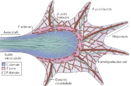
The growth cone consists of a dynamic subcellular structure located at the tip of axons that is highly sensitive and responsive to the extracellular environment. Growth cones are responsible for guiding axons to their targets by enabling axonal steering, extension as well as retraction (Lowery and Van Vactor, 2009). Its function requires the tight orchestration and crosstalk between different processes such as actin cytoskeleton dynamics, adhesion complex assembly and disassembly as well as membrane trafficking (Vitriol and Zheng, 2012). The growth cone has a complex architecture that can be subdivided into different domains based on its cytoskeletal components (Figure 1) (Lowery and Van Vactor, 2009). The peripheral domain (P) is the most dynamic and consists of filopodia which can be described as finger-like projections containing long bundles of actin filaments (F-actin) as well as lamellipodia, located between filopodia, consisting of mesh-like branched F-actin networks (Figure 1). Individual microtubules (MT) can also be seen within filipodia. The central (C) domain consists of stable microtubule bundles originating from the axon shaft as well as various vesicles organelles and F-actin bundles (Figure 1). Positioned at the interface between the central and peripheral domains is the transition zone where actomyosin contractile structures are formed and allow F-actin retrograde flow, a process required for axon elongation (Figure 1) (Lowery and Van Vactor, 2009).
19
Figure 1 The structural and spatial divisions in a typical growth cone
Diagram depicting the spatial subdivisions and structural components of a typical growth cone. (C) central domain, (T) the transition zone and (P) the peripheral domain. Reprinted by permission from Macmillan Publishers Ltd: Nature Publishing Group (Lowery and Van Vactor, 2009)
Growth cone motility and guidance depends largely upon actin dynamics. The spatiotemporal balance between actin polymerization in the P domain and myosin-based actin retrograde flow in the T domain is key in providing directionality to outgrowth (Vitriol and Zheng, 2012).
Rho family GTPases are key regulators of actin dynamics and link surface receptors to actin and microtubule organisation (Stankiewicz and Linseman, 2014). Rho GTPases cycle between their inactive GDP-bound and active GTP-bound states, their active states enable the activation of a variety of downstream regulators and effectors that act upon the actin cytoskeleton. The spatiotemporal regulation of Rho GTPases has been attributed to guanine nucleotide exchange factors (GEFs) and GTPase activating proteins (GAPs). While the GTP bound state of Rho GTPases is promoted by GEFs, the GDP bound state is promoted by (GAPs) (Stankiewicz and Linseman, 2014). The most extensively studied Rho GTPases members are RhoA,
20
Rac1 and Cdc42. Rac1/Cdc42 are typically attributed to growth cone development and axon extension while RhoA is typically attributed to axon retraction and growth cone collapse (Stankiewicz and Linseman, 2014). Nevertheless, evidence suggests that the activation balance of Rho GTPases rather than the activation state of a member is required for proper guidance (Barallobre et al., 2005).
3 Guidance cues
There are four classical families of guidance cues which comprise of the Netrins, ephrins, semaphorins and slits (Kolodkin and Tessier-Lavigne, 2011). Slits are secreted cues, ephrins are membrane bound and Netrins and semaphorins can be either membrane bound or secreted (Kolodkin and Tessier-Lavigne, 2011; Lai Wing Sun et al., 2011) . Whereas Netrins, ephrins and semaphorins can act as bidirectional cues, capable of eliciting both attraction and repulsion in a context dependent manner, slits have been solely associated with repulsion (Morales and Kania, 2016). More recently, a growing list of morphogens as well as growth factors and cell-adhesion molecules have also been implicated in axon guidance (Kolodkin and Tessier-Lavigne, 2011). Guidance molecules function in other neural functions such as cell migration and synaptogenesis as well as in non-neuronal tissues in processes such as angiogenesis, organogenesis, immune function as well as cancer biology (Hinck, 2004; Kolodkin and Tessier-Lavigne, 2011). The present work will focus on Netrins and ephrins for their relevance in the original work described in chapters III - Results.
21 4 Netrin-1
Netrins are a family of laminin-related extracellular proteins with homologues in all assayed organisms with bilateral symmetry (Lai Wing Sun et al., 2011). Mammals express three secreted netrins (Netrin-1-3) and two GPI-anchored netrins (Netrin-G1, Netrin-G2) (Lai Wing Sun et al., 2011). Netrins have been implicated in a variety of neural processes including cell and axon migration, axon arborisation,
synaptogenesis as well as oligodendrocyte development (Lai Wing Sun et al., 2011). Outside the nervous system, Netrins have been implicated in the development of the pancreas, lungs, mammary gland as well as in angiogenesis (Cirulli and Yebra,
2007). Netrin-1, the most extensively studied Netrin member, can illicit axon attraction or repulsion depending on the expression of Netrin-1 receptors by neurons
(Colamarino and Tessier-Lavigne, 1995; Hong et al., 1999; Lai Wing Sun et al., 2011; Poliak et al., 2015). The first ascribed function for Netrin-1 was the guidance of
commissural axons (Kennedy et al., 1994; Serafini et al., 1994; Serafini et al., 1996).
4.1 Netrin-1 in commissural axon guidance
Commissural neurons are responsible for connecting both sides of the CNS. In vertebrates, Netrin-1 was shown to be required for commissural neurons in the
developing brain and dorsal spinal cord to extend axons to an intermediate target, the ventral floor-plate (Kennedy et al., 1994; Serafini et al., 1994; Serafini et al., 1996). The guidance effect of Netrin-1 on commissural neurons was originally proposed to occur through long-range chemotaxis whereby secretion of Netrin-1 by floor plate
22
cells was proposed to establish a gradient of Netrin-1 ranging from high ventral to low dorsal. The long-range chemotaxis theory for Netrin-1 attraction was substantiated by
in vitro culture experiments showing dorsal commissural axons being attracted
towards floor-plate explants, positioned as far as ~250 μm away. This experiment suggested that Netrin-1 could act at a distance by diffusing away from its source cells (Tessier-Lavigne et al., 1988; Serafini et al., 1994). More recently, the requirement for long-range diffusion of Netrin-1 from the floor-plate has been challenged. Evidence in Drosophila shows that in both the midline and the visual system, a tethered Netrin is sufficient to rescue Netrin loss of function phenotypes (Brankatschk and Dickson, 2006; Timofeev et al., 2012). It was also demonstrated that the mechanical attachment of Netrin-1 to a substrate is required for axon outgrowth for mouse
commissural axons in vitro (Moore et al., 2012). Furthermore, biochemical analysis of Netrin-1 shows that it is tightly bound to the membrane fraction and can only be extracted at very high salt concentrations, making it unlikely to be able to diffuse to long-distance within tightly packed cells, at least in the context of passive diffusion (Serafini et al., 1994). Recently, two independent groups demonstrated that
progenitors at the ventricular zone, rather than cells from the floor plate, were the major source of Netrin-1 in guiding commissural axons (Dominici et al., 2017; Varadarajan et al., 2017). Both groups also highlight that Netrin-1 produced by progenitors at the ventricular zone is concentrated at the pial surface that lines the periphery of the spinal cord as well as on the progenitor radial processes encountered by commissural axons when projecting dorsally. Taken together, the evidence
short-23
range haptotactic cue rather than through long-range chemotaxis. It remains unresolved whether there is a requirement for the establishment of a Netrin-1
concentration gradient in the guidance of commissural axons in vertebrates, a mouse harbouring a mutation where Netrin-1 is tethered to the membrane may be useful in resolving this question.
4.2 Netrin-1 receptors
Netrin-1 receptors are members of the immunoglobulin superfamily (IgSF), proteins that contain domains homologous to immunoglobulins (Lai Wing Sun et al., 2011). There is an abundance of evidence implicating IgSF members in CNS
development, a particular subfamily, the IgSF CAMs (cell adhesion molecules) have over 50 members expressed in the mammalian nervous system (Gu et al., 2015). Through homophilic and heterophilic interactions with other CAMs as well as with other receptors and elements of the ECM (extracellular matrix), they influence cell-cell adhesions and regulate cell processes such as cell migration, axon extension, neurite branching, synaptogenesis and plasticity (Leshchyns'ka and Sytnyk, 2016). The receptors identified as eliciting attraction towards Netrin-1 in vertebrates are Dcc, Neogenin and DsCAM (Figure 3A) (Lai Wing Sun et al., 2011). The repulsive effects of Netrin-1 are mediated by members of the UNC5 family which include four members in mammals; UNC5A-D (Figure 3B) (Leonardo et al., 1997; Hong et al., 1999).
4.3 DsCAM
DsCAM (down syndrome cell adhesion molecule) can be alternatively spliced to over 19,000 isoforms. Binding of self-isoforms was shown to elicit repulsion
24
required for dendrite and axon self-avoidance (Schmucker et al., 2000; Wojtowicz et al., 2007; Hattori et al., 2009). Although there is evidence for DsCAM being implicated in axon attraction to Netrin-1 in Drosophila and in chick, complete loss of DsCAM in the mouse does not result in any commissural guidance defects in vivo, or alters commissural axon responses to Netrin-1 in vitro (Andrews et al., 2008; Ly et al., 2008; Liu et al., 2009; Palmesino et al., 2012). These discrepancies may reflect different requirements for DsCAM among species. It is also possible that the full knock-out of DsCAM in the mouse may bring about compensatory mechanisms not available in the context of chick DsCAM knockdown by shRNA.
4.4 Dcc
Unlike DsCAM, loss of Dcc dramatically alters commissural axon guidance, closely phenocopying mice lacking Netrin-1 (Fazeli et al., 1997). Because of defects in commissural axon guidance, human individuals with mutations in Dcc display mirror movements whereby contralateral involuntary movements mirror voluntary ones (Srour et al., 2010). Dcc (deleted in colorectal carcinoma) was named for its absence in most colorectal carcinomas and was proposed to be a putative tumor suppressor gene (Fearon et al., 1990). It’s role as a tumor suppressor has been substantiated by evidence of Dcc being a death receptor, in prolonged absence of Netrin-1, Dcc initiates a caspase cascade leading to apoptosis (Mehlen et al., 1998; Castets et al., 2011). It’s role as a dependence receptor is not ubiquitous, in mice completely lacking Netrin-1, no difference in apoptosis can be seen within the
25
developing CNS (Bin et al., 2015). It’s role as a dependence receptor likely requires specific adaptors as exemplified by DIP13α, proposed to modulate the pro-apoptotic effects of Dcc (Liu et al., 2002).
4.5 Neogenin
Closely related to Dcc is Neogenin, first identified in the chicken and named so for its high expression during neural differentiation (Vielmetter et al., 1994). Neogenin is a receptor for members of the repulsive guidance molecule (RGM) family which in vertebrates includes RGMa, RGMb and RGMc, as well as a receptor for Netrins (Keino-Masu et al., 1996; Rajagopalan et al., 2004; Siebold et al., 2017).
4.6 Neogenin as a receptor for RGMs
During mouse development, whereas RGMa and RGMb are expressed in and out of the CNS in a rather complementary fashion, RGMc expression is excluded from the CNS (Niederkofler et al., 2004; Oldekamp et al., 2004). RGMs are not only ligands for Neogenin but have also been shown to be BMP co-receptors (Babitt et al., 2005; Samad et al., 2005; Babitt et al., 2006). The binding of RGMa to Neogenin in
cis is proposed to enhance BMP downstream signaling (Healey et al., 2015).
BMP/RGM/Neogenin have been involved in a variety of biological processes such as iron homeostasis, bone formation and astrocyte differentiation (Babitt et al., 2006; Zhou et al., 2010; Huang et al., 2016). RGMc/Neogenin signalling is required for iron
26
homeostasis, both Neogenin and RGMc null mice suffer from hepatic iron overload, reduced hepcidin expression and defective BMP signaling (Lee et al., 2010).
In the chick optic tectum and the Xenopus forebrain, RGMa acts as a repulsive cue for Neogenin expressing axons (Monnier et al., 2002; Rajagopalan et al., 2004; Wilson and Key, 2006). RGMa induced growth cone collapse is independent of BMP signaling and involves RhoA, the Rho-associated kinase ROCK as well as PKC (Conrad et al., 2007). Furthermore, RGMa induced growth cone collapse was shown to require Unc5b as well as the GEF LARG (Hata et al., 2009). Unc5 proteins are involved in the repulsion from both Netrin-1 and RGMa by associating with Dcc and Neogenin respectively (Hong et al., 1999; Hata et al., 2009). The convergence of Netrin-1 and RGMa signalling pathways at Unc5 receptors suggests a possible enhancement of repulsive signaling where both cues overlap. In mammals, RGMa inhibits axon outgrowth following spinal cord injury, RGMa inhibition using a blocking antibody results in an increase in axon outgrowth and improves functional recovery (Hata et al., 2006). In contrast, RGMa was shown to have neuroprotective capabilities for retinal ganglion cells (RGCs), following optic nerve transection, the intraocular injection of RGMa reduces RGC death (Koeberle et al., 2010). RGMa/Neogenin were also shown to be required for neural tube closure in Xenopus and mice by regulating neuroepithelium morphology (Niederkofler et al., 2004; Kee et al., 2008).
Misexpression of RGMa in the developing chick hind-brain was shown to influence neural differentiation through an unknown mechanism (Matsunaga et al., 2006).
27
RGMb is the least studied member of the RGM family. There is evidence RGMb acting as both a tumor suppressor as well as promoting cancer progressing by regulating BMP signaling(Shi et al., 2015; Li et al., 2016). In the developing olfactory epithelium (OE), loss of RGMb and Neogenin both result in neural differentiation defects (Kam et al., 2016). The authors propose that progenitors expressing
Neogenin bind RGMb on neighbouring cells and that these interactions regulate cell cycle kinetics and exit (Kam et al., 2016).
Thus, RGM/Neogenin signaling is involved in a wide variety of processes during development and beyond. RGM/Neogenin signalling outside the CNS often involves the regulation of BMP signaling, which to my knowledge, has not been demonstrated within the CNS. Whereas Netrin-1 binds Neogenin on its fibronectin type III (FNIII) domains 4 and 5, RGMs bind Neogenin on the FNIII domains 5 and 6 (Figure 3) (Yang et al., 2008; Xu et al., 2014). In vitro, Netrin-1 inhibits RGMa
induced growth cone collapse of dorsal root ganglion neurons, suggesting that Netrin-1 and RGMa compete in binding Neogenin (Bell et al., 20Netrin-13). This provides evidence for the cross-regulation potential of Netrin-1 and RGMa signalling pathways.
4.7 Neogenin as a receptor for Netrin-1
In mice, whereas the loss of Neogenin alone seemingly does not result in spinal commissural guidance defects, the loss of both Neogenin and Dcc exacerbates the guidance phenotype of Dcc mutants to a degree comparable to Netrin-1 mutants. This suggests that both Dcc and Neogenin collaborate in guiding commissural axons
28
in the mouse (Xu et al., 2014). Neogenin has been shown to be implicated in adult neurogenesis in mice. Loss of Neogenin results in neuroblast migration defects that are presumed to result from a loss of Netrin-1 attraction as well as aberrant
differentiation resulting from impaired cell cycle kinetics (O'Leary et al., 2015). In the supraoptic tract of the developing Xenopus forebrain, knock down of either Neogenin or Netrin-1 results in similar guidance defects, suggesting they may be acting as a ligand/receptor pair in guiding supraoptic tract axons (Wilson and Key, 2006).
4.8 Chick Neogenin
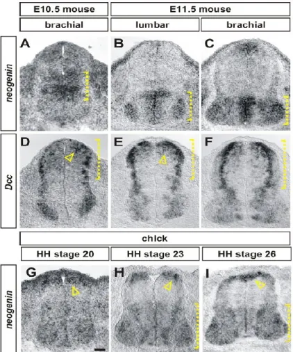
Whereas mammals express both Dcc and Neogenin, the chicken does not appear to have a Dcc gene and only expresses Neogenin (Phan et al., 2011). Within the developing chick spinal cord, expression of Neogenin closely resembles the combined expression of Dcc and Neogenin in the mouse spinal cord, suggesting that chick Neogenin may be capable of accounting for the functions of both mouse Dcc and Neogenin (Figure 2) (Phan et al., 2011). In the chick spinal cord, knockdown of Neogenin by in ovo electroporation of an shRNA construct directed against Neogenin results in commissural axon guidance defects reminiscent of the Netrin1 and Dcc knock-out mice (Phan et al., 2011).
29
(A–H) In situ hybridization experiments for Neogenin (A– C, G–I) and Dcc (D–F) on transverse sections of the spinal cord of E10.5 (A, D) and E11.5 (B, C, E, F) mouse embryos and HH stage 20 (G), 23 (E) and 26 (I) chicken embryos. (A–C) In mouse, Neogenin is expressed first in the intermediate spinal cord (bracket, A). By E11.5,
Neogenin is present at high
levels in the ventral
ventricular zone as well as broadly in motor neurons (brackets, B, C) (D–F) Mouse
Dcc is expressed at highest
levels in the dorsal-most neural progenitors
(arrowhead, D, E) and in post-mitotic neurons
(brackets, D–F) throughout the dorsal spinal cord as well as at lower levels in a broad population of motor neurons. (G–I) The distribution of chicken Neogenin is a
composite of the expression patterns of both mouse Dcc and Neogenin. At all stages, the highest levels of Neogenin expression is in the dorsal-most spinal cord, in dorsal neural progenitors (arrowheads, G, I) and in a population of post-mitotic dorsal neurons whose position is consistent with their being commissural neurons
(arrowhead, H). Neogenin is also present at lower levels in both the ventral ventricular zone and in motor neurons (brackets, H, I). Scale bar: A, D, G: 30 mm, B, C, E, F, H, I: 40 mm. Taken from (Phan et al., 2011)
Figure 2 Chick Neogenin expression in the spinal cord closely reassembles the combined expression of Dcc and Neogenin in the mouse
30 4.9 Neogenin/Dcc structure
Dcc and Neogenin form a subfamily of IgSFs and share the same overall structure with their extracellular portions containing four immunoglobulin domains followed by six fibronectin type III repeats and three intracellular P domains (P1-3) (Figure 3) (Keino-Masu et al., 1996). Whereas the expression of Dcc is mostly restricted to the CNS, Neogenin is highly expressed in the CNS and in mesoderm derived tissue during development as well as in adulthood (Meyerhardt et al., 1997). Netrin-1 has two binding sites for Dcc and Neogenin and both receptors have 2 binding sites for Netrin-1 within their FNIII4 and FNIII5 domains (Figure 3A)
(Meyerhardt et al., 1997; Xu et al., 2014). The crystal structure of Netrin-1 bound to Dcc and Neogenin reveals that two distinct architectures are possible depending on splice variants, 2:2 heteromers or a continuous ligand/receptor assembly (Figure 3C, D) (Xu et al., 2014). The Netrin-1 induced multimerization of Dcc results in the
association of Dcc intracellular P3 domains and is required for Netrin-1 induced attraction (Stein et al., 2001).
31 Figure 3 Netrin-1 signaling
The vertebrate transmembrane receptors Dcc and Neogenin share the same overall structure consisting of four immunoglobulin domains (Ig), followed by six fibronectin type III (FN1-6) repeats and three intracellular P domains (P1-3). Both receptors have two Netrin-1 (yellow) binding sites within their FN4 and FN5 domains. A. The
expression of Dcc and Neogenin in growth cones results in Netrin-1 induced axon attraction and growth promotion. B. Repulsion from Netrin-1 relies on the binding of Netrin-1 to heteromers of Dcc (Neo) and members of the Unc5 family and may also be possible through Unc5s alone. C. Simplified diagram of Netrin-1/Dcc downstream signaling. The binding of Netrin-1 to Dcc induces the phosphorylation (red dots) of the P3 domain leading to receptor multimerization and phosphorylation of kinases such as, FAK and SFKs and association with adaptor proteins such as Nck-1. Activation of SFKs leads to the activation of GEFs which regulate Rho GTPases resulting in
cytoskeletal rearrangements. D, E. Diagrams depicting the two distinct Netrin-1/Dcc and Netin-1/Neogenin signaling assemblies resulting form differences in linker lengths between FN4 and FN5 domains among Dcc and Neogenin splice variants. D. The short isoforms of either Dcc or chNeogenin allow for a continuous assembly of Netrin-1/Dcc (chNeogenin). E. The long isoforms of Dcc and chick or mouse Neogenin results in 2:2 heteromers of Netrin-1/Dcc (Neogenin). A-C were adapted from (Lai Wing Sun et al., 2011), D and E were adapted from (Xu et al., 2014)).
32 5 Netrin-1 signaling
Netrin-1 dependent signaling has been extensively studied, mostly focusing on Dcc as its receptor. A thorough review of the literature investigating Netrin-1
downstream signaling events would exceed the scope of this thesis, nevertheless, here are some of the key findings. Stimulation with Netrin-1 induces the tyrosine phosphorylation of Dcc in its P3 domain resulting in receptor multimerization (Figure 3C) (Meriane et al., 2004; Lai Wing Sun et al., 2011; Xu et al., 2014). In rodent neuroblastoma cells (NG108-15), ectopic expression of Dcc induces a Netrin-1 dependent increase in surface area and filipodia number. These effects were shown to require the activation of the Rho GTPases Cdc42 and Rac1 (Shekarabi and Kennedy, 2002). Two Rho GEFs, Trio and Dock180, have been implicated in mediating Rac1 activation downstream of Netrin-1/Dcc (Briancon-Marjollet et al., 2008; Li et al., 2008). Furthermore, through a Dcc dependent mechanism, Netrin-1 inhibits RhoA which in turn increases the levels of Dcc at the plasma membrane, thereby sensitizing rat commissural neurons to Netrin-1 outgrowth and guidance in
vitro (Moore et al., 2008). The tyrosine kinase adaptor protein Nck-1 binds directly to
Dcc in rat commissural neurons, the expression of a dominant negative Nck-1 inhibits Dcc’s ability to induce Netrin-1 dependent neurite outgrowth and Rac1 activation in N1E-115 cells and fibroblasts respectively (Li et al., 2002).
Three independent groups provided evidence that focal adhesion kinase (FAK), a cytosolic protein tyrosine kinase, binds to Dcc and is phosphorylated upon Netrin-1 treatment and is required for Netrin-1 dependent outgrowth and attraction (Li
33
et al., 2004; Liu et al., 2004; Ren et al., 2004). FAK has also been implicated in signaling downstream of Neogenin in the context of growth cone collapse and
myogenesis (Bae et al., 2009; Endo and Yamashita, 2009). FAK itself is regulated by phosphorylation of multiple tyrosine residues in and outside the nervous system, it is implicated in establishing cell morphology, maturation of adhesive structures and migration (Chacon and Fazzari, 2011). FAK has been implicated in signaling
downstream of semaphorins, ephrins as well as Netrins and is involved in both axon attraction and repulsion (Chacon and Fazzari, 2011). Furthermore, FAK has been shown to interact with both activators and inhibitors of Rho GTPases and can
phosphorylate as well as be phosphorylated by Src family kinases (SFK) (Mitra et al., 2005).
SFKs have been implicated in multiple aspects of neural development including differentiation, axon outgrowth, fasciculation and guidance (Morse et al., 1998; Hoffman-Kim et al., 2002; Kuo et al., 2005; Robles et al., 2005; Kao et al., 2009). Members of SFKs bind Dcc and are phosphorylated upon Netrin-1 stimulation, and SFK function is required for Netrin-1 dependent commissural axon outgrowth and guidance in vitro (Li et al., 2004; Meriane et al., 2004).
To summarize, Netrin-1 binding to Dcc/Neogenin induces the tyrosine
phosphorylation within their P3 domains and receptor multimerization. These events enable the activation of adaptor proteins such as Nck-1 and signaling kinases such as FAK and SFKs. Association with FAK promotes traction to the extracellular
34
The spatiotemporal regulation of GEF activation in turn regulates Rho GTPases and the downstream cytoskeletal remodeling behind growth cone turning and axon outgrowth (Figure 3C).
6 Modulators of Netrin-1 signaling
6.1 Heparin sulfate proteoglycans
Heparin sulfate proteoglycans (HSPG) are polysaccharides of the
glycosaminoglycan family and have been shown to bind to hundreds of proteins (Esko and Lindahl, 2001). HSPGs consist of core proteins mainly consisting of syndecans, glypicans, perlecan and agrin, on to which are attached highly charged heparin sulfate side chains (Lee and Chien, 2004). HSPGs can regulate the
distribution of growth factors as well as enhance the binding of several ligands implicated in axon guidance (Lee and Chien, 2004). Netrin-1 binds heparin with high affinity and its extraction from the heparin fraction during chromatography requires high salt concentrations. (Serafini et al., 1994). Dcc also interacts with heparin via its fifth fibronectin domain, a domain also implicated in binding Netrin-1(Bennett et al., 1997; Xu et al., 2014). Spinal commissural neurons have a cell-autonomous
requirement for heparin sulfate in vivo and in vitro. Dorsal spinal neurons that are genetically deficient for Heparin sulfate have commissural guidance defects comparable to Netrin-1-/- and Dcc-/- mice in vivo and are unresponsive to Netrin-1
dependent outgrowth in vitro (Matsumoto et al., 2007). It has been suggested that heparin sulfate could act as a coreceptor for Dcc in binding Netrin-1, therefore the
35
expression and distribution of HSPGs may play a vital role in modulating the strength of Netrin-1 signaling by promoting Dcc/Netrin-1 interactions (Matsumoto et al., 2007).
6.2 Slit/Robo
Once spinal commissural neurons reach the floor-plate, they continue extending contralaterally, away from the floor-plate, before turning rostrally and extend along the rostral-caudal axis of the spinal cord. Post-crossing neurons
expressing the receptors Robo1/2 and Neuropilin-2, are repelled from the midline by Slit and Semaphorin3B respectively, expressed by the floor-plate (Zou et al., 2000; Long et al., 2004). To avoid stalling at the midline and recrossing the floor plate, commissural axons must become desensitized to Netrin-1 still expressed at the midline (Stein and Tessier-Lavigne, 2001). Xenopus spinal axons in culture are attracted towards a Netrin-1 gradient, when Slit is added to the Netrin-1 gradient, axons become unresponsive to Netrin-1 attraction while retaining the axon outgrowth promoting effect of Netrin-1 (Stein and Tessier-Lavigne, 2001). Furthermore,
Xenopus spinal neurons exogenously expressing a truncated Robo receptor, without its intracellular tail, remain attracted towards Netrin-1 in the presence of Slit whereas axons expressing full-length Robo remain unresponsive (Stein and Tessier-Lavigne, 2001) These result suggest that Slit binding to Robo somehow desensitizes axons to Netrin-1 attraction. Experiments where Xenopus axons express chimeric Robo and Dcc receptors, revealed that the cytoplasmic tails of Robo and Dcc were required in Slit dependent desensitization to Netrin-1. Furthermore, co-immunoprecipitation experiments show that Dcc Robo interaction is Slit dependent. Together, these in
36
vitro results suggest that spinal commissural axons expressing Robo receptors
become insensitive to Netrin-1 attraction when they encounter Slit at the floor-plate (Stein and Tessier-Lavigne, 2001).
In vitro, thalamocortical axons (TCAs) are repelled by Slit1 and are
unresponsive to Netrin-1 (Bielle et al., 2011). For a subset of TCAs, the rostral TCAs, Slit1 and Netrin-1 provided in combination results in attraction towards Netrin-1, even when Slit1 is homogeneously distributed (Bielle et al., 2011; Dupin et al., 2015). Slit-1 induced attraction to Netrin-1 is PKA dependent and proposed to result from an increase in Dcc surface levels (Leyva-Diaz et al., 2014). The permissive effect of Slit1 on rostral TCA attraction to Netrin-1 requires sub-threshold concentrations of Slit1, insufficient for inducing repulsion (Dupin et al., 2015). This effect requires the expression of the Slit1 receptor Robo1 and its coreceptor FLRT3 by rostral TCAs (Leyva-Diaz et al., 2014).
FLRT proteins can promote cell adhesion through homophilic interactions and promote repulsion by binding Unc5 proteins in trans (Karaulanov et al., 2009;
Seiradake et al., 2014). They have also been implicated in FGF signaling and synaptogenesis as well as neural migration. (Bottcher et al., 2004; O'Sullivan et al., 2012; Del Toro et al., 2017). Slit1/Robo interactions silence Netrin-1 attraction in commissural axons and allow Netrin-1 attraction in TCAs. The mechanism behind these opposite responses is currently unknown but may by in part due to differences in the expression of coreceptors such as FLRT.
37 6.3 Draxin
Draxin is a secreted protein recently identified as a guidance cue that shares no homology with any other guidance cue families (Islam et al., 2009). Draxin-/- mice
display a mild fasciculation defects in spinal commissural axons and severe forebrain commissural axon guidance defects comparable to Dcc-/- mice (Fazeli et al., 1997;
Islam et al., 2009). Draxin was shown to bind to the Netrin-1 receptors Dcc,
Neogenin, DSCAM and Unc5A-C as well as to Netrin-1 (Ahmed et al., 2011; Gao et al., 2015). It is also noteworthy that Draxin and Netrin-1 have distinct binding domains on Dcc (Ahmed et al., 2011) In vitro, depending on the concentration and neuronal type, Draxin can either stimulate axonal outgrowth or inhibit outgrowth and induce growth cone collapse (Islam et al., 2009; Ahmed et al., 2011; Shinmyo et al., 2015).
Draxin-/- mice also have thalamocortical projection defects and these defects are
comparable in severity to the defects seen in (Draxin+/- ; Dcc-/- ), (Draxin+/- ; Neo1GT/G), or (Dcc-/- ; Neo1GT/G), suggesting that Draxin genetically interacts with Dcc and
Neogenin in the context of axon guidance (Shinmyo et al., 2015). The role of Draxin in thalamocortical projections occurs independently from Netrin-1 since Netrin-1-/- mice don’t have thalamocortical projections defects (Shinmyo et al., 2015). On the other hand, the similar forebrain commissure phenotype seen in Netrin-1-/-, Dcc-/-, and Draxin-/- mice raises the possibility that Draxin, by binding to both Netrin-1 and Dcc, may enhance Netrin-1/Dcc interactions (Ahmed et al., 2011; Gao et al., 2015)
38 6.4 Dcc/Neogenin cleavage
Dcc immunoprecipitations done on supernatant collected from cell cultures overexpressing Dcc reveals that Dcc’s ectodomain is cleaved near the
transmembrane domain (Galko and Tessier-Lavigne, 2000). Furthermore, it was shown that the in vitro ectodomain cleavage of Dcc can be blocked by a broad spectrum metalloprotease inhibitor (Galko and Tessier-Lavigne, 2000). The in vitro treatment of dorsal spinal explants with metalloprotease inhibitors results in an increase in Dcc expression and a potentiation towards Netrin-1 dependent axonal outgrowth (Galko and Tessier-Lavigne, 2000). It was also demonstrated that Netrin-1 treatment increases the ectodomain cleavage of Dcc (Bai et al., 2011).
The ectodomain of Neogenin can be cleaved by the protease ADAM17 and was proposed to modulate RGMa induced repulsion by regulating surface Neogenin (Okamura et al., 2011). Lrig2, a transmembrane protein that binds Neogenin, can regulate ADAM17 proteolysis of Neogenin (van Erp et al., 2015). The binding of RGMa to Neogenin inhibits Lrig2/Neogenin interactions and allows ADAM17 cleavage of Neogenin. In vitro, Lrig2 is required for the RGMa dependent RhoA activation and growth cone collapse. In vivo, knockdown of Ligr2 results in the promotion of optic nerve outgrowth following experimental optic nerve crush (van Erp et al., 2015). Considering the homology between Dcc and Neogenin, as well as evidence of Dcc cleavage by metalloproteases, it is likely that Dcc may also undergo ectodomain cleavage by ADAM17 (Galko and Tessier-Lavigne, 2000).
39
Several transmembrane proteins such as E and N-Cadherin, Notch and amyloid precursor protein (APP), have their ectodomains cleaved, which
subsequently leads to the cleavage of their remaining intracellular fragments by γ-secretase (De Strooper et al., 1998; Naruse et al., 1998; De Strooper et al., 1999; Struhl and Greenwald, 1999; Marambaud et al., 2002). Dcc and Neogenin both undergo cleavage by γ-secretase, resulting in the release of their intracellular domains (ICD) (Taniguchi et al., 2003; Goldschneider et al., 2008).The ICDs of Dcc and Neogenin are capable of translocating to the nucleus and have been suggested to act as transcription regulators (Taniguchi et al., 2003; Goldschneider et al., 2008).
In Drosophila, the ICD of the Dcc/Neogenin homologue Frazzled, acts as a transcriptional activator of commissurless, an antagonist to Slit/Robo midline
repulsion (Neuhaus-Follini and Bashaw, 2015). The ICD of Neogenin interacts directly with the transcription coactivator LMO4 (LIM-only protein 4) and can translocate to the nucleus where it can presumably regulate transcription (Goldschneider et al., 2008; Banerjee et al., 2016). RGMa induced outgrowth inhibition of RGC axons is lost when LMO4 is knocked down. This suggests that NeoICD/LMO4 may in part regulate repulsion from RGMa by regulating transcription (Banerjee et al., 2016)
Presenilin-1 (PS1) is an essential catalytic component of the γ-secretase complex, PS1-/- mice have a subset of spinal commissural axons that fail to cross the floor-plate, accompanied by spinal motor neurons that aberrantly extend axons to and across the midline (Bai et al., 2011). In early post-mitotic motor neurons, the
40
coexpression of Slit and Robo by motor neurons is proposed to silence motor neuron axon attraction to Netrin-1 at the midline. In Robo1/2-/- mice, a subset of motor neuron axons aberrantly cross the floor-plate as in PS1-/- mice (Bai et al., 2011).
Co-immunoprecipitation experiments show that Robo1 interacts with full-length but not to a truncated Dcc, lacking its extracellular fragment, referred to as Dcc stub (Bai et al., 2011) In PS1-/- mice, the accumulation of Dcc stubs that bind to full-length Dcc at the membrane is thought to inhibit Robo/Dcc interaction, thereby enabling Netrin/Dcc signaling and attraction of motor neuron axons to the midline (Bai et al., 2011)
Thus, the strength and outcome of Netrin-1 signaling can be modulated by cell extrinsic factors, such as the presence of Draxin and Slit in the extracellular
environment, as well as cell intrinsic factors, such as the expression of HSPGs, Robo1, FLRT3 and members of the Unc5 protein family. Netrin-1 signaling may also be modulated by the availability of downstream effectors such as Src, FAK, GEFs and GAPs. Exposure to guidance cues that share common signaling components could potentially attenuate Netrin-1 signaling by sequestering common downstream effectors. Conversely, exposure to other cues could potentiate Netrin-1 signaling by bringing downstream effectors in proximity to Netrin-1 signaling complexes.
7 An introduction to Eph/ephrin signaling
Eph receptors are a subfamily of the receptor tyrosine kinases (RTKs) that include 14 members sub classified into two groups, A and B, based on their general preference for binding either GPI anchored ligands (ephrin-As) or transmembrane
41
ligands (ephrin-Bs) (Gale et al., 1996; Kania and Klein, 2016). Exceptions to these classifications are EphB2 which can bind ephrin-A5 and EphA4, capable of binding ephrin-Bs (Gale et al., 1996; Himanen et al., 2004). Eph/ephrin signaling has vast implications in neural and non-neural tissues during development and adulthood as well as in cancer and neurodegenerative diseases (Kania and Klein, 2016)
A particularity of these receptor ligand pairs is that ephrin ligands also act as receptors for Ephs and that Eph/ephrin interactions can elicit downstream signaling cascades in the cells expressing ephrins (Klein, 2004). Forward signaling describes signaling occurring downstream of an Eph subsequent to ephrin binding whereas reverse signaling describes the reciprocal situation. Efficient signaling requires clustering of Ephs and ephrins and activation induces receptor multimerization (Gale et al., 1996; Taylor et al., 2017).
Both forward and reverse signaling involves the phosphorylation of conserved tyrosine residues and the activation of kinases such as SFKs and the modulation of Rho GTPases through GEFs and GAPs (Klein, 2004). Eph/ephrin forward and reverse signaling have been shown to elicit the cleavage of ephrins and Eph by members of the A disintegrin and metalloproteinase (ADAM) and by matrix
metalloproteinases (MMPs) respectively (Hattori et al., 2000; Janes et al., 2005; Lin et al., 2008). Eph/ephrin signaling also leads to the trans-endocytosis of Eph/ephrin complexes in both forward and reverse signaling (Zimmer et al., 2003). Eph/ephrin endocytosis regulates cell-cell detachment and in some contexts, is required to terminate signaling (Marston et al., 2003; Zimmer et al., 2003; Janes et al., 2009).
42
Although most Eph/ephrin interactions result in cellular segregation, axon repulsion and growth cone collapse, there are several Eph/ephrin signaling events resulting in cell adhesion, axon outgrowth and arborisation (Castellani et al., 1998; Holmberg et al., 2000; Kania and Klein, 2016)
Ephs and ephrins are membrane tethered, therefore, their actions are thought to require cell-cell interactions (Kania and Klein, 2016). Recently, a novel Eph/ephrin signaling mechanism was described whereby cells secreting extracellular vesicles containing EphB2 illicit Eph/ephrin reverse signaling in nearby cells expressing
ephrins. This raises the possibility that Eph/ephrin signalling may occur at a distance, without the need for direct cell-cell contact (Gong et al., 2016).
7.1 Eph/ephrin signaling in axon guidance
Ephs/ephrins are the prototypical example of proteins regulating topographic projections. Many axonal projections form topographic maps whereby the spatial relation among neuron cell bodies is maintained at their axonal targets. Topographic projections enable the efficient processing and integration of spatial relations.
Topographic projections are exemplified by the projections of RGC axons from the retina to the optic tectum and the superior colliculus in the chick and mouse
respectively (Triplett, 2014). Eph/ephrin signaling plays a crucial role in establishing topography in this system. To briefly summarize, RGCs in the retina display a graded expression of EphAs and EphBs, while the optic tectum and superior colliculus
display graded expressions of ephrin-As and ephrin-Bs. These reciprocal expression patterns contribute in RGCs maintaining their relative positions at their target sites
43
and involves both forward and reverse signaling (Frisen et al., 1998; Thakar et al., 2011; McLaughlin et al., 2014).
Eph/ephrin signaling has also been implicated in establishing the axonal projections required for binocular vision. Whereas most RGCs project contralaterally across the midline at a region where axons from both eyes converge, termed the optic chiasm, a sub-population of RGCs project ipsilaterally in animals endowed with binocular vision (Petros et al., 2008). The expression of EphBs by a subset RGCs allows projecting axons to be repelled by ephrin-Bs at the optic chiasm thereby
inhibiting midline crossing and allowing these axons to continue projecting ipsilaterally (Williams et al., 2003).
Like Netrin-1, ephrins have also been implicated in regulating axonal midline crossing in the spinal cord. The brain and spinal cord are connected via ascending and descending spinal tracts. Loss of EphA4 and similarly, loss of ephrin-B3, results in the aberrant crossing of the midline of both ascending and descending axonal tracts (Paixao et al., 2013). The expression of EphA4 on ascending and descending axons allows them to be repelled from ephrin-B3 expressed at the midline (Paixao et al., 2013). Functionally, EphA4 KO mice display a kangaroo-like hopping gait (Dottori et al., 1998). Similarly to guidance defects described for ascending and descending axonal tracts, the hopping phenotype was shown to originate from the aberrant midline crossing of excitatory spinal neurons, no longer repelled by ephrin-B3 expressed at the midline (Kullander et al., 2003; Borgius et al., 2014). The
44
involvement of Eph/ephrins in LMC axon guidance will be discussed in detail in the sections 10.1-10.3.
8. Spinal motor neuron development
Spinal motor neurons (SpMNs) are situated in the ventral portion of the spinal cord and make synaptic connections with muscle fibers in the periphery. The
orchestrated activity of spinal motor neurons is behind the muscle contractions required for movement. SpMNs are cholinergic and receive inputs from motor neurons situated in the cortex, sensory neurons as well as by spinal interneurons (Stifani, 2014). SpMNs subtypes are arranged in discrete columns along the rostral/caudal axis of the spinal cord and are positioned vis-à-vis their peripheral targets (Bonanomi and Pfaff, 2010). Medial motor column (MMC) neurons span the entire spinal cord and innervate axial muscles. Hypaxial motor column (HMC) neurons innervate body wall muscles. Preganglionic motor column (PGC) neurons innervate sympathetic ganglia and are mainly restricted to the thoracic spinal cord. Lateral motor column (LMC) neurons innervate the forelimbs and hindlimbs and are positioned at the brachial and lumbar levels respectively (Figure 4 A, B) (Bonanomi and Pfaff, 2010).
During development, the neural tube, derived from endoderm, defines the structure that eventually gives rise to the spinal cord. The neural tube is populated by neural progenitors that differentiate into the diverse neurons that make up the spinal cord (Jessell, 2000). The mechanisms by which early neural subtype specification is
45
achieved have been studied in great detail (Jessell, 2000; Briscoe and Small, 2015). Within the neural tube, members of the wingless-type MMTV integration site family (WNT) and of the bone morphogenic protein family (BMPs) as well as some of their regulators, are expressed in a decreasing dorsal to ventral gradient. This is
accompanied by a decreasing ventral to dorsal gradient of the morphogen sonic hedgehog (shh) expressed by the notochord and neural tube floor plate (Jessell, 2000). Along with retinoic acid (RA) provided by the paraxial mesoderm surrounding the neural tube, exposure to variable concentrations of morphogens results in the differential expression of homeodomain transcription factors by progenitor domains along the dorso-ventral axis of the neural tube (Jessell, 2000). The class identity of post-mitotic neurons is determined by the specific expression of homeodomain transcription factors expressed by the progenitors from which they derive (Jessell, 2000). All SpMNs originate from the same progenitor domain (pMN) defined by the expression of a combination of transcriptions factors including NKX6.1, PAX6, Olig2 and Hb9 (Figure 4C, D) (Briscoe and Ericson, 2001; Briscoe and Small, 2015).
Expression and cross-repression of homeobox (Hox) proteins along the rostral/caudal axis have been implicated in defining the columnar extent of motor neuron subtypes. For example, Hox6 paralogs define the rostral/caudal extent of the brachial LMC, Hox9 paralogs define the PGC at thoracic levels while Hox10 paralogs define the extent of lumbar LMC (Dasen et al., 2003; Bonanomi and Pfaff, 2010).
` Neural progenitors lining the ventricular zone extend radial processes that terminate at the outer limits of the neural tube. At the apical surface of the ventricular
46
zone, neural progenitors remain adhered through N-Cadherin containing adherens junctions while their end-feet remain adhered to the periphery through integrin-laminin interactions (Meng and Takeichi, 2009; Rousso et al., 2012). Once they exit the cell cycle, new daughter neurons detach from the neuroepithelium at the ventricular zone and differentiate while migrating away perpendicularly from ventricular zone.
The expression of Hox genes leads to the expression of accessory factors by pMNs such as the forkhead transcription factors Foxp2 and Foxp4 (Rousso et al., 2012). In the chick neural tube, premature expression of Foxp2/4 leads to early detachment and differentiation of pMNs, while silencing Foxp2/4 results in a
differentiation delay (Rousso et al., 2012). The Foxp2/4 induced detachment of pMNs from the neuroepithelium was shown to occur via the downregulation of N-Cadherin at the ventricular zone through direct binding of Foxp4 to a regulatory region of the N-Cadherin gene (Rousso et al., 2012). N-N-Cadherin, a homophilic adhesion molecule promoting cell-cell adhesion, was also shown to be downregulated as neural
progenitors delaminate from the ventricular zone in the cortex (Zhang et al., 2010) As LMC neurons settle into the most ventrolateral portion of the neural tube, they begin expressing Foxp1 (Figure 4C) (Dasen et al., 2008; Rousso et al., 2008). Although LMC neurons maintain a generic motor neuron identity in Foxp1 KO mutants, their axonal projections within the developing limbs appears randomized (Dasen et al., 2008)
47
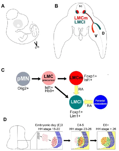
Figure 4 Early development of spinal motor neurons of the LMC
A. Depiction of an E5 chick embryo, the dashed line at hindlimb level indicates the location of the embryo cross-section depicted in (B). B. LMCm and LMCl neurons reside in the ventral spinal cord (SC) at limb-level with LMCm neurons occupying the medial LMC and projecting axons to the ventral limb and LMCl neurons occupying the lateral LMC and projecting axons to the dorsal limb. C, D. LMC neurons arise form the pMN progenitor domain at the ventricular zone that express the transcription factor Olig2. When pMN cells exit the cell cycle, they express HB9 and Isl1 while migrating laterally. Differentiated LMCm neurons occupying the LMC express Foxp1 and Isl1and synthesize retinoic acid (RA, yellow). Later born prospective LMCl neurons express Isl1 and HB9 as they exit the cell cycle. They extinguish Isl1 and begin expressing Lim1 as they migrate in proximity to LMCm neurons. The switch from Isl1 to Lim1 expression is induced and maintained RA provided at first by LMCm neurons and later by all LMC neurons as well as by the paraxial mesoderm adjacent to the ventral spinal cord.
48 8.1 LMC neuron differentiation
LMC neurons differentiate into two subtypes, neurons occupying the medial portion of the LMC (LMCm), project axons to the ventral portion of developing limbs, neurons occupying the lateral portion of the LMC (LMCl), project axons into the dorsal part of the developing limb (Figure 4B) (Landmesser, 1978b). LMCm neurons are generated first, the later born LMCl neurons migrate past LMCm neurons and settle at the most ventrolateral position of the neural tube, adjacent to LMCm neurons (Figure 4C, D) (Hollyday and Hamburger, 1977). LMC subtype specification involves the differential expression of LIM homeodomain transcription factors. LMCm and LMCl neurons express Isl1 and Isl2 as they exit the cell cycle. While LMCm neurons retain the expression of Isl1/2, LMCl neurons replace expression of Isl1 with Lim1 as LMCl neurons migrate past LMCm neurons (Figure 4C, D) (Tsuchida et al., 1994). Retinoic acid signaling provided by LMCm neurons as well as by the paraxial mesoderm adjacent to the neural tube is necessary for LMCl neurons to switch from Isl1 to Lim1 expression (Figure 4C, D) (Sockanathan and Jessell, 1998; Ji et al., 2006). Lim1 and Isl1 are cross repressive, overexpression of either in chick LMC neurons leads to the loss of the other (Kania and Jessell, 2003). Lim1 and Isl1 expression contributes to the settling positions of LMC neurons as evident by the medialization and
lateralization of LMC neurons overexpressing Isl1 and Lim1 respectively (Kania and Jessell, 2003).
Later in development, LMC neurons become subdivided into motor pools with each pool innervating a specific muscle (Landmesser, 2001). Muscle innervation