Université de Montréal
The Role of Cardiotrophin-Like Cytokine Factor 1
on the Development of Atherosclerosis
par
Inna Verlan
Département de pharmacologie et physiologie Université de Montréal
Faculté de médecine
Mémoire présenté
en vue de l’obtention du grade de Maîtrise (M.Sc.) En
Pharmacologie
Option Pharmacologie Moléculaire
Octobre, 2017
Université de Montréal
Faculté des études supérieures et postdoctorales
Ce mémoire intitulé
The Role of Cardiotrophin-Like Cytokine Factor 1
on the Development of Atherosclerosis
Présenté par
Inna Verlan
a été évalué par un jury composé des personnes suivantes :
Dr Guy Rousseau
,
Président-rapporteurDr Martin Sirois
,
Membre de juryDr Jean-François Gauchat
,
Directeur de rechercheDr Gaétan Mayer
,
Co-directeur de rechercheRésumé
Le syndrome métabolique représente un problème majeur de la santé publique mondiale ; le taux augmente constamment en dépit de la technologie médicale innovante et des avancées thérapeutiques. Les maladies cardiovasculaires associées à la dyslipidémie demeurent la principale cause de décès et de morbidité à l'échelle mondiale en dépit d'une vaste recherche médicale et d'un large éventail de médicaments ciblant l'athérosclérose, l'obésité, le diabète, et autres. Des recherches récentes suggèrent l'existence et la persistance d'une inflammation de
faible intensité dans la pathogenèse des pathologies comme l'athérosclérose et l'obésité. La « symbiose » entre le système métabolique et le système immunitaire est substantielle et
toute perturbation contribue au développement des conditions métaboliques altérées qui aboutissent finalement à des troubles tels que l'obésité et l'athérosclérose.
L'objectif général de ma maîtrise était de caractériser l'effet de la cardiotrophin-like
cytokine factor 1 sur le développement de l'athérosclérose et de valider le dérivé de cardiotrophin-like cytokine factor 1 couplé avec le fragment Fc de l’immunoglobuline G qui
possède une longue durée de vie.
Cardiotrophin-like cytokine factor 1 est une cytokine du système immunitaire avec
activité immunorégulatrice. Cardiotrophin-like cytokine factor 1 appartient à la famille d’interleukine 6. Cardiotrophin-like cytokine factor 1 est efficacement sécrétée en présence de
cytokine receptor like factor 1, un récepteur soluble de la cytokine. Cardiotrophin-like cytokine factor 1 possède des activités neurotrophiques médiées par le récepteur du ciliary neurotrophic factor. Cardiotrophin-like cytokine factor 1 est également un ligand à haute affinité pour la
sortiline. Des variantes du gène Sort1 codant pour ce récepteur ont été associés à l’hyperlipidémie et au risque d’infarctus du myocarde dans plusieurs études d’associations génomiques. Il a été observé que la cardiotrophin-like cytokine factor 1 lie et active les transfectants co-expriment de la sortiline et récepteur du leukemia inhibitory factor. Les deux récepteurs sont exprimés par les cellules myéloïdes et le récepteur du leukemia inhibitory factor est un puissant inducteur de la polarisation des macrophages de type M2 (anti-inflammatoire).
L'objectif général du projet était d'étudier si la cardiotrophin-like cytokine factor 1 ou la
macrophages des plaques d’athéroscléroses et réduirait la formation de cellules spumeuses et le développement de la plaque. Nos travaux montrent que l'expression de cardiotrophin-like
cytokine factor 1 dans le modèle murin d’athérosclérose LDLR-/- ne diminue pas le
développement de la plaque. Cependant, certains résultats ont révélé une contribution significative de la cardiotrophin-like cytokine factor 1 dans le gain de masse corporelle sans modification de l'apport calorique.
Mots-clés : système immunitaire, cardiotrophin-like cytokine factor 1, sortiline, syndrome
Abstract
Metabolic syndrome represents a major global health problem. Its rate is constantly increasing. Cardiovascular diseases emerging from dyslipidemia conditions are a worldwide leading cause of death and morbidity, despite extensive medical research and wide range of drugs targeting atherosclerosis, obesity, diabetes etc. Recent findings suggest the existence and persistence of low-grade inflammation in pathogenesis atherosclerosis and obesity. The “symbiosis” between metabolic and immune system is substantial and any perturbation contribute to the development of altered metabolic conditions that ultimately culminate in such disorders as obesity and atherosclerosis.
The overall goal of my Master internship was to characterise the effect of cardiotrophin-like cytokine factor 1 on development of atherosclerosis and validate a long half-life derivative of cardiotrophin-like cytokine factor 1 coupled with Fc fragment of immunolglobulin G.
Cardiotrophin-like cytokine factor 1 is a cytokine of the immune system with immunoregulatory activity. Cardiotrophin-like cytokine factor 1 belongs to the interleukin 6 family of monomeric cytokines. Cardiotrophin-like cytokine factor 1 is efficiently secreted in the presence of cytokine receptor like factor 1, a soluble cytokine receptor. Cardiotrophin-like cytokine 1 possesses neurotrophic activities mediated through the receptor of ciliary neurotrophic factor. Cardiotrophin-like cytokine factor 1 is a high affinity ligand for sortilin. Genome-wide association studies indicated that plasma low-density lipoprotein cholesterol levels and cardiovascular disease are associated with single nucleotide polymorphisms variants regulating sortilin expression. It was observed that cardiotrophin-like cytokine factor 1 binds and activates transfectants co-expressing sortilin and receptor of leukemia inhibitory factor. Both receptors are expressed by myeloid cells and leukemia inhibitory factor is a potent inducer of anti-inflammatory M2 macrophage differentiation.
The overall objective was to investigate if the interaction of cardiotrophin-like cytokine factor 1 or cardiotrophin-like cytokine factor 1 coupled with Fc fragment of immunoglobulin G with atherosclerotic plaque macrophages will reduce the foam cell formation and development of plaque. Our work shows that cardiotrophin-like cytokine 1 expression in mice on LDLR
revealed a significant contribution of cardiotrophin-like cytokine factor 1 in gain of body mass without changes in food intake.
Keywords: immune system, cardiotrophin-like cytokine factor 1, sortilin, metabolic syndrome,
Table of contents
Résumé ... iii
Abstract ... v
Table of contents ... vii
List of tables ... x List of figures ... x List of Abbreviations ... xi Acknowledgements ... xiii Chapter 1: Introduction ... 1 1.1 Immune system-generality ... 1
1.1.1 The innate immune system ... 1
1.1.2 The adaptive immune system... 2
1.1.3 Cytokines ... 3
1.2 Cardiotrophin-like cytokine factor 1-generality ... 3
1.2.1 Identification of cardiotrophin-like cytokine factor 1 ... 3
1.2.2 Cytokine structure ... 5 1.2.3 CLCF1 tissue expression ... 7 1.2.4 CLCF1 secretory pathway ... 7 1.2.5 CLCF1 receptors ... 8 1.2.5.1 CNTFR ... 8 1.2.5.2 Sortilin... 8 1.2.6 Signaling pathways ... 11
1.2.7 CLCF1 knock out and mutation ... 13
1.2.8 Biological activity ... 14
1.2.8.1 Immunomodulatory function ... 14
1.2.8.2 Neurotrophic function ... 14
1.3 Metabolic syndrome-generality ... 16
1.3.1 Link between immunity and metabolic syndrome ... 17
1.4 Atherosclerosis ... 18
1.4.1 Generality ... 18
1.4.2 Lipoprotein metabolism ... 21
1.4.2.1 Lipoproteins ... 21
1.4.2.2 Exogenous lipoprotein metabolism... 23
1.4.2.3 Endogenous lipoprotein metabolism... 24
1.4.2.4 LDLR -/- mice model ... 25
1.4.3 Monocyte fate in atherosclerosis ... 26
1.4.4 Macrophage and foam cell formation ... 26
1.4.5 Macrophage polarization and atherosclerosis ... 27
1.5 Hypothesis and objectives... 29
Chapter 2: Materials and Methods ... 31
2.1 In vitro assay ... 31
2.1.1 Cell culture of 3T3-L1 preadipocytes and standard differentiation into mature adipocytes ... 31
2.1.2 Foam cell formation and flow cytometry analysis ... 32
2.1.3 Biological activity of recombinant FcCLCF1... 32
2.2 In vivo assay ... 33
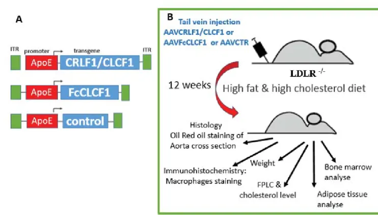
2.2.1 AAV vector construction and virus generation as performed in the lab of Dr. Gaetan Mayer ... 33
2.2.2 Animal treatment ... 36
2.2.3 Experimental setup... 37
2.2.3.1 Bone marrow derived macrophages and spleen cells isolation ... 38
2.2.3.2 Flow cytometry cell staining ... 38
2.2.3.3 Analysis of plasma ... 39
2.2.3.4 Atherosclerotic lesion analysis ... 39
2.2.3.5 Adipose tissue morphometry ... 40
2.2.3.7 Statistical Analysis ... 40
Chapter 3: Results ... 41
3.1 In vitro assay ... 41
3.1.1 Biological activity of recombinant FcCLCF1 protein ... 41
3.1.2 CLCF1 and foam cells formation ... 41
3.1.3 CLCF1 and adipocyte differentiation ... 42
3.2 In vivo assay ... 43
3.2.1 Validation of efficient liver transduction by adeno-associated virus serotype 8 (AAV8) ... 43
3.2.2 Body and organ weight ... 44
3.2.3 Quantification of atherosclerosis lesions ... 45
3.2.4 Effects of CLCF1 expression on plasma lipids... 46
3.2.5 Analysis of bone marrow and spleen cells population by flow cytometry ... 47
Chapter 4: Discussion ... 48
Chapter 5: Conclusion... 52
Chapter 6: Figures ... 54
List of tables
Table 1. Preparation of iodixanol gradient solutions. ... 35
List of figures
Figure 1. The secondary and tertiary structure of CLCF1. ... 6Figure 2. The structure of CNTFR and Sortilin receptor. ... 10
Figure 3. Main signaling pathways activated by CLCF1. ... 12
Figure 4. Role of inflammatory cells and immune responses in atherogenesis. ... 19
Figure 5. The composition and size of different lipoproteins. ... 23
Figure 6. Overview of AAV Plasmid System. ... 34
Figure 7. Animal experimental overview. ... 37
Figure 8. Proliferation assay of Ba/F3 CNTFR cells with increasing concentrations of cytokines. ... 54
Figure 9. CLCF1 and foam cell formation. ... 55
Figure 10. The assessment of cholesterol influx in cultured macrophages. ... 56
Figure 11. CLCF1 decrease adipocyte differentiation in vitro. ... 57
Figure 12. Validation of construct expression. ... 58
Figure 13. Effect of CLCF1 on diet-induced weight gain and body organs weight. ... 59
Figure 14. Effect of CLCF1 on visceral adiposity. ... 60
Figure 15. Effect of CLCF1 on subcutaneous adiposity. ... 61
Figure 16. Effect of CLCF1 on atherosclerotic lesion development. ... 62
Figure 17. Effect of CLCF1 on lipoprotein metabolism. ... 63
Figure 18. Effect of CRLFCLCF1 and FcCLCF1 expression on LDLR-/- mice spleen cells population measured by flow cytometry. ... 65
Figure 19. Effect of CRLFCLCF1 and FcCLCF1 expression on LDLR-/- mice bone marrow cells population measured by flow cytometry. ... 67
List of Abbreviations
APC: antigen-presenting cell; ApoA: apolipoprotein A; ApoB: apolipoprotein B; ApoC:
apolipoprotein C; ApoE: apolipoprotein E; ABCA(G): ATP-binding cassette(A), (G); ATM: adipose tissue macrophage; BMDM: bone marrow derived macrophage; DC: dendritic cells;
CNTF: ciliary neurotrophic factor; CNTFR: receptor of CNTF; CT-1: cardiotrophin-1; CLCF1: cardiotrophin-like cytokine factor 1; CRLF1: cytokine receptor like factor 1; CHD:
coronary heart disease; CVD: cardiovascular atherosclerotic diseases; C/EBPδ: CCAAT/enhancer-binding protein-δ; CD 36: cluster of differentiation 36; CE: cholesterol-ester; CETP: cholesterol ester transfer protein; FAS: fatty acid synthase; FA: fatty acid;
gp130: glycoprotein 130; AGE: advanced glycation end product; HFHC: high-fat, high-
cholesterol diet; Th: helper T lymphocyte; HDL: high-density lipoproteins; HL: hepatic lipase;
IFN: interferon; IL: interleukin; Ig: immunoglobulin; LCAT: lethicin-cholesterol
acyltransferase; LIF: leukemia inhibitory factor; LIFR: receptor of LIF; LDL: low-density lipoproteins; LPL: lipoprotein lipase; LRP: LDL receptor related protein; MHC: major histocompatibility complex; MPO: myeloperoxydase; MMP: metalloproteinase; MTP: microsomal triglyceride transfer protein; MetS: Metabolic syndrome; M-CSF: macrophage colony-stimulating; NPY: neuropeptide Y; NK: natural killer; NP: neuropoetin;
NNT-1/BSF-3: novel neurotrophin-1/B cell-stimulating factor-3; OSM: oncostatin-M; oxLDL: oxidized
LDL; PRR: pattern recognition receptors; PAMP: pathogen associated molecular pattern; r: recombinant; sCNTFR: soluble CNTFR; SOCS 3: suppressors of cytokine signaling-3;
SREBP1: sterol regulatory element binding protein-1; SMC: smooth muscle cell; SRAI/II:
scavenger receptor A I/II; STAT: signal transducer and activator of transcription; TLR: Toll-like receptors; TG: triglyceride; TGF-β: transforming growth factor-β; TNF-α: tumor necrosis factor α; Vsp10p-D: domain vacuolar protein-sorting 10 protein; VLDL: Very low-density lipoproteins
This mémoire is dedicated to my parents,
my husband, my daughter and my son!
Acknowledgements
First of all, I would like to express my sincere thanks to my scientific director Dr. Jean-François Gauchat for welcoming me in his laboratory. Thank you for your support, advice, patience and constant encouraging to not stop believing that at «the end of the tunnel will be the expected light»! I am grateful to the fate that I met notably you at the beginning on my scientific carrier. You have taught me to see the positive things instead of negative and never give up.
Equally, I would like to thank my co-director Dr. Gaetan Mayer. Without you my project could not be realized. Thank you for welcoming me in your laboratory, thank you for your kindness and professional support.
Special thanks to my lab mates: Sarah Pasquin, Salma Chehboun and Samaneh Samami. You were encouraging me all the time and due to all of you, you made my graduate studies unforgettable experience. You are amazing girls and I cannot imagine what I could do without you!!!
I would also like to mention and thank all my teachers from Pharmacology department for their professionalism, who largely have contributed to my academical progression.
I am grateful to all the members of the jury for taking their precious time to read and correct this memoire.
The most importantly, I have to thank my wonderful family: my husband Oleg Lapsin and my children Anastasia and Roman. Without your support and understanding, it would be impossible to path this difficult way and reach all goals.
Thank you to my parents, my parents-in-law and my brother for supporting me from abroad.
Chapter 1: Introduction
1.1 Immune system-generality
The immune system is an interactive network of lymphoid organs, cells, humoral factors and cytokines with specialized roles in defending against infection. Immunity is divided into two parts determined by the speed and specificity of the reaction. These are named the innate and the adaptive responses. Innate (natural) responses occur to the same extent, however, many times the infectious agent is encountered; whereas acquired (adaptive) responses improve on repeated exposure to a given infection.
The innate responses use phagocytic cells (neutrophils, monocytes, and macrophages), cells that release inflammatory mediators (basophils, mast cells, and eosinophils), and natural killer cells. The molecular components of innate responses include complement, acute-phase proteins, and cytokines such as the interferons (1, 2).
Acquired responses involve the proliferation of antigen-specific B and T cells, which occurs when the surface receptors of these cells bind to antigen. Specialized cells, called antigen-presenting cells (APC), display the antigen to lymphocytes and collaborate with them in response to the antigen. B cells secrete immunoglobulins, the antigen-specific antibodies responsible for the eliminating extracellular microorganisms. T cells help B cells to make antibodies and eradicate intracellular pathogens by activating macrophages and by killing virally infected cells. Innate and acquired responses usually work together to eliminate pathogens (1). All these cells develop from pluripotent stem cells in the fetal liver and in bone marrow and then circulate throughout the extracellular fluid. B cells reach maturity within the bone marrow, but T cells must travel to the thymus to complete their development (1).
1.1.1 The innate immune system
The innate immune system is comprised of a variety of cells, including antigen-presenting dendritic cells (DCs), phagocytic macrophages and granulocytes, cytotoxic natural killer (NK) cells, and gamma delta (γδ) T lymphocytes (3). Once an infectious agent penetrates physical and chemical barriers (e.g. epithelia), pattern recognition receptors (PRRs) of the innate immune
system sense their molecular structures so-called pathogen associated molecular patterns (PAMPs). Bacterial PAMPs often include cell wall components such as lipopolysaccharides (LPS), peptidoglycan and lipoteichoic acids. Additionally, viral proteins or unmethylated CpG motifs in bacterial and viral nucleic acids can be recognized (3). The best characterized PRRs are the Toll-like receptors (TLR), whose activation leads to pathogen uptake by phagocytic cells (including macrophages, DCs, neutrophils). After pathogen detection, APC migrate to the local lymph node, where they are involved in the activation and shaping of adaptive immunity (4).
1.1.2 The adaptive immune system
The two main cell populations of the adaptive immune system are T and B lymphocytes (T and B cells), which recognize a high diversity of antigens. The B cells originate from bone marrow and their maturation is accompanied by the expression of membrane bound immunoglobulins, called B cell receptors (BCRs). Activation of B cells by antigen leads to their differentiation into either memory or antibody-secreting plasma cells. Antibodies are composed of different classes (IgG, IgM, IgA, IgE, IgD) (5) and therefore mediate a variety of specific responses including antigen neutralization, opsonisation of pathogens or activation of the complement system (6).
The T cells also arise from bone marrow but migrate to the thymus to complete their maturation. Their receptors, T cell receptors (TCRs), recognize antigens only when displayed as peptides bound to self-major histocompatibility complex (MHC). Based on the class of MHC molecules they bind to, T cells are divided into CD4+ (bind to MHC class II) or CD8+ subsets (bind to MHC class I), TCD4+and TCD8+,respectively. To fully activate T cells, additional costimulatory signals are necessary, which are displayed by antigen-presenting cells (APCs). The activation of naïve TCD4+ cells in the presence of an antigen induces their proliferation and differentiation into several subtypes of helper T lymphocytes (Th1, Th2, Th17, Treg and Tfh) (5). Each subtype of cells secretes specific cytokines involved in different biological function. Each subpopulation of Th has a negative influence on other populations. For example, interferon-γ (IFN-γ) produced by Th1 inhibits Th2 differentiation and Th17, whereas the interleukin 4 (IL-4) produced by Th2 inhibits the proliferation of Th1 and Th17 (7). The Treg
helps to prevent autoimmune T cell proliferation and decrease the intensity of the immune response (8).
By secreting cytokines, the T cells can act on the bone marrow to increase the production of monocytes (macrophages precursors that circulate in the blood) and neutrophils (9); they can activate endothelial cells lining blood vessels to express cell adhesion molecules that cause monocytes and neutrophils in the blood to adhere there. By secreting chemokines, they can direct the migration of adherent cells out the blood stream into the site of infection (10). The immune system is a very complex network that monitoring the balance between pro- and anti-inflammatory signals (cells, cytokines). Once this balance is disrupted, such disorders as autoimmune disease, chronic inflammation, cancer may appear.
1.1.3 Cytokines
Cytokines are small molecular weight glycoproteins (8 to 70 kDa), comparable to hormones and growth factors, secreted by specific cells of immune system to alter the behaviour of itself or another cell. Cytokines send intracellular signals by binding to specific cell-surface receptors. Cytokines are produced by virtually all cells and have a wide variety of functions. The biological effect depends on the cytokine and the cell involved, but typically these molecules will affect cell activation, division, apoptosis, or migration (11, 12). They are able to act locally, as autocrine, juxtacrine or paracrine response modifier (13). Cytokines produced by leukocytes and having effects mainly on other white cells are termed interleukins (14). Cytokines that have chemoattractant activity are called chemokines (15). Those that cause differentiation and proliferation of stem cells are called colony-stimulating factors (16). Those that interfere with viral replication are called interferons (17, 18).
1.2 Cardiotrophin-like cytokine factor 1-generality
1.2.1 Identification of cardiotrophin-like cytokine factor 1
Cardiotrophin-like cytokine factor 1 (CLCF1) is a member of the IL-6 family of cytokines which encompass interleukin 6 (IL-6), ciliary neurotrophic factor (CNTF), leukemia inhibitory factor
(LIF), cardiotrophin-1 (CT-1), oncostatin-M (OSM), neuropoetin (NP) and interleukin 31 (IL-31) (19-21). All of them have the common signal transducing receptor molecule glycoprotein (gp130). Most of them are widely studied and their biological effects are well known. CNTF was tested in the treatment of neurodegenerative disease and obesity (22, 23). CT-1 is known to be a key regulator of energy homeostasis, as well as glucose and lipid metabolism (24). CT-1 enhances regeneration of cirrhotic liver remnants after hepatectomy through promotion of angiogenesis and cell proliferation (25). CT-1 has been already granted Orphan Drug Status by the European Medicine Agency for the prevention of the ischemia/reperfusion injury associated with solid organ transplantation and it is also obtained FDA Orphan Drug Status for protecting the liver from ischemia/reperfusion injury inherent to the procedure of transplantation (http://www.dignabiotech.com/newsdetail.asp?id=151).
Cardiotrophin-like cytokine factor 1 is one of the least defined cytokines that signal through the gp130 receptor subunit. Until 10 years ago, its main roles were thought to be restricted to the regulation of motor neuron development and regulation of immune system by targeting B-cells (26, 27). However, more recent work has identified potential activities in adult physiology, degenerative conditions, cancer, renal pathology (28-30). CLCF1 has several alternate names that are still commonly in use: CLC, CLCF1, NNT-1/BSF-3 (31, 32). In 1999, Senaldi et al. identified a novel member of the gp130 cytokine family using a sustraction cDNA library constructed from activated Jurkat human T-cell lymphoma cells (32). Due to neurotrophic and B stimulating effects, it was termed novel neurotrophin-1/B cell-stimulating factor-3 (NNT-1/BSF-3). Shi et al. independently characterized the same protein using an algorithm incorporating neural network algorithms, applied to a large EST database. Due to its homology to CT-1, this group named it cardiotrophin-like cytokine (CLC) (31).
CLCF1 is a 225-aa protein: the primary structure of CLCF1 contains a putative conventional signal peptide spanning from amino acid 1 to amino acid 27 (31, 32), like IL-6, IL-11, LIF and OSM (33). There are four cysteine residues, two of which located in thesignal peptide (32). CLCF1 contains one potential N-linked glycosylation site (amino acid 29) (32) and the mature CLCF1 is predicted to be a 198-aa peptide of 22 kDa (31, 32). The amino acid sequence of human CLCF1 has 27% homology with CT-1, 24% with IL-11, 23% with CNTF, 21% with LIF and 19% with IL-6 and OSM (32). Mouse CLCF1 displays a high amino acid homology of 96% to human CLCF1 and is also a 225-aa protein with a 27-aa signal peptide and
one potential N-linked glycosylation site (amino acid 29) (32), indicating a strong conservation throughout evolution.
1.2.2 Cytokine structure
CLCF1 is a Class-I helical cytokine, is folded in a four anti-parallel alpha-helices bundle. The four helices are arranged so that the helices A and B run in the same direction and C and D in the opposite direction. Linking the helices in this arrangement is made possible by a long loop joining the A and B helices, a short one between B and C and finally a second-long connection between C and the fourth main helix D (34). The tertiary structure of the cytokine (Figure 1) allows three binding sites (site 1, site 2, site 3) for its interaction with a heterotrimeric receptor composed of alpha (CNTFRα) and beta chains (gp130 and LIFRβ) (35).
Figure 1. The secondary and tertiary structure of CLCF1.
A. The secondary structure of CLCF1 is formed by four anti-parallel alpha-helices bundles. B. Interaction sites 1,2,3 are the binding interface between cytokine and parts of complex receptor, comprising chains: CNTFRα, gp130 and LIFRβ, respectively.
1.2.3 CLCF1 tissue expression
The highest level of human CLCF1 mRNA expression is in lymph nodes and spleen,peripheral blood leukocytes/lymphocytes, bone marrow and fetal liver (31, 32). Moderate levels are expressed in ovary, placenta and kidney, whereas low but detectable CLCF1 mRNA levels were observed in the colon, heart, lung and pancreas (31). Various mouse tissues demonstrated strong CLCF1 mRNA expression in lymph nodes, spleen, liver, lung, uterus and ovary (32). Moderate to low levels were detected in thymus, heart, skin, adrenal and testis (32). CLCF1 has been detected in normal mouse pituitary tissue (20), fetal neuroepithelial cells (36) and in skeletal muscle fibers of embryonic mice (27). Thus, CLCF1 mRNA is expressed in a variety of tissues, suggesting an important biological role in numerous cellular functions.
1.2.4 CLCF1 secretory pathway
Despite the fact that the protein contains a putative signal peptide (32), its secretion is inefficient unless co-expressed with cytokine receptor like factor 1 (CRLF1), a protein that contains no transmembrane or cytoplasmic domain (37, 38). Alternatively, soluble CNTFR (sCNTFR) may act as a chaperone, in the same manner as CRLF1 (34).The site on CLCF1 that binds CRLF1 (site 3) is distinct from the tryptophan hotspot (site 1) through which it binds CNTFRα, but it is the same site 3 through which CLCF1 interacts with LIFRβ (35), suggesting the main role of CRLF1 in facilitating transport and secretion of CLCF1 (see the structure of receptor further in section 2.5.1).
Additional evidence that CRLF1 is not required for CLCF1 signaling comes from the finding that recombinant CLCF1 is biologically active (27, 32, 39).The clinical manifestations of CRLF1 deficiency have therefore been ascribed to the resulting decrease in CLCF1 secretion. However, a recent study (40) has provided compelling evidence to a more complex function. Thus, it appears that some CRLF1 mutations may cause disease without seriously affecting the cellular secretion of CLCF1, which implies that CRLF1 is more than just a facilitator of CLCF1 secretion.
Recent findings suggest that CRLF1 may have separate function and alternative partners (41-43). Thus, CRLF1 has been reported to complex with other cytokine components (44). Larsen and Petersen show that CRLF1 has additional and more important functions implicating both the signaling and turnover of CLCF1 and CNTFRα. Thus, it appears that CRLF1 contains three independent binding sites: one for its well-known binding to CLCF1; one that mediates direct binding to the CNTFRα; and a third site for interaction with VPS10P domain receptors, such as sorLA and sortilin (43, 45).
1.2.5 CLCF1 receptors
1.2.5.1 CNTFRCLCF1 represents a developmentally important second secreted ligand for CNTF receptor (46). CNTFR is a tripartite receptor complex consisting of a non-signaling CNTFRα chain and two signal transducing beta chain receptors: gp130 and LIFRβ (Figure 2) (47). CLCF1 binds to its receptor in a stepwise manner by first recruiting the CNTFRα chain. The CLCF1/CNTFRα complex subsequently binds to gp130 and finally recruits the LIFRβ chains (37). The heterodimerization of signal-transducing chains gp130 and LIFRβ induce the activation of numerous signaling pathways (Figure 3) leading to the activation of gene transcription regulating cell proliferation, survival, migration etc (37, 38, 48).
1.2.5.2 Sortilin
Petersen and colleagues recently showed that both CNTF and CRLF1/CLCF1 strongly bind to the protein of the vacuolar protein-sorting 10 protein (Vsp10p) domain that contains sortilin (45), which recruits the gp130 and LIFRβ receptors to form a heterotrimeric signaling complex. This interaction provides rapid cellular uptake and endocytosis of the ligand, and second, it facilitates induction of gp130/LIFRβ signaling by CRLF1/CLCF1 in the presence of soluble CNTFRα (sCNTFRα) (45). Interestingly, the interaction of CRLF1/CLCF1 with sortilin in the presence of sCNTFRα resulting in increase of phospho-STAT3 levels, which was much higher than interaction of CRLF1/CLCF1 with sCNTFRα alone, without sortilin (45) (Figure 2). In genome wide association studies, the sortilin locus Sort1 was associated with elevated LDL
levels and elevated risks of myocardial infarction (49). Sortilin deficiency protects against atherosclerosis by reducing uptake of native LDL by macrophages and foam cell formation (50). Targeting sortilin in immune cells attenuates inflammation by influencing IL-6 secretion from activated macrophages without changing lipoproteins metabolism, macrophages recruitment, or foam cell formation (51). Studies by several groups support an important role for sortilin in lipoprotein metabolism (50-55). However, the effect of sortilin on plasma cholesterol and its role in the secretion of hepatic lipoproteins remains controversial. Some groups support a role for sortilin in inhibiting lipoprotein export whereas other studies suggest that sortilin facilitates lipoprotein export (55, 56).
Figure 2. The structure of CNTFR and Sortilin receptor.
A, The trimeric receptor consists of non-signaling chains (CNTFRα) which binds the cytokine
(CLCF1 or CRLF1/CLCF1), leading to the heteromerization of two signaling subunits (LIFRβ and gp130) with subsequent trans-phosphorylation and activation of tyrosine kinases.
B, CRLF1/CLCF1 can bind soluble CNTFRα, and via CRLF1 can bind Sortilin to facilitate
signaling and endocytosis of ligands and receptor.
1.2.6 Signaling pathways
CLCF1, like many cytokines of the IL-6 family, mainly activates the JAK/STAT pathway (Figure 3). More specifically, CLCF1 activates JAK1, JAK2 and to a lesser extent TYK2 tyrosine kinases (38), leading to tyrosine phosphorylation of gp130 and LIFRβ subunits (37, 38). CRLF1/CLCF1 or CLCF1/sCNTFR induce downstream signaling events involving tyrosine phosphorylation of STAT3 and to a lesser extent STAT1 (34, 37, 38).
In addition to Jak/STAT signaling, other intracellular signal pathways which mediate functional responses to CLCF1 include SHP-2-mediated action of the PI3K/Akt pathway and the ERK1/2 MAPK pathway (57).
The earliest defined actions of CLCF1 included the induction of B cell hyperplasia, the mechanism by which this occurs remains obscure since B cells do not express the CNTFR (58). It has been suggested that a 3rd potential receptor is utilized by CLCF1 rather than CNTFR since
binding of CLCF1 to B cells has been confirmed (59). Either CLCF1 can act on periphery by forming a complex with soluble CNTFRα (34, 45).
While gp130 and LIFRβ are broadly distributed, CNTFRα is mainly expressed in the brain, retina, skeletal muscles (60-62), and kidney cells (58). Recent proteomics analysis further demonstrated a substantial expression of CNTFRα in the bone, the gut and adipocytes (freely available at https://www.proteomicsdb.org/).
Figure 3. Main signaling pathways activated by CLCF1.
CLCF1, CRLF1/CLCF1 and CNTF bind CNTFRα, induce heterodimerization of gp130 and LIFRβ that trigger trans-phosphorylation and activation of tyrosine kinases JAK1, JAK2. Activated JAKs induces STAT1/3/5A phosphorylation, leading to STATs homo or heterodimerization, translocation to nucleus and activation of gene transcription. CLCF1-elicited activation of JAKs also induces PI3K phosphorylation leading to PDK1 activation and subsequently AKT phosphorylation. Phosphorylated AKT induces mTOR activation leading to upregulation of survival and proliferation. Alternatively, activated JAKs can phosphorylate SHP2 inducing a stepwise activation of Ras, Raf, MEK and ERK 1 or 2. ERK1/2 potentiates the signaling of transcription factors such as STATs.
1.2.7 CLCF1 knock out and mutation
Unlike CNTF, neither the CNTFRα nor CLCF1:CRLF1 is physiologically redundant and in contrast to CNTF-deficient mice which appear normal and healthy (63), CLCF1, CRLF1, or CNTFRα deficient mice suffer from a decreased number of neurons notably in the nucleus facialis and in motor neurons and die shortly after birth due to a failure to suckle (27, 64-66). Other factors may also contribute, such as atrophy of the facial muscles, and a defect in palate development, but these possibilities have not been explored, and it is not known whether they are a primary cause of the defect, or result from the loss of motor neurons.
Similar observations have been made in humans. CRLF1 (CRLF1) and CLCF1(CLCF1) gene mutations in humans both lead to a variety of syndromes that include “cold-induced sweating” (CISS) and Crisponi syndrome, with profuse sweating after exposure to cold, suckling problems during infancy and feeding difficulties in adult life manifesting with marked disinterest in food, as well as musculoskeletal abnormalities including spinal kyphoscoliosis, contracture of the muscles around the elbows, palatal and frontonasal malformations (67, 68). Patients with Crisponi syndrome tend to follow a more severe clinical manifestation and most die within the first year of life during high grade fever episodes (69).
The existing different grade of severity is explained by different degrees of mutated CLCF1 or CRLF1 secreted into media. Herholz and colleagues found that the patients with the most severe disease exhibited mutations that led to the least secretion of CLF1 into media (40). In contrast, the patients with the mildest manifestations of disease (originally characterized as CISS patients), showed relatively greater secretion of CLF1 (40). Since human mutations in CNTF are common, but are not associated with any notable health problems (70), this indicates that CLCF1 and CRLF1 are most biologically important ligands for CNTFR in human development.
1.2.8 Biological activity
1.2.8.1 Immunomodulatory function
CLCF1 is an integral molecule of the immune system. CLCF1 and CRLF1 are expressed in sites of hematopoiesis and immune cell maturation: bone marrow, adult spleen, thymus, and lymph node (31, 32, 71). CLCF1 has effects on adaptive immunity, directly stimulating B cells to proliferate and produce antibodies (Ab) with preference of Th2 over Th1 types (26).
The regulatory effects of CLCF1 was assessed using engineered transgenic mice with aberrantly expressing CLCF1 in the liver under control of the apolipoprotein E promoter. The cytokine was secreted into blood circulation (26). These mice show a phenotype consisting of B cell hyperplasia, hypergammaglobulinemia with anti-dsDNA Ab, and glomerulopathy with mesangial Ig deposition. Interestingly, B cell hyperplasia and hypergamma-globulinemia (IgG and IgM are increased) are also the main abnormalities seen in normal mice given a daily injection of recombinant CLCF1 for 7 days (32), indicating that the effects of CLCF1 supplementation, because of either transgenic expression or pharmacologic administration, are mainly to regulate immunity by stimulating B cell function and Ab production.
1.2.8.2 Neurotrophic function
CLCF1 has important functions within the nervous system, as indicated by its neurotrophic properties in vitro (32, 34, 37) and by the phenotype of mice lacking either CRLF1 or CNTFRα, two molecules assisting in CLCF1 secretion and signal transduction (64, 66). CLCF1 supports the survival of embryonic motor and sympathetic neurons (32, 37). Furthermore, CLCF1 may induce astrocyte differentiation at fetal neuroepithelial cells (36). In vivo injection of CLCF1 showed a regionally specific effect, increasing the number of lumbar spinal cord but not brachial or thoracic motoneurons (27).
1.2.8.3 Metabolic function
The non-redundant roles for the cytokines of the LIF family (LIF, CNTF, CT-1, OSM) was demonstrated in adipogenesis-regulation, insulin-signaling and metabolism (72-74). They exert significant yet diverse effects on lipogenesis in adipocytes and hepatic cells (72, 73, 75). Furthermore, through its central and peripheral action LIFRβ/gp130 signaling induces anorexic
effect and body weight regulation. CNTF administration was leading to a dose-dependent decrease in food intake and weight loss in leptin deficient ob/ob mice (76, 77), due to a hypothalamic inhibition of the orexigenic signal mediated by neuropeptide Y (NPY) (78). CNTF anorexic effect was also observed in patients, suffering of amyotrophic lateral sclerosis, receiving prolonged administration of the cytokine, triggering the interest of the potential of the cytokine in treatment of obesity (79). A recombinant (r) human CNTF derivative (Axokine) was tested in a phase II clinical study resulting in significant weight loss in obese patients (23, 74). Peripheral side effects and the development of neutralizing antibodies during the phase III trial led to the suspention of the development of this molecule (74). Chronic injection of rCLCF1 at high dose was also shown to induce 8% body weight loss in mice (32). Since CNTF and CLCF1 act through the same receptor complex, CLCF1 likely shares the mechanistic pathway with CNTF for body weight regulation.
Like CNTF, CT-1 was recently shown to have an important effect on body weight. The lack of CT-1 in mice leads to adult onset obesity (80). Moreover, chronic rCT-1 administration reduces body weight and fat accumulation in diet-induced or genetically obese rodents (73). These observations can be explained by the potential activation of the hypothalamic anorexigenic pathway by CT-1, as previously observed with CNTF (78). Similarly, daily intraperitoneal injections of OSM in diet-induced and ob/ob obese mice induced beneficial effects in these models of obesity and metabolic syndrome (81). LIF administration led also to a significant reduction of food intake and body weight gain (75). In vitro and in vivo studies have demonstrated that LIF can significantly modulate the release of the orexigenic neuropeptide NPY (82).
Moreover, cytokines of LIF family act directly on periphery to modulate adipogenesis. For example, CT-1 can regulate lipolysis in murine adipocyte cell lines and in the mouse white adipose tissue (WAT) by increasing adipose triglyceride lipase activity (83). OSM upregulates STAT 5 phosphorylation and ERK signaling pathway in murine preadipocytes attenuating C/EBPβ-induced adipogenesis (75).
CNTF directly regulates adipocyte metabolism. CNTF induces STAT3 phosphorylation in cultured 3T3-L1 adipocytes and rodent primary adipocytes (72). CNTF was shown to trigger the AKT pathway in preadipocytes, while activation of MAPK-Erk1/2 was observed in mature adipocytes (82).
LIF activates STATs 1 and 3 in preadipocytes and mature adipocytes (83). LIF inhibits triacylglyceride (TAG) accumulation during adipogenesis. Acute treatment with LIF resulted in increased expression of suppressors of cytokine signaling-3 (SOCS3) and CCAAT/enhancer- binding protein-δ (C/EBPδ) mRNA in 3T3T-L1 adipocytes. Chronic treatment resulted in decreased protein levels of sterol regulatory element binding protein-1 (SREBP1) and fatty acid synthase (FAS) (83).
1.3 Metabolic syndrome-generality
Metabolic syndrome (MetS) is a complex disorder with high socioeconomic cost that is considered one of the most alarming public health issues facing the world today (84). MetS is defined by a cluster of interconnected factors that directly increase the risk of coronary heart disease (CHD), other forms of cardiovascular atherosclerotic diseases (CVD), and diabetes mellitus type 2 (DMT2). Its main components are dyslipidemia (elevated triglycerides and apolipoprotein B (apoB)-containing lipoproteins, and low high-density lipoproteins) (85), elevation of arterial blood pressure and dysregulated glucose homeostasis (86).
Besides the multiple components and clinical implications of MetS, there is still no universally accepted pathogenic mechanism, but it seems to be largely attributable to insulin resistance with excessive flux of fatty acid (86).
Recently, it has become evident that systemic low-grade inflammation in the liver, muscle, and adipose tissue is a major contributor to the development of obesity and insulin resistance (87-91). The term “para-inflammation” was proposed by Ruslan Medzhitov (92) to
characterize immune responses in which persistent tissue stress by a variety of stimuli, such as advanced glycation end products (AGEs) (glucose forms covalent adducts with the plasma proteins, lipids and nucleic acids which play an important role in the pathogenesis of diabetic complications like retinopathy, nephropathy, neuropathy, cardiomyopathy), free fatty acid and oxidized lipoproteins, triggers maladaptive chronic non-resolving immune activation, which is capable of resetting homeostatic set-points and thereby inhibiting insulin signaling pathways and prompting the development of insulin resistance, ultimately driving all components of the metabolic syndrome (93, 94). Hence, insulin resistance could serve as a transient adaptive mechanism, diverting blood glucose to leukocytes and other cell types required for the
preservation of homeostasis and tissue repair upon acute infection. However, the long-term consequences of such adaptive mechanism may be metabolically detrimental, particularly in cases of chronic non-resolving inflammation. Multiple lines of evidence link chronic activation of pro-inflammatory pathways to obesity-related insulin resistance (91, 95, 96). Cytokines and chemokines such as IL-6, IL-1β, MCP-1 and TNF-α are released by both adipocytes and immune cells, thereby contributing to the development of obesity (97). Thus, inflammation may be one of the links between obesity and insulin resistance, and may also promote endothelial dysfunction and early atherogenesis.
1.3.1 Link between immunity and metabolic syndrome
The cross-talk between the immune and metabolic systems is pivotal in promoting “metabolic health” throughout life. Perturbation of such “symbiosis” contributes to the tendency to develop altered metabolic states that may culminate in such well-known disorders as obesity and atherosclerosis (98).
The pathogen-associated molecular patterns (PRRs) are components of the innate immune system well known for their ability to sense foreign molecules and initiate a defense response with consequent release of pro-inflammatory cytokines (99). However, the PRRs are capable also to sense endogenous ligands formed in pathologic state which could be a trigger of inflammation in metabolic disorders (100, 101). Of these PRRs, TLR4 has received the most attention, as this receptor can be activated by free fatty acids (FAs) to generate pro-inflammatory signals and activate NF-κB (81). TLR4-deficient mice are protected from the inflammatory activation induced by obesity and demonstrate protection from insulin resistance induced by lipid infusion (102). The modified low-density lipoproteins (LDL), such as oxLDL can also activate TLR2, TLR4. The intracellular cholesterol crystals in foam cells activate the inflammasome NLRP3 (NLR family, pyrin domain containing 3) and trigger the secretion of IL-18 and IL-1β (pro-inflammatory cytokines) (103, 104).
Moreover, all metabolic tissues contain resident populations of leukocytes, presented even in lean healthy animals, indicating that the immune system is ready to respond to nutrient-derived signals (105, 106). For example, the extent of adipose tissue macrophage (ATM) infiltration is dynamically altered with lipid flux in adipocytes in lean and obese states, and may
serve to suppress lipolytic signals (107). ATMs are recruited to adipose tissue when chemokine or lipid release (lipolysis) are triggered and may function to promote lipid storage by suppressing lipolysis. These events could be classified as an inflammatory response, as it involves the acute recruitment of leukocytes to fat, but it lacks many of the cardinal signs of classic inflammation (dolor, rubor, calor, and tumor) (90).
1.4 Atherosclerosis
1.4.1 Generality
Atherosclerosis gives rise to cerebrovascular disease and coronary artery disease (CAD) through a slowly progressing lesion formation and luminal narrowing of arteries. Upon plaque rupture and thrombosis, these most common forms of cardiovascular disease manifest as acute coronary syndrome (ACS), myocardial infarction or stroke (108, 109). Coronary artery disease arising from atherosclerosis is still the leading cause of death and morbidity worldwide despite the availability to a wide range of drugs, targeting multiple and diverse pathological pathways (110). There is still an enormous need for additional therapies and to discover the new risk factors of cardiovascular disease (CVD).
Atherosclerosis is considered not only a disorder of lipid accumulation in the arterial wall as it was traditionally thought, but also a chronic inflammatory disease that contains components of both innate and adaptive immune systems (108, 111-113), implicated in all disease stages (Figure 4).
Figure 4. Role of inflammatory cells and immune responses in atherogenesis.
LDL is deposited in the subendothelial space, and the accumulated LDL is oxidized to oxLDL that activates the endothelium. Coronary risk factors also activate the endothelium and induce the adhesion molecules. Monocytes migrate into the subendothelial space using the adhesion molecules, differentiate into macrophages, take up oxLDL, and change to foam cells. The protein components of the oxLDL particle are processed and presented as antigens to T cells by macrophages and dendritic cells (DCs). Other self and foreign antigens may also trigger similar immune reactions. T cells differentiate into effector T cells (Th1, Th2, and Th17) and release cytokines and chemokines, and stimulate the migration of smooth muscle cell (SMC) and other inflammatory reactions. Migrated SMCs change their phenotype from contractile SMCs to synthesized ones that produce cytokines. Synthesized SMCs and foam cells contribute to form the atherosclerotic plaques including the lipid core and fibrous cap formation. Proatherogenic cytokines including IFN-γ secreted by Th1, and IL-12 secreted by DCs and macrophages deteriorate the lesion formation, might be associated with destabilizing the plaque, and induce the plaque rupture. Regulatory T cells (Tregs) suppress effector T cell activation, the differentiation of naïve T cell into effector T cells, and downregulate antigen presentation of DCs via secretion of anti-inflammatory cytokines including IL-10 and transforming growth factor (TGF)-β. Tolerogenic DCs, characterized by downregulated expressions of CD80/CD86, maintain the tolerance to self-antigens by inducing Tregs or by inhibiting effector T cells. Immunoglobulins produced by B cells are also thought to play a role in atherogenesis. apo B, apolipoprotein B; CRP, C-reactive protein; HSP, heat shock protein; IFN, interferon; Ig, immunoglobulin (114).
The first step preceding the atherosclerotic lesion formation is endothelial activation or dysfunction and structural alterations, including the absence of a confluent luminal elastin layer and exposure of proteoglycans (115), which permit subendothelial accumulation of low-density lipoprotein (LDL), which are mediated by coronary risk factors such as dyslipidemia, hypertension (116), diabetes mellitus (117), and smoking (118). Secondly, elevated levels of circulating cholesterol transported by apolipoprotein B100 (apoB100)-containing LDL binds to negatively charged extracellular matrix proteoglycans which leads to retention of LDL particles in the intima, where they are susceptible to oxidative modification by reactive oxygen species or enzymes such as myeloperoxidase or lipoxygenases released from inflammatory cells (119-121). The resultant formation of oxidized LDL (oxLDL) has been suggested to be the critical event in inducing inflammation in vascular wall (122). Thirdly, oxidized lipids retained in the subendothelial space promotes activation of endothelial cells. Activated endothelial cells have increased expression of monocyte interaction/adhesion molecules (VCAM-1, ICAM-1 and P-selectin), proinflammation receptors (Toll-like receptor 2, TLR 2) and chemoattractants (MCP-1 and IL-8) leading to attachment and transmigration of monocytes into intimal space ((MCP-123- (123-125). In addition, other cells contribute to development of lesions including dendritic cells (126, 127), mast cells (128), T cells (129) and B cells (130). Subsequently, monocyte-derived macrophages take up oxLDL via scavenger receptor leading to the formation of lipid-laden foam cells (131, 132). Following such steps, the initial fatty streaks contain lipids and numerous immune cells such as macrophages, dendritic cells (DCs), T lymphocytes etc. Advanced atherosclerotic lesions involve the migrating smooth muscle cells (SMCs), debris, apoptotic cells, and extracellular matrix such as collagen and proteoglycans (113).
Fatty streaks do not result in clinical complications and can even undergo regression. However, once smooth muscle cells infiltrate, and the lesions become more advanced, and such vulnerable plaque regression is less likely to occur (133, 134). Small populations of vascular smooth muscle cells (VSMCs) already present in the intima proliferate in response to growth factors produced by inflammatory macrophages (135). In addition, macrophage-derived chemoattractants cause tunica media smooth muscle cells to migrate into the intima and proliferate. Critical smooth muscle cell chemoattractants and growth factors include platelet-derived growth factors (136), matrix metalloproteinases (137), fibroblast growth factors (138), and heparin-binding epidermal growth factor (139). The accumulating VSMCs produce a
complex extracellular matrix composed of collagen, proteoglycans, and elastin to form a fibrous cap over a core comprised of foam cells (140). As lesions advance, substantial extracellular lipid accumulates in the core, in part due to large cholesterol-ester (CE)-rich particles arising from dead macrophage foam cells (141, 142). As the intimal volume enlarges due to accumulating cells, the fibrous cap becomes weak. The advanced plaque rupture leads to acute exposure of procoagulant and prothrombotic factors from the necrotic core of the lesion to platelets and procoagulant factors in the lumen, thereby causing thrombus formation (143). Thrombus formation at sites of plaque rupture accounts for the majority of clinical events with acute occlusive lumenal thrombosis causing myocardial infarction, unstable angina, sudden cardiac death, and stroke (144, 145). Calcifications are also common in advanced atherosclerotic lesions and increase with age (146). The necrotic core can completely calcify with time and calcifications can constitute most of plaque volume (147). Osseous metaplasia is sometimes seen in human lesions but these are rare and only occur in arteries that are already heavily calcified (148).
1.4.2 Lipoprotein metabolism
Whole body lipid homeostasis is maintained through a balance between exogenous uptake and endogenous synthesis of fatty acids and cholesterol and the pathway of reverse cholesterol transport. The transport of these lipid in the blood occurs in macromolecular complexes is called lipoproteins.
1.4.2.1 Lipoproteins
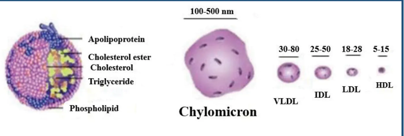
All lipoproteins are spherical, soluble lipid carriers comprised of a hydrophobic triglyceride (TG) and cholesterol-ester (CE) rich core encapsulated by a hydrophilic monolayer of phospholipids, free cholesterol and apolipoproteins (149). The classification of lipoproteins is based on their density, lipid composition and apolipoprotein association (150) (Figure 5). In addition to provide structural support to lipoprotein complexes, apolipoproteins also determine the interaction of lipoproteins with cell surface receptors as well as their rate of catabolism (151, 152). The largest lipoproteins are chylomicrons which are formed in the intestine and transport
low-density lipoproteins (VLDL), secreted by hepatocytes, carry endogenous TG with modest amounts of endogenous as well as exogenous CE on an apoB100- backbone, and are associated with apoEs and apoCs. The catabolism of VLDL results in VLDL-remnant lipoproteins called intermediate density lipoproteins (IDL), which transport roughly equal partitions of TG and CE. Further catabolism of IDL followed by a series of modifications and lipid exchanges with various lipoproteins and peripheral tissue, results in the formation of CE-rich low-density lipoproteins (LDL) particles (149).
LDL carries about 60-70% of serum cholesterol (154). It transports cholesterol from the liver to the peripheral tissues. High levels of LDL cholesterol are harmful because it can build up on the arterial walls to initiate the formation of atherosclerotic plaques (85). Cholesterol is also transported by high-density lipoproteins (HDL), the smallest and densest lipoprotein particle. HDL is the key lipoprotein involved in reverse cholesterol transport and the transfer of cholesteryl esters between lipoproteins (85, 155). Recent studies discovered a number of pleotropic atheroprotective effects of HDL which include antioxidative (156, 157), anti-inflammatory (158-160), antiapoptotic (161), antithrombotic properties (162-164). HDL cholesterol is commonly known as the “good” cholesterol (85), as high levels of HDL cholesterol are associated with reduced levels of CVD. This concept was first demonstrated in the Framingham study in the 1970s and 1980s (165). However, recent human studies have cast some doubt on the “good cholesterol” HDL hypothesis (110).
HDL cholesterol concentration was identified several years ago, as an ideal target for lowering the incidence of atherosclerosis (166-168). However, the atheroprotective properties of HDL may not be directly related to its concentration in plasma (110, 169-171). It was suggested that the cholesterol efflux (reverse cholesterol transport) capacity of an individual’s plasma is a better predictor of CVD status than HDL cholesterol concentration alone (172). Moreover, sometimes HDL does not appear protective and it might be classified as dysfunctional, a term associated with HDL particles that have been modified and thus, are no longer protective (173, 174). Accumulating data suggest that HDL can easily be modified and lose its antiatherogenic activities through multiple mechanisms. Based on the nature of modification, the alterations were classified into three types: spontaneous oxidative modification (175-177), due to the presence of free metal ions and free radicals in the atherosclerotic plaques, similar to the oxidation of LDL; enzyme-induced modification,
including myeloperoxidase (MPO), chymase-tryptase, matrix metalloproteinases (MMPs), PMN-associated enzyme, endothelial lipase, and so on (178-183). These enzymes can degrade or oxidize apolipoproteins without significant changes in lipid moiety. Metabolic modification, such as glycation (184-186) that occurs under hyperglycemic conditions, and acute phase reactants-induced modification during inflammation (169, 187, 188).
Figure 5. The composition and size of different lipoproteins.
On the left, the lipoproteins form a sphere consisting of cholesterol-esters and triglycerides enveloped by a membrane formed of a monolayer of phospholipids and free non-esterified cholesterol and apolipoproteins. On the right, a drawing showing the size of different lipoproteins ranging from 100 to 500 nm for chylomicron down to 5 to 15 nm for HDL (150).
1.4.2.2 Exogenous lipoprotein metabolism
Absorption of dietary lipids occurs in the jejunal portion of the small intestine, and upon internalization into intestinal enterocytes, TG-rich chylomicron particles are formed (189). Chylomicrons, upon entry into the lymphatics and into plasma, become associated with apoEs and apoCs, both of which are required for the metabolism of chylomicron particles (153). Following TG hydrolysis by lipoprotein lipase (LPL) anchored on capillary endothelial cells, the chylomicron-derived FAs and glycerol are taken up predominantly by adipose tissue and re-esterified into TGs for storage. The resultant chylomicron remnant particles are efficiently taken up by the liver through LDLR- and LDL receptor related protein (LRP)- mediated uptake by recognition of apoE (190), concluding the exogenous lipid metabolism pathway.
1.4.2.3 Endogenous lipoprotein metabolism
The endogenous lipoprotein pathway begins with hepatic de novo synthesis of lipoproteins. Approximately 70% of circulating cholesterol is produced in hepatocytes, making the liver the primary regulator of whole body cholesterol and lipid homeostasis (191). VLDL are particles that transport TG and cholesterol from the liver for redistribution to various tissues. Assembly of VLDL particles occurs within the secretory pathway of liver cells. This is a two steps process (192). The first series of events in VLDL assembly process occurs in the rough endoplasmic reticulum (193, 194). This process gives rise to a partially lipidated form of apoB (pre-VLDL) (195). The pre-VLDL formation is highly dependent on MTP (196). Microsomal triglyceride transfer protein (MTP) is essential for VLDL formation, and lack of it leads to a total loss of apoB-containing lipoproteins from plasma (197, 198). MTP transfers lipids: mainly triglycerides, but also cholesterol-esters and phospholipids. The second step, which occurs outside the rough ER in a smooth membrane compartment, pre-VLDL associates with the major proportion of lipids, forming a bona fide VLDL (real VLDL) (192). This step depends on ADP-ribosylation factor 1 (199) and its activation of phospholipase D (200). So, once VLDL particles are formed and secreted by hepatocytes, in capillary beds of target tissues such as adipose and muscle, endothelial cell lipoprotein lipase (LPL) hydrolyzes the VLDL liberating FA and glycerol which are taken up via soluble passive diffusion or CD36-mediated uptake in order to be re-esterified into TGs in adipose tissue or used for fatty acid oxidation in muscle (201). Following LPL-mediated hydrolysis of VLDL particles, VLDL-remnants or IDLs are formed, which can be taken up by recognition of apoE by the LDLR or the LRP following hepatic lipase (HL) modification (202). Alternatively, IDL can be hydrolyzed by HL resulting in the formation of LDL particles (203). The principal mechanism of LDL uptake is receptor-mediated endocytosis via LDL receptor (204). The LDLR is expressed ubiquitously. It accounts for approximately one third of LDL uptake by extrahepatic tissues. However, the liver is the primary site of LDLR expression and therefore regulates most of the clearance of circulating LDL particles (204).
Cholesterol is also transported by HDL, the smallest and densest lipoprotein particle. HDL is a heterogeneous collection of lipoprotein particles. Two-dimensional electrophoresis of
plasma lipoproteins separates the HDL based on their size and charge. Five major HDL particles have been identified: pre-β-1 HDL, α-4 HDL, α-3 HDL, α-2 HDL, and α-1 HDL (205, 206). The overwhelming majority of HDL particles contain apoAI, it comprises roughly 70% of the HDL mass (207, 208). Many HDL particles also contain apoAII, second most abundant protein in HDL. The metabolism of HDL initiates with apoAI synthesis in the liver and intestine, but in order to form HDL, lipid-free apoAI must interact with cells expressing ATP-binding cassette transporter A1 (ABCA1) that moves cellular lipids across the bilayer in a process requiring hydrolysis of ATP (209, 210). Nascent HDL released by ABCA1 expressing cells contains cellular phospholipids (PL) and free cholesterol (FC), and this particle is the substrate for lethicin-cholesterol acyltransferase (LCAT) which esterifies FC into cholesteryl ester (CE), building up the hydrophobic core necessary to generate spherical alpha HDL particles (211, 212). Additional lipidation of HDL can take place with ABCG1 which has been reported to lipidate mature, spherical HDL (213). Another enzyme-cholesterol ester transfer protein (CETP) moves the cholesteryl ester to apoB-containing lipoproteins in exchange to triglycerides. The concerted action of CETP-mediated cholesteryl ester transfer and hepatic lipase-mediated hydrolysis of triglycerides and phospholipids leads to formation of smaller HDL particles that are the preferred binding partners for scavenger receptor B type I (SR-BI), the major HDL receptor in hepatic cells. Thus, in humans, HDL-cholesterol can be returned to the liver via two pathways: direct hepatic uptake by scavenger receptor B1 (SR-B1) (214, 215); or through CETP exchange of HDL-CE for TG in apoB-containing lipoproteins, followed by hepatic uptake of these apoB-containing particles by the LDL receptor (216). In the liver the cholesterol is converted to bile acids for subsequent excretion (217).
1.4.2.4 LDLR -/- mice model
Deletion of the Ldlr in C57Bl/6J mice disrupts normal murine lipoprotein metabolism, resulting in elevated plasma cholesterol, particularly in the LDL fraction as a result of defective LDL clearance (218). Furthermore, these animals have increased cholesterol in both VLDL and IDL when fed a high-fat diet (219). LDLR-/- mice fed in a high-fat high-cholesterol (HFHC) diet for
12 weeks recapitulate many features of the metabolic syndrome such as dyslipidemia, hyperinsulinemia, insulin resistance and hepatic steatosis (220). Furthermore, these animals
develop atherosclerotic lesions that are relatively advanced, exhibiting significant lipid accumulation, increased macrophage and smooth muscle cell infiltration and enhanced collagen deposition (220).
1.4.3 Monocyte fate in atherosclerosis
Monocytes are essential for the development and exacerbation of atherosclerosis. They arise from proliferating and differentiating hematopoietic stem and progenitor cells in the bone marrow. Increased production of bone marrow monocytes in experimental models of atherogenesis has been reported in hypercholesterolemic swine, rabbits, and rodents (221, 222). It was also shown that hypercholesterolemia induces monopoiesis in extramedullary organs, including the spleen (223). There are several subtypes of monocytes based on their cell surface expression of the glycoprotein Ly6C in mice. Ly6Chigh monocytes are short-lived, transport
tissue antigens to lymph nodes (224), and accumulate at sites of inflammation (225) where they differentiate to macrophages and dendritic cells. Ly6Clow monocytes are longer lived, patrol the
vasculature, respond early to infection (226), and survey endothelial integrity (227). The
activation of endothelial cells by components of oxLDL, and possibly also by the turbulent blood flow at the arterial branching points, lead to the expression of adhesion molecules such as E-selectin and VCAM-1 on the endothelial surface of atherosclerotic artery and promotes the recruitment of circulating monocytes. These act in synergy with chemokines which attract monocytes, dendritic cells and T cells into the intima (228). Monocytes in the intima are stimulated by macrophage colony-stimulating (M-CSF) factor produced by activated endothelial cells to differentiate into macrophages; this process is necessary for the development of atherosclerosis (229).
1.4.4 Macrophage and foam cell formation
A central hallmark of atherosclerosis is the cholesterol-loaded macrophage or foam cell. This process is initiated by the ingestion and processing of LDL in both its native and modified forms (230). Native LDL uptake occurs via the LDL receptor (LDLR), which undergoes negative feedback regulation under high intracellular sterol concentrations (231). Although this pathway
certainly contributes to foam cell development (231), the predominant LDL uptake pathway by macrophages in lesions is that of modified LDL via the scavenger receptors cluster of differentiation (CD) 36 and scavenger receptor A I/II (SRAI/II). Upon entry into the intima, LDL particles undergo oxidative modification rendering them high-affinity scavenger receptor ligands (131). Unlike the native LDLR, scavenger receptors do not undergo negative feedback regulation in response to intracellular sterol accumulation (131). Consequently, macrophage uptake of modified LDL particles in lesions can persist indefinitely, and is only limited by substrate availability and cell viability. There is also an alternative mechanism for macrophages foam cell formation; receptor-independent uptake of unmodified native LDL called macropinocytosis, by which cells ingest fluids within vacuoles (232).
Hence, the macrophages uptake of modified LDL is mediated by scavenger receptors such as SR A I/II and CD36. However, deletion of scavenger receptors does not reduce macrophage oxLDL uptake. Even in CD36-/- , SR A-/- double knock out mice there are abundant
lipid laden macrophages in the vessel wall and these mice can develop atherosclerosis (233), although lesions complexity is reduced (234). Moreover, targeting macropinocytosis by PI3K inhibitors and Cytochalasin D (inhibitor of actin polymerization) reduced uptake, but residual LDL uptake still take place (233), suggesting existence of another additional pathway leading to foam cell formation.
Genome-wide association studies for coronary artery disease reported noncoding genetic variants at chromosome 1p13 to be significantly associated with CVD (235) and plasma level of LDL cholesterol (56, 236, 237). The SORT1 gene, encoding the protein sortilin, seems to be the causal gene at the locus regulating LDL cholesterol levels (53, 55, 238). Sortilin is involved in macrophage LDL uptake. Increased concentrations of extracellular LDL cause an upregulation of macrophage Sort1 mRNA and protein (50), promoting continuous uptake of LDL by Sortilin. Sortilin deficiency led to reduced atherosclerosis in ApoE-/-, LDLR-/- mice
models (50, 51).
1.4.5 Macrophage polarization and atherosclerosis
Macrophages are heterogeneous cell population. Several classes of macrophages have been described based on their expression of markers, the production of specific factors, and their