Int. J. Mol. Sci. 2021, 22, x. https://doi.org/10.3390/xxxxx www.mdpi.com/journal/ijms
Article 1
Cerebral Apolipoprotein D exits the brain and accumulates in
2peripheral tissues
3Frederik Desmarais1,3,†, Vincent Hervé2,3,†, Karl-F. Bergeron1, Gaétan Ravaut1, Morgane Perrotte2, Guillaume Fyfe- 4
Desmarais1,3, Eric Rassart3, Charles Ramassamy2* and Catherine Mounier1* 5 1 Laboratoire du métabolisme moléculaire des lipides, Centre de recherches CERMO-FC, Département des 6 sciences biologiques, Université du Québec à Montréal (UQAM), 141 av. du Président-Kennedy, H2X 1Y4, 7
Montréal, Québec, Canada 8
2 Centre Armand-Frappier Santé Biotechnologie, Institut National de la Recherche Scientifique (INRS), 531 9
boul. des Prairies, H7V 1B7, Laval, Québec, Canada 10
3 Laboratoire de Biologie Moléculaire, Département des Sciences Biologiques, Université du Québec à Mon- 11 tréal (UQAM), 141 av. du Président-Kennedy, H2X 1Y4, Montréal, Québec, Canada 12 * Correspondence: Catherine Mounier, [email protected]; Charles Ramassamy, 13
† Co-first authors 15
Abstract: Apolipoprotein D (ApoD) is a secreted lipocalin associated with neuroprotection and lipid 16
metabolism. In rodent, the bulk of its expression occurs in the central nervous system. Despite this, 17
ApoD has profound effects in peripheral tissues, indicating that neural ApoD may reach peripheral 18
organs. We endeavour to determine if cerebral ApoD can reach the circulation and accumulate in 19
peripheral tissues. 3 hours was necessary for over 40% of all the radiolabeled human ApoD (hA- 20
poD), injected bilaterally, to exit the CNS. Once in circulation, hApoD accumulates mostly in the 21
kindeys/urine, liver, and muscles. Accumulation specificity of hApoD in these tissues was strongly 22
correlated with the expression of lowly glycosylated basigin (BSG, CD147). hApoD was observed 23
to pass through bEnd.3 blood brain barrier endothelial cells monolayers. However, cyclophilin A 24
did not impact hApoD internalisation rates in bEnd.3, indicating that ApoD exit from the brain is 25
either independent of BSG or relies on additional cell types. Overall, our data shows that ApoD can 26
quickly and efficiently exit the CNS and reach the liver and kidneys/urine, organs linked to the 27
recycling and excretion of lipids and toxins. This indicates that cerebral overexpression during neu- 28
rodegenerative episodes may serve to evacuate neurotoxic ApoD ligands from the CNS. 29
Keywords: Apolipoprotein D; Basigin; Protein accumulation; Blood-brain barrier 30 31
1. Introduction 32
Apolipoprotein D (ApoD) is a 25-30 kDa glycosylated protein belonging to the 33 lipocalin superfamily of hydrophobic molecule carriers [1-4]. ApoD is known for its abil- 34 ity to bind various ligands, including arachidonic acid (ARA) and progesterone [1, 5, 6]. 35 ApoD can bind ARA and mediate its release from cell membranes. Furthermore, since 36 ARA is the precursor of the eicosanoid class of lipid inflammation mediators, ApoD’s ca- 37 pacity to bind ARA allows it to modulate the production of eicosanoids and attenuate 38 inflammation [7, 8]. ApoD can also limit inflammation and oxidative stress by reducing 39 oxidized lipids [1, 9-12]. Because it is overexpressed during many neurodegenerative dis- 40 eases and stresses, ApoD is considered an important actor in brain protection and repair 41 [9, 11, 13-15]. ApoD is also a member of the apolipoprotein family as it associates with 42 lipoproteins (mainly high-density lipoproteins) in the blood. In humans, contrary to most 43 apolipoproteins, ApoD is minimally produced in the liver and intestines [1, 5]. Rather 44 mRNA expression is found, although in varying quantities, in several tissues including 45 the central nervous system (CNS), mammary glands, spleen, adipose tissues, adrenals and 46
Citation: Lastname, F.; Lastname, F.;
Lastname, F. Title. Int. J. Mol. Sci.
2021, 22, x.
https://doi.org/10.3390/xxxxx Academic Editor: Firstname Last-name
Received: date Accepted: date Published: date
Publisher’s Note: MDPI stays
neu-tral with regard to jurisdictional claims in published maps and institu-tional affiliations.
Copyright: © 2021 by the authors.
Submitted for possible open access publication under the terms and con-ditions of the Creative Commons At-tribution (CC BY) license (http://crea-tivecommons.org/licenses/by/4.0/).
skin [1, 16, 17]. In contrast to human, in mice and rats, ApoD mRNA expression is mainly 47 restricted to the CNS [1, 2, 18-20]. However, moderate to high protein levels are found in 48 peripheral tissues including the liver suggesting that the protein could cross the blood 49
brain barrier (BBB) [1, 21]. 50
In support to the hypothesis that apoD can exit the CNS, the overexpression of hu- 51 man ApoD (hApoD) in the CNS of transgenic mice leads to elevated level of apoD in the 52 livers, likely originating from expression in the CNS [19]. These transgenic mice develop 53 hepatic and muscular steatosis [8, 19, 22] probably because apoD modulates the metabo- 54
lism of these tissues. 55
Beside its ability to circulate as part of lipoproteins, up to 10% of plasma ApoD is 56 found in the protein phase, indicating that ApoD may also circulate freely in its soluble 57 form [23]. Three recent studies have also highlighted the presence of ApoD in extracel- 58 lular vesicles [24-26]. ApoD has been shown to be transported to neurons by extracellular 59 vesicles originating from astrocytes [26]. ApoD can also be found associated with cerebro- 60 spinal fluid lipoproteins [27-29]. ApoD was also observed in urine [30]. Together, the 61
results show that ApoD transport is polyvalent and complex. 62
The Basigin (BSG) receptor, also known as cluster of differentiation 147 (CD147), me- 63 diates ApoD internalisation in neurons [14]. BSG is also responsible for the internalisation 64 of other proteins such as caveolin with its affinity being dependent on BSG glycosylation 65 level [31]. BSG is expressed in a wide range of tissues and cell types [32] (data available at 66 https://www.proteinatlas.org/ENSG00000172270-BSG/tissue). Circulating ApoD, through 67 BSG-mediated internalisation, could therefore accumulate in many different structures. 68 BSG is expressed in mouse brain endothelial cells, the main component of the blood-brain 69 barrier (BBB) [33]. The BBB is the largest barrier separating the peripheral circulation from 70 the CNS [34]. It is a highly selective barrier, composed of tightly linked endothelial cells 71 that cover the bloodstream capillaries. Astrocytes and pericytes also participate in the ar- 72 chitecture and function of the BBB. In addition to BSG, apolipoprotein receptors such as 73 Low-density lipoprotein receptor (LDLR), LDLR-related protein (LRP1) and Megalin are 74 also expressed in BBB endothelial cells and are involved in transporting ligands across the 75
barrier [35]. 76
The mouse brain microvascular endothelial cell line bEnd.3 is the most characterized 77 in terms of tight junction proteins [36, 37] and is thus often used to study the endothelial 78 component of the BBB [38]. These cells express the tight junction proteins ZO-1, occludin 79 and claudin-5 [39] and possess fluorescein permeability similar to those of primary mouse 80 endothelial cells [40]. These mouse brain capillary endothelial cells contains several recep- 81 tors present on the surface of endogenous BBB endothelial cells, including apolipoprotein 82 receptors such as LDLR and LRP1. These cells also possess receptors that participate in 83 the transcytosis process of proteins such as transferrin and lactoferrin [35]. 84 In this study, we endeavour to determine if cerebral ApoD can reach the bloodstream 85 from the brain and if BSG plays a role in this process. We also set out to determine in 86 which peripheral organs ApoD preferentially accumulates once in circulation. 87
2. Results 88
2.1. hApoD exits the central nervous system and reaches peripheral tissues 89 We endeavored to establish whether hApoD can exit the brain and reach the blood- 90 stream and accumulate in peripheral organs. For this, the cerebral ventricles of mice were 91 injected with radiolabelled hApoD or albumin as a control. Injected proteins were allowed 92 to diffuse from the ventricles and tissue/fluid for 3 or 6 hours. Samples were subsequently 93 taken throughout the mouse body. During this procedure, mice were perfused with a sa- 94 line solution to avoid contamination in tissues stemming from potential radiolabelled pro- 95 teins in the blood. Also, BBB integrity was confirmed by circulating an Evans blue solution 96 in perfused mice (Supp. Fig.3). As expected, 3 hours after ICV injection of radiolabeled 97 proteins, most of the recovered albumin (93%; Fig.1A), remained sequestered in the brain. 98
Within this time frame, 41% of the recovered hApoD was found in the periphery, showing 99 that hApoD can exit the brain compartment. Interestingly, a significant proportion of hA- 100 poD accumulated in the liver (6.06%) but the major fraction was found in urine (28.87%). 101 Radioactivity levels in the bloodstream were very low, indicating that hApoD was rapidly 102 cleared from the circulation. Electrophoresis of urine and brain protein samples in dena- 103 turing conditions confirmed that the radiation detected corresponded to native hApoD 104
molecules migrating at 30 kDa (Fig.1B). 105
106
107
Figure 1. hApoD injected in the brain accumulates in urine and liver. Radiolabeled proteins (Bo- 108
vine Serum Albumin (BSA) and hApoD) were injected in mouse cerebral ventricles. Blood, urine 109
and – after perfusion – organs were collected 3h post-injection. A) Radioactivity recovered in flu- 110
ids and tissue homogenates (%) compared to the total radioactivity recovered in the whole body. 111
Statistical significance was evaluated via a Student t-test: *p<0.05, **p<0.01 (n=3 animals). B) Radio- 112
labelled proteins extracted from brain and urine and visualized by radiography. A representative 113
electrophoresis gel is shown. 114
2.2. hApoD accumulates in specific tissues 115
ICV injections were limited to a total volume of 10 μL (5 μL per ventricle), adding up 116 to approximately 2.5 μg of radiolabeled proteins. Under these experimental restrictions, 117 hApoD could only be shown to accumulate in the kidneys and liver (Fig.1A). The radia- 118 tion found in other organs (muscles, pancreas, spleen, intestines, and others) was too close 119 to background radiation levels to confidently affirm that hApoD had accumulated in these 120 organs. To determine if hApoD can target other tissues, we injected a greater amount of 121
radiolabeled protein (approximately 10μg) directly in the bloodstream. Note that this 122 higher dose of hApoD was still within physiological parameters for circulating ApoD 123 (Rassart et al. 2000). Albumin was again chosen as a control because of its propensity to 124 remain in the circulation (Andersen et al. 2014). Again, the blood and its radioactive con- 125 tent were eliminated by perfusion with a saline solution before tissue samples were taken. 126 The amount of recovered radiolabeled hApoD was evaluated 3h and 6h post-injection 127 (Fig.2A) and as a function of the albumin control (Fig.2B). Accumulation of hApoD oc- 128 curred at a higher rate than albumin in the kidneys (5.6-fold), liver (30.0-fold), pancreas 129 (3.1-fold), caecum (2.8-fold), spleen (3.3-fold), mesenteric adipose tissue (MAT; 4.2-fold) 130 and muscles (2.4-fold) at least at either 3 or 6 hours. The kidneys emerged as the tissue 131 that most readily accumulates hApoD with 14% of recovered radioactivity per gram of 132 tissue. This accumulation was probably due to hApoD within the nephrons since a major 133 part of the radioactivity was found in urine (Fig.3A). The biggest difference between al- 134 bumin and hApoD accumulation however was found in the liver (Fig.2B), indicating a 135 very strong specificity of hApoD for this tissue. Interestingly, the accumulation of hApoD 136 in muscles was stronger than albumin 3h after injection. However, after 6h, hApoD levels 137 diminished (from 1.6 to 1.0% per gram of tissue) while albumin levels increased (from 0.7 138 to 1.4% per gram of tissue). Conversely, hApoD accumulation in the MAT appeared to be 139 more persistent than albumin. MAT radiation from hApoD only diminished by 0.10% per 140 gram of tissue in 3h. In comparison, radiation from albumin was reduced by 0.96% per 141 gram of tissue in that same interval. Very interestingly, hApoD within the bloodstream 142 did not appear to enter the brain suggesting that hApoD can only cross the BBB in one 143
direction. 144
Figure 2. Circulating hApoD accumulates in specific peripheral organs. Radiolabeled proteins 146
(Bovine Serum Albumin (BSA) and hApoD) were injected intravenously in mice. Blood, urine and 147
– after perfusion – organs were collected 3h and 6h post-injection. A) Results are presented as the 148
percentage of radioactivity recovered per gram of tissues. EAT: epididymal adipose tissue, SAT: 149
subcutaneous adipose tissue, MAT: mesenteric adipose tissue, and BM: bone marrow. B) The heat 150
map shows the accumulation of hApoD relative to albumin. Statistical significance was evalu- 151
ated via a Student t-test: *p<0.05, **p<0.01, ***p<0.001 (n=4 animals). 152
To establish a general portrait of hApoD biodistribution when it exits the blood- 153 stream, we extrapolated the total radioactivity found in each organ (or fluid) according to 154 their total weight (or volume). Since we did not measure the total weight of adipose tis- 155 sues nor of bone marrow, these tissues were excluded from this analysis. Albumin was 156 mainly found in circulation at both 3h (91%) and 6h (83%) after injection (Fig.3A). As ex- 157 pected from our previous results, hApoD exited the bloodstream at a higher rate than 158 albumin. After 6 hours, half of the injected hApoD had left the circulation and a significant 159 fraction was again found in urine (33%; Fig.3A). Globally, hApoD accumulation level in 160 tissues was higher at 3h (25.2%) compared to 6h (18.1%), suggesting a progressive shift of 161 hApoD from blood to tissues and finally to urine. The principal target of hApoD at 3h 162 (Fig.3B) was the muscles (10.67%) followed by the liver (8.93%), kidneys (3.09%), pancreas 163 (1.24%) and spleen (0.41%). Because of their mass, muscles accounted for a large propor- 164 tion of hApoD accumulation despite their poor apparent affinity for hApoD (Fig 2). For 165 instance, while the liver accumulated hApoD at a much higher rate (6.0 % of recovered 166 radioactivity per gram of tissue), it was still outclassed in total hApoD accumulation by 167 the muscles despite their low accumulation rate (1.6 % of recovered radioactivity per gram 168
of tissue; see Fig.2A). 169
170
Figure 3. Relative accumulation of hApoD in fluids and tissues. Radiolabeled proteins (Bovine 171
Serum Albumin (BSA) and hApoD) were injected intravenously in mice. Blood, urine and – after 172
perfusion – organs were collected 3h and 6h post-injection. Results are presented for each A) 173
fluid and B) organs as their average respective percentage of the total radioactivity recovered in 174
the animals. Statistical significance was evaluated via a Student t-test: *p<0.05, **p<0.01, ***p<0.001 175
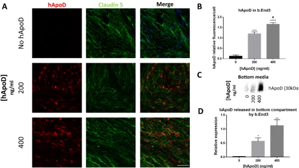
2.3. hApoD transcytoses through bEnd.3 brain endothelial barrier cells 177 Since hApoD can exit the brain compartment, we explored the possibility that hApoD 178 might be able to pass through a BBB model consisting of a monolayer of the endothelial 179 bEnd.3 cells. The impermeability of this monolayer model to passive diffusion was vali- 180 dated by electrical resistance measurement (≥35 Ohms/cm2) and Dextran permeability as- 181 say (Supp. Fig.2). Expression of Claudin-5 between adjacent cells was also validated, con- 182 firming the formation of tight junctions (Fig.4A). Within 24h, exogenous hApoD applied 183 to the top compartment of the monolayer was internalized (Fig.4A) and was found in the 184 bottom medium (Fig.4B), indicating that hApoD is subject to transcytosis through bEnd.3 185 cells in a dose-dependent manner. However, bEnd.3 cells are not polarized [41, 42]. There- 186 fore, our results may underestimate the amount of hApoD that actually underwent 187 transcytosis since part of the internalized protein could be released back to the top com- 188 partment where it was first internalised and not contribute to the buildup of ApoD con- 189
centration in the bottom compartment. 190
191
Figure 4. hApoD is subject to endocytosis and transcytosis through bEnd.3 cells. The top side of 192
bEnd.3 cell monolayers was exposed to different concentrations of hApoD for 24h. A) Cells were 193
immunostained for hApoD (red) and a tight junction marker (Claudin-5, green). Nuclei were 194
stained with Hoechst (blue). Scale bar: 50μm. B) Bottom media proteins were immunoblotted for 195
hApoD. Representative images are shown (n=3 independent experiments). Statistical significance 196
for panels B and D was evaluated via a Student t-test: *p<0.05, **p<0.01,***p<0.001, ****p<0.0001 197
against the 0ng/ml (*) and 200 ng/ml (#) conditions. 198
2.4. Cyclophilin A competition does not reduce hApoD internalization in brain endothelial 199
barrier cells 200
Having confirmed that hApoD can cross through bEnd.3 brain endothelial cells, we 201 next endevoured to determine if the BSG receptor was implicated in this process. We used 202 cyclophylin A, a BSG ligand known to compete with ApoD for BSG-dependent internali- 203 zation [14]. We first confirmed that cyclophilin A was non-toxic at the concentrations used 204 (Fig.5A). Surprisingly, internalization and transcytosis of hApoD by bEnd.3 monolayers 205 were unchanged despite the presence of various molar ratios of competing cyclophilin A 206 (up to 10-fold excess relative to hApoD; Fig.5B-D). We then determined if BSG was ex- 207 pressed in bEnd.3 cells in comparison to control tissues (brain and liver). Our analysis 208 shows that BSG was expressed in bEnd.3 cells at a comparable level to the liver, the tissue 209
with the highest specific affinity for hApoD. Interestingly, BSG expression was much 210 stronger in bEnd.3 than in whole brain lysate. Additionally, the level of BSG glycosylation 211 varied greatly between samples (Fig.5E). BSG glycosylation can influence its protein-pro- 212 tein interactions [31]. Interestingly, the ratio of lowly-glycosylated BSG (LG-BSG) relative 213 to total BSG expression appeared to align with our observed cellular affinity for hApoD 214 internalization in vivo (Figs.5F and 2A). The liver, one of the tissues that most readily 215 accumulate hApoD, had a high LG-BSG /total ratio, while the bEnd.3 cells had a lower 216 ratio and needed 24 hours to internalise and excrete hApoD to the bottom media. This 217 suggested a greater role of BSG in peripheral tissues than in endothelial cells for hApoD 218
internalization. 219
220 221
Figure 5. Cyclophilin A competition does not reduce hApoD internalization in bEnd.3 cells. A) 222
Viability of bEnd.3 cells in the presence of cyclophilin A, at 1, 5 and 10 times the molar concentra- 223
tion of hApoD (13.33nM) used in the experiment. H2O2 was used as a control for the loss of cell 224
viability. B) The top side of bEnd.3 cell monolayers were exposed to hApoD (13.33nM) for 24h. 225
Cyclophilin A was included from the beginning of the experiment at a molar equivalent (1:1) or in 226
molar excess (1:10) relative to hApoD. Cells were immunostained for hApoD (red). Nuclei were 227
stained with Hoechst (blue). Scale bar: 20 μm. Representative confocal sections are presented. C) 228
Average hApoD signal per cell (5 images taken for each monolayer). Fluorescence was normalized 229
by the number of cell nuclei. D) Bottom media proteins immunoblotted for hApoD. E) Immunob- 230
lot of BSG in bEnd.3 and control tissues (HG-BSG, Highly glycosylated BSG; LG, lowly glycosyl- 231
ated BSG). Panels D and E are representative results. F) Quantification of LG-BSG expression rela- 232
tive to total BSG expression as compared to the brain (*) and liver (#). Statistical significance for 233
panels A, C and F was evaluated via a Student t-test: ***p<0.001, ****p<0.0001 (n=3 independent 234
experiments). 235
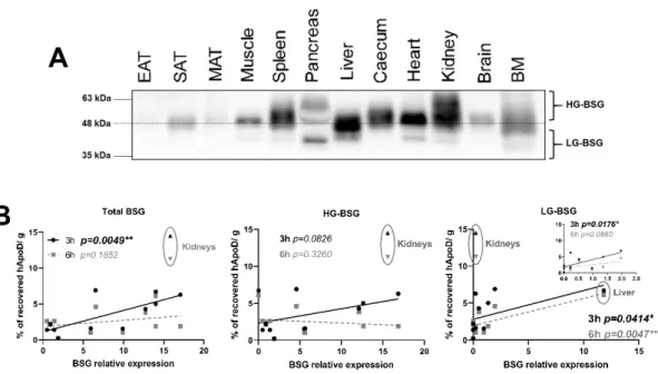
2.5. Relationship between BSG glycosylation and hApoD accumulation 236 To further explore if BSG glycosylation was linked to the degree of tissue hApoD 237 internalization, we studied the expression of the multiple glycosylated forms of BSG in 238 mice tissues. BSG expression, per mg of tissue, was heterogeneous between tissues, with 239 the liver and heart presenting an especially high overall BSG expression and adipose tis- 240 sues having a low expression (Fig.6A). BSG bands were observed at multiple size and in 241
varying patterns according to the tissue type. The SAT, muscles and the brain only ex- 242 pressed the highly-glycosylated form of BSG (HG-BSG, >48kDa). The liver and pancreas, 243 however, expressed multiple forms of BSG (Fig.6A). We next performed correlations be- 244 tween BSG expression by glycosylation levels and the specific accumulation of hApoD in 245 each tissue. As expected, the total expression of BSG in tissues was positively correlated 246 with hApoD accumulation. However, the correlation was only statistically significant for 247 the 3-hour time point (Fig.6B). Interestingly, expression of the HG-BSG form was not 248 found to be associated with hApoD specific accumulation. In opposition, LG-BSG expres- 249 sion was correlated with hApoD specific accumulation at both time points, but especially 250 6 hours after injection (p=0.0047**). Kidneys were outliers relative to all other tissues in- 251 vestigated and were therefore excluded from these correlation analyses (presented as sep- 252 arate data set)(Fig.6B). It is very likely that hApoD does not require to be internalized in 253 kidney cells during urine excretion and it is probably passively filtered through the glo- 254
merular pores (discussed later). 255
256
Figure 6. hApoD tends to accumulate in tissues expressing underglycosylated Basigin. A) Im- 257
munoblot of Basigin (BSG) in its multiple glycosylated forms (HG, highly glycosylated; LG, lowly 258
glycosylated) in various tissues, including epididymal adipose tissue (EAT), subcutaneous adipose 259
tissue (SAT), mesenteric adipose tissue (MAT) and bone marrow (BM). Each well contained whole 260
protein extracts from 20μg of tissue. A representative immunoblot is shown (n=4 animals). B) Cor- 261
relations between average hApoD accumulation (% of recovered radioactivity per gram of tissue) 262
and average expression level of different classes of glycosylated Basigin in various tissues, 3h 263
(black) and 6h (grey) after IV injection. Inset: liver data eliminated from the correlation. Pearson’s 264
product moment correlations are presented: *p<0.05, **p<0.01 (n=4 animals). 265
3. Discussion 266
Our results demonstrate that injected hApoD can exit the brain and reach peripheric 267 tissues. These observations were made by comparing the amount of hApoD that escaped 268 the CNS to an albumin control. The rate at which hApoD leaves the brain was observed 269 to be higher than that of albumin. Albumin is a natural component of the cerebro-spinal 270 fluid (CSF). Its concentration in the CSF is lower than 1/125th of the one found in circula- 271 tion (CSF/serum ratio < 8x10⁻³) [43] and the CNS barriers greatly limit albumin’s ability 272 to transfer bilaterally between the brain and the circulation. Similarly to other components 273 of the CSF, albumin is constantly eliminated from the brain by the brain’s waste clearance 274 system [43, 44]. The elimination of albumin from the CNS is a passive process that does 275
not require an active transport across CNS barrier. In contrast, the hApoD’s higher exit 276 rate strongly suggest that it might be actively transported. 277 The two major circulating hApoD recipient organs are the kidneys/urine and the 278 liver, the first being implicated in lipid metabolism and recycling and the latter being 279 mostly implicated in waste removal. This corroborated our previous hypothesis that cer- 280 ebral hApoD reaches the liver [8]. We previously showed that hApoD overexpression in 281 the brain results in the development of an hepatic and muscular steatosis in mice [19]. The 282 presence of hApoD as well as an increase in ARA proportion in the liver suggested that 283 hApoD derived from the brain was responsible for the added efflux of ARA into the liver 284
[8, 19, 22]. 285
Surprisingly, a large part of the hApoD recovered outside the CNS was found in the 286 urine. It was previously shown that ApoD can pass through the kidneys and accumulate 287 in urine in monomeric and dimeric form [45]. Multiple factors including protein size and 288 charge determine which molecules can pass through the kidneys by glomerular filtration. 289 It is generally believed that proteins and polymers in the range of 30-50kDa can pass 290 through the glomerular pores. However, the diameter and shape of the molecules are also 291 important as elongated molecules such as 350-500 kDa nanotubes with 1 nm diameter 292 have been shown to be efficiently cleared by glomerular filtration [46]. Glomerular pores 293 have been described as cuboids with an average dimension of 4 nm (40 Å) by 14 nm (140 294 Å) in a cross section, and 7nm (70 Å) in length [47]. Albumin is a 66-69 kDa flexible, ellip- 295 soid-shaped protein with a 3.8 nm diameter and a length of 15 nm. Despite its size, ap- 296 proximately 3.3g of albumin is filtered daily in the human kidney [48]. Therefore, it was 297 not surprising to find albumin in urine. ApoD is a 29-32 kDa, 4.5 nm (45 Å) by 4.0 nm (45 298 Å) protein [49]. Considering its size, it is likely that hApoD is also able to directly pass 299 through the glomerular pores and end up in urine without the assistance of active 300 transport. This idea is supported by the fact that kidneys were outliers in our correlation 301 between BSG expression and hApoD accumulation in tissues (Fig.6B). This indicated that 302 BSG was likely not implicated in this process or that another mechanism was far more 303 important for hApoD accumulation in kidney\urine. However, the physiological rele- 304 vance ApoD’s capacity to be filtered by the kidneys remain elusive. 305 One possible interpretation is that ApoD could also participate in the elimination of 306 harmful waste products (oxidized lipids) and pro-inflammatory molecules (ARA) from 307 the CNS or other targeted tissues. ApoD is overexpressed in periods of neurodegenerative 308 stress [15, 50-55]. ApoD is known to facilitate myelin clearance, extracellular matrix re- 309 modeling and axon regeneration after nerve injury [17]. A large quantity of hydrophobic 310 and pro-inflammatory molecules is released from damaged neurones and myelin sheets 311 after nerve injury. ARA and lysophospholipids (LPC) are two of the major products re- 312 sulting from PLA2-mediated myelin cleavage and are both ApoD ligands. In fact, ApoD- 313 null mice have higher basal levels of free ARA and LPC in intact nerves as well as a lower 314 free LPC level than wildtype mice in injured nerves [17]. The lack of ApoD during nerve 315 injury appears to cause a heightened inflammatory response, delayed inflammation re- 316 sorption and slower healing process [17]. It is believed that this occurs, at least in part, 317 because of a lack of proper management and clearance of free ARA and LPC. Previous 318 studies on ApoD’s role in managing these substances only looked at the possibility that 319 these harmful compounds were sequestered by ApoD and reabsorbed locally by cells in 320 the CNS. Considering our present results, however, it is possible that ApoD might redirect 321 part of the released lipids away from the neurons and into the kidneys and liver during 322 these episodes. This may prevent further damage caused by peroxidized lipids or pro- 323
inflammatory ARA-derived eicosanoids in the brain [17, 56]. 324
325 Our results also show that hApoD is rapidly depleted from the circulation in favor 326 of tissular accumulation or excretion via the kidneys/urine. Interestingly, while ApoD is 327 known to be increased in the CSF of patients with Alzheimer’s disease [51], its plasmatic 328
levels do not appear to be affected [57]. This could be due to the quick pace at which hA- 329 poD is depleted from plasma. Unfortunately, no data exist on ApoD concentration in the 330 urine and peripheral tissues from Alzheimer and other neurodegenerative diseases pa- 331
tients. 332
333 In addition to tissues with lipid recycling and excretory functions, a number of other 334 tissues were targeted by circulating ApoD. The skeletal muscles were also an important 335 reservoir of hApoD accumulation, despite their poor specific affinity for hApoD (Fig.3A) 336 and low expression of the highly glycosylated BSG (Fig.6A). They only remained an im- 337 portant hApoD reservoir because of their large mass. Additionally, accumulation of hA- 338 poD over albumin was only significant 3h post-injection and tended to be lower than al- 339 bumin at 6h post-injection. It is worth mentioning that, while albumin largely stays in 340 circulation, it can also accumulate in muscles [58] and intestinal cells [59]. This could ex- 341 plain why hApoD accumulation was stronger than albumin in these tissues at only one 342 time point. Another evidence that ApoD targets the skeletal muscles is that transgenic 343 mice overexpressing hApoD in their CNS develop muscle steatosis [19]. The function of 344 ApoD in the skeletal muscles is likely unrelated to ApoD’s hypothetical function of re- 345 moving harmful molecules from the CNS. Other authors described ApoD as a lipid trans- 346 porter with a very large diversity of ligands. In fact, ApoD can bind cholesterol and preg- 347 nenolone bringing these essentials lipids in various organs where they can be used. Pre- 348 vious studies also mentioned that increased expression of ApoD in the CNS during brain 349 injury can increase the availabilities of essential lipids for the synthesis of new cellular 350
membranes (Reviewed in Rassart et al 2020 [1]). 351
352 As of now, the literature provides little insight into ApoD’s potential role in skeletal 353 muscles. One study reported that ApoD, followed by the leptin receptor (LEPR), are the 354 two most upregulated transcripts in muscle disuse atrophy. Meanwhile, genes responsi- 355 ble for energy metabolism, mitochondrial function, cell cycle regulation, stress response, 356 sarcomere structure, cell growth/death, and protein turnover were downregulated [60]. 357 ApoD is also upregulated in age-related skeletal muscle cells senescence, where it is ex- 358 pected to play anti-oxidative and anti-inflammatory roles [1, 9, 61]. Our results indicate 359 that circulating ApoD could also be implicated in these mechanisms. 360 361 The spleen also appears to be targeted by circulating hApoD. The spleen is the major 362 cite of heme recycling from senescent red blood cells. There, heme is converted to bilirubin 363 and then transported to the liver to be secreted into bile in the intestines [62]. Interestingly, 364 ApoD can bind to bilirubin [63] and has long been suspected to participate in heme recy- 365
cling [5]. 366
367 hApoD appeared to also have affinity for the MAT. We previously showed that 368 ApoD protein levels in adipose tissue are linked to improved metabolic parameters in 369 obese women. Specifically, protein levels of ApoD in the human MAT were associated 370 with reduced circulating TNF-α and improved insulin sensitivity (QUICKY index). These 371 correlations did not exist with ApoD mRNA expression [64] suggesting that circulating 372 ApoD, after adipose tissue internalization, was specifically responsible for this associa- 373 tion. This hypothesis is reinforced by our present results, which indicate that circulating 374 hApoD can accumulate in the MAT. However, the role of exogenous ApoD in adipose 375 tissues remains unclear. In this tissue, ApoD may exert anti-inflammatory effects through 376 its ability to modulate ARA metabolism [7, 8, 22], to reduce oxidative stress [9, 10, 65-67] 377 and to disrupt osteopontin function, a protein implicated in macrophage recruitment [1, 378
68]. 379
380 We used an in vitro model of the endothelial part of the BBB, bEnd.3 cell monolayers, 381 to confirm that hApoD can be subject to internalization and transcytosis. Beside the BBB, 382
there are also other barriers between the periphery and the CNS through which ApoD 383 could cross. The blood-CSF barrier (BCSFB), for instance, separates the blood from the 384 CSF. It is formed by epithelial cells and the tight junctions of the choroid plexus. ApoD 385 could potentially cross this barrier, enter the bloodstream and then be distributed to pe- 386 ripheral organs. However, the BBB area in the human brain is 10 times greater than the 387 BCSFB area [69]. Furthermore, since ApoD is mainly secreted by glial cells [1], in proxim- 388 ity to the BBB, its preferred exit route is likely to be the BBB. This motivated the use of the 389
bEnd.3 cell line as our in vitro model. 390
391 Considering that ApoD internalization is a BSG-dependent, cyclophilin A-sensitive 392 mechanism in neurons and HEK 293T [14], we expected BSG to be implicated in hApoD 393 internalization by bEnd.3 cells. Though BSG is expressed by bEnd.3 cells, cyclophilin A 394 did not limit hApoD internalization/transcytosis in our hands. Interestingly, the BSG gly- 395 cosylation pattern in bEnd.3 cells is different from the one reported in 293T cells. The 293T 396 cells appear to possess a greater amount of LG-BSG (especially 30-35 kDa). Furthermore, 397 hApoD internalization by 293T cells was reported as soon as 4h after administration [14], 398 while that process necessitated 24h to be detectable in our bEnd.3 cell assays (data not 399 shown). Our in vivo data shows that HG-BSG expression is not correlated to hApoD in- 400 ternalization in tissues, contrary to the LG-glycosylated forms. Higher levels of glycosyl- 401 ation present on a protein can hinder its protein-protein interactions [70]. In agreement 402 with this observation, deglycosylation of BSG was reported to increase its interaction with 403 Caveolin-1 [31]. It is likely that a similar phenomenon occurs between BSG and ApoD. 404 This is supported by the fact that underglycosylated BSG expression is strongly associated 405 with hApoD specific accumulation in peripheral tissues (Fig.6B). Taken together, these 406 results suggest that, while BSG may be an important factor in peripheral hApoD cellular 407 internalization, it may not be implicated in hApoD’s capacity to exit the brain. 408 409 These results may also imply that BSG is not the exclusive receptor for ApoD. Inhib- 410 iting LDLR has previously been shown to reverse the synaptogenic effects of ApoD in 411 dorsal root ganglion cell cultures [68]. LDLR is present at the BBB as well as in bEnd.3 412 cells [35] and is a major ApoE receptor in the brain [72, 73]. LDLR could therefore also 413 participate in the passage of the ApoD through the BBB. The lack of competition by cyclo- 414 philin A could be due to an LDLR-mediated internalization of hApoD instead of a BSG- 415
mediated one in bEnd.3 endothelial cells. 416
417 While we showed the passage of hApoD through endothelial cell monolayers, our 418 model lacked the full complexity of the BBB. Though endothelial cells are the main com- 419 ponents of the BBB, the mechanisms regulating the crossing of proteins through the BBB 420 are more complex and involve other cell types. The BBB is also composed of pericytes and 421 astrocytes in an architecture that is difficult to achieve in vitro. In fact, addition of glial 422 cells to bEnd.3 cells in a co-culture model enhances the barrier function of the endothelial 423 monolayer [38]. To note, the presence of hApoD in the plasma was rapidly detectable in 424 vivo (3 hours), a kinetic that was not replicable in our in vitro model. It appears highly 425 probable that other cell types are implicated in hApoD exit from the brain. 426 427 In our experiments, we used free soluble hApoD monomer purified from cystic fluid. 428 However, in physiological conditions, it is not excluded that ApoD may be able to pass 429 through the BBB via extracellular vesicles (EVs). Indeed, ApoD is found in EVs [24-26], 430 including EVs from serum and CSF [24]. Recently, EVs secreted from astrocytes and con- 431 taining ApoD were shown to be internalized in neurons and to contribute to the survival 432 of neurons under oxidative stress [26]. The passage of EVs from the periphery to the CNS 433 has also been shown, notably in a drug delivery context where EVs were shown to cross 434 the BBB in both directions [74]. Therefore, the high rate at which hApoD was able to exit 435 the brain in vivo may be due to an inclusion on ApoD into EVs by glial cells prior to its 436
passage through the BBB. This could also be a factor explaining the slow rate of internali- 437 zation observed in vitro in endothelial bEnd.3 cells, where glial cells were absent. 438 439 In our study, circulating hApoD did not appear to enter the brain from the circula- 440 tion. This could indicate that hApoD transport through the CNS barriers is unidirectional 441 or simply follows its concentration gradient (from brain to blood) under normal condi- 442 tions. It is also possible that the quick pace at which hApoD is internalized by other tissues 443 depletes the circulating stocks of hApoD in a manner that prevents its return to the brain. 444 It is worth mentioning that our data do not definitely prove that ApoD is incapable of 445 entering the brain from the circulation. Our study was aimed at characterizing the biodis- 446 tribution of hApoD upon release from the brain. Injected radiolabeled proteins that do 447 enter the brain often do so at a low percentage (e.g. approximately 1% of the total injected 448 amount for transferrin) [75]. Therefore, studies centered on the brain often employ meth- 449 odologies different that used here. These include the use of a positive control and the in- 450 jection of proteins into the left jugular vein instead of the tail vein [76], with a possible 451 recirculation of radiolabelled proteins [77]. Further steps will be required to formally as- 452 certain if ApoD can enter the brain from the circulation. ApoD exist in different forms. 453 Much like BSG, ApoD also possess multiple glycosylation levels which are likely to influ- 454 ence its protein-protein interactions [2]. Glycosylation plays an important role in the ther- 455 mostability, folding, as well as the overall charge of a protein and can influence its protein- 456 protein interactions [70]. The injection of different forms of ApoD, including monomers, 457 dimers, tetramers, and EV containing ApoD – as well as differently glycosylated ApoD – 458
could also give different results. 459
460 In conclusion, our study shows for the first time that hApoD can efficiently exit the 461 brain, most likely by active passage through the BBB and reach peripheral tissues with 462 functions related to lipid and glucose homeostasis (liver, muscles, pancreas, adipose tis- 463 sues and intestines, together known as metabolic organs) as well as excretion (Kid- 464 neys/urine). The characterization of hApoD’s distribution pattern represents an important 465 advance in the understanding of ApoD function and correlates well with its known func- 466 tions. The high amount of hApoD found in the liver and urine in our experiments suggests 467 that ApoD may have a role in the excretion and recycling of brain lipids. Our results also 468 suggest that ApoD accumulation in peripheral tissues is likely dependent on BSG and 469 influenced by its glycosylation levels. ApoD’s capacity to leave the brain is likely more 470 complex and might not depend on BSG. However, it remains unclear what receptor other 471 than BSG participates in this process. Furthermore, ApoD’s own glycosylation and incor- 472 poration into EVs is also likely to influence ApoD cell internalization and tissue distribu- 473
tion. 474
4. Materials and Methods 475
4.1. Animals 476
Experiments were carried out on C57BL/6 male mice of 3 to 4 months of age. Animals 477 were housed under standard conditions at constant temperature (20-22°C) and humidity 478 (50-60%), under a 12h light/dark cycle with free access to water and food (standard rodent 479 chow; Charles River #5075). Experimental procedures were approved by the Animal Care 480 and Use Committee of Université du Québec à Montréal (protocol #913). 481
4.2. Protein radiolabelling 482
Human ApoD (hApoD), purified from breast cyst fluid [4] and bovine serum albu- 483 min (BSA) were both radiolabelled with iodine-125 in the form of iodine monochloride 484 following the method described by A.S. McFarlane [78]. Unbound Iodine-125 was re- 485 moved by exclusion chromatography using Bio-Spin 6 gel columns (BioRad #732-6002). 486 The eluted protein concentration was assessed by Bradford assay (Bradford 1976). Specific 487
radioactivity ranged from 0.11 to 0.16 μCi/μg of protein. Radiolabeled proteins were vis- 488 ualised after denaturing gel electrophoresis followed by revelation using a Molecular Dy- 489 namics Storage Phosphor Screen (Kodak Storage Phosphor Screen SO230) and Typhoon 490
FLA9500 (Supp. Fig.1). 491
4.3. Intracerebroventricular and intravascular injections 492
For intracerebroventricular (ICV) injection, ketamine/xylazine anesthetized animals 493 were placed on a stereotaxic table (Stoelting #51600). Approximately 2.5 μg of radio- 494 labeled hApoD or BSA (5 μl volume; 7 x 10⁵ CPM) was bilaterally injected into the lateral 495 ventricles at a rate of 0.2 μl/min (Hamilton syringe 1701 N) as previously described [79, 496 80]. The bregma coordinates used for injection were: 1.0 mm lateral, −0.3 mm posterior 497 and −2.5 mm below, as previously described [81]. The needle was gently removed 5 498 minutes after the end of each injection. For intravascular injections, 100μL PBS (pH 7.4) 499 containing 3x10⁶ CPM (1.35 μCi) of either radiolabeled hApoD or BSA was administered 500
via the tail vein (26G syringe). 501
4.4. Tissue and fluid sample preparation 502
To collect urine, the bladder was emptied with a 26G needle and syringe. Blood was 503 collected by cardiac exsanguination from the left ventricle with a 22G needle and syringe 504 without damaging the inferior vena cava. Immediately after, the blood was flushed from 505 the circulation by performing a whole-body perfusion. Briefly, a catheter was inserted into 506 the inferior vena cava from the right auricle of the heart and maintained in place with 507 chirurgical sutures. The lower part of the right ventricle was perforated to allow the blood 508 to escape. A buffer solution (128mM NaCl, 4.0mM KCl, 0.62mM H2PO4, 1.1mM 509 NaH2PO4, 11.1mM Dextrose, 10.1mM HEPES, 1.1mM MgCl2, 0.42mM MgSO4, 1.5mM 510 CaCl2, pH 7.4, 37ºC) was then circulated with a peristaltic pump at 2 mL/min for at least 511 5 minutes, until the buffer-diluted blood coming out was clear. Organs and tissues were 512 then collected: brain, liver, adductor and medial hamstring muscles, kidneys, heart, 513 spleen, pancreas, caecum, bone marrow, as well as omental, mesenteric and subcutaneous 514
adipose tissues. 515
Plasma was prepared by centrifugation (15 minutes; 2,000 g) of 500μL blood mixed 516 with 50μL EDTA 10%. Whole organ weight was determined whenever possible (for brain, 517 liver, kidneys, heart, spleen, pancreas and caecum). Because only samples of adipose tis- 518 sues and bone marrow were obtained, their whole organ weight was not determined. Tis- 519 sue samples were homogenised in a lysis buffer (2μL/mg of tissue) suitable for tissues 520 with high a lipid content (50mM Tris-HCl pH 7.4, sucrose 250mM, 100mM NaF, 10mM 521 sodium pyrophosphate, 1mM EDTA, 1mM DTT, 1mM sodium vanadate, 1mM PMSF) 522 using a Dounce tissue grinder (Wheaton, 357421). Tissue homogenate and fluid (plasma 523 and urine) radioactivity was measured by scintillation counting. Individual organ radio- 524 activity was extrapolated by multiplying the specific radioactivity (CPM/g of tissue sam- 525 ple) by the weight of the organ. To extrapolate blood radioactivity, plasma was considered 526 to account for 58% of the blood volume (Feher 2012) and the total blood volume was con- 527 sidered to be 2mL for a 25g mouse [82]. Pelleted blood cells had radioactive levels approx- 528 imately half of that of the plasma itself according to Geiger counter readings. Therefore, 529 blood cell specific radioactivity (BCSR) was estimated from the plasma specific radioac- 530
tivity according to the following equation: 531
𝐵𝐵𝐵𝐵𝐵𝐵𝐵𝐵 =𝑃𝑃𝑃𝑃𝑃𝑃𝑃𝑃𝑃𝑃𝑃𝑃 𝑃𝑃𝑠𝑠𝑠𝑠𝑠𝑠𝑠𝑠𝑠𝑠𝑠𝑠𝑠𝑠 𝑟𝑟𝑃𝑃𝑟𝑟𝑠𝑠𝑟𝑟𝑃𝑃𝑠𝑠𝑟𝑟𝑠𝑠𝑟𝑟𝑠𝑠𝑟𝑟𝑟𝑟2 532
Total blood radioactivity (TBR) was calculated as follows: 533
Similarly, whole muscle radioactivity was extrapolated from the combined adductor 535 and medial hamstring muscles specific radioactivity and the animal body weight. Total 536 wet skeletal muscle mass was considered to be 6.9g for a 25g mice, consisting of 1.8g of 537 dry weight [83] and 5.1g of water (water composition of 75%) [84]. Total muscle radioac- 538
tivity was calculated as follows: 539
𝑇𝑇𝑟𝑟𝑟𝑟𝑃𝑃𝑃𝑃 𝑃𝑃𝑚𝑚𝑃𝑃𝑠𝑠𝑃𝑃𝑠𝑠 𝑟𝑟𝑃𝑃𝑟𝑟𝑠𝑠𝑟𝑟𝑃𝑃𝑠𝑠𝑟𝑟𝑠𝑠𝑟𝑟𝑠𝑠𝑟𝑟𝑟𝑟 = 𝑀𝑀𝑚𝑚𝑃𝑃𝑠𝑠𝑃𝑃𝑠𝑠 𝑟𝑟𝑃𝑃𝑟𝑟𝑠𝑠𝑟𝑟𝑃𝑃𝑠𝑠𝑟𝑟𝑠𝑠𝑟𝑟𝑠𝑠𝑟𝑟𝑟𝑟 ×6.9𝑔𝑔25𝑔𝑔 × 𝐵𝐵𝑟𝑟𝑟𝑟𝑟𝑟 𝑤𝑤𝑠𝑠𝑠𝑠𝑔𝑔ℎ𝑟𝑟 540 In addition to measuring radioactivity from the urine contained in the bladder, the 541 bottom of each cage was swabbed with a humid absorbing paper and this radioactivity 542 (measured by scintillation counting) was added to the urine fraction. The total recovered 543 radioactivity was determined by adding all tissues and fluids together. 544
4.5. Cell culture 545
The bEnd.3 brain endothelial cell line was obtained from the American Type Culture 546 Collection (ATCC, CRL-2299). Cells were cultured in Dulbecco’s modified Eagle’s me- 547 dium (DMEM), with 10 % fetal bovine serum (FBS), 1% sodium pyruvate (1 mM) and 548 antibiotics (100 U/mL penicillin and 100 μg/mL streptomycin) from Wisent Bioproducts, 549
in a humidified incubator at 37°C with 5% CO2. 550
To generate monolayers, 30,000 bEnd.3 cells were seeded on the upper surface of 551 Falcon cell culture inserts (Corning, #08770) placed within the wells of a 24-well plate 552 (Corning, #09761146). The culture medium (top and bottom compartments) was replaced 553 every two days. After 7-10 days, monolayer imperviousness was systematically validated 554 via transendothelial electrical resistance (TEER) (Supp. Fig.2) measurements using a volt- 555 age and resistance meter (EVOM2, World Precision Instruments Inc.) equipped with a cell 556 culture cup chamber electrode (Endohm-6, World Precision Instruments Inc. USA). The 557 background value measured on a cell-free insert (insert resistance) was subtracted from 558 the each raw TEER measurement. Resistivity (Ohms.cm2) was calculated as follows: 559 𝑇𝑇𝑇𝑇𝑇𝑇𝐵𝐵 = �𝐵𝐵𝑠𝑠𝑃𝑃𝑠𝑠𝑃𝑃𝑟𝑟𝑃𝑃𝑅𝑅𝑠𝑠𝑠𝑠 (Ω) − 𝐼𝐼𝑅𝑅𝑃𝑃𝑠𝑠𝑟𝑟𝑟𝑟 𝐵𝐵𝑠𝑠𝑃𝑃𝑠𝑠𝑃𝑃𝑟𝑟𝑃𝑃𝑅𝑅𝑠𝑠𝑠𝑠 (Ω)� × 𝐵𝐵𝑚𝑚𝑟𝑟𝑠𝑠𝑃𝑃𝑠𝑠𝑠𝑠 𝑃𝑃𝑟𝑟𝑠𝑠𝑃𝑃 (𝑠𝑠𝑃𝑃2) 560 Monolayers with TEER values of at least 35 Ohms/cm2 (Supp. Fig.2) were retained 561 for transcytosis experiments (Wuest, Wing, and Lee 2013; Clark and Davis 2015). 562 The monolayers were also systematically validated by assessing permeability to Dex- 563 tran-FITC (10kD, Sigma-Aldrich). Briefly, 10μl of of Dextran-FITC (1mg/mL; in serum- 564 free, phenol red-free culture media) were added to the media on the top of the insert. After 565 a 1h incubation, 150μl of culture medium was sampled from the bottom compartment and 566 analyzed on a 96-well plate reader (Tecan, Switzerland). Apparent permeability index 567
(Papp) was calculated from the following equation: 568
𝑃𝑃𝑎𝑎𝑎𝑎𝑎𝑎=∆𝑟𝑟 𝐵𝐵𝑉𝑉𝑅𝑅∆𝐵𝐵𝑅𝑅
𝑖𝑖𝑖𝑖𝑖𝑖𝐵𝐵𝐷𝐷 569
With Papp apparent permeability (cm.s-1); VR, volume of receiving compartment 570 (cm3), ΔCR, change in concentration in the receiving compartment (µM); Δt, time in sec- 571 onds (s); Sins: surface of the insert (cm2) and CD: concentration in the donor compartment 572 (μM) [35]. Monolayers with Dextran-FITC apparent permeability values (Papp) below 10- 573 6 cm.s-1 were used for transcytosis experiments [85]. Monolayers were further validated 574 following transcytosis experiments by verifying the presence of Claudin 5-positive tight 575
junctions between cells [38]. 576
For our transwell assays, monolayer cells were treated 24h on the top side with hA- 577 poD (purified from human cystic fluid) at 200 or 400 ng/ml with or without recombinant 578 human cyclophilin A (R&D systems, 3589-CAB). Cell viability (Supp. Fig.2) was con- 579
4.6. Immunofluorescence 581 Monolayer cells were fixed with 4% paraformaldehyde 15 minutes at room temper- 582 ature, permeabilized with 0.2% Triton X-100 in PBS and blocked with 5% no-fat dry milk 583 in PBS. Primary antibodies rabbit anti-Claudin 5 (Thermofisher #34-1600) and mouse anti- 584 ApoD (2b9 1:100) [86] were used with the dilution (1:100). Secondary antibodies rabbit 585 Alexa Fluor 488 (Invitrogen #A11008) and mouse Alexa Fluor 594 (Invitrogen #A11005) 586 were used with the dilution 1:500. The nuclei were stained with Hoechst 33258 (10μg/ml, 587 Sigma-Aldrich #861405) 15 minutes at room temperature. Then, inserts were placed on a 588 slide with mounting medium (Prolong™ Gold Antifade Mountant). Mounted inserts were 589 examined on a Zeiss LSM780 system confocal microscope equipped with a 30mW 405 nm 590 diode laser, a 25mW 458/488/514 argon multiline laser, a 20mW DPSS 561 nm laser and a 591 5mW HeNe 633 nm laser mounted on Zeiss Axio Observer Z1, as well as a Plan-Apochro- 592 mat 63x oil DIC 1.4NA objective. Images (1.6x zoom scans) were acquired with Zen 2011 593 software (Zeiss, Germany). Images were analyzed with ImageJ software (version 1.52p). 594
4.7. Immunoblotting 595
For tissue sample analysis, lysates were incubated 30 minutes at 4oC, cleared by cen- 596 trifugation (10,000 g, 15 minutes) and the lipid layer was discarded. For bEnd.3 transwell 597 media analysis, media proteins from top and bottom compartment were concentrated 598 with Amicon Ultra-0.5 10kDa centrifugal filters (Millipore Sigma #UFC501024). 599 For both sample types, protein concentration was assessed by Bradford assay (Brad- 600 ford 1976). Proteins (20μg, unless otherwise indicated) were separated on 10% SDS-PAGE 601 gels and transferred on PVDF membranes. Blocking was performed using 5% milk, 1h at 602 room temperature. Membranes were then incubated with primary antibodies overnight 603 at 4oC. Antibodies used were against Basigin (Abcam, ab188190 1:5,000), ApoD (MyBio- 604 Source #2003092 1:1,000), and goat anti-rabbit HRP conjugated IgG (Sigma-Aldrich 605 #A6154 1:10,000 or Abcam ab6721 1:4,000). Bands were visualized using chemilumines- 606 cent HRP substrate (Millipore, WBKLS0500) in a FUSION FX7 Imaging system and ana- 607
lyzed with the gel analyzer function of Image J software. 608
4.7. Statistics 609
Our histogram results are presented as mean ± standard error of the mean. The sta- 610 tistical analysis were performed with the GraphPad 7 software. For histograms, statisti- 611 cally significant differences from control values were determined by using a one-tailed 612 Student’s t-test and a Welch’s correction was applied when variances between groups 613 were unequal. Variance equivalence between groups was determined by Fisher’s f-test. 614 Associations between variables of interest were quantified by linear regression using 615 Pearson’s product moment correlation coefficients analysis. Statistical significance was 616
considered reached if the p-value was <0.05. 617
Supplementary Materials: The following are available online at www.mdpi.com/xxx/s1, Figure S1: 618
Protein integrity after radiolabeling, Figure S2: bEnd.3 model validation, Figure S3: HEPES circula- 619
tion protocol validation. 620
Author Contributions: FD: VH, GR, MP and GDF performed the experiments. FD, VH and KFB 621
wrote the manuscript. CM CR, KFB and ER edited the manuscript and supervised the study. FD 622
and VH are co-first authors. CR and CM are co-last authors. All authors have read and agreed to 623
the published version of the manuscript. 624
Funding: FD was supported by a Fonds de recherche du Québec-Nature et Technologie (FRQNT) 625
fellowship 626
Institutional Review Board Statement: The study was conducted according to the guidelines of the 627
Declaration of Helsinki, and approved by the Animal Care and Use Committee of Université du 628
Data Availability Statement: In this section, please provide details regarding where data support- 630
ing reported results can be found, including links to publicly archived datasets analyzed or gener- 631
ated during the study. Please refer to suggested Data Availability Statements in section “MDPI Re- 632
search Data Policies” at https://www.mdpi.com/ethics. You might choose to exclude this statement 633
if the study did not report any data. 634
Acknowledgments: We are grateful for the financial support provided by the NSERC, the Louise & 635
André Charron Research Chair on Alzheimer disease and the Quebec Network on Aging-RQRV 636
(CR). We are also grateful for the work accomplished by our interns: Doriane Gisquet, Annabelle 637
Boudy and Audrey Girardeau. 638
Conflicts of Interest: The authors declare no conflict of interest. The funders had no role in the 639
design of the study; in the collection, analyses, or interpretation of data; in the writing of the manu- 640
script, or in the decision to publish the results. 641
Appendix A 642
643 Supplementary Figure 1. Protein integrity after radiolabeling 644 645 646
647 Supplementary Figure 2. bEnd.3 model validation. A) Representative schematic of TEER 648 measurement in a bEnd.3 endothelial monolayer model disposed on insert. B) Transen- 649 dothelial electrical resistance (TEER) of bEnd.3 cells after seeding. TEER value for bEnd.3 650 cells above 30 Ω.cm2 means that the monolayer is established. C) Permeability coefficient 651 (Papp) values of Dextran 10kDa measured in bEnd.3 culture or without cells. Values are 652 taken 12 days after seeding. D) Viability of bEnd.3 in presence of ApoD at 200 ng/ml and 653 400 ng/ml for 24h. Statistical significance for panels C and D was evaluated via a Student 654 t-test: ****p<0.0001. n=3 independent experiments for all panels. 655
656 Supplementary Figure 3. HEPES circulation protocol validation. HEPES was circulated 657 with a peristaltic pump at 2 mL/min for at least 5 minutes, until the buffer-diluted blood 658 coming out was clear. An Evans Blue buffer was then circulated for 5 minutes, showing a 659 proper coloring of tissues and circulatory system A-C) with exception to the brain B) andC) 660
which remained clear. 661
References 662
1. Rassart, E., et al., Apolipoprotein D. Gene, 2020. 756: p. 144874. 663
2. Li, H., et al., Cerebral Apolipoprotein-D Is Hypoglycosylated Compared to Peripheral Tissues and Is Variably Expressed in 664
3. Kielkopf, C.S., et al., Identification of a novel tetrameric structure for human apolipoprotein-D. J Struct Biol, 2018. 666
4. Balbin, M., et al., Apolipoprotein D is the major protein component in cyst fluid from women with human breast gross cystic 667
disease. Biochem J, 1990. 271(3): p. 803-7. 668
5. Rassart, E., et al., Apolipoprotein D. Biochim Biophys Acta, 2000. 1482(1-2): p. 185-98. 669
6. Morais Cabral, J.H., et al., Arachidonic acid binds to apolipoprotein D: implications for the protein's function. FEBS Letters, 670
1995. 366(1): p. 53-56. 671
7. Thomas, E.A., R.C. George, and J.G. Sutcliffe, Apolipoprotein D modulates arachidonic acid signaling in cultured cells: impli- 672
cations for psychiatric disorders. Prostaglandins, Leukotrienes and Essential Fatty Acids, 2003. 69(6): p. 421-427. 673
8. Desmarais, F., et al., Apolipoprotein D overexpression alters hepatic prostaglandin and omega fatty acid metabolism during 674
the development of a non-inflammatory hepatic steatosis. Biochim Biophys Acta Mol Cell Biol Lipids, 2019. 1864(4): p. 522-531. 675
9. Ganfornina, M.D., et al., Apolipoprotein D is involved in the mechanisms regulating protection from oxidative stress. Aging 676
Cell, 2008. 7(4): p. 506-15. 677
10. Bhatia, S., et al., Selective reduction of hydroperoxyeicosatetraenoic acids to their hydroxy derivatives by apolipoprotein D: 678
implications for lipid antioxidant activity and Alzheimer's disease. Biochem J, 2012. 442(3): p. 713-21. 679
11. Dassati, S., A. Waldner, and R. Schweigreiter, Apolipoprotein D takes center stage in the stress response of the aging and de- 680
generative brain. Neurobiol Aging, 2014. 35(7): p. 1632-42. 681
12. Bhatia, S., et al., Increased apolipoprotein D dimer formation in Alzheimer's disease hippocampus is associated with lipid con- 682
jugated diene levels. J Alzheimers Dis, 2013. 35(3): p. 475-86. 683
13. Do Carmo, S., et al., Neuroprotective effect of apolipoprotein D against human coronavirus OC43-induced encephalitis in mice. 684
J Neurosci, 2008. 28(41): p. 10330-8. 685
14. Najyb, O., L. Brissette, and E. Rassart, Apolipoprotein D Internalization Is a Basigin-dependent Mechanism. J Biol Chem, 2015. 686
290(26): p. 16077-87. 687
15. Najyb, O., et al., Apolipoprotein D Overexpression Protects Against Kainate-Induced Neurotoxicity in Mice. Mol Neurobiol, 688
2017. 54(6): p. 3948-3963. 689
16. Drayna, D., et al., Cloning and expression of human apolipoprotein D cDNA. J Biol Chem, 1986. 261(35): p. 16535-9. 690
17. Garcia-Mateo, N., et al., Schwann cell-derived Apolipoprotein D controls the dynamics of post-injury myelin recognition and 691
degradation. Front Cell Neurosci, 2014. 8: p. 374. 692
18. Séguin, D., M. Desforges, and E. Rassart, Molecular characterization and differential mRNA tissue distribution of mouse 693
apolipoprotein D. Molecular Brain Research, 1995. 30(2): p. 242-250. 694
19. Do Carmo, S., et al., Human apolipoprotein D overexpression in transgenic mice induces insulin resistance and alters lipid 695
metabolism. Am J Physiol Endocrinol Metab, 2009. 296(4): p. E802-11. 696
20. Cofer, S. and S.R. Ross, The murine gene encoding apolipoprotein D exhibits a unique expression pattern as compared to other 697
species. Gene, 1996. 171(2): p. 261-263. 698
21. Boyles, J.K., et al., Identification, characterization, and tissue distribution of apolipoprotein D in the rat. J Lipid Res, 1990. 31(12): 699
p. 2243-56. 700
22. Labrie, M., et al., Apolipoprotein D Transgenic Mice Develop Hepatic Steatosis through Activation of PPARgamma and Fatty 701
Acid Uptake. PLoS One, 2015. 10(6): p. e0130230. 702
23. Camato, R., et al., Protein polymorphism of a human plasma apolipoprotein D antigenic epitope. J Lipid Res, 1989. 30(6): p. 865- 703
75. 704
24. Cheow, E.S., et al., Plasma-derived Extracellular Vesicles Contain Predictive Biomarkers and Potential Therapeutic Targets for 705
Myocardial Ischemic (MI) Injury. Mol Cell Proteomics, 2016. 15(8): p. 2628-40. 706
25. Jiang, R., et al., Differential proteomic analysis of serum exosomes reveals alterations in progression of Parkinson disease. Med- 707
icine (Baltimore), 2019. 98(41): p. e17478. 708
26. Pascua-Maestro, R., et al., Extracellular Vesicles Secreted by Astroglial Cells Transport Apolipoprotein D to Neurons and Me- 709
diate Neuronal Survival Upon Oxidative Stress. Front Cell Neurosci, 2018. 12: p. 526. 710
27. Patel, S.C., et al. Astrocytes synthesize and secrete the lipophilic ligand carrier apolipoprotein D, Neuroreport, 1995. 6: p. 653- 711
657 712
28. Koch, S., et al. Characterization of four lipoprotein classes in human cerebrospinal fluid, J Lipid Res., 2001. 42(7): p. 1143-51 713
29. Mahley, R.W., Central Nervous System Lipoproteins. ApoE and Regulation of CHolesterol Metabolism, Arterioscler Thromn 714
Vasc Biol, 2016. 36(7): p. 1305-1315 715
30. Sun, H., et al., Differential urinary proteins to diagnose coronary heart disease based on iTRAQ quantitative proteomics. Anal 716
Bioanal Chem, 2019. 411(11): p. 2273-2282. 717
31. Tang, W., S.B. Chang, and M.E. Hemler, Links between CD147 function, glycosylation, and caveolin-1. Mol Biol Cell, 2004. 15(9): 718
p. 4043-50. 719
32. Uhlen, M., et al., Proteomics. Tissue-based map of the human proteome. Science, 2015. 347(6220): p. 1260419. 720
33. Zuchero, Y.J., et al., Discovery of Novel Blood-Brain Barrier Targets to Enhance Brain Uptake of Therapeutic Antibodies. Neu- 721
ron, 2016. 89(1): p. 70-82. 722
34. Pardridge, W.M., CSF, blood-brain barrier, and brain drug delivery. Expert Opin Drug Deliv, 2016. 13(7): p. 963-75. 723
35. Wagner, S., et al., Uptake mechanism of ApoE-modified nanoparticles on brain capillary endothelial cells as a blood-brain bar- 724
36. Rahman, N.A., et al., Immortalized endothelial cell lines for in vitro blood-brain barrier models: A systematic review. Brain Res, 726
2016. 1642: p. 532-545. 727
37. Eigenmann, D.E., et al., Comparative study of four immortalized human brain capillary endothelial cell lines, hCMEC/D3, 728
hBMEC, TY10, and BB19, and optimization of culture conditions, for an in vitro blood-brain barrier model for drug permeability 729
studies. Fluids Barriers CNS, 2013. 10(1): p. 33. 730
38. Yang, S., et al., Identification of two immortalized cell lines, ECV304 and bEnd3, for in vitro permeability studies of blood-brain 731
barrier. PLoS One, 2017. 12(10): p. e0187017. 732
39. Rabanel, J.M., et al., Transport of PEGylated-PLA nanoparticles across a blood brain barrier model, entry into neuronal cells 733
and in vivo brain bioavailability. J Control Release, 2020. 328: p. 679-695. 734
40. Watanabe, T., et al., Paracellular barrier and tight junction protein expression in the immortalized brain endothelial cell lines 735
bEND.3, bEND.5 and mouse brain endothelial cell 4. Biol Pharm Bull, 2013. 36(3): p. 492-5. 736
41. Helms, H.C., et al., In vitro models of the blood-brain barrier: An overview of commonly used brain endothelial cell culture 737
models and guidelines for their use. J Cereb Blood Flow Metab, 2016. 36(5): p. 862-90. 738
42. Brown, R.C., A.P. Morris, and R.G. O'Neil, Tight junction protein expression and barrier properties of immortalized mouse 739
brain microvessel endothelial cells. Brain Res, 2007. 1130(1): p. 17-30. 740
43. Tumani, H., A. Huss, and F. Bachhuber, The cerebrospinal fluid and barriers - anatomic and physiologic considerations. Handb 741
Clin Neurol, 2017. 146: p. 21-32. 742
44. Jessen, N.A., et al., The Glymphatic System: A Beginner's Guide. Neurochem Res, 2015. 40(12): p. 2583-99. 743
45. Blanco-Vaca, F. and H.J. Pownall, Disulfide linked dimers of apolipoprotein D in urine. Electrophoresis, 1993. 14(10): p. 1086-7. 744
46. Ruggiero, A., et al., Paradoxical glomerular filtration of carbon nanotubes. Proc Natl Acad Sci U S A, 2010. 107(27): p. 12369-74. 745
47. Rodewald, R. and M.J. Karnovsky, Porous substructure of the glomerular slit diaphragm in the rat and mouse. J Cell Biol, 1974. 746
60(2): p. 423-33. 747
48. Tojo, A. and S. Kinugasa, Mechanisms of glomerular albumin filtration and tubular reabsorption. Int J Nephrol, 2012. 2012: p. 748
481520. 749
49. Eichinger, A., et al., Structural insight into the dual ligand specificity and mode of high density lipoprotein association of 750
apolipoprotein D. J Biol Chem, 2007. 282(42): p. 31068-75. 751
50. Bhatia, S., et al., Apolipoprotein D Upregulation in Alzheimer's Disease but Not Frontotemporal Dementia. J Mol Neurosci, 752
2019. 67(1): p. 125-132. 753
51. Terrisse, L., et al., Increased levels of apolipoprotein D in cerebrospinal fluid and hippocampus of Alzheimer's patients. J Neu- 754
rochem, 1998. 71(4): p. 1643-50. 755
52. Kalman, J., et al., Apolipoprotein D in the aging brain and in Alzheimer's dementia. Neurol Res, 2000. 22(4): p. 330-6. 756
53. Desai, P.P., et al., Apolipoprotein D is a component of compact but not diffuse amyloid-beta plaques in Alzheimer's disease 757
temporal cortex. Neurobiol Dis, 2005. 20(2): p. 574-82. 758
54. Thomas, E.A., et al., Increased CNS levels of apolipoprotein D in schizophrenic and bipolar subjects: implications for the path- 759
ophysiology of psychiatric disorders. Proc Natl Acad Sci U S A, 2001. 98(7): p. 4066-71. 760
55. Ong, W.Y., C.Y. Hu, and S.C. Patel, Apolipoprotein D in the Niemann-Pick type C disease mouse brain: an ultrastructural 761
immunocytochemical analysis. J Neurocytol, 2002. 31(2): p. 121-9. 762
56. Bazinet, R.P. and S. Laye, Polyunsaturated fatty acids and their metabolites in brain function and disease. Nat Rev Neurosci, 763
2014. 15(12): p. 771-85. 764
57. Perrotte, M., et al., Blood-based redox-signature and their association to the cognitive scores in MCI and Alzheimer's disease 765
patients. Free Radic Biol Med, 2019. 130: p. 499-511. 766
58. Smith, P.C., et al., Albumin uptake by skin, skeletal muscle and lung in living and dying patients. Ann Surg, 1978. 187(1): p. 31- 767
7. 768
59. Hashem, L., M. Swedrowska, and D. Vllasaliu, Intestinal uptake and transport of albumin nanoparticles: potential for oral de- 769
livery. Nanomedicine (Lond), 2018. 13(11): p. 1255-1265. 770
60. Chen, Y.W., et al., Transcriptional pathways associated with skeletal muscle disuse atrophy in humans. Physiol Genomics, 2007. 771
31(3): p. 510-20. 772
61. Do Carmo, S., et al., Modulation of apolipoprotein D and apolipoprotein E mRNA expression by growth arrest and identifica- 773
tion of key elements in the promoter. J Biol Chem, 2002. 277(7): p. 5514-23. 774
62. Nowell, S.A., et al., Identification of enzymes responsible for the metabolism of heme in human platelets. J Biol Chem, 1998. 775
273(50): p. 33342-6. 776
63. Peitsch, M. and M. Boguski, Is apolipoprotein D a mammalian bilin-binding protein? New Biol., 1990. 2(2): p. 197-206. 777
64. Desmarais, F., et al., High ApoD protein level in the round ligament fat depot of severely obese women is associated with an 778
improved inflammatory profile. Endocrine, 2018. 61(2): p. 248-257. 779
65. Charron, J.B., et al., The plant Apolipoprotein D ortholog protects Arabidopsis against oxidative stress. BMC Plant Biol, 2008. 780
8: p. 86. 781
66. Muffat, J., D.W. Walker, and S. Benzer, Human ApoD, an apolipoprotein up-regulated in neurodegenerative diseases, extends 782
lifespan and increases stress resistance in Drosophila. Proc Natl Acad Sci U S A, 2008. 105(19): p. 7088-93. 783
67. Bajo-Graneras, R., et al., Apolipoprotein D mediates autocrine protection of astrocytes and controls their reactivity level, con- 784
68. Jin, D., M. El-Tanani, and F. Campbell, Identification of apolipoprotein D as a novel inhibitor of osteopontin-induced neoplastic 786
transformation. International Journal of Oncology, 2006. 787
69. Takeshita, Y. and R.M. Ransohoff, Inflammatory cell trafficking across the blood-brain barrier: chemokine regulation and in 788
vitro models. Immunol Rev, 2012. 248(1): p. 228-39. 789
70. Arey, B., The Role of Glycosylation in Receptor Signaling, in Glycosylation, S. Petrescu, Editor. 2012, IntechOpen. p. 273-286. 790
71. Kosacka, J., et al., Apolipoproteins D and E3 exert neurotrophic and synaptogenic effects in dorsal root ganglion cell cultures. 791
Neuroscience, 2009. 162(2): p. 282-91. 792
72. Bell, R.D., et al., Transport pathways for clearance of human Alzheimer's amyloid beta-peptide and apolipoproteins E and J in 793
the mouse central nervous system. J Cereb Blood Flow Metab, 2007. 27(5): p. 909-18. 794
73. Deane, R., et al., apoE isoform-specific disruption of amyloid beta peptide clearance from mouse brain. J Clin Invest, 2008. 795
118(12): p. 4002-13. 796
74. Alvarez-Erviti, L., et al., Delivery of siRNA to the mouse brain by systemic injection of targeted exosomes. Nat Biotechnol, 2011. 797
29(4): p. 341-5. 798
75. Vavere, A.L. and M.J. Welch, Preparation, biodistribution, and small animal PET of 45Ti-transferrin. J Nucl Med, 2005. 46(4): p. 799
683-90. 800
76. Banks, W.A., S.A. Farr, and J.E. Morley, Permeability of the blood-brain barrier to albumin and insulin in the young and aged 801
SAMP8 mouse. J Gerontol A Biol Sci Med Sci, 2000. 55(12): p. B601-6. 802
77. Fishman, J.B., et al., Receptor-mediated transcytosis of transferrin across the blood-brain barrier. J Neurosci Res, 1987. 18(2): p. 803
299-304. 804
78. McFarlane, A., Efficient trace-labelling of proteins with iodine. Nature, 1958. 182(4627): p. 53. 805
79. Xiang, J., et al., Delta-secretase-cleaved Tau antagonizes TrkB neurotrophic signalings, mediating Alzheimer's disease patholo- 806
gies. Proc Natl Acad Sci U S A, 2019. 116(18): p. 9094-9102. 807
80. Sasaki-Hamada, S., M. Ikeda, and J.I. Oka, Glucagon-like peptide-2 rescues memory impairments and neuropathological 808
changes in a mouse model of dementia induced by the intracerebroventricular administration of streptozotocin. Sci Rep, 2019. 809
9(1): p. 13723. 810
81. Chen, Y., et al., Intracerebroventricular streptozotocin exacerbates Alzheimer-like changes of 3xTg-AD mice. Mol Neurobiol, 811
2014. 49(1): p. 547-62. 812
82. Harkness, J. and J. Wagner, Biology and husbandry., in The biology and medicine of rabbits and rodents, J. Harkness and J. 813
Wagner, Editors. 1989, Lea & Febiger: Philadelphia. p. 372. 814
83. Griffin, G.E. and G. Goldspink, The increase in skeletal muscle mass in male and female mice. Anat Rec, 1973. 177(3): p. 465-9. 815
84. Yang, B., et al., Skeletal muscle function and water permeability in aquaporin-4 deficient mice. Am J Physiol Cell Physiol, 2000. 816
278(6): p. C1108-15. 817
85. Li, G., et al., Permeability of endothelial and astrocyte cocultures: in vitro blood-brain barrier models for drug delivery studies. 818
Ann Biomed Eng, 2010. 38(8): p. 2499-511. 819
86. Terrisse, L., et al., Structure-function relationships of human apolipoprotein D an immunochemical analysis. Life Sci, 2001. 820
70(6): p. 629-38. 821