HAL Id: pastel-00004594
https://pastel.archives-ouvertes.fr/pastel-00004594
Submitted on 19 Jan 2009
HAL is a multi-disciplinary open access archive for the deposit and dissemination of sci-entific research documents, whether they are pub-lished or not. The documents may come from teaching and research institutions in France or abroad, or from public or private research centers.
L’archive ouverte pluridisciplinaire HAL, est destinée au dépôt et à la diffusion de documents scientifiques de niveau recherche, publiés ou non, émanant des établissements d’enseignement et de recherche français ou étrangers, des laboratoires publics ou privés.
Regulation of mucosal immune responses by anthrax
edema toxin derivatives.
Alexandra Duverger
To cite this version:
Alexandra Duverger. Regulation of mucosal immune responses by anthrax edema toxin derivatives.. Life Sciences [q-bio]. AgroParisTech, 2007. English. �NNT : 2007AGPT0044�. �pastel-00004594�
1 N° /__/__/__/__/__/__/__/__/__/__/
Thèse pour obtenir le grade de
Docteur de l’Institut des Sciences et Industries du Vivant et de l’Environnement (Agro Paris Tech)
Spécialité : Immunologie
présentée et soutenue publiquement par
Alexandra DUVERGER
Le 12 décembre 2007
Régulation des réponses immunes des muqueuses
par les dérivés de la toxine œdémateuse de l’anthrax.
Regulation of mucosal immune responses
by anthrax edema toxin derivatives.
Directeur de thèse : Prosper N. BOYAKA
Responsable scientifique Agro ParisTech: Daniel TOMÉ
Devant le jury : M. Daniel Tomé, Professeur, AgroParistech, Président
Mme Morgane Bomsel, Directeur de Recherche, CNRS, Rapporteur Mme Nathalie Kapel, Professeur, Université Paris V, Rapporteur
M. Prosper Boyaka, Professeur, The Ohio State University, Examinateur M. Colin Tinsley, Professeur, AgroParistech, Examinateur
2
Acknowledgments
I would like to thank the people who helped me accomplish this thesis, either directly by advising or by assisting me in my research work or indirectly by encouraging me to pursue it. Dr. Prosper N. Boyaka, my mentor, for his enthusiasm and endless knowledge. Thank you for sharing your ideas with me, for your patience with my ignorance in immunology and for guiding me progressively toward more independency. And thanks for trying to teach me to be patient (!). I wish you the best for this newly-setup lab at OSU; I have no doubts you will achieve breakthroughs in the near future! Keep in touch, at least to maintain your French!
Dr. Daniel Tomé, professor of Human Nutrition at the AgroParisTech and head of the laboratory of Nutrition Physiology and Feeding Behavior at INRA / AgroParisTech in Paris. Without you, this thesis would not have happened. Thank you for opening your address book to me, when I was looking for a PhD.
The faculties and the staff of the Microbiology Department, Immunobiology Vaccine Center (at the time) at UAB: Dr. Kohtaro & Keiko Fujihashi, Raymond Jackson, Frederik Van Ginkel, Rioky Kobayashi, Shinichi Sekine, Tatsuya Fukuiwa, Romy Fisher, Angela Tafaro and Rebeka Sims. I would especially like to thank Ray and Frits for their collaboration on the nasal tracking studies. Thank you all for your friendly welcome, and for the amount of time you spent explaining experimental techniques to me.
Dr. Zina Moldoveanu of the Microbiology Department at UAB. Thank you for your guidance during the Mucosal Immunology Journal Clubs, for graciously sharing your knowledge with me, and for being so friendly.
Dr. Craig L. Maynard, who recently graduated from the department of Pathology of UAB. Thank you Craig for your kindness and patience in training me on intracellular cytokine staining. INA-PG / AgroParistech Master’s students: Cécilia Vuillard, Edith Crenn, Elodie Murat, Chloé Manificat, Juliette Trépreau and Jeanne-Marie Carré. Through your questions, you have motivated me to go more into details in certain topics that were still unclear to me. Thank you for the friendly atmosphere you brought to the lab! Thanks a lot, Jeanne-Marie, for lending a helping-hand these last few weeks!
Janelle Henderson, Graduate Program Coordinator in the Department of Veterinary Biosciences at OSU. Thank you for your warm welcome when I arrived to Columbus, for being so patient
3 and by answering my numerous questions, and for your professionalism in dealing with my immigration paperwork.
Jan Sally, the building manager of the Department of Veterinary Biosciences. Thank you Jan for being such a good friend and helping me when I had to move! I was really lucky to have such a kind and dedicated chauffeur to pick me up at and bring me to the airport!
My family in France, my grand-ma, my parents and my aunt, for encouraging me through my thesis and especially, in my decision to live abroad, as hard as it can be for them. Thank you for listening to me, and for being there, whenever I need you. A thought for my papy, who passed away in May, but who, I know, continues watching me evolve. My new family in the US. Thank you all for your unmeasured support, and your kindness.
Barry, my husband. Thank you for believing in me and encouraging me in those moments of despair. Without you, I could not have done it. I cannot wait to be together again and start a family with a soon-to be-born little boy!
4
Remerciements
Je souhaite remercier les gens qui m’ont aidé à accomplir cette thèse, soit directement en me conseillant ou m’assistant dans mon travail de recherche, soit indirectement en m’encourageant à le poursuive.
Pr. Prosper N. Boyaka, mon directeur de thèse, pour son enthousiasme et son inépuisable savoir. Merci d’avoir partagé tes idées avec moi, pour ta patience avec mon ignorance en immunologie et pour m’avoir guidée progressivement vers plus d’indépendance. Et merci d’avoir essayé de m’apprendre à être patiente ! Je te souhaite une bonne continuation et la réussite pour ton nouveau labo à l’OSU; je ne doute pas que tu vas accomplir des exploits dans un futur proche ! Restons en contacts, si ce n’est pour maintenir ton français un minimum !
Pr. Daniel Tomé, mon responsable-scientifique, professeur de Nutrition Humaine à l’AgroParisTech et directeur de l’UMR Physiologie de la Nutrition et Comportement Alimentaire l’INRA / AgroParisTech à Paris. Sans vous, cette thèse n’aurait pas eu lieu. Merci de m’avoir ouvert votre carnet d’adresses lorsque je cherchais une thèse.
Les professeurs-chercheurs et l’équipe du département de Microbiologie, Immunobiology Vaccine Center (à l’époque) à l’UAB : Dr. Kohtaro & Keiko Fujihashi, Raymond Jackson, Frederik Van Ginkel, Rioky Kobayashi, Shinichi Sekine, Tatsuya Fukuiwa, Romy Fisher, Angela Tafaro and Rebeka Sims. J’aimerais remercier tout spécialement Ray et Frits pour leur collaboration sur les études de ciblage nasal. Merci à tous pour votre accueil si amical, et pour tout le temps que vous avez passé à m’expliquer les techniques expérimentales.
Dr. Zina Moldoveanu du département de Microbiologie à l’UAB. Merci pour vos conseils pendant les Mucosal Immunology Journal Clubs, d’avoir partagé votre savoir avec moi et d’avoir été si amicale.
Dr. Craig L. Maynard qui a récemment reçu son doctorat, au department de pathologie à l’UAB. Merci pour ta gentillesse et ta patience à me montrer le marquage intracellulaire en cytokines. Les étudiants-ingénieur de l’INA-PG / AgroParistech: Cécilia Vuillard, Edith Crenn, Elodie Murat, Chloé Manificat, Juliette Trépreau and Jeanne-Marie Carré. A travers vos questions, vous m’avez motivée à approfondir certains aspects qui étaient encore flous pour moi. Merci d’avoir mis une ambiance sympa au labo ! Jeanne-Marie, merci pour le coup de main final ! Janelle Henderson, coordinatrice du programme des doctorants au département des Biosciences vétérinaires à l’OSU. Merci de ton accueil si chaleureux à mon arrivée à Columbus, d’avoir été si
5 patiente et répondu à mes nombreuses questions et aussi, pour ton professionnalisme lorsque tu as traité mes papiers d’immigration.
Jan Sally, le gérant du bâtiment du département des Biosciences vétérinaires à l’OSU. Merci Jan d’être un si bon ami et de m’avoir aidée avec mes déménagements. J’ai eu beaucoup de chance d’avoir eu un chauffeur si gentil et dévoué pour venir me chercher et me déposer à l’aéroport. Ma famille en France, ma mamie, mes parents et ma tante, pour m’avoir encouragée tout au long de cette thèse, et surtout, dans ma décision de rester vivre à l’étranger, aussi dur que cela puisse être pour eux. Merci de m’avoir écoutée, et d’être toujours présents pour moi. Une tendre pensée pour mon papy qui est parti en mai dernier, mais qui je sais, continue à me regarder progresser. Ma nouvelle et formidable famille aux Etats-Unis. Merci à vous pour votre soutien incommensurable aussi, et votre bonté.
Barry, mon mari. Merci d’avoir cru en moi et de m’avoir encouragée dans les moments difficiles. Sans toi, je n’y serais pas arrivée. J’ai hâte que nous soyons à nouveau ensemble et que nous commencions notre vie de famille avec, bientôt, un nouveau petit arrivant !
6
Résumé substantiel
Contrairement aux vaccins injectables, les vaccins des muqueuses s’administrent facilement et s’accompagnent rarement d’effets secondaires. De plus, comme ils favorisent de fortes réponses humorale et à médiation cellulaire au niveau des muqueuses, seuls les vaccins des muqueuses offrent une protection optimale contre les pathogènes qui souvent empruntent les portes d’entrée telles que la bouche, le nez, le tractus génital ou rectal ou la peau pour envahir l’hôte. Cependant, des difficultés pour developper des adjuvants des muqueuses efficaces et sans effets secondaires ont ralenti la mise sur le marché de nouveaux vaccins administrables par voie des muqueuses. Les entérotoxines, comme la toxine cholérique de Vibrio cholerae (CT), sont des adjuvants des muqueuses très puissants. Néanmoins, leur toxicité exclut toute utilisation dans des vaccins chez l’homme. En tant que modèles expérimentaux, ces entérotoxines ont permis de souligner le rôle de l’activité enzymatique et l’importance du type de récepteurs auxquels les adjuvants se fixent et donc d’élucider certaines propriétés des adjuvants et leur toxicité inhérente.
Suite à l’observation que l’administration nasale de doses subléthales de la toxine œdémateuse de Bacillus anthracis (EdTx) n’inhibait pas mais plutôt augmentait les réponses immunes à un vaccin nasal protéique contenant CT comme adjuvant, nous nous sommes demandés si EdTx pouvait agir comme adjuvant des muqueuses. Nous avons démontré qu’EdTx, qui se fixe aux récepteurs des toxines de l’anthrax, est un adjuvant capable de promouvoir une immunité systémique et des muqueuses à des vaccins protéiques administrés par voie nasale, et génère une gamme de réponses immunes différente de CT, qui elle, se fixe aux gangliosides. Contrairement à CT, les dérivés d’EdTx ne ciblent pas les tissus du système nerveux central après administration nasale. Nous avons montré qu’EdTx améliore les fonctions de présentation de l’antigène par les macrophages, mais ne stimule pas la production de cytokines pro-inflammatoires. NGF (Nerve Growth Factor), un facteur inné qui a un rôle dans l’induction de l’immunité des muqueuses par CT, n’affecte pas les réponses immunes des muqueuses engendrées par EdTx in vivo, mais est un élément-clef dans la production d’IgA par des lymphocytes B stimulés par EdTx ou CT in vitro. Récemment, il a été rapporté que CT peut promouvoir une réponse immune dans le tractus intestinal ou même génital après immunisation transcutanée. Nous avons voulu tester l’efficacité de l’adjuvant EdTx par les voies transcutanée et sublinguale, voies très attractives pour la délivrance de vaccins. Il est apparu qu’EdTx est
7 capable d’induire des réponses immunes systémiques par ces deux voies, mais que seule la voie sublinguale permet le développement d’immunoglobulines A dans les muqueuses.
8
Abstract
Mucosal vaccines can be easily administered and favor humoral and cell-mediated immunity at mucosal sites. Thus, mucosal vaccines most likely offer optimal protection against pathogens that invade the host via mucosal surfaces. However, licensing of new mucosal vaccines is hampered by the lack of safe and efficacious mucosal adjuvants. The enterotoxin cholera toxin (CT) is a powerful adjuvant but its toxicity precludes its use in humans. As experimental models, enterotoxins have highlighted the importance of the enzymatic activity and targeted receptors in their adjuvanticity. Our work stems from the observation that sublethal doses of Bacillus
anthracis edema toxin (EdTx) do not inhibit immune responses to nasal vaccines containing CT
as adjuvant. We have now shown that EdTx derivatives represent a new class of adjuvants, which promote mucosal and systemic immune responses to vaccines given via multiple routes. We demonstrated that EdTx is capable of inducing systemic and mucosal immunity to protein vaccines administered nasally. Unlike the ganglioside-binding CT, EdTx, which bind the anthrax toxin receptors, do not target central nervous system tissues after nasal delivery. Furthermore, the innate factor nerve growth factor, which plays a role in the induction of mucosal immune responses by CT, did not affect the adjuvant activity of EdTx in vivo. We have established that the adjuvant activity of EdTx involves the enhancement of antigen presenting cell functions. Finally, we showed that EdTx is an effective adjuvant via both the transcutaneous and sublingual routes, although it could give rise to mucosal IgA responses only after sublingual immunization. Key words:
Anthrax, toxins, adjuvant, nasal, cutaneous, sublingual, mucosal, immunity, IgA Research laboratory:
The Ohio State University, Department of Veterinary Biosciences VMAB 339, 1900 Coffey Road
9
Résumé
Les vaccins des muqueuses s’administrent facilement et favorisent une immunité humorale et cellulaire au niveau des muqueuses. Ainsi, ils offrent une protection optimale contre les pathogènes envahissant par les muqueuses. Cependant, la mise sur le marché de nouveaux vaccins des muqueuses est entravée par le manque d’adjuvants des muqueuses efficaces et sans effets secondaires. La toxine cholérique (CT) est une entérotoxine à fort pouvoir adjuvant mais sa toxicité empêche son utilisation chez l’homme. En tant que modèle expérimental, elle a permis de comprendre l’importance de l’activité enzymatique et des récepteurs dans l’adjuvanticité. Notre travail se base sur l’observation que des doses sublétales de la toxine œdémateuse de Bacillus anthracis (EdTx) n’inhibent pas la réponse immune à des vaccins « nasaux » contenant CT comme adjuvant. Nous avons montré que les dérivés EdTx représentent une nouvelle classe d’adjuvants qui donnent des réponses systémiques et des muqueuses à des protéines vaccinales administrées par de multiples voies, notamment nasale. Contrairement à CT qui se fixe aux gangliosides, EdTx se fixe aux récepteurs des toxines de l’anthrax et ne cible pas les tissus du système nerveux central après administration nasale. Le facteur inné nerve growth factor intervient dans les réponses des muqueuses induites par CT mais n’affecte pas l’adjuvanticité d’EdTx in vivo. L’activité adjuvante d’EdTx implique aussi l’augmentation des fonctions de présentation de l’antigène. Enfin, nous avons montré qu’EdTx est un adjuvant efficace par les voies transcutanée et sublinguale, bien que les IgA des muqueuses ne soient induits qu’après immunisation sublinguale.
Mots-clés :
10 TABLE OF CONTENTS
1 FIGURES AND TABLES ... 13
2 ABBREVIATIONS... 16
3 INTRODUCTION ... 18
3.1 The mucosal immune system ... 18
3.1.1 The mucosal surfaces ... 18
3.1.2 Compartimentalization of the mucosal immune system ... 19
3.2 Mucosal vaccines and adjuvants ... 32
3.2.1 Mucosal vaccines against infections... 32
3.2.2 Vaccine adjuvants and mucosal adjuvants ... 36
3.2.3 Current mucosal adjuvants... 38
3.3 Anthrax ... 48
3.3.1 Bacillus anthracis infection and anthrax disease... 48
3.3.2 Mode of action of anthrax toxin complexes... 49
3.3.3 Use of anthrax toxins in immune therapy ... 52
4 AIM OF THE STUDY ... 55
4.1 General aim... 55
4.2 Specific aims ... 56
5 RESULTS... 58
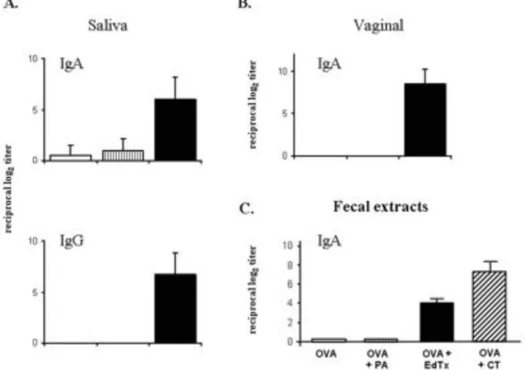
5.1 Anthrax edema toxin is an adjuvant for nasal vaccine antigens and promotes SIgA antibodies ... 59
5.2 Anthrax edema toxin is an adjuvant for its binding subunit and promotes anti-PA antibodies, which protect macrophages against LeTx-induced death... 61
5.3 EdTx as a nasal adjuvant induces antigen-specific Th1 and Th2 cell cytokine responses ... 63
5.4 Nasal EdTx does not exhibit the safety issues associated with ganglioside-binding enterotoxins... 64
5.5 Role of the binding subunit PA in the nasal adjuvanticity of EdTx in vivo... 65
5.6 An enzymatically active EF is needed for the nasal adjuvanticity of EdTx in vivo ... 68
5.7 Involvement of innate immunity components in edema toxin adjuvanticity... 69
5.7.1 Proinflammatory cytokines do not play a major role in the adjuvant activity of EdTx derivatives... 69
5.7.2 Effect of EdTx subunits and mutant derivatives on costimulatory molecule expression and antigen uptake by macrophages ... 71
5.7.3 Role of Nerve Growth Factor in the adjuvant activity of EdTx ... 73
5.8 Edema toxin acts as an adjuvant for serum Ab responses to topically co-applied vaccine antigens ... 78
5.9 Antibody responses induced by EdTx as a transcutaneous adjuvant in saliva and mucosal secretions... 80
5.9.1 CT and EdTx differentially affect immune cells in the GI tract after TCI application ... 81
11
5.11 The transcutaneous adjuvant edema toxin promotes mixed Th1 and Th2 cytokine and delayed-type
hypersensitivity responses ... 84
5.12 Sublingual immunization with EdTx as adjuvant ... 85
5.13 EdTx is an adjuvant for systemic vaccines... 88
6 DISCUSSION ... 91
6.1 The ATR-binding EdTx is an adjuvant for non-invasive vaccines... 91
6.1.1 Requirement for enzymatic activity of EF ... 92
6.1.2 Potential adverse effects... 93
6.2 Quality of immune responses induced by EdTx as adjuvant for non-invasive vaccines ... 93
6.2.1 EdTx induces Abs that neutralize anthrax toxin in vitro... 93
6.2.2 T helper cytokine and DTH responses ... 94
6.2.3 Mucosal IgA Ab responses ... 95
6.3 Mechanisms of adjuvanticity of EdTx for non-invasive vaccines... 95
6.3.1 Contribution of innate immune factors ... 95
6.3.2 Imprinting of homing properties on immunocytes ... 96
7 GENERAL SUMMARY AND CONCLUSION ... 99
8 LIST OF PUBLICATIONS* ... 102
12
Figures and tables
13
1 Figures and tables
Figure 1. The Mucosa-Associated Lymphoid Tissue (MALT). ...20
Figure 2. Homing receptors and addressins for mucosal homing of immune effector cells...22
Figure 3. Structure of human IgA Abs and transepithelial transport of secretory IgA Abs...25
Figure 4. Human and mouse T helper cell subsets. ...29
Figure 5. Enterotoxins CT and LT-I cell entry. ...39
Figure 6. GM1-binding enterotoxin adjuvants CT and LT-I. ...40
Figure 7. Bacillus anthracis life cycle...48
Figure 8. Anthrax toxins cell entry. ...51
Figure 9. Anthrax toxin derivatives as antigen delivery system. ...52
Figure 10. Serum OVA-specific IgG1 Abs after nasal CT. ...58
Figure 11. Serum OVA-specific Abs after nasal immunization with EdTx. ...60
Figure 12. Mucosal SIgA Ab responses following nasal immunization with EdTx. ...61
Figure 13. PA-specific Abs after nasal immunization with EdTx. ...62
Figure 14. EF-specific Abs after nasal immunization with EdTx. ...63
Figure 15. EdTx does not target CNS tissues. ...65
Figure 16. Inflammatory cytokine responses to nasal EdTx in vivo...65
Figure 17. Native Bacillus anthracis toxin subunits and mutant derivatives. ...66
Figure 18. F1-V-specific Abs after nasal immunization with PA mutants and wild-type EF. ...67
Figure 19. PA-specific Abs after nasal immunization with PA mutants and wild-type EF...68
Figure 20. F1-V-specific Abs after nasal immunization with wild-type PA and EF mutants. ...69
Figure 21. In vitro IL-6 cytokines after EdTx treatment in macrophages...70
Figure 22. In vitro costimulatory molecule expression on macrophages after EdTx treatment. ...71
Figure 23. Costimulatory molecule expression in macrophage cell lines after treatment with EdTx derivatives. ...72
Figure 24. Ovalbumin uptake by macrophage cell lines after treatment with EdTx derivatives...73
Figure 25. Salmonella uptake by macrophage cell lines after treatment with EdTx, CpG or CT. ...73
Figure 26. Anti-NGF inhibits Ag-specific responses in vivo. ...74
Figure 27. Anti-NGF inhibits CTb-specific responses in vivo...74
Figure 28. Nerve growth factor is a nasal adjuvant. ...75
Figure 29. Anti-NGF does not inhibit nasal EdTx-induced OVA-specific responses. ...75
Figure 30. Anti-NGF does not inhibit nasal EdTx-induced F1-V-specific responses...76
Figure 31. Anti-NGF does not inhibit nasal EdTx-induced PA-specific responses. ...77
Figure 32. Anti-NGF effect on CT or EdTx-treated B cell IgA secretion. ...78
Figure 33. OVA-specific Ab responses after TCI with EdTx...79
Figure 34. OVA-specific Abs in the secretions after TCI with EdTx. ...80
Figure 35. Long-lasting OVA-specific Abs after TCI with EdTx. ...81
Figure 36. Intestine weights 16 hours after oral or transcutaneous administration of CT. ...82
Figure 37. PA-specific Ab responses after TCI with EdTx. ...83
Figure 38. EF-specific Ab responses after TCI with EdTx: IgG1/ IgG2a ratio...83
Figure 39. OVA-specific Th cell cytokine responses after TCI with EdTx...84
Figure 40. Delayed-Type Hypersensitivity responses after TCI with EdTx...85
Figure 41. Structures of the skin and the buccal epithelia...85
Figure 42. F1-V-specific Ab responses after sublingual immunization with EdTx. ...86
Figure 43. PA-specific Ab responses after sublingual immunization with EdTx...87
Figure 44. EF-specific Ab responses after sublingual immunization with EdTx: IgG1/ IgG2a ratio. ...88
Figure 45. I.p. injection of OVA with EdTx in BALB/c and C57BL/6 mice...88
14 Figure 47. Schematic representations of immune responses after transcutaneous and sublingual
immunizations. ...100
Table 1. Mucosal vaccines against infections. ...36
Table 2. Non-GMI-targeting bacterial toxins and derivatives tested as adjuvant for mucosal vaccines. ...42
Table 3. Cytokines and chemokines as mucosal adjuvants...46
Table 4. Neutralizing anti-PA Ab responses after nasal EdTx. ...62
Table 5. Antigen-specific CD4+ T cell cytokine responses after nasal EdTx. ...64
15
Abbreviations
16
2 Abbreviations
Ab ___________Antibody Ag ___________Antigen
CLN _________Cervical lymph nodes CMI __________Cell-mediated immunity CNS _________Central nervous system CT ___________Cholera toxin
CTb __________Binding subunit of CT DC __________Dendritic cell
ECs __________Epithelial cells
EdTx _________Bacillus anthracis edema toxin (wild-type PA plus wild-type EF) EdTx S414N ___Wild-type PA plus EF S414N
EF ___________Bacillus anthracis edema factor (wild-type)
EF S414N _____Mutant of EF with 10% residual enzymatic activity FAE _________Follicle-associated epithelium
GALT ________Gut-associated lymphoid tissue GI ___________Gastrointestinal
i.p. ___________Intra-peritoneal
IFN-!/"/# ______Interferon alpha/ beta/ gamma IL ___________Interleukine
LeTx _________Bacillus anthracis lethal toxin (wild-type PA plus wild-type LF) LF ___________Bacillus anthracis lethal factor (wild-type)
LP ___________Lamina propria
MALT ________Mucosa-associated lymphoid tissues MHC _________Major histocompatibility complex MLN _________Mesenteric lymph nodes
MØ __________Macrophage
NALT ________Nasal-associated lymphoid tissue NGF _________Nerve growth factor
NK cells ______Natural killer cells OB __________Olfactory bulbs
ON/E _________Olfactory nerves/epithelium OVA _________Ovalbumin
PA ___________Wild-type PA
PA ___________Bacillus anthracis protective antigen
PA dFF E308D _Mutant of PA unable to translocate EF or LF to the cytosol PA-U7 ________Mutant of PA lacking the furin cleavage site
PBS __________Phosphate-buffered saline pIgR _________Polymeric Ig receptor PP ___________Peyer’s patch
SED _________Sub-epithelial dome SIgA _________Secretory-IgA Abs
TCI __________Transcutaneous immunization
TGF-!/"_______Transforming growth factor alpha/ beta Th ___________T helper
TLR __________Toll-like receptor wt____________Wild-type
17
Introduction
18
3 Introduction
Direct blood-borne infections are rare and only occur through injections, transfusions and bites. Most human pathogens enter the body through the mucosal surfaces, mainly the gastrointestinal, genitourinary and respiratory tracts. Cold viruses, influenza, food poisoning agents, tuberculosis, sexually transmitted diseases, cholera, diphtheria and plague are just a few examples. While it is widely accepted that provision of appropriate immunity at mucosal surfaces would be of great benefit for host immunity, only a few current vaccines specifically target the mucosal immunity. Vaccines delivered by mucosal (i.e., oral, nasal or rectal) routes most efficiently promote immunity in both mucosal tissues and the systemic compartment and thus, for the last two decades, great efforts are being undertaken to make mucosal immunization the benchmark method for mass immunization. These efforts include the development and characterization of adjuvants, which could be added to mucosal vaccines and enhance the magnitude and / or the quality of systemic and mucosal immune responses to co-inoculated vaccine antigens.
3.1 The mucosal immune system
3.1.1 The mucosal surfaces
Mucous membranes cover epithelial surfaces and comprise the linings of the digestive, the respiratory and the urogenital systems as well as eye conjunctiva, the inner ear and the ducts of all exocrine glands (salivary, gastric and sweat glands, prostate, liver). Overall in human, they cover the equivalent of one and a half tennis courts or a surface area of approximately 400m$ [1].
The epithelium of mucosal surfaces is protected by a number of mechanisms including the mucus, antibodies, lipids and other non-mucin factors, such as antimicrobial peptides or defensins [2]. The mucosa hosts and selectively regulates an abundant microbiota and antigens. The interactions with commensal microbes and food antigens require the integrity of the mucosal barrier to be preserved. In the event the homeostasis is altered and protective immunity is needed, the epithelial cells lining the mucus membrane will help release secretory IgA (SIgA) antibodies, which are the hallmark of mucosal immunity.
19 3.1.2 Compartimentalization of the mucosal immune system
Highly sophisticated immune mechanisms help maintain the integrity of the mucosal surfaces and prevent the dissemination of pathogens and allergens in the (quasi) sterile systemic compartment. Thus, in addition to the innate mechanisms described above, mucosal surfaces defense arsenal involves well-orchestrated antigen-specific adaptive immune mechanisms. The later is possible by the accumulation in or the transition of immune cells between inductive sites and effector sites. Inductive sites for mucosal immunity consist in defined lymphoid structures, or mucosa-associated lymphoid tissues (MALT), which lack afferent lymphatics, as well as local draining lymph nodes. The effector sites constitute more diffuse lymphoid tissues, including the lamina propria (LP) of various mucosae, the stroma of exocrine glands and surface epithelia. In addition, specific homing receptor / addressin couples orchestrate homing of effector cells from inductive sites to distinct effector sites.
3.1.2.1 Induction of mucosal immunity in the MALT
Gut-associated lymphoid tissue (GALT) comprise the Peyer’s patches (PP), the appendix, solitary lymphoid nodules, crytopatches [3] and isolated lymphoid follicles [4]. In humans, inductive sites of the upper respiratory tract, and oral / nasal cavities are represented by the unpaired nasopharyngeal tonsil or adenoids, the paired palatine tonsils and other smaller lymphoid structures of Waldeyer’s pharyngeal ring, collectively named nasal- or nose-associated lymphoid tissue (NALT). While tonsils are not present in rodents, NALT structures exist and are localized on the cartilaginous soft palate (Figure 1). The GALT has been the object of extensive studies and often serves to describe how immune responses are initiated in inductive sites. Intestinal antigens can be taken across the epithelial cell layer through specialized membrane/microfold or M cells located in the follicle-associated epithelium (FAE) of the PP. Antigens are then captured by APCs of the subepithelial dome (SED), which are mostly dendritic cells (DCs). This event triggers DC to mature and migrate to the interfollicular region (IFR) of PP, and later MLN, where they present antigens to T cells. Those T cells can in turn interact with adjacent B cells in B cell follicles. More recently, it became clear that antigens can also be taken up by dendritic cell extending dendrites in between intestinal epithelial cells (ECs) [5]. As described above for antigens taken up by M cells, those taken up by extending dendrites will ultimately be presented to T cells in MLN for subsequent induction of T and B cell responses. Nasal immunization studies in mice have demonstrated that the NALT serves has the inductive
20 site for immune responses initiated via the airways. Like the GALT, the NALT is covered by M cells and cervical lymph nodes are believed to play the same role as MLN in the gastrointestinal (GI) tract. The skin is not a strict mucosa. However, it is an epithelium in contact with the external environment. The interest for the skin as a “mucosal” tissue has steadily grown since the observation that transcutaneous immunization can induce immunity in the GI tract [6].
During the last few months and while I was working on this document, the first reports were published indicating that the murine sublingual surface is a distinct and previously unsuspected inductive site for mucosal immunity [7, 8]. As we will see later in this dissertation, our new findings support this notion.
Figure 1. The Mucosa-Associated Lymphoid Tissue (MALT).
The mucosal immune system is divided into inductive sites and effector sites. Inductive sites consist of organized mucosa-associated lymphoid tissue (MALT) as well as local and regional draining lymph nodes, whereas effector sites consist of distinctly different histological compartments, including the lamina propria (LP) of various mucosae, stroma of exocrine glands and surface epithelia. Based on anatomical regions, three most studied subdivisions of the MALT are represented: the NALT or nasal-associated lymphoid tissue, the GALT or gut-associated lymphoid tissue, and the SALT or skin-associated lymphoid tissue. The recently described sublingual site of induction is not depicted. Antigens are sampled at mucosal ifferentiate into memory or effector cells. Not depicted inductive sites and presented to local T and B cells. Sensitized B and T cells leave the site of original encounter of the Ag, transit through the lymph to reach the general circulation and then disseminate between selected mucosal effector sites.
21
3.1.2.2 Homing to mucosal effector sites
After initial Ag exposure in inductive sites such as the GALT, activated B and T cells home via the thoracic duct and the blood circulation to distant mucosal effector sites, where they differentiate into effector and memory cells to elicit a mucosal immune response (Figure 1). It is important to note that the homing of effector B and T lymphocytes is governed by specific mucosal homing/tissue receptors, which impose restrictions on their recirculation between mucosal sites. In this regard, !4"7 binds to MAdCAM-1 (mucosal addressin cell adhesion
molecule-1), the major homing receptor ligand in the intestinal lamina propria. Chemokines are also directly involved in mucosal homing of effector B and T cells. CCL25 or TECK (thymus-expressed chemokine) is (thymus-expressed in the small intestine and attracts CCR9+ IgA-commited B cells. Beside CCR9, CCR10 is another chemokine receptor implied in lymphocyte trafficking to the small intestine expressing its ligand CCL28 or MEC (mucosa-associated epithelial chemokine). Mucosal DCs are increasingly recognized as key players in the acquisition of mucosal homing capabilities by effector B and T cells. Thus, retinoic acid produced by GALT DCs (i.e. PP and MLN) appears to be the determinant factor for controlling the expression of integrin !4"7 and CCR9 by B and T cells [9]. As a consequence of the pattern of homing receptors
and addressins expressed by cells sensitized in the GI tract, oral immunization induces strong antibody responses in the small intestine, ascending colon and mammary and salivary glands but not in the distal segments of the large intestine, tonsils, and the female genital tract (Figure 2).
22
Figure 2. Homing receptors and addressins for mucosal homing of immune effector cells.
The selective homing of cells sensitized in a particular inductive site is determined by site-specific integrins or homing receptors they express on their surface and corresponding mucosal tissue-specific receptors or addressins displayed on vascular tissue. Chemokines produced locally by certain tissues also play a role in attracting cells in inductive sites especially by regulating their surface expression of integrins. Interestingly, factors like retinoic acid produced by mucosal DCs located in the MLN or PP can influence the expression of the integrin !4"7 and the
chemokine receptor CCR9 on T and B cells, thereby modulating their migration toward MAdCAM-1 and the gut-associated chemokine TECK/CCL25. Those factors have yet to be identified for inductive sites like the NALT and the SALT. The fact that cytokines, chemokines, integrins are differentially expressed between mucosal tissues explains the compartmentalization of the mucosal immune system which preferentially associates a certain mucosal inductive site with a particular effector site (like the NALT and the respiratory tract, or the GALT and the gastrointestinal (GI) tract).
Rectal and vaginal immunizations give rise to Ab responses essentially restricted to the rectum and the female genital tract mucosa, respectively [10]. One could assume that signals not yet identified, probably provided by local DCs, govern and limit the development of immune responses to these particular sites.
In contrast to the GALT, homing from the NALT or other inductive sites is less well documented. Compelling evidence suggest that !4"7 is not an important homing molecule in the
airways, or the urogenital tract. On the other hand, CCR10, !4"1, and L-selectin seem to control
23 only low to intermediate levels of !4"7, a homing receptor profile that does not favor homing to
the small intestinal mucosa. Lack of L-selectin was shown to impair immune responses in the head and neck tissues and the saliva [11]. It has been shown that NALT-derived B cells express CCR10 and !4"1-integrin at high levels, which allows them to migrate to the respiratory and
genito-urinary tracts. Interestingly, high levels of antigen-specific IgA and IgG Abs could be induced in cervicovaginal secretions after intranasal immunization in mice, monkeys [12] or humans.
As indicated earlier, interest for the skin as a “mucosal” tissue has steadily grown since the observation that transcutaneous immunization can induce immunity in the GI tract [6]. In this regard, the skin-associated lymphoid tissue (SALT) contains keratinocytes, Langerhans cells, skin trophic T cells, and lymphatic endothelial cells of the skin, but few or no B cells [13]. Lymphocyte homing to the skin is regulated by various homing receptors, including the cutaneous lymphocyte-associated antigen. The cutaneous lymphocyte-associated antigen is a carbohydrate epitope present on memory/effector T cells that infiltrate inflamed skin, where it can interact with both P- and E-selectins. Skin-homing effector T lymphocytes also express chemokine receptors such as CCR4 and/or CCR10, and high levels of !4"1.
The fact that E-selectin is highly expressed on endothelial cells of inflamed skin, female genital tract and oral mucosa suggests that transcutaneous immunization could also promote immune responses in the genital tract [14] and saliva, although this point has not been thoroughly
investigated. Retinoic acid, produced by mucosal DCs from the MLN and the PP, favors the expression of gut homing receptors and was shown to block the expression of P- and E-selectin ligands [15]. Vitamin D3 is generated in the skin after sun exposure and converted to the biologically active form 1,25(OH)2D3 through an enzymatic cascade involving
25-hydroxyvitamin D 1 alpha-hydroxylase produced by keratinocytes. In contrast with RA, vitamin D3 suppresses the expression of the gut homing receptors !4"7 and CCR9 and signals T cells to
express CCR10 and thus, enables them to respond to CCR10-ligand CCL27 and to migrate to epidermis [16].
The recent publications on sublingual immunization in mice showed broad immune responses with induction of Ab responses in both the GI and respiratory tracts [7, 8]. However, mucosal homing receptors and addressins involved and their regulation remain to be elucidated.
24 Conclusions from the compartimentalization of the mucosal immune system: The previously described common mucosal immune system is in fact very compartmentalized and this point should be taken in consideration when devising new non-invasive vaccines and immune therapy to be administered via mucosal or cutaneous routes. For instance, immunization via the oral route gives rise to mucosal immune responses that are constraint mainly to the intestinal mucosa, while nasal immunization covers a broader spectrum by inducing mucosal effector cells in the upper respiratory tract, the general circulation and the genitor-urinary tract. In addition, while most basic mechanisms (e.g. lymphoid structures, homing molecules) to enhance secretory immunity are common between mice and humans, some differences may explain inconsistencies between immune responses of mice and humans to mucosal vaccines. Studies in mice have ‘set’ the nasal route of immunization as an effective method of vaccination to induce immune responses in the systemic and distant mucosal effector compartments; its major asset being to induce optimal and long-term protection in the genital tract, which is not achieved after intra-vaginal immunization or oral immunization in mice. Nevertheless, clinical studies in humans have not shown such consistency in terms of protective mucosal Abs found in the reproductive tract and memory responses. Therefore, new immunization strategies for induction of targeted mucosal immunity are still needed.
3.1.2.3 Polymeric IgA antibodies and mucosal immune responses
Mucosal B cells differ from their systemic counterparts and represent a first line of defense at mucosal surfaces through the active production and exportation of secretory IgA (SIgA) Abs and to a lesser extent secretory IgM (SIgM) Abs.
Polymeric IgA and secretory IgA. The daily production of IgA is greater than any other immunoglobulin class. In contrast with mice, which only have one type of IgA, two subclasses of IgA Abs are present in humans (Figure 3A). IgA1 Abs represent the majority of serum IgA (84 %); they are also present in the tonsils and nasal mucosa. IgA2 Abs predominate in the large intestine and the female genital tract. These IgA Abs consist of a dimer (or a tetramer), linked by a J-chain polypeptide, and covered by a polypeptide chain called the secretory component (SC)
[17] (Figure 3A). The SC is derived from the polymeric Ig receptor (pIgR) present on the
basolateral membrane of most mucosal epithelia (digestive, respiratory, genital) and glandular epithelia in the mammary, respiratory and lacrimal glands and is responsible for the transport of
25 polymeric IgA across ECs [18] (Figure 3B). Both IgA subclasses are well exported by pIgR, and give secretory IgA (SIgA). However, IgA2 Abs are more resistant to bacterial degradation than IgA1 Abs [17]. Dimeric IgA secreted by plasma cells in subepithelial tissues binds to pIgR and is internalized by receptor-mediated endocytosis[18]. The complex receptor-IgA is transported into vesicles across the EC to the luminal membrane where the vesicle fuses with the plasma membrane. The pIgR is enzymatically cleaved from the membrane to become the SC, which is bound to and released together with pIgA into the mucous. The same mechanisms apply for the transport of pentameric IgM (Immunoglobulin also containing a J chain) to mucous secretions.
Figure 3. Structure of human IgA Abs and transepithelial transport of secretory IgA Abs.
A. Schematic representations of human immunoglobulins A (IgA). Heavy chains are shown in purple, light chains in blue, J chain in orange, secretory component in yellow. N-linked glycans are shown in green and O-linked glycans by small light blue circles. Top left: IgA1. Top right: IgA2 (Only one allotypic form of IgA2 is represented). Bottom: Dimeric SIgA. B. Schematic representation of the transport of pIgA through a polarized epithelial cell. Polymeric IgA (pIgA) is secreted by plasma cells in the lamina propria at a mucosal membrane. pIgA binds to its receptor, pIgR, on the basolateral surface of an epithelial cell. The complex is endocytosed and transcytosed in endosomes toward the apical surface. There, the pIgR is cleaved, releasing secretory IgA into the lumen. [Adapted from Woof J.M. and J. Mestecky. Mucosal immunoglobulins, 2005. Immunol. Rev. 206 (1), 64–82].
Although not considered as a secretory immunoglobulin, IgG is also present in most external secretions at levels comparable to those of IgM. However, in some secretions like the urine, seminal fluid and female genital tract secretions, IgG Abs outnumber SIgA Abs. The IgG Ab can be passively released into mucosal surfaces complexed to IgA or IgM Ab transported across epithelial cells. Other mechanisms independent of pIgR govern the entrance of IgG into
26 secretions, including passive diffusion. IgE Abs may be carried into mucous membranes and secretions by mast cells and cause their degranulation with local histamine release [19].
Development of SIgA responses. The IgA-secreting B cells are primarily generated through the aid provided by helper T cell to B cell precursors for class-switch from surface (s)IgM to (s)IgA. More precisely, the % to ! isotype switch is mediated by an essential cytokine, TGF-" along with IL-4 and IL-10. The interactions of CD40 on B cells with CD40 ligand on T cells [20], and of ICOS (inducible costimulatory) on activated Th cells with ICOS-L constitutively expressed on B cells [21] are also required for the immunoglobulin switch. This process occurs within the germinal center (GC) of mucosal inductive sites, like the PP. Class-switched B cells expand while homing via the lymph and the blood to the effector sites such as the intestinal lamina propria. There, they continue their differentiation into IgA-producing plasma cells with the aid of IL-5 and IL-6. Because of this dependence on Th2 cytokines, SIgA antibody responses have initially been considered as limited to Th2-inducing stimuli. This notion has been revisited in the last decade, with the observation that SIgA antibodies are also induced by Th1-inducing pathogens/vaccines (i.e., IL-12 [22, 23] and oral Salmonella vectors [24, 25]). In addition, Th1 and inflammatory cytokines (IFN-# and TNF-!) regulate the expression of pIgR on the basolateral membrane of ECs [26] and the expression of SC [27] and thus, the transepithelial transport of
polymeric immunoglobulins. Interestingly, it has been shown that the upregulation of expression of pIgR by TNF-! is enhanced by retinoic acid [28].
The T cell-dependent mechanism for induction of IgA Abs can be further enhanced by a crosstalk between intestinal epithelial cells (IEC) and dendritic cells [29]. IEC sampling antigens in the gut lumen release thymic stroma lymphopoietin (TSLP), a signal sensed by gut DCs. In turn, DCs are able to prime and polarize CD4+ Th cells into Th2 cells producing IL-4 and IL-10. IL-6, which is a plasma cell-inducing cytokine, is also a mediator released by these DCs. Thus, together with TGF-", IL-4 and IL-10 drive mucosal B cells to undergo an IgA-class switch. T cell-independent (TI) pathways for induction of IgA responses to intestinal flora have been described. In mice, these TI responses involve B1 cells derived from the peritoneal cavity. The mechanism relies on B-lymphocyte stimulator/B-cell-activating factor of the TNF family (BLyS/BAFF) and a proliferation-inducing ligand (APRIL), factors secreted by activated antigen-presenting cells (APCs) (e.g. after exposure to bacteria) and involved in peripheral B cell survival, maturation, and differentiation. Humans lack B1 or B2 cells but a similar mechanism to
27 mice for the induction of IgA has recently been unveiled. BAFF and APRIL expressed by DCs and mucosal epithelial cells, sensing bacteria in the gut lumen, can trigger a T cell-independent IgA2 class switch in human mucosal B cells [29].
Biological activities of SIgA. More pIgA is transported to the human gut lumen in a day (40 mg/kg body weight, which represents approximately 3 g of SIgA) than the total production of IgG (30 mg/kg). The high avidity due to multiple antigen-binding sites of IgA (and IgM) and the amount of SIgA produced in the gut compensate for their low affinity. SIgA also possess a feature that makes them resistant to proteolytic degradation in harsh environments such as the gut lumen and oral cavity: the secretory component, acquired during the transepithelial transport of dimeric IgA. Furthermore, SC enhances both stability and effector functions of pIgA. As one would expect, SIgA exert functions that differ from those mediated by IgG in the general circulation and that are adapted to the unique environment where these Abs operate [19].
The main function of antibodies secreted in the lumen is to perform immune exclusion of exogenous antigens, by inhibiting uptake of soluble or particulate Ag. By coating commensal as well as pathogenic microorganisms without stopping their growth, SIgA effectively reduce their access to the epithelial surface and at the same time, maintain the balance of the intestinal flora by preventing the outgrowth / invasion of a population over another. Another non-specific mechanism results from glycans present on SIgA and on free SC (Figure 3), which interfere with microbial adherence to mucosal surfaces. Factors present in the mucous can enhance the protective effects of IgA. For instance, the mucin binds SIgA and thus enhances entrapment of SIgA-coated bacteria in the mucus layer. It has become recently appreciated that SIgA adheres selectively to M cells in intestinal Peyer's patches to mediate the transepithelial transport of the Ab molecule and the bound antigen to underlying GALT [30].
Ag-specific functions of SIgA are illustrated by their role in protection against virus infection, such as influenza or human immunodeficiency viruses (HIV). Elegant studies with HIV have shown that pIgA antibodies transiting in epithelial cells during pIgR-mediated transport can neutralize intracellular viruses [31]. SIgA can neutralize various Ag like viruses, by binding to specific epitopes in mucosal secretions. In addition, SIgA can eliminate immune complex formed inside ECs. The role of SIgA in protection against bacteria is more complex. As mentioned above, SIgA do not interfere with commensal or pathogenic bacteria numbers. However, SIgA are necessary for protection of intestinal surfaces against secreted bacterial toxins.
28 Below the epithelial surface, SIgA display anti-inflammatory properties, which counteract inflammation resulting from complement activation by IgG Abs. However, SIgA can also interact with complement to enhance opsonization of Ag [32].
Summary on secretory antibodies: The SIgM and IgG antibodies can participate in the clearance of mucosal infections, notably in the upper-respiratory and the genital tracts. Nonetheless, SIgA are the major antibodies that contribute to the defense of mucosal surfaces. Serum IgG Abs, can play a major role in the protection of mucosal tissues only in sites where loose epithelial barrier (stratified) allows their leakage from the blood stream. In contrast, SIgA have the unique ability of interfering with the uptake of viruses and macromolecules without requiring or causing tissue damage or alteration of the integrity of the mucosal epithelium. They have been shown to protect against many bacterial toxins and control the initial epithelial colonization of bacterial pathogens, which helps prevent horizontal oral-fecal spread of pathogens and provides herd protections in populations. Thus, it is clear that the induction of specific secretory Abs by a vaccine formulation is a crucial goal for vaccinologists as they could help combat most mucosal infections.
3.1.2.4 Mucosal T cell responses: helper, cytotoxic and regulatory functions
T helper cells in the mucosa. As mentioned above, cytokines secreted by T helper cells play a crucial role in the development of IgA secreting plasma cells. The differentiation of CD4+ T helper cells can be induced in mucosal tissues by APCs that migrate to the interfollicular area of the PP and MLN. It has also been suggested that ECs could process and present antigens themselves to T cells via MHC class I or II molecules and produce cytokines that control the differentiation of Th cell subsets [33]. Like their counterpart in the systemic compartment, two lineages of CD4+ Th cells were described in mucosal tissues based on cytokines produced. Thus, Th1 cells produce IFN-# and TNF-!, while Th2 cells produce IL-4, IL-6, IL-10 and IL-13 (Figure 4). Recently, it has been shown that the repertoire of CD4+ Th cells is more diverse and that IL-17-producing Th cells or Th17 exist alongside with the previously described Th1 and Th2 lineages (Figure 4). Indeed, studies of autoimmune diseases such as experimental autoimmune encephalitis (EAE) and collagen-induced arthritis (CIA) have highlighted the roles of two cytokines, IL-23 and IL-17, rather than Th1 cytokines as traditionally believed. IL-17 is produced by cells involved in the adaptive branch of the immune system but possesses effector
29 functions that are characteristic of the innate system. In fact, IL-17 acts like IL-1", TNF-!, Toll-like receptor (TLR)-4-agonists and as such, represents a link between the innate and adaptive branches of the immune system to induce inflammation. IL-17 also regulates intestinal barrier functions through the induction of tight junction formation [34]. Finally, it has been suggested that TGF-" and IL-6 can induce the differentiation of Th17 cells from naturally occurring CD4+ CD25+ T regulator cell (Treg) precursors [35].
Figure 4. Human and mouse T helper cell subsets.
T helper (Th) cells arise from the thymus. When they acquire CD4, they become precursor Th (pTh) cells. Upon encounter with antigen and stimulation with appropriate cytokines, they start to proliferate. Depending on the nature of the stimulation they can enter the Th1, the Th2 or the Th17 (or IL-17-producing Th cells) pathway. Th1 cells produce IFN-#, and regulate antigen presentation and cellular immunity. Interleukin-12 (IL-12), which is produced by activated antigen-presenting cells (APCs), drives the process of Th1-cell differentiation. Th2 cells produce IL-4, IL-5 and IL-13, which are important regulators in humoral immunity and allergic responses. IL-4 is required for Th2-cell differentiation. Th17 cells, which require IL-23 for differentiation and produce IL-17, as well as IL-6 and TNF-!, have a dominant role in the development and maintenance of autoimmune inflammation. [Adapted in part from Laurence A. and O’Shea JJ. T(H)-17 differentiation: of mice and men, 2007. Nat. Immunol. 8: 903].
Mucosal cytotoxic T lymphocytes (CTLs). Cytotoxic functions of mucosal T cells represent a crucial backup mechanism for mucosal protection in case of a defective or insufficient IgA Ab
30 response development against a viral or a bacterial infection. A majority of mucosal CTLs are CD8+!" T cells and are mostly found at effector sites, such as the intestinal lamina propia. Just like IgA-producing plasma cells, mucosal CTLs derive from precursors in inductive sites (i.e., PP and MLN). CTLs differentiate with the aid IL-2 produced by Th1 cells into effector CTLs and their maintenance and survival is dependent on IL-7 and IL-15. Mucosal CTLs recognize antigen on MHC class I molecule and are then able to kill their target or a virus-infected cell through the secretion of perforin, a protein that forms pores in infected cell membranes, and cytolytic granules full of granzyme proteases. It has been established that CTLs can be induced in systemic and mucosal tissues after oral [36], nasal [37], rectal [38], vaginal and after transcutaneous immunizations [6]. For example, oral immunization with a live recombinant
Mycobacterium bovis BCG vectors expressing SIV nef gives rise to the development of
antigen-specific CTL responses in inductive sites (PP and MLN), in effector tissues (intestinal LP), as well as in systemic lymphoid tissues [39]. Studies with knockout mice have demonstrated the requirement of CD4+ Th cells for induction of CTLs; however, cytotoxic T-cell responses were also reported in vivo in the absence of CD4 helper T cells [40].
Regulatory T cells. Most of the time, the mucosal immune system ignores external microorganisms (i.e., commensal bacteria) and antigens (i.e., food antigens) and does not mount an immune response against them. Mucosal homeostasis can also be maintained by specifically suppressing a cellular and/or a humoral immune response, which would be detrimental for the integrity of the mucosal surfaces. Mucosal tolerance concerns mainly the T cell compartment as delayed-type hypersensitivity (DTH) and in vitro T cell proliferation responses are diminished
[41].Various active mechanisms are involved in mucosal tolerance, including activation-induced
cell death, deletion and/or anergy of Ag-specific T cells, induction of regulatory T cells and production of suppressive cytokines [41].
To date, four types of regulatory T cells were described: 1) the antigen-specific CD4+ Th2-like cells that produce IL-4 and IL-10 and antagonize the activity of Th1 effector cells [42]; 2) the CD4+CD45RBlow Tr1 cells producing IL-10 [43]; 3) the TGF-"-producing Th3 cells [44]; 4) and CD4+CD25+ regulatory T cells or Treg cells [45, 46]. Tregs are naturally occurring regulatory T cells; they are CD4+ and express the IL-2-receptor ! chain (CD25). Because they are anergic, they do not proliferate after allogeneic or polyclonal stimulation and produce small amounts of cytokines. Once activated, they block proliferation of and cytokine production in CD4+ or CD8+
31 T cells in a cell contact-dependent manner. Notably, Treg cells immediately confer suppressive properties to CD4+ T cells, a process that has been termed “infectious tolerance”. In turn, these induced-Th suppressor cells display suppressive activities that are cell contact-independent (cytokine-mediated).
Recent studies have provided a link between Tr1 cells, Th3 cells and Tregs. Indeed, it has been shown that there are two subsets of Tregs with different properties. This dichotomy is based on their expression of one of the two integrins, !4"7 and !4"1, one being a homing receptor to
mucosal site, the other to inflamed tissues, respectively. Studies have demonstrated that co-culture of !4"1+ Tregs with conventional CD4+ T cells induce Th3-like TGF-" secreting cells
while !4"7+ Tregs in the same culture conditions induce IL10-producing Tr1 like cells and the
induction of Foxp3, a transcription factor thought to be expressed solely on Tregs [47].
While the induction of mucosal tolerance may be useful to treat allergies or autoimmune diseases, the development of a regulatory mechanism in response to a mucosal vaccine against an infectious disease is certainly not desirable. Therefore, appropriate vaccine formulations with optimal antigen / adjuvant / delivery system combinations are needed to overcome suppressive responses and elicit strong immune responses.
Summary on mucosal T cells and relevance for vaccine strategy: Regulatory T cells prevent the host from developing excessive reactions the myriade of microorganisms presnt in mucosal surfaces. Cytotoxic T cells can also provide mucosal tissues with specific and non specific protection of against pathogens. These responses as well as the magnitude and quality of Ab responses are orchestrated by T helper cells. Therefore, a major challenge for vaccinologists is to induce the T helper subset(s) that will more effectively support the needed immune responses for optimal protection. In this regard, the following parameters have important implications on the nature of T helper cell subset responses: (1) the adjuvant, (2) the vaccine carrier, (3) the presence of regulatory cytokines, (4) the route of administration, and (5) the quantity and nature of the antigen.
32
3.2 Mucosal vaccines and adjuvants
3.2.1 Mucosal vaccines against infections
As mentioned earlier, the most common sites of entry for infectious agents are mucosal surfaces. Because only immune responses initiated in mucosal tissues give rise to effector cells with mucosal homing capabilities, local protection of these sites can only be achieved by mucosal vaccination. Other major advantages of mucosal vaccinations are the following: 1) The administration is painless and does not require trained medical personnel; 2) These needle-free procedures eliminate the risk of cross-infections by blood-borne pathogen though contaminated needles.
The development of mucosal adjuvants has long been empirical, with variable degree of success. It has now become clear that the design of effective mucosal vaccines and delivery systems should take into account specific hurdles associated with the mucosal route considered. For example, intact proteins are usually destroyed in the GI tract or their uptake is limited, proteolysis of vaccine antigen by enzymes, and interferences with local antigens and flora are the main obstacles in the case of oral delivery. Regardless of the route of immunization, it is important to confer the adequate immunostimulating properties to the mucosal vaccine for the induction of adaptive immunity. This is usually the role of the adjuvant. To date, several mucosal vaccines are either currently used or under development for mass vaccination.
3.2.1.1 Oral polio vaccine
The live-attenuated orally administered polio vaccine (Sabin OPV), introduced in 1961, is the first known oral vaccine. The OPV was superior at inducing mucosal Ab responses than the parenteral inactivated polio vaccine (Salk IPV) released in 1955. An enhanced formulation of the parenteral vaccine (e-IPV) was later found to induce some IgA Abs in nasal secretions, although to a lower extent than after OPV or infection with wild-type virus. This observation raised the debate that some mucosal immunity could be induced by parenteral immunization. However, this information is contentious because data was collected among populations who could also receive OPV or who were located in poliovirus endemic areas. Therefore, it is still unclear whether parental e-IPV can induce mucosal immunity. However, it is unambiguous that individuals that have been either primed by OPV or naturally exposed to the live poliovirus can develop a strong memory mucosal IgA response [48]. The OPV remains the vaccine of choice for use against
33 epidemic outbreaks of poliomyelitis due to its ability to rapidly induce development of intestinal immunity, and to provide indirect vaccination for those coming into contact with immunized patients. Nonetheless, better attenuation of the vaccine strain would be needed to prevent potential reversion to wild-type and subsequent risk of vaccine-associated paralytic poliomyelitis
[49, 50].
3.2.1.2 Vaccines against enteric infections
The World Health Organization (WHO) estimates that diarrheal diseases account for 15-34 % of all deaths in certain countries and cause 4 to 6 million deaths a year, mostly children, eldery and immunocompromised people. While efficacious vaccines exist against V. cholerae, S. typhi and rotavirus, none are available against enterotoxigenic E. coli (ETEC) and Shigella (Table 1). Cholera vaccines. Cholera is one of the best-studied mucosal infections. Until recently, the only vaccine available consisting of phenol-killed whole cells of Vibrio cholerae, was administered parenterally in two doses at two-week interval and reached a 50% protection in individuals that lasted no longer than 6 months. Presently, two oral vaccines are available. Dukoral is made of killed whole cells Vibrio cholerae O1 combined with recombinant binding subunit CTb. It has proven to be safe and efficient in several trials. The live attenuated vaccine CVD 103-HgR, containing a genetically manipulated classical V. cholerae O1 Inaba strain, with a deletion in the gene encoding the enzymatic subunit cholera toxin A, is the second internationally licensed oral cholera vaccine. While some studies have shown very high levels of protection (>90%), a large field trial in Indonesia yielded only 24% protection in the first year [51].
Typhoid fever vaccines. Typhoid fever is caused by Salmonella enterica, serovar typhi, commonly called Salmonella typhi. It is responsible for 600,000 deaths a year according to the WHO. The parenteral heat phenol inactivated whole cell vaccine is still in use in some developing countries but is associated with fever and systemic reactions. There are now two other vaccines available against typhoid fever, both very well tolerated. An orally administered, live-attenuated vaccine made from the Ty21a strain of Salmonella typhi, and a Vi capsular polysaccharide vaccine (ViCPS) (Sanofi Pasteur) for intramuscular use in one dose. None provide total protection but both reduce typhoid fever. Presently, efforts are being made to search more immunogenic strains for oral vaccines [52].
Rotavirus vaccines. Rotaviruses are the most common cause of severe diarrhea in children. It is responsible of about 600,000 deaths annually worldwide. Two virus surface proteins, VP4 and
34 VP7, are targets of neutralizing antibodies and either antibody can mediate protection. In 1998, the highly efficacious vaccine Rotashield, a live, orally administrated,tetravalent human-simian reassortant (RRV-TV) vaccine containing four serotypically distinct VP7 components, was licensed but withdrawn shortly after because of elevated cases of intussusception (i.e. intestinal invagination resulting in obstruction). In 2006, two new vaccines were introduced. Rotarix is a monovalent live-attenuated human rotavirus vaccine (made of a tissue-culture-adapted human P1A[53]G1, VP6 subgroup II and NSP4 geno-group B strain), administered in two doses orally in infants of 2 and 4 months of age. RotaTeq is a pentavalent bovine-human reassortant vaccine. It contains several pieces of nucleic acid from human and bovine parent rotavirus strains; it is composed of the five strains, each containing a human rotavirus gene encoding the VP7 neutralizing protein from different serotypes. This vaccine is given orally in a three dose-schedule in infants who are 2, 4 and 6 months old. Rotarix and RotaTeq are considered equivalent in their protective effects [54, 55].
3.2.1.3 Vaccines against respiratory infections
Influenza virus. A trivalent live attenuated cold-adapted vaccine FluMist has been developed for intranasal spray delivery and licensed in the USA for vaccination of persons from 5–49 years of age [56]. The vaccine is safe, effective, and shows remarkable genetic stability, but has to be kept at -18°C. When compared with injectable influenza vaccine, live attenuated influenza vaccine has demonstrated broader serum antibody responses, particularly against antigenically mismatched influenza A strains [57]. A new, heat-stable version of the vaccine has recently been developed, and has shown remarkable efficacy in clinical trials. Other influenza vaccines, currently being developed, include a synthetic peptide influenza vaccine for nasal administration. In an attempt to elicit a higher antibody response, PowderJect (USA) developed a needle-free delivery method to administer influenza vaccine called epidermal powder immunization and shown elevated humoral immune responses in humans [58]. Virosomes are small (150 nm) spherical vesicles composed of a lipid membrane with integrated envelope proteins derived from influenza virus, predominantly hemagglutinin. They differ from virus-like particles in that they are semisynthetic particles reconstituted in vitro from lipids and from virus-derived proteins. An intranasal virosomal subunit influenza vaccine with heat labile toxin from Escherichia coli (LT-I) as the adjuvant elicited strong systemic and mucosal Ab responses in healthy adults, with the presence of neutralizing secretory IgA Abs in nasal secretions [59].
35 Respiratory Syncytial Virus (RSV). The RSV causes lower respiratory tract infection in infants and reinfections can occur in all age groups. To date, there is no licensed vaccine against RSV. Most attenuated live RSV strains tested in humans caused mild to moderate congestion in the upper respiratory tract of infants of one to two months old and, therefore, were considered as insufficiently attenuated for early infancy. Intranasally delivered, genetically engineered attenuated or vectored vaccines are currently most promising for newborns. Maternal vaccination may be the optimal strategy to protect the very young infants [60].
3.2.1.4 Vaccines against genitourinary infection
Human Papillomavirus (HPV) serotypes 16 and 18 are associated with the development of cervical cancer [53, 61]. HPV is a nonenveloped, encapsulated, double-stranded DNA virus. The HPV L1 capsid protein can generate virus-like particles (VLP) when expressed in heterologous system such as yeast. VLPs are non-infectious and immunogenic without adjuvant, can induce neutralizing antibodies and be protective against viral challenge. A tetravalent vaccine composed by virus-like particles (VLPs) of HPV 16, HPV 18, HPV 6 and HPV 11 has been approved in the USA [62]. The VLPs are manufactured in Saccharomyces cerevisiae and administered with aluminum adjuvant in three intramuscular injections. Research efforts are now focusing on an alternative route of administration and on the induction of immunity in the genital mucosa to overcome the variation of Ag-specific antibodies in the cervix seen in ovulating women. Nasal administration of HPV16 VLPs along with LT-I in mice has been shown to elicit strong systemic and mucosal Ab responses, with the presence of SIgA in vaginal secretions, as well as cell-mediated immunity against VLPs [63].