Université de Sherbrooke
Effects of apelin mimetic compounds on sodium current in rat ventricular cardiomyocytes
By
Alexandria Tiffinger
Department of Pharmacology-Physiology
Thesis presented to the Faculty of Medicine and Health Sciences to obtain the degree of Master of Science (M. Sc.)
in Physiology
Sherbrooke, Québec, Canada January 2018
Members of Evaluation Jury
Robert Dumaine, Ph.D., Pharmacology-Physiology Ahmed Chraïbi, Ph.D., Pharmacology-Physiology Michel Grandbois, Ph.D., Pharmacology-Physiology
knowledge will sooner or later play its part in the service of man.” -Ernest Starling
Effets des composés mimétiques de l'apéline sur le courant sodique dans les cardiomyocytes ventriculaires chez le rat
Par
Alexandria Tiffinger
Programme de Pharmacologie-physiologie
Mémoire présenté à la Faculté de médecine et des sciences de la santé en vue de l’obtention du diplôme de maitre ès sciences (M. Sc.) en Physiologie, Département de
Pharmacologie-physiologie, Faculté de médecine et des sciences de la santé, Université de Sherbrooke, Sherbrooke, Québec, Canada, J1H 5N4
L'hypertension est la principale cause de décès au monde. L'hypertension artérielle commence avec le rétrécissement des vaisseaux sanguins, suivi par une augmentation de la pression artérielle. Il y a une augmentation compensatoire de la contractilité et du rythme cardiaque due à une stimulation adrénergique accrue. Cette période initiale d'augmentation de l’adrénaline est suivie d'une désensibilisation des récepteurs bêta-adrénergiques et de l’apparition d’arythmies et d’hypertrophie cardiaque. L'hypertrophie cardiaque est un mécanisme physiologique caractérisé par une augmentation de la taille des cardiomyocytes et de la masse musculaire. Les effets inotropes négatifs de l’hypertrophie cardiaque conduisent à une réduction du débit et une accélération du rythme cardiaque. Les traitements de l'hypertension devraient réduire la tension artérielle tout en augmentant la contractilité cardiaque. L'apéline, un peptide endogène, possède ces qualités. Elle existe sous trois formes actives, soit l'apéline-13, -17 et -36. L'apéline se lie à son récepteur (APJ), un récepteur couplé aux protéines G (GPCR), et augmente la contractilité et l’excitabilité cardiaque. Nous avons déjà montré que l'apéline-13 augmente de 35% l’amplitude du courant sodique cardiaque (INa) (Chamberland et al., 2010). Les effets de l'apéline sont transitoires et sa demi-vie est courte. Dans le but d'améliorer son potentiel thérapeutique, notre groupe a remplacé le résidu C-terminal de l'apeline-13 par les acides aminés non naturels 2Nal et Tyr(OBn) (Murza et al., 2012, 2015). Les composés mimétiques ont une plus grande affinité pour APJ par rapport à l'apéline-13. 2Nal et Tyr(OBn) peuvent être des alternatives utiles aux thérapies anti-adrénergiques actuelles dans le traitement de l'hypertension et la prévention de l'hypertrophie. Ce mémoire étudie les effets de l'apéline-13, du 2Nal et du Tyr(OBn) sur l’INa dans des cardiomyocytes ventriculaires du rat, dans le but d'évaluer la capacité des composés mimétiques à moduler la contractilité cardiaque. 2Nal et Tyr(OBn) n'ont eu aucun effet sur l’amplitude d’INa. Leurs effets au niveau de l'activation, de l'inactivation ainsi que sur le courant de fenêtre (IW), qui se produit entre ces deux états, suggèrent qu'ils peuvent augmenter la contractilité cardiaque en diminuant l'activité de l’échangeur Na+/Ca2+ (NCX). Les effets de 2Nal et de Tyr(OBn) pourraient donc être thérapeutiques ou pro-arythmiques. Mots clés : INa, récepteur APJ, 2Nal, Tyr(OBn), cardiovasculaire, arythmie, hypertension, hypertrophie cardiaque
2
A
BSTRACTEffects of apelin mimetic compounds on sodium current in rat ventricular cardiomyocytes
By
Alexandria Tiffinger Pharmacology-Physiology
Thesis presented to the Faculty of Medicine and Health Sciences to obtain a Master degree diploma (M.Sc.) in Physiology, Department of Pharmacology-Physiology, Faculty of Medicine and Health Sciences, Université de Sherbrooke, Sherbrooke, Québec, Canada,
J1H 5N4
Hypertension is the leading cause of death globally, and its prevalence continues to rise. Arterial hypertension starts with the narrowing of blood vessels, leading to increased blood pressure. Consequently, there is a compensatory increase in cardiac contractility and rhythm due to increased adrenergic stimulation. This initial period of increased epinephrine levels is followed by desensitization of beta-adrenergic receptors, leading to arrhythmias and cardiac hypertrophy. Cardiac hypertrophy in cardiovascular disease is a pathological mechanism in which there is an increase in the size of cardiomyocytes, leading to an increase in muscle mass. Ultimately, the negative inotropic effects of cardiac hypertrophy lead to cardiac dysfunction, characterized by a reduced cardiac output and accelerated heart rate. In order to help prevent the development of pathological cardiac hypertrophy, medications for the treatment of hypertension should reduce blood pressure while increasing cardiac contractility. Apelin, an endogenous peptide, possesses these qualities. Patients with hypertension have an increased level of circulating apelin, which exists in three active forms: apelin-13, -17, and -36. Apelin binds to the APJ receptor, a G protein coupled receptor (GPCR), and increases cardiac contractility and excitability. Our lab has previously shown that apelin-13 increases the amplitude of the cardiac sodium current (INa) by 35% (Chamberland et al., 2010). The effects of apelin are, however, transient, and its half-life is short. With the goal of improving upon apelin to further study its therapeutic potential for cardiovascular disease, our research group replaced the C-terminal residue of apelin-13 with unnatural amino acids 2Nal and O-benzyl-Tyrosin (Tyr(OBn)) (Murza et al., 2012, 2015). The resulting mimetic compounds have higher affinity for the APJ receptor compared to apelin-13. 2Nal and Tyr(OBn) may therefore be useful alternatives to current anti-adrenergic therapies in the treatment of hypertension and prevention of hypertrophy. This study investigates the effects of apelin-13, 2Nal, and Tyr(OBn) on sodium current in isolated rat ventricular cardiomyocytes, with the goal of assessing the ability of the mimetic compounds to modulate cardiac contractility through INa. In contrast to apelin-13, 2Nal and Tyr(OBn) had no effect on INa amplitude. Their actions on INa activation and inactivation, and on the window current (IW) that occurs between these states, suggest that they may nonetheless increase cardiac contractility via a decrease in Na+/Ca2+ exchanger (NCX) activity. The effects of 2Nal and Tyr(OBn) may therefore be therapeutically beneficial, or pro-arrhythmic. Keywords: INa, APJ receptor, 2Nal, Tyr(OBn), cardiovascular, arrhythmia, hypertension, cardiac hypertrophy
1 Résumé en Français ... iv 2 Abstract ... iv 3 Table of Contents ... vi 4 List of Figures ... ix 5 List of Tables ... x 6 List of Abbreviations ... xi 1 1. Introduction ... 1
1.1 An overview of the heart and cardiovascular system ... 1
1.2 The heart: cardioelectrophysiology ... 2
1.2.1 The cardiac conduction system ... 2
1.2.2 Cardiac action potentials, ionic channels, and currents ... 3
1.2.2.1 Cardiac sodium channel and current ... 6
1.2.3 The basis of contraction... 8
1.3 The heart: cardiovascular disease ... 10
1.3.1 Hypertension and hypertrophy ... 10
1.3.2 Cardiac arrhythmias ... 11
1.4 The apelin receptor, APJ... 12
1.4.1 Structure ... 12
1.5 Apelin ... 13
1.5.1 Structure ... 13
1.5.2 Signaling ... 14
1.5.2.1 G protein signaling pathways ... 14
1.5.2.2 Biased signaling ... 16
1.6 The apelinergic system ... 16
1.6.1 Apelin and the cardiovascular system ... 17
1.6.1.1 Apelin has a cardioprotective role ... 17
1.6.1.2 Apelin decreases cardiac pressure and improves cardiac contractility ... 19
1.6.1.3 Apelin modulates sodium currents ... 21
1.7 Apelin-13 C-terminal modifications affect its binding, signaling, and hypertensive action ... 23
1.7.1 Apelin mimetic compounds 2Nal and Tyr(OBn) ... 24
1.8 Hypothesis ... 26
1.8.1 Objective ... 27
2 2. Materials and Methods ... 28
2.1 Animal models ... 28
2.1.1 Isolation of rat ventricular cardiomyocytes ... 28
2.1.1.1 Preparation of animals and surgical procedure ... 28
2.1.1.2 Enzymatic dissociation ... 29
2.2 Whole-cell patch clamp ... 30
2.2.1 Setup and micropipettes ... 30
2.2.2. Data recording ... 30 2.2.2.1 INa activation protocol ... 31 2.2.2.2. INa inactivation protocol ... 31 2.3 Electrophysiological solutions ... 31 2.3.1 Extracellular solution ... 32 2.3.2 Intracellular solution ... 32 2.4 Mimetic compounds ... 32
2.4.1 Synthesis of mimetic compounds... 32
2.4.2 Drug treatments ... 32
2.5 Data analysis and statistics... 33
3 3. Results ... 34
3.1 Effects of apelin-13 on cardiac sodium current ... 34
3.1.1 Effect of apelin-13 on INa amplitude ... 34
3.1.2 Effect of apelin-13 on INa steady state activation... 36
3.1.3. Effects of apelin-13 on INa steady state inactivation ... 37
3.1.4 Effect of apelin-13 on sodium window current ... 39
3.2 Effects of 2Nal on cardiac sodium current ... 40
3.2.1 Effects of 2Nal on INa amplitude ... 40
3.2.2 Effect of 2Nal on INa steady state activation ... 41
3.2.3 Effect of 2Nal on INa steady state inactivation ... 42
3.3 Effects of Tyr(OBn) on cardiac sodium current ... 43
3.3.2. Effect of Tyr(OBn) on INa steady state activation ... 45
3.3.3 Effect of Tyr(OBn) on INa steady state inactivation ... 46
3.4 Effects of 2Nal and Tyr(OBn) on window current ... 47
4 4. Discussion ... 49
4.1 Summary of results ... 49
4.2. The effects of apelin-13 and its mimetic compounds Tyr(OBn) and 2Nal on cardiac sodium current in isolated rat ventricular cardiomyocytes ... 50
4.2.1 Concentration-dependent effects ... 50
4.2.1.1 Apelin-13 ... 50
4.2.1.2 2Nal and Tyr(OBn) ... 51
4.2.2 Potential effects on excitability ... 51
4.2.3 Potential effects on channel availability ... 52
4.2.4 Effects on sodium window current ... 53
4.2.5 Potential effects on calcium homeostasis ... 54
4.2.6 Potential effects on intracellular signalling ... 56
4.2.6.1 Potential implication of the cAMP-PKA pathway ... 56
4.2.6.2 Potential implication of biased signaling ... 57
4.3 Study limitations ... 58
4.3.1 The rat model ... 58
5 5. Conclusions and Perspectives ... 61
5.1 Conclusions ... 61 5.2 Perspectives ... 61 6 6. Acknowledgments ... 63 7 7. List of References ... 64 8 8. Appendix ... 75 8.1 Protocol adjustments ... 75
8.1.1 Enzymatic dissociation protocol ... 75
Figure 1. General anatomy of the heart ... 1
Figure 2. The cardiac conduction system ... 3
Figure 3. Cardiac currents ... 4
Figure 4. Structure of the voltage-gated sodium channel NaV1.5 ... 6
Figure 5. The molecular basis of contraction ... 8
Figure 6. Schematic representation of the crossbridge cycle of contraction ... 9
Figure 7. Schematic representation of apelin, its three active forms, and its receptor ... 13
Figure 8. Schematic representation of apelin-APJ receptor signaling pathways ... 15
Figure 9. Apelin has positive inotropic effects ... 19
Figure 10. Apelin increases cardiac excitability ... 20
Figure 11. Apelin increases cardiac contractility and modulates cardiac sodium current .... 21
Figure 12. The C-terminal residue of apelin-13 appears to be essential to its effects ... 23
Figure 13. Apelin-13 mimetic compounds 2Nal and Tyr(OBn) ... 25
Figure 14. Patch clamp recording protocols ... 30
Figure 15. Effect of apelin-13 on cardiac sodium current amplitude ... 35
Figure 16. Apelin-13 increases INa in a concentration-dependent manner ... 36
Figure 17. Effect of apelin-13 on cardiac sodium current steady state activation ... 37
Figure 18. Effect of apelin-13 on cardiac sodium current steady state inactivation ... 38
Figure 19. Effect of apelin-13 on cardiac sodium window current ... 39
Figure 20. Effect of 2Nal on cardiac sodium current amplitude ... 41
Figure 21. Effect of 2Nal on cardiac sodium current steady state activation ... 42
Figure 22. Effect of 2Nal on cardiac sodium current steady state inactivation ... 43
Figure 23. Effect of Tyr(OBn) on cardiac sodium current amplitude ... 44
Figure 24. 2Nal and Tyr(OBn) do not have a concentration-dependent effect on INa amplitude ... 45
Figure 25. Effect of Tyr(OBn) on cardiac sodium current steady state activation ... 46
Figure 26. Effect of Tyr(OBn) on cardiac sodium current steady state incativation ... 47
Table 1: Changes made to tyrode solution ... 76
Table 2: Changes made to intracellular solution ... 76
Table 3: Changes made to extracellular solution ... 77
4-AP AC ACE Akt AP APJ AT1a ADP ATP ATII BDM BSA cAMP CHO EC50 EGTA DAD DAG DNA DT EAD ENa eNOS ERK1/2 GNa GNa,max GPCR h IC50 ICaL IKr IKs IK1 IL-8 INa INa,max 4-Aminopyridine Adenylate cyclase Angiotensin-converting enzyme Protein kinase B Apelin
Angiotensin-like putative (receptor) Angiotensin II receptor type 1a Adenosine diphosphate
Adenosine triphosphate Angiotensin II
2,3-Butanedione monoxime Bovine serum albumins
Cyclic adenosine monophosphate Chinese hamster ovary
Half maximal effective concentration Egtazic acid Delayed afterdepolarization Diacylglycerol Deoxyribonucleic acid Developed tension Early afterdepolarization
Reversal potential for sodium current Endothelial nitric oxide synthase Extracellular signal-related kinase 1/2 Sodium current conductance
Maximum sodium current conductance G protein coupled receptor
Steady state inactivation Inhibitory constant Calcium current
Rapid-activating delayed rectifier potassium current Slow-activating delayed rectifier potassium current Inward rectifier potassium current
Interleukin-8 Sodium current
Ito IW IP3 IV LTCC LVDP m mRNA MAP MAPK MCP-1 MEK MIF mTOR NaV NCX NHE NO Pi PI3K PIP2 Phe Phe13 PCR PKA PKC PLC RyR SEM SERCA SCN5A TTX Tyr(OBn) V1/2 Vm Vmax Vrest
Transient outward potassium current Window current
Inositol triphosphate
Current-voltage (relationship) L-type calcium channel
Left ventricular developed pressure Steady state activation
Messenger ribonucleic acid Mean arterial pressure
Mitogen-activated protein kinase Monocyte chemoattractant protein-1 MAP/ERK kinase
Migration inhibitory factor Mechanistic target of rapamycin Voltage-gated sodium channel Na+/Ca2+ exchanger Na+/H+ exchanger Nitric oxide Inorganic phosphate Phosphoinositide 3-kinase Phosphatidylinositol-4,5-biphosphate Phenylalanine
C-terminal phenylalanine residue of apelin-13 Polymerase chain reaction
Proteine kinase A Protein kinase C Phospholipase C Ryanodine receptor
Standard error of the mean
Sarco/endoplasmic reticulum Ca2+-ATPase
Sodium voltage-gated channel alpha subunit 5 (gene) Tetrodotoxin
O-benzyl-Tyrosin
Mid-activation or -inactivation potential Membrane potential
Speed of depolarization Resting membrane potential
1.1 An overview of the heart and cardiovascular system
The heart is a muscular organ responsible for pumping blood throughout the body via rhythmic contractions. It is composed of four chambers, two ventricles and two atria (Figure 1). The myocardium, the thick middle layer of the heart wall, comprises the muscle tissue of
Figure 1. General anatomy of the heart
The heart is composed of four compartments: the left and right atria, and left and right ventricles. Separating the right atrium and ventricle is the tricuspid valve, while the mitral valve separates the left atrium and ventricle. The pulmonary valve separates the right ventricle and pulmonary arteries, and the aortic valve separates the left ventricle and aorta. Inset: The heart wall has three distinct layers. The myocardium, the muscular middle layer, is composed of cardiomyocytes and is responsible for contraction of the heart. It is surrounded by the pericardium, the outermost layer, and the endocardium, the innermost layer. Image adapted from Nerbonne and Kass, Physiol Rev 2005 and Servier Medical Arts.
the heart and provides structure for the heart’s chambers. It is composed of cardiomyocytes and is responsible for the contraction of the heart (Boron and Boulpaep, 2017).
The three main components of the human circulatory system are the heart, blood, and blood vessels. Contraction of the heart pumps blood throughout the body, and cardiac output volume and arterial pressure fluctuate with each heartbeat. Arterial pressure reaches a maximum systolic pressure with ventricular contraction and minimum diastolic pressure with ventricular relaxation (Boron and Boulpaep, 2017). Mean arterial pressure (MAP) represents the arterial pressure of one cardiac cycle over its duration (Boron and Boulpaep, 2017). The diameter of blood vessels and cardiac output blood volume can change blood pressure – MAP increasing with vasoconstriction or increased blood volume, or decreasing with vasodilation or decreased blood volume (Boron and Boulpaep, 2017).
1.2 The heart: cardioelectrophysiology
1.2.1 The cardiac conduction system
The heart contracts and relaxes in a rhythmic and coordinated manner. Three main structures – the generating sinoatrial and atrioventricular nodes, and impulse-propagating His-Purkinje system – are responsible for coordinating cardiac contraction (Figure 2). Cardiac muscle cells, or cardiomyocytes, within the myocardium posses a property known as automaticity, which is the ability to generate their own action potentials, enabling electrical impulses to be transmitted rapidly throughout the myocardium. Electrical synapses, or gap junctions, between cardiomyocytes allow the propagation of action potentials from cell to cell within cardiac muscle tissue. The His-Purkinje system allows for the simultaneous contraction of the ventricles via conducting fibres composed of electrically excitable cells. The system consists of the common Bundle of His, its left and right branches, and Purkinje fibres. Together, these specialized tissues are known as the cardiac conduction system.
Cardiac contraction is initiated by action potentials that originate spontaneously in the sinoatrial node, rather than via neuronal stimulation. The sinoatrial node is a group of specialized cardiomyocytes located in the right atrium near the superior vena cava, and is
under both parasympathetic and sympathetic nervous control. It is colloquially known as the natural pacemaker of the heart, as it intrinsically initiates action potentials at a steady rate (Boron and Boulpaep, 2017; Boyett, 2009; Park and Fishman, 2011).
1.2.2 Cardiac action potentials, ionic channels, and currents
Action potentials are transient electrical impulses in which the membrane potential (Vm) of excitable cells depolarizes, or becomes more positive than resting membrane potential (Vrest), before returning, or repolarizing, to Vrest (Boron and Boulpaep, 2017). Depolarization of the cellular membrane above a minimum threshold potential is required for the initiation of an action potential (Boron and Boulpaep, 2017). In ventricular
Figure 2. The cardiac conduction system
There are three main components of the cardiac conduction system: the sinoatrial node, atrioventricular node, and His-Purkinje system. Cardiac action potentials are generated spontaneously at the sinoatrial node, which is located in the upper right atrium. The atrioventricular node, located in the lower portion of the right atrium, receives the signal from the sinoatrial node; it is responsible for conduction between the atria and the ventricles. After a brief delay, the signal is conducted from the atrioventricular node to the apex of the heart via the common bundle of His, the left and right bundle branches, and finally the Purkinje fibres. Imaged adapted from Nerbonne and Kass, Physiol Rev 2005 and Servier Medical Arts.
cardiomyocytes, action potentials have five distinct phases occurring as a result of the
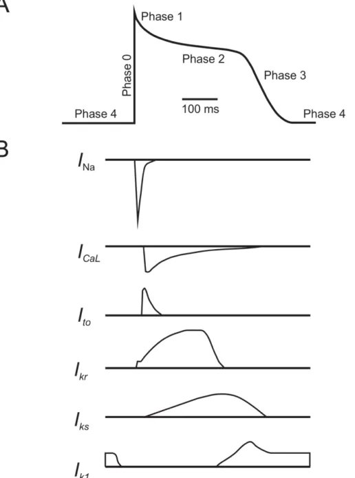
Figure 3. Cardiac currents
A. Ventricular cardiomyocyte action potential has 5 distinct phases. Phase 0 is an initial rapid depolarization of the cell (upstroke). Phase 1, a transient repolarization, follows immediately afterwards and leads to a voltage plateau (Phase 2) characteristic of ventricular action potentials. Phase 3 repolarization returns the cell to normal resting membrane potential, where the cell remains in Phase 4. B. Depolarizing and repolarizing currents contributing to each phase. INa: inward sodium current; ICaL: inward slow calcium current; Ito, Ikr, Iks, and IK1: outward potassium currents. Modified from Nerbonne and Kass, Physiol Rev 2005.
coordination of depolarizing (inward) and repolarizing (outward) currents (Figure 3A). The activation and inactivation of different ionic currents are responsible for each phase (Figure 3B). Phase 0 is the rapid depolarizing upstroke and is associated to activation of the inward sodium current (INa). Partial repolarization in Phase 1 follows immediately, due to the activation of transient outward potassium current (Ito) and inactivation of INa. Phase 2, the plateau phase, is particularly prominent in ventricular cardiomyocytes, and is responsible for excitation-contraction coupling. The plateau is a result of residual INa and activation of the calcium current (ICaL), balanced with the activation of outward rapid- (IKr) and slow-activating (IKs) delayed rectifier potassium currents. As ICaL inactivates, IKr and IKs favour repolarization, initiating Phase 3, which is completed by activation of the inward rectifier potassium current (IK1). The cell is thus returned to its resting potential, the diastolic resting membrane potential of Phase 4 (Boron and Boulpaep, 2017; George, 2013; Nerbonne and Kass, 2005). Phase 4 occurs when the cell is at rest, before the initiation of a new action potential, where the membrane most permeable to K+ (I
K1) and membrane potential remains around approximately -90 mV (Grunnet, 2010; Santana et al., 2010).
Electrical activity in cardiomyocytes is mediated by several ionic currents. INa is generated by activation of voltage-gated NaV1.5 sodium channels. It is responsible for the rapid depolarization of Phase 0 in atrial and ventricular cardiomyocytes. The transient repolarization that immediately follows in Phase 1 is due, in part, to the inactivation of these sodium channels, and to the activation of fast and slow voltage-gated Ito channels. The depolarization of the membrane activates voltage-gated L-type Ca2+ channels. I
Ca,L, paired with residual INa and counteracted by emerging outward K+ currents, is responsible for the characteristic plateau of Phase 2 (Nerbonne and Kass, 2005; Schmitt et al., 2014). This influx of calcium into cardiomyocytes triggers contraction (Bers and Perez-Reyes, 1999). ICa,L inactivates, increasing K+ efflux and eventually favouring repolarization via I
Kr and IKs, respectively. This leads to Phase 3 repolarization which is completed via IK1, which helps to maintain resting membrane potential throughout Phase 4 (George, 2013; Nerbonne and Kass, 2005; Schmitt et al., 2014).
1.2.2.1 Cardiac sodium channel and current
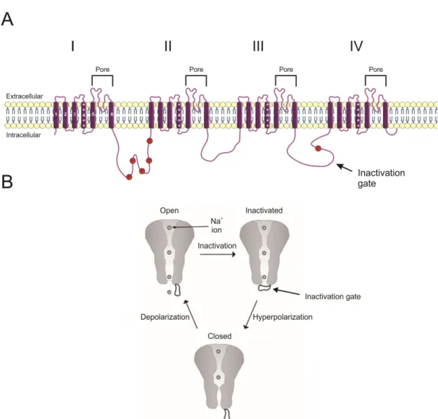
Figure 4. Structure of the voltage-gated sodium channel NaV1.5
A. Schematic representation of the structure of NaV1.5. Roman numerals (I-IV) indicate subunits, each composed of 6 transmembrane -helices (numbered in white). Transmembrane segment 4 of each subunit (numbered and indicated by ++++) are the voltage-sensors. The ion-selective pore is formed by the space between segments 5 and 6 of each subunit. The transmembrane loop between subunits III and IV forms the channel’s inactivation gate. Red circles indicate phosphorylation sites. B. NaV1.5 has three conformational states: open, inactivated, and closed (resting). (A) Adapted from Yu et al., Genome Biol 2003 and Servier Medical Arts. (B) Modified from Bagal et al., Channels (Austin) 2015.
In excitable cells, action potentials are initiated and propagated by voltage-gated sodium channels (NaV). A number of NaV isoforms exist, and are localized to different cell types. NaV1.1, 1.2, 1.3, and 1.6 isoforms are localized primarily to central nervous system neurons, while NaV1.7, 1.8, and 1.9 are predominant in the peripheral nervous system. In skeletal muscle, NaV1.4 is the primary NaV isoform (Catterall, 2012). In the heart, the cardiac sodium current is generated by the opening of tetrodotoxin (TTX)-insensitive NaV1.5 channels. All sodium channels share a basic structure of four transmembrane subunits and additional subunits (Figure 4A). Each subunit has 6 transmembrane -helices, with segment 4 (S4) acting as voltage sensor. An ion-selective space is found between segments 5 and 6 (S5 and S6) of each domain and corresponds to the pore of the channel. An extracellular loop between domains III and IV binds to the S6 segment and inactivates the channels upon depolarization (Catterall, 2000; McPhee et al., 1995; Yu and Catterall, 2003). Mutations in the gene SC5NA coding for the cardiac sodium channel that affect inactivation are linked to cardiac arrhythmias associated with long QT and Brugada syndromes (Brugada et al., 2014; Dumaine and Antzelevitch, 2002).
Voltage-gated sodium channels have three conformational states depending on the position of the activation and inactivation gates: closed, open, and inactive (Figure 4B) (Bagal et al., 2015). At resting membrane potential, NaV1.5 is in its closed, or resting, state. From the channel’s closed state, the activation gate opens rapidly in a multi-step process in response to depolarization of the cellular membrane above threshold potential. This allows for an influx of Na+ and the upstroke of the action potential (Catterall, 2000). Inactivation, on the other hand, is a slower, single-step process following depolarization. During inactivation, the inactivation gate – the intracellular loop between segments III and IV – closes by binding to the intracellular pore. The inactivation of of sodium channels gradually decreases the number of available channels as the resting membrane potential of the cells is depolarized, or the channels open, thus reducing the amplitude of INa. In the inactive state, the channels become impermeable to Na+ and insensitive to membrane depolarization. Upon return to resting membrane potential, the inactivation gate unbinds and the channel returns to its closed state, in which it is once again sensitive to membrane depolarization (Armstrong, 2006; Bagal et al., 2015; Eaholtz et al., 1994; Hille, 1992).
1.2.3 The basis of contraction
At an intracellular level, the coordination and interaction of a number of ion channels, exchangers, and other molecules is required for contraction (Figure 5). Calcium is the ultimate driver of contraction, with the entry of calcium into the cell triggering contraction (Bers, 2002). Calcium enters the cell via L-type calcium channels (LTCC), which are activated when a propagating action potential reaches the T-tubule of a cardiomyocyte. LTCCs are the voltage sensors in excitation-coupling, the process by which an increase in intracellular calcium concentration triggers contraction of a cardiomyocyte (Meissner, 2004). Figure 5. The molecular basis of contraction
Calcium ions (Ca2+) enter the cardiomyocyte via L-type calcium channels (LTCC), stimulating ryanodine receptors (RyR) on the sarcoplasmic reticulum membrane to release Ca2+ into the cytoplasm. Epinephrine binds to -adrenergic and modulates intracellular Protein kinase A (PKA) signalling. Ca2+ is eliminated from the cytoplasm via the sodium-calcium exchanger (NCX) on the cellular membrane and SERCA on the sarcoplasmic reticular membrane. Adapted from Servier Medical Arts.
The entry of calcium via LTCCs activates ryanodine receptors (RyRs) on the sarcoplasmic reticulum (SR) membrane, releasing calcium into the cytoplasm and further increasing the intracellular concentration of calcium (Meissner, 2004). Epinephrine can also facilitate cardiomyoctye contraction via the type 2 -adrenergic receptor, a G-protein coupled receptor (GPCR). Binding of epinephrine triggers an adenylate cyclase (AC)-mediated pathway via the stimulatory Gs subunit. Downstream protein kinase A (PKA) further stimulates both LTCC and RyR directly to increase intracellular Ca2+ concentration.
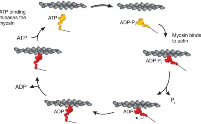
Figure 6. Schematic representation of the crossbridge cycle of contraction
The crossbridge cycle is the basic mechanism of myocyte contraction. (Clockwise) The hydrolysis of ATP on an unbound myosin head (yellow) to ADP-Pi allows the formation of a crossbridge between the activated myosin head (red) and actin (grey). The release of Pi strengthens the crossbridge, and a conformational change in the myosin head produces the power stroke. The filament remains in rigor until the ADP molecule is released and a new ATP molecule binds to the myosin head, releasing myosin. Modified from Sivaramakrishnan et al., JCTR 2009.
Myocytes are composed of functional units called myofibrils, which themselves are composed of a thick filament (myosin) and thin filament (composed of troponin, tropomyosin, and actin). Following the increase in intracellular calcium as described above, calcium binds and activates troponin C. Calcium-troponin C binding allows conformational changes in troponin T and troponin I that permit the interaction of myosin and actin. A crossbridge forms between the myosin head and actin following the hydrolysis of an adenosine triphosphate (ATP) molecule on the myosin head. When inorganic phosphate (Pi) is released, forming adenosine diphosphate (ADP), the crossbridge strengthens, and the power stroke of contraction occurs as a conformational change in the myosin head pulls actin and myosin filaments past one another. The release of ADP at the end of the cycle leaves the myofilament in a rigid state until ATP again binds to the myosin head and dissociates the actin-myosin complex (Figure 6) (Huxley, 1969; Sivaramakrishnan et al., 2009).
When the contraction is complete, Ca2+ is eliminated from the cytoplasm via two mechanisms involving the sodium-calcium exchanger (NCX) on the cell membrane, and the sarco/endoplasmic reticulum Ca2+-ATPase (SERCA) on the SR membrane. NCX is an electrogenic transporter which passively imports three sodium (Na+) ions in exchange for the export of one Ca2+, using the electrochemical gradient of Na+ as the driving force to maintain ionic gradients. In contrast, SERCA is an active transporter which uses ATP to pump Ca2+ from the cytoplasm to the SR following stimulation and contraction (Boron and Boulpaep, 2017; Periasamy and Kalyanasundaram, 2007).
1.3 The heart: cardiovascular disease
1.3.1 Hypertension and hypertrophy
Hypertension, or high blood pressure affects 40% of the world’s adult population and is the leading cause of death around the world (Padwal et al., 2016). It begins with the narrowing of blood vessels (Mayet and Hughes, 2003). This can be initially caused by a number of factors, including a diet high in salt and/or fat, a sedentary lifestyle, and genetics (Ashley and Niebauer, 2004; Blaustein et al., 2012). An elevated level of salt (sodium) can lead to an increased concentration of intracellular calcium via reduced turnover of the NCX.
Elevated calcium levels, in turn, causes contraction of smooth muscle and, ultimately, vasoconstriction (Coleman and Khalil, 2002). Increased sodium concentration can result in reduced synthesis of vasodilating nitric oxide (NO), further increasing vasoconstriction (Edwards and Farquhar, 2015).
A primary consequence of hypertension is elevated arterial pressure, requiring the heart to work harder in order to produce the same cardiac output. As such, epinephrine levels rise to increase cardiac function. Initially, epinephrine increases cardiac contractility and, consequently, cardiac rhythm, by increasing cardiac current via LTCC and myofilament sensitivity (Lohse et al., 2003). Following this initial period of compensation, however, the sustained stimulation of -adrenergic receptors by epinephrine results in their desensitization. Consequently, contractility is reduced and the heart responds less to adrenergic stimulation (Lohse et al., 2003; Packer, 1988; Vidal et al., 2012). In this manner, hypertension can lead to hypertrophy, a condition characterized by an increase in cardiac muscle mass (Shimizu and Minamino, 2016). Cardiac hypertrophy is a physiological mechanism in which there is an increase in the number of myofilaments, and therefore an increase in cardiomyocyte size, resulting in cardiac dysfunction (Schiattarella and Hill, 2015; Shimizu and Minamino, 2016). Hypertrophy is characterized by the gradual desensitization of myofilaments. Theoretically, pathological hypertrophy as a result of hypertension could be prevented by the treatment of hypertension with a drug that increases cardiac contractility, while decreasing blood pressure. Apelin possesses these qualities.
1.3.2 Cardiac arrhythmias
Pharmacological treatments of cardiovascular disease, including hypertension, often alter cardiac contractility via modulation of cardiac currents. As such, when investigating and developing new cardiovascular therapeutic molecules, it is imperative to assess their potential to cause cardiac arrhythmias. Cardiac arrhythmia is a condition in which the heart beats with an abnormal rhythm. Changes in ionic currents can result in transient afterdepolarization cardiac arrhythmias, of which there are two subtypes: early and delayed afterdepolarizations (EADs and DADs, respectively). Abnormal repolarization in phases 2 or 3 of the cardiac action potential results in EADs (Antzelevitch and Burashnikov, 2011). EADs are a potentially-lethal form of ventricular arrhythmia in cardiac disease. Action
potentials generated by EADs can induce additional EADs as they propagate, potentially leading to the development of sustained arrhythmias such as Torsades des Pointes, which can cause sudden cardiac death (Weiss et al., 2010). DADs occur following complete repolarization, before initiation of a new action potential (Antzelevitch and Burashnikov, 2011). Similarly to EADs, DADs above a minimum threshold can trigger cardiac action potentials, in turn causing premature ventricular contraction and triggering further arrhythmias (Liu et al., 2015). Though changes to intracellular concentrations of sodium and calcium, or to the kinetics of the ionic channels, can have therapeutic potential in the treatment of cardiovascular disease, they may alternatively cause negative secondary effects.
1.4 The apelin receptor, APJ
1.4.1 Structure
Interested in identifying deoxyribonucleic acid (DNA) sequences coding for vasopressin receptors, O’Dowd et al. (1993) discovered a polymerase chain reaction (PCR) fragment within the human genome which encoded a novel protein with seven hydrophobic transmembrane domains common to GPCRs (O’Dowd et al., 1993). As much as 54% commonality was found between the amino acid sequence of the novel protein’s transmembrane domains and those of the angiotensin II (ATII) receptor type AT1a (O’Dowd et al., 1993). The new protein was therefore named the angiotensin-like putative (APJ) receptor (O’Dowd et al., 1993). Although the APJ receptor shares a similarity of 30% with AT1a receptor, it does not bind ATII (O’Dowd et al., 1993). The APJ receptor was shortly thereafter identified in amphibians, rats, and in murine embryonic tissue (Devic et al., 1996, 1999; Habata et al., 1999).
The APJ receptor protein displays a high level of evolutionary conservation. In the years since its discovery, APJ orthologues have been found in a number of other vertebrate models, including rhesus macaque, bovine, zebrafish, and dog (Chamberland et al., 2010; Devic et al., 1996; Margulies et al., 2001; Schilffarth et al., 2009; Tucker et al., 2007). Genomic analysis has also shown an amino acid homology of 92% between human and murine APJ, and 90% between human and rat APJ (Devic et al., 1999; O’Carroll et al., 2000).
1.5 Apelin
1.5.1 Structure
Initially tagged as an “orphan GPCR” receptor, it was later found that APJ receptors were actually binding apelin, an endogenous peptide (O’Dowd et al., 1993; Tatemoto et al., Figure 7. Schematic representation of apelin, its three active forms, and its receptor Apelin is an endogenous protein formed form the proteolytic cleavage of pre-pro-apelin. There are three active forms of apelin: apelin-13, apelin-17, and apelin-36. All three forms bind to the angiotensin-like putative (APJ) receptor. Apelin exhibits a high degree of homology between vertebrate species. Solid black outlines indicate residues conserved between human, bovine, mouse, and rat. Image adapted from O’Carroll et al., J Endocrinol 2013; Zhong et al., BBA 2017; and Servier Medical Arts.
1998). Apelin is a 36-amino acid peptide, which was first isolated from bovine stomach extract (Tatemoto et al., 1998). Despite the structural similarities between the APJ and AT1a receptors, there are no significant structural or sequential similarities between apelin and ATII (Lee et al., 2000; Tatemoto et al., 1998).
Apelin is an endogenous peptide, produced by the proteolytic cleavage of pre-pro-apelin into one of three active forms: pre-pro-apelin-36, pre-pro-apelin-17, or pre-pro-apelin-13, the most common form in circulation (Tatemoto et al., 1998). Similar to the APJ receptor, apelin possesses a high level of evolutionary conservation, indicating that apelin may have an important physiological role (O’Carroll et al., 2013) (Figure 7). The C-terminal 27 amino acids are identical in human and bovine pre-pro-apelin (Tatemoto et al., 1998). Furthermore, the isoforms of apelin exist with a high degree of homology across a number of vertebrate species, including human, rat, mouse, and bovine, with 100% homology of the final 23 C-terminal amino acids of apelin-36 (Habata et al., 1999).
1.5.2 Signaling
1.5.2.1 G protein signaling pathways
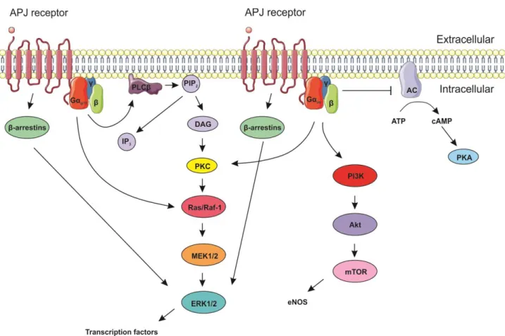
The binding of apelin to the APJ receptor stimulates intracellular signalling via a number of GPCR-mediated pathways (Figure 8). Apelin binding to the APJ receptor elicits a downstream intracellular signaling response that depends on the type of G protein coupled to the receptor (Masri et al., 2002). Via Gq/11, apelin can activate the phospholipase C (PLC) pathway to hydrolyze phosphatidylinositol bisphosphate (PIP2) to inositol triphosphate(IP3) and diacylglycerol (DAG) (O’Carroll et al., 2013). In turn, DAG can activate the phosphokinase C (PKC) pathway and downstream extracellular signal-related kinase (ERK) cascade (Masri et al., 2002). ERK can also be activated via β-arrestin signaling (Shenoy et al., 2006). Apelin can also activate the ERK cascade via direct stimulation of Ras, independent of PKC. The ERK pathway, in turn, can lead to the activation of various transcription factors (Daviaud et al., 2006). Via Gi/o, apelin binding to the APJ receptor can inhibit cyclic adenosine monophosphate (cAMP) production from adenosine triphosphate (ATP) and adenylate cyclase (AC), reducing protein kinase A (PKA) production (Bai et al., 2008; Medhurst et al., 2003; Tatemoto et al., 1998). Apelin can also activate the PKC pathway described above via Gi/o. PKA- and PKC-mediated pathways are both known to
increase INa (Herbert et al., 1990; Lu et al., 1999; Smith and Goldin, 1992; Zhou et al., 2000). Apelin has been shown to increase INa, however in a PKC-dependent and PKA-independent manner (Chamberland et al., 2010). Via Gi/o, apelin can also activate the phosphoinositide 3-kinase (PI3K) pathway (Zeng et al., 2010). The PI3K pathway results in downstream activation of endothelial nitric oxide synthase (eNOS) to produce nitric oxide (NO) (Azizi et
Figure 8. Schematic representation of apelin-APJ receptor signaling pathways
Apelin binding to its receptor, the APJ receptor, activates a number of G-protein coupled receptor (GPCR)-mediated pathways that vary depending on the type of coupled G protein. PLC (phospholipase C ); PIP2 (phosphatidylinositol-4,5-biphosphate); IP3 (i nositol triphosphate); DAG (Diacylglycerol); PKC (protein kinase C); MEK1/2 (MAP/ERK kinase 1/2); ERK1/2 (extracellular signal-related kinase 1/2); AC (adenylate cyclase); ATP (adenosine triphosphate); cAMP (cyclic AMP); PKA (protein kinase A); PI3K (phosphoinositide 3-kinase); Akt (protein kinase B); mTOR (mechanistic target of rapamycin); eNOS (endothelial nitric oxide synthase). Arrows indicate stimulation while the blunted arrow indicates inhibition. Adapted from J Endocrinol 2013 and Servier Medical Arts.
al., 2013; Ishida et al., 2004; Jia et al., 2007; Tatemoto et al., 2001). Finally, apelin also recruits -arrestins (Evans et al., 2001; Lee et al., 2010; Zhou et al., 2003).
1.5.2.2 Biased signaling
Recent studies indicate that apelin-APJ receptor signalling may occur via a biased mechanism (Ceraudo et al., 2014; Kumar et al., 2016; Read et al., 2016; Yang et al., 2015). Biased signaling is the tendency of a ligand binding to its receptor to activate one GPCR signaling pathway over another. APJ receptor variants with mutations at various conserved extracellular sites responsible for calcium mobilization preferentially favoured one of Gq, Gi, or β-arrestin signaling (Kumar et al., 2016). Modulation of the signaling bias of apelin could affect the kinetics of contraction. As described above, calcium plays an essential role in excitation-contraction coupling of cardiomyocytes. For example, a bias towards Gq/11 could result in an increase in IP33. IP3 stimulates calcium release from the SR, therefore increasing the concentration of intracellular calcium, and potentially increasing cardiomyocyte contractility. Furthermore, PKC signalling occurs via either Gi/o or Gq/11 signaling. As PKC increases INa, apelin signaling via the APJ receptor could increase cardiomyocyte contractility in this manner. Finally, β-arrestin signaling can also induce ERK1/2 signaling, which has been shown to be activating by apelin in a PLC-independent manner (Masri et al., 2002). As β-arrestin signaling also promotes GPCR endocytosis, favouring these pathways may, alternatively, increase the internalization of the APJ receptor, potentially minimizing the effects of apelin.
1.6 The apelinergic system
The apelinergic system is widely distributed throughout the vertebrate central nervous system and peripheral tissues and has numerous roles. It is found in the spinal cord and hypothalamo-nerohypophysial axis of the central nervous system, as well as in a number of peripheral tissues such as the pancreas, kidney, lung, and heart (Lee et al., 2000; Medhurst et al., 2003; Murza et al., 2016; O’Carroll et al., 2000; Yu et al., 2014). The apelin/APJ pathway plays a regulatory role in homeostasis (Galanth et al., 2012), metabolism (Bertrand et al.,
2015; Boucher et al., 2005), and, most notably, the cardiovascular system (Chamberland et al., 2010; Dalzell et al., 2015; Farkasfalvi et al., 2007; Szokodi et al., 2002).
1.6.1 Apelin and the cardiovascular system
In the circulation of healthy adult humans, apelin is present at nanomolar concentrations (Zhen et al., 2013). In individuals with chronic heart failure, however, apelin levels in the plasma are decreased, while ventricular apelin expression is upregulated (Chong et al., 2006; Földes et al., 2003). Furthermore, in rats with end-stage heart failure, the apelin/APJ receptor signaling pathway is also suppressed (Koguchi et al., 2012). Apelin has been shown to have both direct and indirect effects at the cardiac level, acting to increase contractile force and vasodilation (Ashley et al., 2005; Salcedo et al., 2007; Szokodi et al., 2002). Additionally, the long-term administration of apelin to hypertensive rats with advanced heart failure has been shown to rescue the activity of the suppressed apelin/APJ pathway, as well as inhibit cardiac dysfunction and remodelling (Koguchi et al., 2012). Apelin, therefore, seems to have beneficial effects in cardiopathological conditions. Our goal in this study is to determine the cardiac effects of apelin, primarily on the electrophysiology of the heart.
1.6.1.1 Apelin has a cardioprotective role
Within the cardiovascular system, the apelinergic system appears to have hypotensive effects (Czarzasta et al., 2016; He et al., 2015; Zeng et al., 2009). Indeed, compared to wild type mice, apelin-deficient mice exposed to chronic hypoxia exhibit more severe pulmonary hypertension (He et al., 2015). Administration of apelin to rats with monocrotaline-induced pulmonary hypertension reduced arterial pressure, right ventricular overload and hypertrophy, and improved biventricular diastolic function (Falcão-Pires et al., 2009).
Apelin seems to also have protective effects against infarction. Diminished cardiac function during an infarction can be improved by the administration of apelin, which, by activating the ERK1/2 pathway, reduces cardiomyocyte apoptosis and oxidative stress (Zeng et al., 2009; Zhong et al., 2017). Furthermore, rats fed high-fat diets following infarction have
been shown to have increased expression of apelin and myocardial APJ receptor messenger ribonucleic acid (mRNA) (Czarzasta et al., 2016).
Apelin may also improve contractility by remodeling the heart contractile machinery. Hypothyroid rats treated with apelin experienced significant increases in left ventricular developed pressure (LVDP) as well as cardiac contractility. When co-administered with the thyroid hormone T4, the effects of apelin on heart contractile performance were further amplified, suggesting that apelin may mimic the non-genomic cardiac effects of thyroid hormones, which are impaired in hypothyroidism (Faraji Shahrivar et al., 2016). The non-genomic effects of thyroid hormones include the modulation of ion flux via voltage-gated sodium channels and NCX function (Hiroi et al., 2006). Additionally, in these hypothyroid rats, apelin was able to increase alpha-myosin heavy chain mRNA expression, effectively reducing associated cardiac dysfunction, though the mechanism by which this occurs remains yet to be investigated (Faraji Shahrivar et al., 2016). As the PI3K/Akt pathway has been implied in the cardioprotective effects of thyroid hormone, it is possible that this pathway via Gi/o biased signaling may similarly be implicated in apelin’s effects on cardiac contractile function and machinery (Faraji Shahrivar et al., 2016; Hiroi et al., 2006; Masri et al., 2004).
Apelin appears to also have a cardioprotective role against cardiac dysfunction during sepsis. In human patients admitted to intensive care with critical illnesses. An increase in blood apelin levels was detected during sepsis (Lesur et al., 2010). Administration of apelin to septic rats and rats with systemic inflammation-induced cardiac dysfunction appears to improve cardiac dysfunction by reducing the production of pro-inflammatory cytokines, including monocyte chemoattractant protein-1 (MCP-1) and interleukin-8 (IL-8) (Chagnon et al., 2017; Pan et al., 2010). In a endotoxemic rat model, apelin-13 also reduces myocardial inflammation, injury, and apoptosis, including migration inhibitory factor (MIF) and inducible nitric oxide synthase, as well as cleaving caspase 3 expression and attenuating myeloperoxidase activity (Chagnon et al., 2017). The apparent protective effect of the apelinergic system within the cardiovascular system supports the idea of a potential therapeutic role of apelin or related compounds.
1.6.1.2 Apelin decreases cardiac pressure and improves cardiac contractility
Intravenous injection of apelin immediately decreases the systolic and diastolic blood pressure and mean arterial pressure (MAP) of anaesthetized rats in a dose-dependent manner (Lee et al., 2000; Tatemoto et al., 2001). Co-administration of a nitric oxide (NO) inhibitor eliminates the effect of apelin on MAP, indicating that apelin may act to reduce blood pressure via a NO-depending mechanism (Tatemoto et al., 2001). It has been shown in vivo in humans that apelin, via an NO-dependent mechanism, causes the vasodilation of splanchnic arteries and forearm resistance vessels, and decreases MAP (Japp et al., 2008; Salcedo et al., 2007).
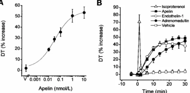
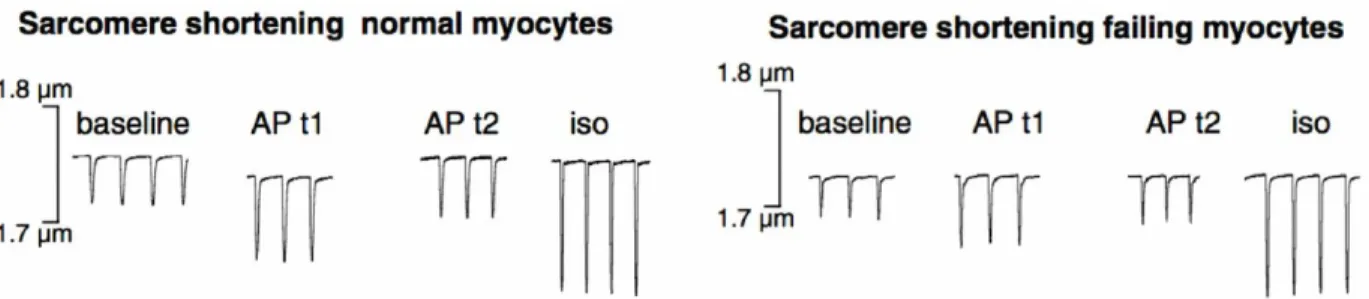
In isolated perfused rat hearts, apelin increases developed tension in a dose-dependent manner, with a half-maximal response of approximately 0.03 nM (Figure 9). Apelin also has a transient effect on isolated perfused rat hearts, with a progressive increase in contractility observed from approximately 2 to 24 minutes of perfusion time (Szokodi et al., 2002). In both healthy and failing cardiomyocytes, apelin induced transient sarcomere shortening (Figure 10) (Farkasfalvi et al., 2007). The positive inotropic effect of apelin has also been
Figure 9. Apelin has positive inotropic effects
A) Apelin increases developed tension (DT) in isolated perfused rat hearts. B) In contrast to isoprotenerol, a β-adrenergic receptor agonist, there is a difference in the time course of DT with apelin. Whereas isoprotenerol produces a peak which quickly returns to basal levels, apelin causes a gradual increase in DT which remains consistent without returning to basal levels. Figure modified from Szokodi et al., Circ Res 2002
observed in vivo, following administration of apelin to both healthy rats and rats with heart failure (Andersen et al., 2011; Berry et al., 2004). Comparison of wild type mice to apelin-deficient littermates showed that, with age, those with apelin deficiency developed reduced cardiac contractility (Andersen et al., 2011; Kuba et al., 2007). Furthermore, the positive inotropic action of apelin appears to be elevated in failing myocardium compared to healthy muscles, possibly due to a higher availability of Ca2+ due to apelin exposure (Dai et al., 2006). Apelin remains one of the most potent endogenous positive inotropic agents presently known (Maguire et al., 2009).
Inhibition of the Na+-Ca2+ exchanger (NCX) reduces the positive inotropic effects of apelin (Szokodi et al., 2002). The driving force behind the NCX, which exports one calcium ion in exchange for three sodium ions under normal conditions, is the sodium concentration gradient across the cellular membrane. A change in intracellular sodium concentration can thus alter the function of the NCX (Shattock et al., 2015). An increase in intracellular sodium as a result of increased INa, for example, would decrease the concentration gradient across the cellular membrane, therefore reducing the rate of turnover of NCX, and thus reducing the extrusion of calcium and prolonging the state of elevated intracellular calcium concentration during contraction. In this manner, apelin may also act to increase contractility of cardiomyocytes via an increase in calcium availability (Dai et al., 2006).
Figure 10. Apelin increases cardiac excitability
Treatment with apelin results in a transient increase in contractility of cardiomyocytes. In normal cardiomyocytes, at time 1 (1 min), there is an increase in sarcomere shortening which returns to approximately baseline levels by time 2 (8 min). Apelin has the same effect on the contractility of failing cardiomyocytes, with an increase in sarcomere shortening at time 1 and a return to baseline levels by time 2. The control treatment with isoprotenerol shows that the cells are still excitable following apelin treatment. Figure modified from Farkasfalvi et al., 2007.
1.6.1.3 Apelin modulates sodium currents
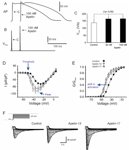
Figure 11. Apelin increases cardiac contractility and modulates cardiac sodium current
A. Treatment with apelin increases the amplitude of cardiac action potentials. B. The speed of depolarization (Vmax), indicating an increase in sodium current in the presence of 100 nM apelin. C. Apelin increases Vmax in a dose-independent manner. D. Apelin increases sodium channel openings at negative voltages. Both apelin-13 and -17 increase the peak amplitude of INa compared to control (no drug), and the threshold of activation is more negative. E. The left shift in steady state activation shows that apelin increases activation at negative voltages. F. Apelin-13 and apelin-17 both gradually increase the amplitude of the sodium current to a maximal level after a period of perfusion of 20 minutes. Figure modified from Chamberland et al., J Mol Cell Cardiol 2010.
The APJ receptor was confirmed to be present in dog cardiomyocytes for the first time in 2010 (Chamberland et al., 2010). Immunofluorescence confocal microscopy shows that APJ receptors are localized at Z-lines of isolated left ventricular cardiomyocytes. Z-lines are areas of high contractility due to the presence of T-tubules and sodium channels, therefore indicating an interaction between apelin, the APJ receptor, and T-tubules to facilitate its inotropic effects (Chamberland et al., 2010).
Using patch clamp technique on the isolated canine cardiomyocytes, it was also shown that at a concentration of 100 nM, both apelin-13 and apelin-17 increase the amplitude of sodium current to a maximal level after 20 minutes of perfusion (Figures 11D and 11F.). Maximum conductance remains unaffected by apelin (Chamberland et al., 2010). Steady-state activation of the sodium current, however, undergoes a negative shift following perfusion with apelin (Figures 11D and 11E.). This indicates an increase in the probability of sodium channels to open at more negative voltage rather than the synthesis or recruitment of additional channels (Chamberland et al., 2010). Furthermore, a change in sodium current amplitude is likely to change the rate of cardiac action potential firing and cardiac excitability. Indeed, apelin, at a concentration of 100 nM, also increases the maximum rate of depolarization of ventricular action potentials (Figures 11A, 11B, and 11C) (Chamberland et al., 2010).
While apelin does not affect sodium current steady state inactivation, it does slow recovery from inactivation. The longer recovery will prolong the refractory period of the cardiac action potential. The longer recovery will prolong the refractory period of the cardiac action potential, which may minimize the risk of tachycardia (Chamberland et al., 2010). Together with the increase in cardiac excitability caused by the specific modulation of sodium current activation, the dual-effect characteristic of apelin indicate that it has strong therapeutic potential for treating arrhythmias as well as cardiovascular diseases such as arterial hypertension and heart failure (Chamberland et al., 2010).
1.7 Apelin-13 C-terminal modifications affect its binding, signaling, and hypertensive action
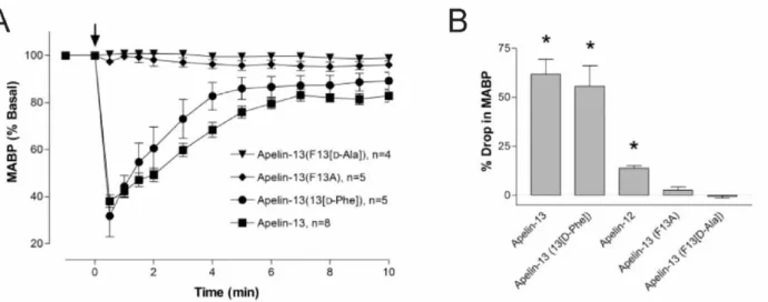
In order to further explore the therapeutic potential of the apelinergic system, a greater understanding of the structure-activity and structure-function relationships of apelin/APJ receptor is required. The interaction between apelin and the APJ receptor was first established by measuring the extracellular acidification rate of Chinese hamster ovary (CHO) cells expressing the APJ receptor, in the presence of apelin isolated from bovine stomach as well as synthetic derivatives. Synthetic apelin-36, apelin-17, and apelin-13 produced from the C-terminal 36, 17, and 13 residues of pre-pro-apelin, respectively, were shown to elicit a response in CHO cells expressing the APJ receptor (Tatemoto et al., 1998). Apelin-36 showed the lowest activity in this study, with a median effective concentration (EC50) of 20 nM, compared to 2.5 nM for apelin-17 and 0.37 nM for apelin-13 (Tatemoto et al., 1998). The shorter isoforms, apelin-17 and apelin-13, produced responses 8 and 67 times greater, respectively, than the response to the longer isoform, apelin-36 (Tatemoto et al., 1998). This was the first indication that the C-terminal region of apelin was involved in its binding to the APJ receptor, and ultimately imperative for its biological functions (Tatemoto et al., 1998). More recently, it has been shown that apelin-13 analogues with the terminal Phe13 residue
Figure 12. The C-terminal residue of apelin-13 appears to be essential to its effects Apelin-13 analogues with the terminal Phe13 residue deleted or replaced by an alanine residue do not induce the observed drop in mean arterial pressure (MABP) that is observed with unmodified apelin-13. Figure modified from Lee et al., Endocrin 2005.
deleted or replaced by an alanine residue do not induce the previously-observed decrease in MAP (Lee et al., 2005) (Figure 12). Therefore, the C-terminal residue of apelin-13 appears to be essential to its hypotensive effect.
Apelin, in summary, appears to have many potentially beneficial characteristics for the treatment of cardiovascular disease, including anti-inflammatory properties, as well as the ability to increase contractility in the heart while decreasing contractility in the vasculature. Many current treatments for cardiovascular disease induce similar contractile effects at both the level of the heart and the vasculature, limiting their efficacy. Ideally, cardiovascular treatments should reduce the arterial pressure while increasing cardiac contractility; apelin possesses these qualities. The apelinergic system is thus an attractive potential option for prevention and treatment of cardiovascular disease.
1.7.1 Apelin mimetic compounds 2Nal and Tyr(OBn)
Apelin increases contractility by modulating INa, as previously described. However, the half-life of apelin is short and its effects are transient. With the ultimate goal of improving the cardiovascular effects to of apelin to further study its potential as a therapeutic agent, the structure-activity relationship between apelin and APJ was modulated by replacing the C-terminal Phe13 residue with a number of unnatural amino acids (Murza et al., 2012, 2015). Testing the affinity of these various synthetic mimetic compounds made it evident that the distance of the C-terminal side chain from the backbone of the apelin-13 molecule is an essential characteristic of the apelin/APJ receptor structure-activity relationship. Compounds with a disubstituted -carbon showed little change in binding affinity compared to apelin-13, suggesting minimal significance of the -carbon in apelin/APJ interaction (Murza et al., 2015). Finally, while electron-poor aromatic residues in the place of Phe13 resulted in an increased affinity for the APJ receptor compared to apelin-13, the highest affinity of a molecule for APJ was identified by increasing electron density and steric hindrance. This led to the development of two novel apelin mimetic compounds, Tyr(OBn) and 2Nal (Figure 13). These mimetic compounds have higher binding affinities to the APJ receptor than apelin. While apelin-13 has an inhibitory constant (IC50) of binding affinity of 1.2 nM, 2Nal has an
IC50 of 0.5nM and Tyr(OBn) has an IC50 of 0.02nM, making it the most potent APJ receptor ligand known to date (Murza et al., 2012, 2015).
Tyr(OBn), developed by replacing the terminal Phe13 with O-benzyl-Tyrosin, has been identified as the most potent APJ receptor ligand known to date, with a binding affinity 60 times greater than that of apelin-13 (Murza et al., 2015). Additional binding assays on truncations of the Tyr(OBn) modification resulted in dramatic reductions of binding affinity compared to both the parent molecule and original modification (Murza et al., 2015).
One of the intracellular pathways triggered by the binding of apelin to the APJ receptor results in inhibited cAMP production (Medhurst et al., 2003). Compared to apelin-13, the Tyr(OBn) modification shows a higher ability to inhibit cAMP production (Murza et al., 2015). The inhibition of cAMP production will, in turn, diminish the production of PKA.
Figure 13. Apelin-13 mimetic compounds 2Nal and Tyr(OBn)
Replacing the C-terminal Phe13 residue of apelin-13 with unnatural amino acids 2Nal and O-benzyl-Tyrosin (Tyr(OBn)) resulted in the development of novel apelin-13 mimetic compounds.
As PKA is known to increase INa, this could suggest that Tyr(OBn) may, in fact, have a decreased ability to increase INa. Furthermore, while Tyr(OBn) appears to activate the Gi1 subunit of the APJ receptor and recruit -arrestin-2 at approximately the same efficacy as apelin-13, it shows an increased efficacy for GoA activation and -arrestin-1 recruitment (Murza et al. 2017). In keeping with these findings, the reduction of MAP by Tyr(OBn) was both more robust and more sustained following intravenous administration of Tyr(OBn) to rats, compared to equivalent doses of apelin-13 (Murza et al., 2015, 2017). These results indicate identified synthetic apelin mimetic compounds may be interesting to further the potential pharmacological and therapeutic aspects of apelin.
1.8 Hypothesis
Heart failure is a complex condition characterized by cardiac abnormalities mainly caused by high blood pressure. In cases of ischemic heart failure, the arteries have become narrowed, resulting in decreased blood flow, and thus a decrease in oxygenation of the heart. Recent studies in humans have shown that apelin, a recently discovered neuropeptide, has inotropic and vasodilating properties that could make it a potent molecule for the development of an antihypertensive drug. It has also been shown that modifications of the C-terminal Phe13 residue of apelin-13 appear to be imperative to its cardiovascular effects. Apelin may have therapeutic potential in treatment and prevention of hypertension and cardiac hypertrophy. Common current treatments for hypertension are angiotensin-converting enzyme (ACE) inhibitors and beta-blockers. ACE inhibitors act reduce vasoconstriction by inhibiting the production of AGTII. While beta-blockers decrease blood pressure, they have negative inotropic effects on the heart, such that they decrease contractility. Ideally, to help prevent the development of hypertrophy, a medication should reduce blood pressure while increasing cardiac contractility. Apelin possesses these qualities. Therefore, the hypothesis of this study is that apelin-13 mimetic compounds Tyr(OBn) and 2Nal, which have modified C-terminal residues, could be useful alternatives to current anti-adrenergic therapies for the prevention of hypertension and hypertrophy.
1.8.1 Objective
This study proposes to investigate, at the cellular level, the effects of apelin-13 mimetic compounds Tyr(OBn) and 2Nal on sodium current in isolated rat ventricular cardiomyocytes. The goal of this project is to determine whether the electrophysiological properties of Tyr(OBn) and 2Nal are likely to lead to cardiac arrhythmias. Specifically, this study aims to determine whether apelin derivatives Tyr(OBn) and 2Nal modulate cardiac contractility by increasing the influx of sodium through INa.
2.1 Animal models
All methods involving the use and care of animals are in accordance with the Canadian Council on Animal Care (protocol number 306-12) and approved by the Université de Sherbrooke Institutional Animal Care Committee. For the purposes of this study, 200-250g male and female Sprague Dawley rats were obtained from Charles River Laboratories (Sherbrooke, QC).
2.1.1 Isolation of rat ventricular cardiomyocytes
Rat ventricular cardiomyocytes were isolated by enzymatic dissociation, according to a protocol based on but modified from that which has been previously described (Chamberland et al., 2010). Modifications were made in a series of steps described in detail in the appendix. All solutions were prepared in advance and stored at -20C for up to one month, however leaving out any required glucose, 2,3-Butadenione monoxime (BDM), collagenase, or Bovine Serum Albumins (BSA). Final reagents were added and pH was adjusted as indicated on the day of use.
2.1.1.1 Preparation of animals and surgical procedure
Rats were given an intraperitoneal injection of 5000 U heparin in the posterior quadrant of the abdomen 10 minutes prior to sedation. They were sedated in an isolated chamber with isoflurane and 2% oxygen. The hearts were excised via thoracotomy and submerged in filtered calcium-free Tyrode solution (pH 7.4 adjusted with NaOH), on ice, for transport to the laboratory. Calcium-free Tyrode solution contained: 135 mM NaCl, 5.4 mM KCl, 1.0 mM MgCl2, 0.33 mM NaH2PO4, 10 mM HEPES, 30 mM Taurine, 10 mM Glucose, and 10 mM BDM.
2.1.1.2 Enzymatic dissociation
Any remaining non-cardiac tissue was dissected out from the hearts prior to cannulation. Rat hearts were cannulated rapidly following excision with a 16G1½ cannula via the aorta, and secured with a suture and surgical clip. Cannulated hearts were placed on a Langendorff perfusion apparatus. At all times during perfusion, hearts were submerged in a 37C bath of the respective perfusion solution. Hearts were perfused at a rate of approximately 20 ml/min with calcium-free Tyrode solution at 37C until cleansed of blood, generally 7.5-8.5 minutes. Perfusion was then switched to calcium-free Tyrode solution at 37C, containing 37820 U/ml collagenase (CLS 2 lots 46B16451, 47K17726A, and 47E17554B; Worthington Biochemical, Freehold, NJ; see appendix for details) for approximately 4.5 minutes. Hearts were then perfused with collagenase-Tyrode solution at 37C containing 10 M CaCl2 until the heart tissue was sufficiently digested. Total perfusion times with collagenase-Tyrode solutions ranged from approximately 8-18 minutes, however most commonly fell within the range of 14-16 minutes. Level of digestion was assessed by verifying under a microscope that cells were sufficiently detached from a sample of ventricular tissue pinched from the heart with forceps. The heart was sufficiently digested when the cardiac tissue appeared to have the consistency of a highly viscous liquid when sampled with forceps; under the microscope, cardiomyocytes were clearly visible along the edges of sampled tissue. Following completion of perfusion, hearts were removed from the Langendorff perfusion system, minced, and agitated at 37C, in a solution containing 50% collagenase-calcium Tyrode solution, and 50% calcium-free Tyrode solution with 0.5% BSA. Cells were collected in fractions every 2-4 minutes and washed with room-temperature calcium-free Tyrode solution containing 0.5% BSA. All cell fractions were stored for up to 36 hours at 4C in filtered Kraft-Bruhe solution (pH 7.2 adjusted with KOH), containing: 100 mM Potassium glutamate, 10 mM L-Aspartic acid potassium salt, 25 mM KCl, 10 mM KH2PO4, 2 mM MgSO4, 20 mM Creatine, 0.5 mM EGTA, 5 mM HEPES, 20 mM Glucose, and 0.1% BSA.
2.2 Whole-cell patch clamp
2.2.1 Setup and micropipettes
Isolated ventricular cardiomyocytes were placed in a chamber (35 mm x 10 mm Petri dish) mounted on the stage of inverted microscope (Olympus) on an air table and in a Faraday cage. Patch micropipettes were pulled with a gravity puller (Model PP-83, Narashige Corp) from Custom 8520 patch glass (outer diameter 1.50 mm, inner diameter 1.16 mm, wall thickness 0.17 mm; Harvard Apparatus). Pulled and fire-polished micropipettes had resistances between 1.2 and 3.5 M when filled with intracellular solution.
2.2.2. Data recording
Patch clamp data were recorded using an Axopatch 200A amplifier and acquired with pCLAMP 10 software (Axon Instruments). Series resistance and whole cell capacitance were
Figure 14. Patch clamp recording protocols
Patch clamp protocols used to measure (A) activation and (B) inactivation (channel availability on isolated rat ventricular cardiomyocytes.