Université de Montréal
Changes in the corticospinal excitability underlying
voluntary wrist movement investigated with the TMS
method
par Liziane Burtet
Sciences Biomédicales, École de réadaptation Faculté de médicine
Mémoire présenté à la Faculté des études supérieures envue de l’obtention du grade de maîtrise
en sciences (M.Sc.) option réadaptation
Avril, 2007
.1I
de Montréal
Direction des bibliothèques
AVIS
L’auteur a autorisé l’Université de Montréal à reproduire et diffuser, en totalité ou en partie, par quelque moyen que ce soit et sur quelque support que ce soit, et exclusivement à des fins non lucratives d’enseignement et de
recherche, des copies de ce mémoire ou de cette thèse.
L’auteur et les coauteurs le cas échéant conservent la propriété du droit
d’auteur et des droits moraux qui protègent ce document. Ni la thèse ou le mémoire, ni des extraits substantiels de ce document, ne doivent être imprimés ou autrement reproduits sans l’autorisation de l’auteur.
Afin de se conformer à la Loi canadienne sur la protection des renseignements personnels, quelques formulaires secondaires, coordonnées
ou signatures intégrées au texte ont pu être enlevés de ce document. Bien
que cela ait pu affecter la pagination, il n’y a aucun contenu manquant.
NOTICE
The authot of this thesis or dissertation has granted a nonexciusive license allowing Université de Montréal to reproduce and publish the document, in
part or in whole, and in any format, solely for noncommercial educational and research purposes.
The author and co-authors if applicable retain copyright ownership and moral rights in this document. Neither the whole thesis or dissertation, nor
substantial extracts from it, may be printed or otherwise reproduced without the author’s permission.
In compliance with the Canadian Privacy Act some supporting forms, contact
information or signatures may have been cemoved from the document. While this may affect the document page count, it does not represent any loss of
Ce mémoire intitulé
Changes in the corticospinal excitability underlying
voluntary wrist movement investigated with the TMS
method
présenté par: Liziane Burtet
a été évalué par un jury composé des personnes suivantes:
Elaine Chapman, président-rapporteur Robert forget, directeur de recherche
Anatol G. Feldman, co-directeur Joyce fung, membre du jury
Résumé
Plusieurs études ont montré une corrélation entre l’activité de neurones dans le cortex moteur et des variables mécaniques décrivant l’output moteur (forces des muscles et trajectoires). Cependant, les corrélations n’impliquent pas nécessairement un lien de causalité entre les variables enregistrées. Nous avons testé l’hypothèse que les signaux corticaux descendants (révélés par les potentiels moteurs évoqués- PMEs) peuvent influencer les variables d’output (activité EMG) tout en demeurant virtuellement indépendantes de ces dernières. La stimulation magnétique transcrânienne (1.2 à 1.4 fois le seuil moteur) a été utilisée pour analyser l’excitabilité corticospinale des voies projetant aux muscles du poignet droit (n= 7 sujets). Les PMEs ont été enregistrés dans deux fléchisseurs et deux extenseurs du poignet avant et après un mouvement volontaire allant de 45 “ de flexion à 25° d’extension et vice-versa. Malgré des niveaux d’EMG de base semblables entre les deux positions angulaires, la modulation des PMEs a montré des changements réciproques dans les influences corticales visant les motoneurons qui activent les fléchisseurs et les extenseurs. De plus, pour chacun des muscles, ce changement relatif dans la taille des PMEs n’a pas varié avec la manière d’atteindre la position finale, mais a démontré des propriétés reliées à la position. Ces résultats démontrent que le cortex moteur est impliqué dans la spécification et le changement de positions-seuils du poignet, plutôt que dans la détermination directe de l’activité EMG ou de forces musculaires. Ces découvertes permettront d’ouvrir de nouvelles perspectives sur la possibilité d’utiliser la SMT con-mie un outil prognostique et diagnostique dans l’évaluation de l’efficacité, de l’intégrité et de la réorganisation des voies descendantes chez les populations cliniques.
Mots-clés : Stimulation magnétique transcrânienne; voies corticospinales; mouvementes du poignet; posture et mouvement; contrôle de la position-seuil; contrôle moteur.
Numerous studies revealed a correlation between the activity of the celis in the motor cortex with mechanical variables describing the motor output (muscle forces and movement trajectories). Correlations, however, do flot imply causality between the recorded variables. We tested the hypothesis that descending cortical signais (revealed by recording motor evoked potential-MEP) can influence the output variables (EMG activity) while remaining virtually independent of them. We used transcranial magnetic stimulation (TM$) to analyze the excitability of corticospinal pathways projecting to the wrist muscles (n 7 subjects). Stimulation was applied before and after voluntary movement from one position to another. The right wrist was placed on a horizontal manipulandum. Single TMS pulses were applied (1.2 to 1.4 times the motor threshold) over the wrist area of the lefi primary motor cortex. MEP was recorded in two wrist flexors (FCR, f CU) and extensors (ECR, ECU) at two static wrist positions before and afier following discrete voluntary movements between 45 ‘
wrist flexion and 25° extension. The tonic EMG activity of muscles at each static position was made close to zero by applying small loads compensating passive muscle tensions. Despite similar EMG levels, the MEP modulation showed a phasic reciprocal change in cortical influences on flexor and extensor motoneurons. Furthermore, relative change in the MEP size for each muscle was consistent regardless of how the position was reached. Corticospinal excitability therefore showed position related properties. The resuits imply that the motor cortex is involved in resetting the thresholds wrist position, rather than in the specification of EMG activity and forces.These findings open the possibility of using TMS as a prognostic and diagnostic tool in assessing the control, integrity and reorganization of descending pathways in clinical populations.
Keywords: Transcranial magnetic stimulation; cortïcospinal pathways; wrist movements; posture and movement; threshold position control; motor control.
Table des matières
Page titre.
Page d’identification du jury ii
Résumé iii
Abstract iv
Table des matières V
Liste des tableaux vii
Liste des abréviations utilisées ix
Dédicace x
Remerciements xi
CHAPTER I
1.0 Introduction, review ofliterature and objectives 1
1.1 Introduction 1
1.2 Review ofliterature 2
1.3.1 Motorcontrolstudies 2
1.3.2 Threshold position control 5
1.3.3 Physiological origin ofthreshold position control 6
1.3 Objectives 17 CHAPTER II 18 2.OMethods 18 2.1 Subjects 18 2.1.1 Inclusion criteria: 18 2.1.2 Exclusion criteria 18 2.2Apparatus 19 2.3 Data recording 22
2.6 Data analysis 25
2.7 Statistics 27
CHAPTER III 28
3.OResults 28
3.1 EMG levels can be the same at different wrist positions 31
3.2 Position-related changes in TMS responses 33 3.3 Position-related changes in TMS responses may be independent of the direction of
movement from one position to another 37
3.4Latency 39 CHAPTERW 41 4.0 Discussion 41 4.1 Study limitations 47 4.2 future directions 47 CHAPTER V 49 5.0 Conclusions 49 6.0 Bibliographie 50 Annexe I. Questionnaires I
Questionnaire pour Information sur sujet et Antécédents Médicaux I Questionnaire for medical and personal subject information II
Questionnaire de Dominance Edinburgh III
Edinburgh Handedness Inventory IV
Liste des tableaux
Table 1: The difference between EMG activity levels at the two wrist positions 32 Table 2 : Maximal and minimal values ofthe MEP ratios for the two wrist positions 38
Figure 1: Rapid elbow flexion movement and reactions of muscles to passive oscillations at
the initial and final positions 7
Figure 2: Physiological origin ofthreshold position control $
Figure 3: Experimental setup 21
Figure 4: Wrist position before and afier voluntary movement 24 Figure 5: Wrist position, EMG activity and MEP ofwrist muscles for movement from
flexion to extension 29
Figure 6: Wrist position, EMG activity and MEP ofwrist muscles for movements from
extension to flexion 30
Figure 7: Mean ME? ratios ofthe muscles for two wrist positions from the transition from
flexion to extension 35
Figure 8: Mean MEP ratios ofthe muscles for two wrist positions from the transition from
Liste des abréviations utilisées
CNS : central nervous system f CR: flexor carpi radjahs
f CU : flexor carpi ulnaris ECR: extensor carpi radialis ECU: extensor carpi ulnaris EMG : electromyographic activity
F—*E : voluntary movement from flexion to extension E—*F : voluntary movement from extension to flexion IC : invariant characteristic
ICF : intracortical facilitation ICI : intracortical inhibition ISI : interstimulus intervals Ml : primary motor cortex MEP: motor evoked potential
TES : transcranial electrical stimulation TMS : transcranial magnetic stimulation
J’aimerais dédier le présent travail principalement à mon père, Jose et à ma mère, Marli, qui ont supporté la distance et qui m’ont donnée tout ce dont j’avais besoin (et davantage). Et aussi, à tous mes frères de sang et de coeur et leurs familles.
“Este trabalho eu dedico aos meus pais, Jose e Marli, que me ensinaram que a vida pode ser simples e a familia é a base de tudo. Eles que sempre fizeram e fazem de tudo para proporcior o que preciso e muito mais (arnor, compreensâo e respeito). Sâo duas pessoas que eu amo, respeito e admiro! E no poderia deixar de citar os meus irmàos
biologicos (Mauricio e Leandro) e de coraco, assim como suas preciosas familias que distantes ou proximos sempre estiveram ao meu lado, me apoiando”
‘Saudade como suportar essa dor fisica dos que estào ou ficarâo fisicamente distantes.
Remerciements
Même si je voulais mentionner le nom de toutes les personnes éloignées par la distance ou le temps qui ont pris part dans ce moment spécial de ma vie, cela me serait impossible. Cependant, une par une, j’aimerais leur dire: merci. Par ailleurs, je suis profondément reconnaissante pour certaines personnes qui ont participé à la réalisation de ma maîtrise, telles que ma cousine Stella Maris Michaelsen qui m’a fait découvrir Montréal et Sheila Schneiberg, ainsi que cette équipe de recherche. J’aimerais aussi remercier Luci Teixeira, Omer Dyer,
Éric
Mapas, JF Pilon, Imen Khelia, Daniele Moro, Ksenia Ustinova, Luis A Knaut, Fang Yang, Nadine Mussampa et d’autresamis
qui m’ont aidée. Je remercie particulièrement Dr Archana Sangole, Helli Rapits et Sibele Melo qui m’ont aidée plus directement dans cette période d’étude. Finalement, je ne pourrais oublier tous les sujets qui ont paticipé dans ce projet.Merci aussi aux gens qui ont permis le déroulement de ce projet: Michel Goyette, Daniel Marineau, et au formidable Valeri Goussev; je dois le remercier plus de mille fois pour ses programmes, ses conseils et sa patience exemplaire.
À
mes directeurs: Dr. Robert Forget, qui m’a acceptée comme étudiante et m’a donnée la chance d’obtenir un diplôme de maîtrise en réadaptation.À
Dr. Anatol G. F eldman, avec qui j’ai vraiment appris à voir le contrôle moteur avec une perspective différente. Merci pour votre patience.À
Dr. Mindy Levin, Merci.J’ai pensé à certains moments qu’il ne serait pas possible de finir tout cela, mais j’ai ensuite décidé d’y aller un jour à la fois.
1.0 Introduction, review of literature and objectives
1.1 Introduction
The question of whether the nervous system controls individual muscles and body segments or the body as a whole system is stiil debatable. It is ofien assumed that control levels of the nervous system are conelated with mechanical variables describing the motor output such as muscle forces, movement direction, velocity and acceleration (Georgopoulos et al. 1986, 1989; Caminiti et al. 1990; Reina et al. 2001; Scott and Kalaskal995, 1997). A widely accepted theory is that the different control levels of the motor system are directly involved in EMG level and force specification (Kawato 1999). Using this assumption it is possible to demonstrate a correlation between mechanical variables and corticospinal excitability. It is however difficuit to explain a causality relationship between the two. Another theory states that motor actions emerge as a resuit of a central resetting of the threshold position of the body segments - the position at which the muscles begin to be recruited (Feldman et al. 2007; Feldman and Orlovsky 1972; Ostry and Feldman 2003). This study investigates corticospinal influences associated with voluntary movement by evaluating changes in the corticospinal excitability using the transcranial magnetic stimulation (TMS) responses to which are recorded muscle evoked potentials (MEPs). The general objective is to examine the relationship between the mechanical variables (EMG activity, position and movement direction) and corticospinal excitability.
1.2 Review of literature
In this study we addressed the question of how motor actions are controlled. We first review the literature that addresses this question. In the present study, in order to evaluate the role of the motor cortex in the control of movements, we analyzed changes in the excitability of corticospinal pathways with the transition from one wrist position to another by using transcranial magnetic stimulation (TMS) of the motor cortex. Therefore, we also review literature devoted to the TMS method.
1.3.1 Motor controistudies
Over the last decades, neurophysiological data have been accumulated to explain how the brain, spinal cord and sensory-motor apparatus are organized to produce effective motor actions. In humans, motor actions were analyzed based on recording of kinematic and kinetic variables as well as electromyographic (EMG) activity. These data were used to formulate different theories explaining the control of movement and posture. These studies were complemented by experiments in animals by recording neural activity in different brain areas.
Numerous studies have found that the activity of celis in the motor cortex is correlated with mechanical variables describing the motor output - muscle forces, torques, movement trajectories and velocities (Georgopoulos and al.1982,1992; Hasan and Karst 1989; $ergio and Kalaskal998; Tax et al.1989; Weijs et al.1999). However, it has also been suggested that cortical signaIs can influence and thus correlate with the output variables
There are conflicting views on how motor actions are controlled. In traditionaÏ views, control levels of the nervous system are directiy programmed by specifying EMG activity (motor commands), forces or torques to produce the desired motor output, for example, a goal-directed trajectory of the hand to an object (Brown and Cooke., 1986; Corcos et al., 1989; Gottlieb et al.1990). Another theory states that motor actions are controiied by regulating the threshold position at which muscles start their recruitment and the EMG activity and muscle forces emerge depending on the difference between the actual and threshoid position ofthe system (Feldman 1986).
Several motor control studies focus on the planning and execution of movement. These studies are also heipfui in the understanding of neurological motor dysfunctions. In many studies, researchers describe motor actions as consisting of several components and try to identify the regions of the brain responsible for generating these components. For example, a simple task such as reaching for and grasping an object requires information about object location relative in space, current ami position as well as information about the size and shape of the object. Studies have shown that during reaching, separate but parallel parieto-premotor channeis process visual and spatial information required for reaching and grasping (Kandel, 2000; Latash, 1998). The motor cortex also plays an essentiai role in the movement preparation and execution (Hoshiyama et al,1996; Bonnard et ai, 2003). In addition, motor actions are produced in a specific environment and sudden changes in the environmental conditions may perturb the motor action resuiting in errors. The neural mechanisms responsibie for minimizing these errors are oflen used on-une. In visualiy guided reach-to-grasp actions, the posterior parietal cortex and cerebellum plays a critical role in the on-une error detection and adaptive control (Tunik et ai., 2005; Kandel, 2000).
According to the traditionaÏ theory, a movement trajectory is planned in terms of spatial coordinates and their derivatives. This information is then transformed into required
forces and torques by using an internai representation of dynamicai equations of motion of the body and its interaction with the environment. The traditional notion that the nervous system directly plans movement variables such as forces has also been supported by extensive literature that demonstrates a reiationship between force, EMG activity and movement parameters. In this context, movement variables such as joint angles (Scott and Kalaska 1995, 1997), muscle forces, movement direction, velocity and acceleration (Georgopoulos et al. 1986, 1989; Caminiti et al. 1990; Reina et al. 2001) are assumed to be coded in the motor cortex. Scott and KaÏaska (1995, 1997) further hypothesized that motor cortex neurons control muscle force and joint angles.
In the framework of the threshold position control theory, movement is generated by regulating the threshold position at which muscles begin to be recruited. EMG activity and muscle forces emerge due to the difference between the actual and threshold position. Feidman and Orlovsky (1972) employed tonic electrical stimulation of different descending systems (cortico-, vestibulo-, and reticulo-spinal). They found that the most adequate measure of these influences is a shifi in the threshold of the tonic stretch reflex, i.e., the position at which motor units of leg muscles begin their recrnitment. These shifis can also 5e visualized as dispiacements of the muscle-reflex characteristic (the dependency of muscle force on muscle length) along the spatial (length) coordinate. Similar characteristics were recorded for the elbow and ankle muscles in humans using the unloading method (Asatryan and f eldman 1965; feldman 1986). In human experiments, resuits showed that a fixed descending command constrains the set of possible equilibrium points of the joint. The equilibrium point is a point on the invariant characteristic, i.e. the combination of the muscle torque and the joint angle associated with an equilibrium state. These points form a torque-angle curve called an invariant characteristic (IC; feldman & Orlovsky 1972; f eldman, 1986). A specific point from this curve is established following interactions between the muscles and with the externai load. Voluntary movements are accomplished by shiffing the IC. It is essential that each IC reflects the property of central command (threshold position) muscle and reflex properties (feidman 1974; 1976; 1986). The
patients with hemiparesis afler stroke (Levin 2000).
If the central command is maintained and so is the IC but the load is suddenly changed, the arm involuntarily moves from one position to another (as is the case during the unloading reflex). In contrast, when the load remains the same, descending systems may reset the muscle activation threshold to elicit a voluntary arm movement to another position. In both cases, EMG modifications and forces emerge following the difference between the actual and the reference threshold arm position (St-Onge et al. 1997). This principle is generalized to movements involving the whole body (St-Onge and Feldman 2004).
Studies from f eldman’s laboratory revealed a monotonie EMG-torque relationship for each IC. In other words, the tonic EMG level associated with the points on each IC was flot constant, but related to the torque (f eldman and Levin 1995). Depending on the extemal forces, the EMG level in one static position of the joint may be the same or different from that in another static position. The same level of tonic EMG activity at two different positions implies that muscle activation is flot the primary variable used by the CNS to choose between the two positions (Feldman 1986; feldman and Levin 1995).
1.3.2 Threshold position control
It is often assumed that the different control levels of the motor system are directly involved in EMG and force specification. This idea suggests that EMG activation in the relevant arm muscles should be different for different arm position even if the movement is not influenced by extemal torques or forces (Kawato 1999). The notion of threshold control, on the other hand, implies that EMG and force result as a shifi in the activation
threshold levels of the muscles from a current to a new position consequently eliciting movement, figure 1. The threshold length for activation of a muscle is called lambda
Q).
Ostry and Feidman (2003) observed the same EMG activity in the elbow muscles (agonist and antagonist), before and afler active or passive movement. The EMG activity levels were close to zero at an initial anu position and returned to - zero at the final arrn position(Figure 1). Similar findings were reported by Foisy et al. (2006) for all arm muscles, when they compared the EMG activity at different instances in reaching movements performed under varying load conditions. The occurrence of similar EMG activity at two different positions can be explained in neurophysiological terms by understanding the physiological interpretation of threshold position control (F eldman and Levin, 1995).
1.3.3 FhysiologicaÏ ongin ofthreshold position controÏ
Motoneuronal activity is usually characterized by electrical units such as membrane potential or currents. When a muscle is stretched quasi-statically from an initial position x1, the motoneuronal membrane potential depolarizes and eventually reaches an electrical threshold V, at which the motomeurons begin to be recmited. The muscle length, at this instance, is regarded as the threshold muscle Iength (2+), Figure 2A. When independent control inputs are added
(t
:depolarization, :hyper-polarization), the same stretch elicits motoneuronal recruitment at a shorter threshold lengthQ.).
A shift in the muscle threshold (2k) length can also occur by shifiing the electrical threshold (V,), Figure 2B. A change in membrane potential precedes the generation of motoneuronal spikes that form EMG bursts underlying motor actions. A shift in threshold position is therefore initiated prior to the onset of EMG activity and force generation (feedforward process). Thus, the motoneuronal activity and therefore muscle EMG activity emerge due to a difference between the actual (x) and the thresholdQ)
muscle length (Feldman et al. 2007).passive - -7 - p . •4 -1 .0 s
Figure 1. Rapid elbow flexion movement (A) and reactions of muscles to passive oscillations at the initial (B) and final (C) positions. Note that the activity of elbow muscles (four lower traces in B) at the initial elbow position is practically zero (background noise level) and, after transient EMG bursts, retums to zero at the final position. Muscles are activated in response to passive oscillations of the arm at the initial (B) and final (C) positions. An elastic connector was used to compensate for the small passive torque of non-active flexor muscles at the initial position of about 140°. The compensation was unnecessary for the final position (about 900) since it is known that at this position the
torque of passive elbow muscle is zero. Reproduced with permission from Ostry and Feidman (2003).
B
A
C
passive ———— “3 ce?!Iat) 4i ic4 0.5 s —-“ Triceps (med) 0.5 sVi±jE
“r’;
Muscle length (x)
Muscle length (x)
(A) (B)
Figure 2. Physiological origin of threshold position control. Each motoneuron (MN) receives afferent influences that depend on the muscle length (x) as well as on central control influences that are independent of muscle length. The MN is recruited when the membrane potential exceeds the electrical threshold (Vt). A: When the muscle is stretched quasi-statically from an initial length (xi) the motoneuronal membrane potential increases from its initial value (Vi) according to afferent length-dependent feedback from the muscle (solid diagonal une). The electrical threshold (Vt) is eventually reached at length 2+, at which the motoneuron begins to be recruited. When independent control inputs are added
(
:depolarization, .:hyper-poÏarization), the same stretch elicits motoneuronal recruitmentat a shorter threshold length (X). B: $hifis in the spatial tbreshold (horizontal arrow) can also result from changes in the electrical threshold (vertical arrow). In both cases (A or B), shifis in the membrane potentials and respective changes in the threshold position are initiated prior to the onset of EMG activity and force generation (a feed-forward process). Thereby, the activity of motoneurons and muscle force emerge depending on the difference between the actual (x) and the threshold (X) muscle length. Reproduced with permission from Pilon et al. (2007).
At a given position, the ami is stabilized by tonic descending facilitation of a and/or ‘y- motoneurons resulting in a small initial EMG activity of appropriate muscles. In order to change the current ami position, the CNS presumably specifies a new level of descending facilitation ofCL- and ‘y-motoneurons that gives rise to an increase in the agonist
muscle EMG activation resulting in shortening of these muscles. Following muscle shortening, proprioceptive feedback will eventually de-facilitate the motoneurons of antagonist muscles, thus neutralizing the surplus excitation induced by supra-spinal inputs, which occurs at a new joint position at which the movement will cease. A comparison of the initial and final states shows similar motoneuronal activity at both positions (Figure 1) except that the tonic level of descending inputs to the agonist motoneurons at the final position is assumed to be bigger than at the initial position. This suggests that the control variables are defined by the descending control influences to the motoneurons regardless of the muscle activation level (Feidman et aI. 2007).
To understand how the CNS influences motor actions it is necessary to measure the corticospinal excitability using TMS and recording MEPs from appropriate muscles. The following section briefly reviews the TM$ method and relevant studies to address questions related to descending influences from the motor cortex and its role in the control of movements.
1.3.4 Transcranial magnetic stimulation (TMS)
It was only in 1874 that Batholow described the movements of the contralateral side of the body during faradic stimulation of the exposed cortex parts of the brain of a woman with an open ulcer on her scalp. It was subsequently reconfirmed by several
neurosurgeons at the tum of the 20th century that the motor response could be elicited or interrupted by electrical stimulations of the brain (see Rothwell et al. 1991). However, it was only in 1980 that Merton and Morton showed that stimulating the primary motor cortex (Ml) with a specialized transcranial electrical stimulation (TES) could produce a twitch in the contralateral body muscles. Rothwell et al. (1987) using the same non invasive technique discovered that stimulation of Ml can evoke EMG responses in all contralateral ami muscles (deltoid, biceps, forearm flexors and extensors, APB and FDI) at short latencies, with an orderly progression from the proximal to the distal muscles.
A major limitation in the use of TES is related to high resistance of the scalp and skull necessitating the use of high voltage stimuli for excitation of the corticospinal tract. This techniques elicits painful sensations. A major portion of the electrical current in TES stimulation is transmitted through the skin and subcutaneous tissues surrounding the bony skull while only a small fraction of the current actually ftows into the brain (Rothwell 1997). The electrical transmission through the scalp contracts the scalp muscles and activates the nociceptive fibres evoking pain sensations and discomfort.
TMS introduced in 1985 by Barker et al. is also a non-invasive technique that produces effects similar to TES. The main advantage of TMS is that it practically painless, compared to TES (Rothwell 1997; Di Lazarro et al. 2004). TMS is produced with a wire coil connected to a large electrical capacitor that is rapidly discharged through the coil to create a magnetic field. When the current is discharged through the coil, the magnetic field rapidly passes into the brain and this perturbation induces electrical fields in the motor cortex in this case (Ml). The perturbation depolarizes the neurones thus evoking repetitive discharges (MEP) in the muscle of the contralateral side of the body (Barker et al.1986), which can be recorded with electromyographic (EMG) equipment (Rothwell 1997).
The effect of motor cortex stimulation can be more easily observed and quantified by factors such as the peak-to-peak magnitude of the MEP, the area under the rectified response signal curve (Kiers et al. 1995) and MEP latency. Latency is defined as the time
it has been used to estimate of speed of propagation of corticospinal signais. Besides conduction lime, latency depends on the position of the recording electrodes, as well as the site of stimulation over the motor cortex (Berardelli et aï. 1990; Fujiki e aï. 1996).
Cortical excitability is also measured by the motor threshold of the stimulation generally described as the lowest stimulus intensity of TMS at which a motor evoked potential (MEP) can be recorded in the target muscle (Pascual-Leone et al. 1994). Using TM$ for mapping of the motor cortical output and evaluating the interhemispheric asymmetries, several authors have described the range of motor threshold stimulation intensities displayed in healthy subjects as being variable (Cicinelli et al. 1997), with significant within-group differences (Bûtefisch et al. 2003) and age-dependency (Matsunaga et al. 1998) . However, in a given individual the cortical output ofthe right and lefi hemispheres presented similar excitability properties (Biitefisch et aI. 2003; Cicinelli et al. 1997) in hand muscles. However, these factors may be inconsistent depending on the height and alertness of the subject. In addition, MEP responses can vary from trial to trial due to fluctuations in environmental noise which alters the influence and the ‘uncertain range’ of cortical excitability (Cicinelli et al. 1997; Burke et al. 1995; Rossini et al. 1991).
In tonically (pre)activated muscles, TMS of the primary motor cortex induces a short-latent MEP (excitatory effect) followed by a temporary suppression of muscle activity in the target muscle - a silent period or decreased EMG activity (Rossini et al.
1994; Rothwell et al. 1991; Kuijk et al.2005). Afler that silent period, the EMG activity retums to its previous level. In healthy subjects, during voluntary tonic activation ofthe lefi and right hand muscles, the duration of the silent period was similar (l 5 0-240 ms) when TMS was applied to the right and to the lefi hemisphere, respectively (Inghilleri et al. 1993; Classen et al. 1997; Cicinelli et al. 1997), irrespective of whether the TM$ was focal or non-focal (Bertasi et al. 2000). The non-focal stimulation (by circular coils or standard
round cous) induces currents in the brain that flow in the annulus and not in the cou centre. Thus, a large volume of neural tissue may be activated by such device. The figure-of-eight coils provide a more focal stimulation, inducing an electric field under the junction region of the 8 that is twice as large as that under the two wings (Rothwell 1997). In addition, neither motor latency nor the silent period duration were correlated with the magnitude of the background tonic activation of the muscle (Catano et al.1997; Uozumi et a. 1992, Bertasi et al. 2000 ; Wu et al. 2000; Classen et al.1997; Day et al.1997).
Paired-pulse TM$ (Kujirai et al. 1993) is oflen used to investigate the modulation in corticospinal excitability (facilitation and inhibition). Depending on the interval between the two stimuli (Kujiray et al. 1993, Ziemann et al. 1998), the effect of the first, conditioning pulse may be excitatory (due to intra-cortical facilitation, ICF; latency between pulses = 10-15 ms) or inhibitory (due to intra-cortical inhibition, ICI; latency between pulses = 1-6 ms). The long-latency effect is thought to result from activation of cortico-cortical glutamatergic excitatory pathways (Lepert et al. 1997) and the short-latency inhibition is attributed to the activation of intracortical GABAergic inhibitory intemeurones (Ziemann et al. 1996).
Findings of Classen et al. (1997) and Matsunaga et al. (199$) further showed that the excitability of motoneurons retumed to the previous level (H-reflex responses, Inghilleri et al. 2003) whereas the silent period continues for up to 40 ms (Bertasi et al. 2000). This prolongation was attributed to intracortical inhibitory intemeurones (Brasil
-Neto et al. 1995; Inghiller et al. 1993, 2003; Bertasi et al. 2000). Changes in cortical excitability (Bûtefisch et al. 2003) or differences in the silent period (Classen et al. 1997; Liepert et al. 2000a) may be relevant for the functional recovery of patients (Nudo et al. 1999) for example, when comparing both hemispheres or in different stages afier the stroke (Classen et al. 1997).
In healthy subjects, TMS has also been use to evaluate changes in descending cortical influences on spinal motoneurons (Rothwell et al. 1991). Several studies have
intensity of stimulation, enhances the size of the MEP (Barker et al.1986; Rothwell et al. 1987). However, when the MEP was associated directly with motor outputs (for example - force, aipha-motoneurons excitability, tonic EMG activity), controversial results were found in the distai and proximal arm muscles. In the distal first dorsal interosseous (FDI) hand muscles, the MEP produced by constant intensity progressively increased with increasing isometric contraction (Di Lazzaro et al. 1998). However, Todd et al (2003; 2004) did flot find parallel changes between force and MEP size in proximal muscles (e.g, biceps brachii) for forces within the range of 50-100% of maximal voluntary contraction. These controversial resuits illustrate that both peripheral and cortical influences can modulate the MEPs. Thus, to evaluate the effect of cortico-spinal influences under different conditions, one needs to be sure that the changes in the MEPs are not related to the differences in the levels ofthe EMG activity under these conditions.
Relations between corticospinal excitability and EMG output were also explored by comparing TMS responses in relaxed and contracting muscles. Di Lazzaro et al. (1998) tried to reproduce the same ME? sizes in different contraction states (active contraction vs rest) for the FDI muscle. They also recorded potentials from electrodes implanted in the high cervical cord (epidural electrodes at C1-C2 levels). They found that, at rest, the TMS intensity needed to generate a given MEP size was higher than that required to stimulate the same muscle during active contraction. The increase in the MEP size with the transition from rest to active contraction was attributed to an increase in excitability of spinal motoneurons rather than to an increase in corticospinal excitability.
There is strong evidence that a voluntary movement is mediated by changes in corticospinal excitability prior to the EMG and movement onset (MacKinnon and Rothwell.2000; Hoshiyama et ai, 1996; Schneider et al. 2004). For example, studies that explored the onset of voluntary movement (after a visual or an auditory cue) found an
increase of the MEP in agonist muscles before the first deflection of the EMG background (Schneider et al. 2004; Hoshiyama et al. 1996; Mackiimon and Rothwell 2000; Reynolds and Ashby 1999; Nikolova et al. 2006). In paired-pulse TMS, the MEP was also facilitated (ISI-13 ms) before the EMG burst signal (Nikolova et al. 2006). The increase of the MEP of the agonist wrist muscle occurred prior to a voluntary movement from neutral to extension and to flexion positions in the absence of any significant changes in EMG activity (Hoshiyama et al. 1996; Mackinnon and Rothwell 2000). In addition, the H-reflex of the FCR muscle did not change during this period, suggesting that the relative size of the subliminal fringe ofmotoneurons remained constant (Mackinnon and Rothwell 2000).
Lewis et al (2001) and Coxon et al (2005) examined the modulation ofcorticospinal excitability during different phases of passive flexion- extension wrist movement. The basal EMG level was reduced during the passive movements. The resuits showed inhibition of the FCR’s MEP during the extensor phase and facilitation during the flexor phase. The MEP modulation during the dynamic condition (passive movement) was greater than in static positions (Lewis et al. 2001).
Corticospinal excitability has been observed at different shoulder static positions (30° adduction and 30° abduction) in distal muscles(abductor digiti minimum- ADM, ECR and FCR) and absence of any change in the EMG activity. (Ginanneschi et al. 2006; 2005). The MEP was significantly smaller for the ECR muscles and higher for the fCR muscles at 30° shoulder abduction. When a paired-TMS was used on the FCR muscles, there was a significant increase in ICF (15 ms I$I) at 30° shoulder abduction, confirming that TMS responses are accompanied by subsequent intra-cortical facilitation. In addition, the H-reflex evoked at the f CR muscle did flot show a significant difference between the motoneurons activity at the two positions (Ginanneschi et al. 2006). The MEP of the distal ADM showed resuits similar to the ECR muscle. The activity ofmotoneurons ofthe ADM muscle decreased at 30° shoulder abduction. With paired-TMS, ICf of the ADM showed a significant decrease at abduction, thus suggesting that the effect of different proxirnal
Kazennikov et al. (2006) used a single TMS pulse for a forearm agonist muscle (biceps brachii) to examine the modulation of the corticospinal excitability during postural adjustment to active unloading. In this bimanual task, they observed a decrease in MEP and EMG activity from the time of static holding ofthe load to the time at which the object was touched by the opposite hand in order to initiate the unloading task. In addition, this was also accompanied by a small displacement, also reported by others (Kaluzny and Wiesendanger 1 992;Forget and Lamarre 1995) and, an activation of the antagonist muscles at time ofthe postural adjustment (f orget and Lamarre 1995).
Modulation in the corticospinal excitability can thus be found at different levels of voluntary contractions (Barker et al.1986; Rothwell et al.1987; Tood et al 2003; 2004; Di Lazzaro et al. 1998), active unloading (Kazennikov et al. 2006) before voluntary movement (MacKinnon and Rothwell.2000; Hoshiyama et al, 1996; Schneider et al. 2004) and sometimes correlated directly with the different levels of tonic EMG activity, thus related the motoneurons activity or torque force. However, in static positions the changes in the corticospinal excitability were at distal muscles and independent of the EMG levels. (Ginanneschi et al. 2005;2006).
By analyzing modulation of corticospinal excitability in relation to the change in peripheral variables such as the muscle length, joint angle, torques one can test the threshold control position concept. Furthermore, this method can also be used for testing the notion that motor actions emerge following the difference between the actual and referent positions (feldmanl986, St-Onge and feldman 2004).
f ollowing the same purpose of Ostry and f eldman (2003), this study investigates whether the tonic EMG activity in the agonist and antagonist muscles of the wrist joint remains the same at two different positions, before and after voluntary and passive
movement. Modulation of the cortico spinal excitability of the wrist joint muscles was measured, at two positions (flexion and extension), using the TMS method, under two conditions before and after voluntary movement - from flexion to extension and from extension to flexion positions.
The general objective of this study was to investigate the changes in the corticospinal excitabiÏity associated with the transition from one wrist position to another. The specific objective of the study was to test 2 hypotheses and its alternatives:
(1) The corticospinal inputs to wrist motoneurons vary with different wrist positions and are independent from the movement direction. $pecifically, when EMG activity is near zero the extensor motoneurons are facilitated when the wrist position is at extension, whereas flexor motoneurons are facilitated when the wrist position is at flexion.
(2) The changes in the corticospinal influences associated with such intentional movements can be expressed as shifis in the threshold wrist position, i.e. the position at which motoneurons of wrist muscles begin to be recrnited. This hypothesis would be supported if the corticospinal excitability at two wrist positions changes when the background excitability of the targeted motoneurons of wrist muscles is maintained constant. Since the MEP amplitude depends on the background excitability of motoneurons, it was necessary to elaborate a special technique to exclude this effect so that MEPs might only reflect changes in the corticospinal influences. Finding such conditions was an additional objective ofthis study.
(3) Alternative to (1) and (2): corticospinal excitability is aiways coupled with the excitability of targeted motoneurons and thus is related to the EMG activity, force and movement direction generated at a given wrist position.
CHAPTER II
2.0 Mcthods
2.1 Subjects
Seven healthy subjects (1 male and 6 female, age 31 ± 5.3 yrs; minimum=25 yrs and maximum=40 yrs) participated in the study after signing an informed consent form approved by the Ethics Committee of the CRIR. Subjects were recruited by an announcement from the Institut de réadaptation de Montréal (IRM). Ail subjects were right-handed as determined by the Edinburgh’s test (Oldfield RC, 1971).
2.1.1 Inclusion criteria:
Healthy subjects between 18 and 50 years old were included in the study if they had no history of neuroiogical diseases or physical deficits involving the upper extremities. A questionnaire for medical and personal subject information was used.
2.1.2 Exclusion criteria:
Subjects were excluded from the study if they had craniotomies or cranium fracture, personal or family history of epilepsy, history of diseases of the peripheral or
They also were excluded if they were treated with antispasmodic, anxiolytic, anticonvulsive drugs, anti-depressants or other drugs that could influence neuronal excitability, or were unable to understand or express themselves in French or English.
2.2 Apparatus
Subjects sat in a reclining armchair that provided support for the head, neck, and torso in a comfortable position allowing them to relax the right ami placed on a table (the elbow angle was about 100°, horizontal shoulder abduction was about 45°). The head and neck were additionally stabilized with a cervical collar. The hand and then forearm were oriented horizontally in a neutral position between pronation and supination. The hand was placed in a plastic split attached to a light horizontal manipulandum that could be rotated freely about a vertical axis. The vertical axis of hand rotation at the wrist joint was aligned with the vertical axis of the manipulandum (see apparatus in figure 3). The motion of the forearm was minimized by foam blocks and Velcro straps attached to the table whereas the hand could be rotated freely.
In the experiments, we compared EMG responses due to TMS at two distinctive wrist positions (25-30° wrist extension and 40-45° wrist flexion relative to the neutral position, 0°). Unlike the neutral position, the torques of passive flexor and extensor muscles at these positions are not balanced so that it is necessary to activate wrist flexor muscles to balance a passive torque of antagonist (extensor) muscles at the flexion position and, vice versa at the extension position. However, to evaluate the effects of corticospinal
influences at these wrist positions, it was necessary to equalize the state ofmotoneurons of wrist muscles in terms of their activity and excitability. In order to do this, we used elastics to compensate the passive extensor torque at the flexion wrist position and the passive flexor torque at the extension position. In such a way, we could bring flexor and extensor motoneurons to a nearly threshold state at each of the two positions. Specifically, two elastics were used, one at each side of the manipulandum. One end of each elastic was attached at a small distance (about 2 cm) from the axis of rotation ofthe manipulandum and the other end to the table (Figure 3). At the neutral (zero) wrist position (figure 3 D), the torques produced by the elastics were balanced. With rotation of the manipulandum from this position, the moment arm of one elastic decreased whereas that of the other elastic increased. At the selected flexion wrist position, the elastics produced a torque assisting flexors and at the extension wrist position (Figure 3 E), a torque assisting extensors. Using this method, we were able in most cases to compensate for the passive torques at the two selected wrist positions and thus exclude position-related changes in EMG activity while testing TMS responses. As a result, subjects could maintain the hand at these positions at a near threshold level (see Resuits).
Figure 3. Wrist manipulandum used in the expenment. The forearm was placed on a horizontal platform (B) and the hand in a plastic spiit oriented vertically (A). The wrist joint could rotate fteely by flexion-extension movements about the vertical axis. Elastics (C) were used to compensate passive muscle torques at the selected flexor and extensor positions. Elastic torque was zero at the neutral position (D) assisted wrist extensors at the selected extensor position (E). F: Schematic diagram
showing compensation of passive muscle torque (solid une) by elastic torque (dashed line). support C. Elastics B. Forearm support Extensor position Neutral position Dj ,/: E Wrist angle F
2.3 Data recording
Eiectromyographic (EMG) activity of 2 wrist flexor and 2 extensor muscles was recorded using four pairs of 10 mm Ag/AgC1 bipolar surface electrodes (about 2-3 cm between the centers) piaced on the bellies of the flexor carpi radjahs (FCR), flexor carpi ulnaris (FCU), extensor carpi radjahs (ECR) and extensor carpi ulnaris (ECU). The electrodes were placed afler standard cleaning the skin surface with aicohol. EMG signais were amplffied (Grass electromyograph), fiitered (20- 500 Hz) and sampled at a rate of 5 kHz. Wrist position was recorded with a precision potentiometer coupled to the shafi of the manipulandum.
2.4 Stimulation techniques
TM$ was produced by a Magstirn 200 stimulator (Magstim Cie, Whales, UK). We used a double coiled electromagnet (eight-shaped, outer diameter 70 mm, 45° between the axes of each coil) for TMS so that the direction of the currents in the two coils were opposite (Pascual-Leone et al. 2002). TM$ was deiivered to the lefi primary motor cortex (Rothwehi 1997; Ziemann et al.1998). The ehectromagnet was placed on the surface of the scalp in such a way that the point of intersections between the two cous (oriented in a frontal plane) was approximately 2 cm anterior and 6 cm haterai to the vertex (Cz), according to the 10-20 system for EEG ehectrode placement (Jasper 1950, see Bonnard et al. 2003). from this position, the cous were moved approximatehy 0.5 cm in the anterior posterior and medial-hateral directions to a position that appeared optimal for eliciting MEPs recorded from the target muscle (Wassermann et al.1992; Byrnes et al.1998). The
neutral position. The optimal spot was marked with a feit pen on the scalp. This served as visual reference of the coil position. To partly compensate the weight of the cou, it was suspended from the ceiling of the room and the experimenter held it in reference to the four marks on the scalp around the perimeter of the cou. Once the motor threshold (MT) was determined, the TMS intensity was enhanced to 20-40% above the threshold (1.2 -1.4 x MT). The apparatus could produce maximal intensity of stimulation of 2 Tesla (measuring the charge of the capacitor used for delivering the current to the coils). An intensity of 1.2 to 1.4 times the MT corresponded to approximately 20 to 40 % of the maximal output of the stimulator. Ten stimuli were delivered at an interval of about 10 seconds at each of the two wrist positions, before and after the movement in order to average, 10 responses at each ofthe two positions.
00
figure 4. Wrist position, before and afier the voluntary movement. In one set of trials, the movement was perforrned from an initial 45° wrist flexion position to a final 25 ° extension position. In another set, the movement was performed in the opposite direction.
450
Afier determining the motor threshold and adjusting stimulation intensity, subjects were asked to establish an extension wrist position of about 25°. At this initial position a single TMS was delivered and afier approximately 2 seconds the subjects were asked to move the hand, in a self-paced speed, to flexion position of about 45°. A second TMS was delivered at this position after the transitional EMG bursts. The interval between the two TM$ was 10-12 s. After 20 s the trial was finished and subjects retumed the hand to the extension position to be prepared for the next trial. In one block of 10 trials, the experimental testing aiways started at the extension position (E—>F sequence). The second block of experimental testing (also 10 trials) started at the flexion position (f—*E sequence). The order ofthese sequences was randomized across subjects.
2.6 Data analysis
The EMG activity and wrist displacement were reordered using a PC and analyzed with LabView and Matlab sofiware specifically adapted to this project. The variables analyzed included
1. The amplitude (peak to peak) of MEPs in the EMG activity of the 4 muscles
2. The latency of MEPs was measured as a time between the onset of the artefact from TMS and the first deflection point above the background noise or, if present, EMG activity.
3. The time between 2 TMS in each trial.
4. The wrist positions at which the TMS were delivered
5. In order to evaluate the difference in the TMS responses at the two wrist positions, we computed the ratio of respective MEP amplitudes for each muscle. For extensor muscles, the amplitude of the ME? response in each extensor muscle was divided by the amplitude of the ME? response in the flexor position in the same trial (E/F ratio). For flexor muscles, FIE ratio was computed in a similar but opposite manner. Mean and standard error (SEM) ofthese ratios for each muscle were computed individually for each ofthe 2 experiments.
6. The level of rectified EMG activity was computed in 200 ms windows before each TMS to estimate whether or flot there was a statistically significant difference between the EMG levels in each muscles at the time when the two TMS were delivered (i.e. before and afier movement). To statistically estimate the condition-dependent differences in the TMS responses, it was necessary to have statistical parameters of empirical probability distribution for two segments of EMG activity (in different positions).The values of these parameters should be statistically independent, so that they should be separated by time that is greater than the correlation interval inside each EMG segment. The coi-relation interval was determined by the lag value of the correlation function, so that the value of the function did not exceed 0.1 of the maximum r. Only correlated values of EMG fragments were taken for statistical comparison. Given the standard deviations for both segments of EMG in each trail, we were able to confirm the hypotheses that the probability distributions for EMG segments were independent.
2.7 Statistics
Effect of joint position on EMG level for a given muscles before and after the movement (i.e. between the two positions) was compared using Student t-test (p< 0.05) for each muscle. The influence of position on the MEP amplitude was assed by calculating the mean and standard deviation of the MEP amplitude at extension position / flexion position for the extensor muscles (ECR and ECU) and the MEP amplitude at flexion position / extension position for the flexor muscles (FCR and FCU) for each muscle. The cases in which EMG activity of muscles was not equalized at the two rest wrist positions were excluded from the analysis of effects of TMS. For each muscle, effects of the position and movement direction on MEP amplitude and on MEP latency were compared using separate Analyses of variance (ANOVA) with repeated measures (to ail subjects). Statistical significance was set at the 5% level. The trials where the subject did not maintain the correct angle or, voluntariiy helped himself to hold the position, were rejected from the analyses.
CHAPTER III
3.0 Resuits
The main goal of this study was to analyse the changes in the corticospinal influences on motoneurons of 4 wrist joint muscles (ECR, ECU, FCR and FCU) at two different positions. In each trial, TMS was produced before and afier the subject performed a voluntary wrist movement from 25° of extension to 45° of flexion and vice-versa.
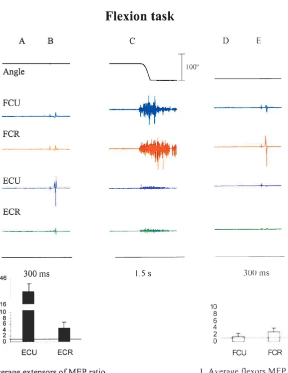
Figures 5 and 6 show typical examples of EMG and wrist position recordings taken from single trials in two subjects (S 3 and S 5, respectively. The movement was performed from a flexion to an extension position (Figure 5) and in the reverse direction (Figure 6). In each figure, the first panel (A) shows the EMG levels at the initial position, 200 ms before TMS. Panel B shows MEPs stimulated by TM$ in the four muscles at the same initial position. The middle panel (C) shows EMG activity during an active motion from the initial to a final position. Panel D shows the EMG activity at the final wrist position, approximately 8 seconds afler the movement offset, just 200 ms before the second TMS. Panel E shows MEPs stimulated by TMS at the final position. One can see that before the movement, the EMG level of the muscles was close to zero. At the final position, aller transient EMG bursts, the EMG activity gradually retumed to the pre-movement, near-zero level. The first TMS was delivered at the initial position, about 2 s before the movement onset. The second TMS was delivered approximateÏy 10 s later, i.e., aller the end of the movernent, when the EMG activity had retumed to the pre-movement level.
Angle FCU FCR ECU ECR FGR
4
ECU1. Average of flexors MEP ratio 2. Average extensors MEP ratio
Figure 5. Wrist position (joint angle), EMG activity of wrist flexors (fCR, FCU) and extensor muscles (ECR,ECU) before (A,B) during (C) and after (D,E) a movement ftom flexion to extension (F-’ E). B, E: MEP before (B) and afier (E) the movement (subject Si). Graphs 1 and 2 are averages ofthe ratio ofTMS responses: for flexors, the average is the ration of MEPs at flexion and extension positions and for extensor the ratio of MEPs at the extension and flexion positions (see also figure 7).
A B
Zr
D E11000
1.5s 4 I tf
300 ms 4 300msA B C D E Angle FCU L FCR
‘km
ECU ECR÷
ECU ECR2. Average extensors ofMEP ratio
10 8
h
FOU FCR
1. Averae fle\ors MEP ratio
Figure 6. Vristposition (joint angle), EMG activity of wrist extensor muscles t E(’R. E(‘I.
and flexors (FCR, FCU) before (A,B) during (C) and after (D.E) u movement Froni extension to flexion (E— F). B, E: MEPs before (B) and aller (E) the moement (subjeci
6). See also figure 8.
1.5 s
46 300ms
l6à
1LLà
amplitude of MEPs changed with the transition from one position to the other (compare B and E in each figure): flexor responses (in the fCR and FCU) were bigger at the flexor wrist position compared to those at the extensor position, and vice versa for extensor (ECR and ECU) responses. The pattems of position-related responses to TMS remained qualitatively similar regardless of whether the movements were made from flexion to extension (f—*E, f ig. 5) or from extension to flexion (E—+F, fig. 6). In other words, the pattems of MEP changes with the transition from one wrist position to the other, whereas the EMG level remained the same, regardless of how these positions were reached in each trial.
The quantitative observations illustrated in Figures 5 and 6 are confirmed by quantitative analysis described below.
3.1 EMG levels can be the same at different wrist
positions
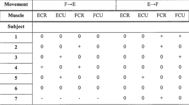
Table 1 shows the effect of wrist joint position on the tonic EMG level (200 ms windows before each stimulation) in the extensors (ECR and ECU) and flexors (f CR and FCU) using Student t-test (p< 0,05) for each muscle.
Movement F—*E E—*F
Muscle ECR ECU FCR FCU ECR ECU fCR FCU
Subject 1 0 0 0 0 0 0 + + 2 0 0 + O O O + O 3 0 + O O O O O + 4 + O + O O O O O 5 0 + O O O + O O 6 0 0 0 0 0 0 0 0 7 - - - -
o
o
+o
Table 1. The difference between EMG activity levels at two wrist positions before and afier the movement (O, insignificant; + significant at p< 0.05 level; -, EMG recording was not
made, for subject 7) in the cases when the hand moved from a flexion to an extension position (condition F—E) and in the reverse direction (condition E—>F) in different subjects.
In 41 cases out of52 the EMG levels were similar and did not show a significant difference between the positions (extension and flexion). In 11 cases the level of EMG was significantly different for the two positions. EMG was not recorded for the remaining cases (4 cases). Conclusion: in most cases, the EMG activity could be equalized at two positions at near zero, threshold level.
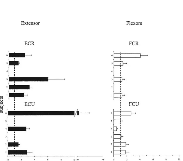
It is known that responses of muscles to TMS are modified depending on the EMG activity of muscles (Di Lazzaro et al. 199$). To exclude this confounding factor in the estimation of position-related corticospinal influences on motoneurons, we compared TMS responses in each muscle only in those cases when the EMG activity levels were equalized at the two positions (zeros in Table 1). In these cases, modulation in corticospinal excitability was investigated by measuring the change in the MEP amplitude at the two positions. The change in TM$ responses for the extensor muscles (ECR and ECU) was characterized by the ratio of their MEP amplitudes at the extension position to that at the flexion position in each trial and then computing the mean ratio and its standard deviation across ail trials. We thus normalized the MEP to compare positional TMS effects in different subjects. This method was more preferable than that based on a direct comparison of positional MEP changes since it somewhat diminishes the influence of the EMG electrode placement and skin resistance on the MEP measurement across subjects. Position reiated changes in TMS responses in flexors muscles were characterized by the ratio of MEP amplitudes at the flexion position to that at the extension position. Note that, according to these definitions, the ratio exceeding 1 for extensor muscles implies that the MEPs of these muscles are bigger at the extension wrist position. In contrast, the ratio exceeding 1 for flexor muscles implies that the MEPs of these muscles are bigger at the flexion wrist position.
Figures 7 and 8 shows MEP ratios for trials in which subjects move the hand from flexion to extension and from extension to flexion, respectively. One cari see that in the cases when the EMG levels were equalized at the two wrist positions, the ratio for extensors typically exceeded 1. Thus, TMS responses in extensors muscles were higher when the wrist was in the extension position. In most cases, the ratio for flexors also
exceeded 1 implying that flexor TMS responses were higher at the flexion wrist position. In other words, with transition from one wrist position to another, TMS responses typically changed reciprocally for flexor and extensor muscles.
35
Extension task
Extensor Flexors ECR FCR I I :1 I 1-.9. ECU:
FCU - I_______ / 6 4 I 4 21H *l’oIFigure 7. The mean MEP ratios (± SEM) for the two wrist positions resulting from the transition from flexion to extension (F —*E), for wrist extensors (right panels) and flexors (lefi panels).
Extensor
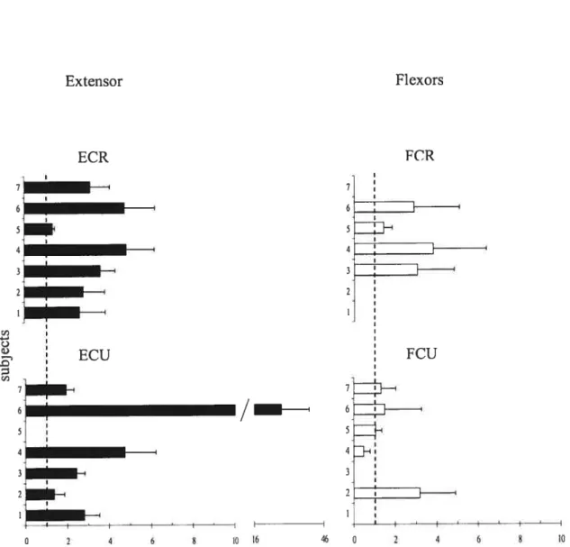
Flexion task
Flexors
Figure 8. The mean MEP ratios (+ SEM) for the two wrist positions resulting from the transition from extension to flexion (E _*F), for wrist extensors (riglit panels) and flexors (left panels).
ECR 7 6 5 4 3 2 ECU 7 6 4 FCR FCU 7 61 4 • 2 /—I 1016 46 0 2 4 6 8 10
and ail trials showed a significant effect (p< 0.05) of position on the ME? amplitude in most cases. The influence of position on MEPs was flot significant in one case for the ECR, ECU and FCR (F= 3.38, p= 0.79 ; F= 3.36, p= 0.76; and F= 1.32, p= 0.26, respectively) and in three cases for the FCU (F= 0,36, p= 0.5$; F= 2.25, p= 0.15; F= 0.12, p= 0.91). Thus, except for few cases, the TMS responses were position-dependent, both for flexor and extensor muscles.
for extensor muscles, the ME? ratio was higher than 1 and the ME? amplitude at the wrist extension position was> 2 tirnes higher than that at the flexion position in 77% of cases (for flexion and extension task). For flexor muscles, except for FCU muscles in one subject (4; see figures 7 and 8
)
the MEP ratio also exceeded 1, implying that the MEP amplitude at the wnst flexion position was higher than that at the extension position. These findings further suggest a strong position-related effect of TMS stimulation, reciprocal for flexors and extensors.3.3 Position-related changes in TMS responses may be
independent of the direction of movement from one
position to another.
Quaiitativeiy, from comparison of Figures 5 and 6, 7 and 8 or from Table 2 one can see that the pattems of position-dependent changes in TMS responses were simiiar for both type of movements from one wrist position to another (F—*E or E—*F). $tatistically, the movement direction effect was insignificant (p> 0.05) for ail four muscles.
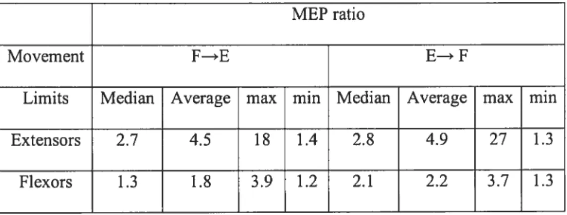
MEP ratio
Movement f—*E E—* F
Limits Median Average max min Median Average max min
Extensors 2.7 4.5 18 1.4 2.8 4.9 27 1.3
flexors 1.3 1.8 3.9 1.2 2.1 2.2 3.7 1.3
Table 2 shows the maximal, minimal and median of the TMS response of the extensors and flexors muscles (characterized by an average of the MEP ratios for each subject and muscles) of subjects group, excluding the FCU responses from subject 4 (in this subject, MEP ratio for FCU muscle, but flot for other 3 muscles, was < 1 in extension and flexion tasks).
3.4
Latency
MEP latency was measured by identifying the first deflection of the EMG trace from the background level afler the stimulus artefact. In ail cases the latency was in the range 14-18 ms (see table 3). ANOVA showed insignificant effects of wrist position, muscle, and movement direction (p>0.05). Table 3 lists the respective mean values for all cases.
o
o
Extension task (F—*E) Flexion task (E —*f) Muscle ECR ECU FCR fCU ECR ECU FCR FCU Position E F E F E f E F E F E F E F E f Subject 1 16.0 16.0 16.7 15.0 17.5 16.5 17.6 16.4 17.7 17.6 17.1 17.3 17.3 16.8 16.6 16.0 2 16.8 15.9 15.2 14.8 16.5 16.0 16.4 16.2 16.9 17.2 15.5 15.1 16.5 15.0 16.1 16.8 3 17.2 16.9 17.7 17.5 16.6 16.4 17.4 17.4 17.6 17.7 18.2 18.2 17.0 17.1 17.7 17.5 4 16.4 17.3 16.9 17.6 17.4 16.7 17.9 18.3 17.6 17.7 18.2 18.2 17.0 17.1 17.7 17.5 5 13.5 14.6 14.1 15.1 15.0 15.1 14.9 15.0 15.7 16.6 17.3 17.6 16.9 16.1 17.8 17.3 6 15.3 16.1 16.4 16.6 16.5 15.9 15.8 16.3 15.1 16.3 16.1 17.1 16.4 15.3 15.7 17.0 7 -12.2 12.4 11.6 11.9 12.0 11.4 12.7 11.6 Mean 15.9 16.1 16.2 16.1 16.6 16.1 16.7 16.6 16.1 16.5 16.3 16.5 16.1 15.5 16.3 16.3 ± SD ±1.3 ±1.0 ±1.3 ±1.3 ±0.9 ±0.6 ±1.2 ±1.1 ±2.0 ±1.9 ±2.3 ±2.3 ± 1.8 ±2.0 ± 1.8 ±2.1 Table 3. Mean MEP (±SD) latency for each muscle at extension and flexion positions for ail subjects (except for the flexion task in subject 7).4.0 Discussion
The general objective of this study was to investigate the changes in corticospinal influences on motoneurons of wrist flexors and extensors following an intentional transition from one wrist position to another. Thereby it was necessary to create conditions when the levels of EMG activity of these muscles at the two positions are equalized, i.e., afler the movement the EMG activity retums to the pre-movement near zero level. We tested two hypotheses. First, the corticospinal influences on motoneurons of wrist muscles may be different even though the EMG activity levels remain the same at different static wrist positions. Second, the position-related changes in corticospinal excitability may not be related to the direction of the movement that brings the wrist from one position to another. In other words, the changes in corticospinal excitability are related to the positions, regardless of whether these positions were achieved by movement from wrist flexion to extension or from extension to flexion.
Our study showed that EMG levels can be the same at different wrist positions. In fact, at the initial position and after the voluntary movement to the new position, the EMG activity was close to zero, suggesting that motoneurons of the muscles recorded at the initial wrist angle were near their activation thresholds and retumed to a near threshold state after establishing the final joint angle. Our findings are thus similar to those by Ostry and f eldman (2003) and Foisy and f eldman (2006) forarm movements and would suggest that the motor cortex resets the threshold position of the body segments when active movements are produced, rather than in direct specification of EMG pattems (see f eldman 2007). Note that in the present study, EMG levels were equalized between positions in most but flot ail trials (see Table 1). One explanation of these exceptions is the presence of co-activation of agonist and antagonist muscles: subjects were not always successful in minimizing this co activation after the end of movement. Co-activation is oflen used to stabilize the wrist joint
by increasing stiffness. Indeed, the wrist anatomy is especially complex, which allows the nervous system to greatly vary the hand shape and range of motion. The presence of multi joint muscles (including those recorded in the present study) and the absence of short muscles directly attaching distal radial and ulnar bones to the carpal bones possibly favour this liberty of movements at the expense of stability. Linked to this explanation is the fact that the wrist muscles are polyfunctional: subjects might combine wrist flexion with some abduction or adduction creating torques that tended to rotate the hand in the splint, resulting in respective changes in the EMG levels at the two wrist position. Yet another explanation is that, using elastics, we were flot always able to fully compensate for passive muscle torques at the two different wrist positions, necessitating activity of some muscles to counteract the residual passive torques of antagonist muscles at each position. These elastics might also be responsible for variability in the stabilization of the hand and fingers positions. This drawback in the present set-up can be overcome by using a torque-motor for compensation of passive torque, as is the case in the new set-up in our laboratory.
When TM$ is used to measure the corticospinal excitability, it is usually correlated with variables characterizing the motor output, such as EMG activity and force (Di Lazzaro et al 1998; Todd et al 2003, 2004). In the current study, we were generally successful in equalizing EMG activity at two wrist positions to support the previous feldman’s hypothesis that changes in the motor cortex may not be involved in direct specification of EMG activity but rather in resetting of the threshold position at which appropriate muscles begins their recruitment.
Our results showed that the corticospinal excitability measured by MEP amplitude of the extensors were higher at the extension position whereas flexors MEP amplitude were higher at the flexion position. This reflected a reciprocal change in the corticospinal influences on flexors and extensors motoneurons with the transition ftom one position to another. Thus, the results suggested a strong position-related change in corticospinal influences. In addition, this finding showed that the pattems of position-related changes in
words, the observed changes in the corticospinal influences due to the position might lead the resetting in the postural state required for the movement production rather than the movement direction itself. In few cases, specifically for the flexor (FCR, f CU) muscles, there were no position-related changes in the MEP amplitude. One argument could be that the stimulation site may facilitate one group of muscles more than another (Georgopoulos et al. 1986, 1989), or can influence a group of multijoint muscles, which cross more than two joints (Graziano et al. 2005). The effect of position in the corticospinal excitability could be variable in multijoint muscles due to the gating of the afferent information obtained ofthe crossed joints, other alternative explanation could be a subtie discrepancy in the hand positions, particularly in the fingers. Therefore, the presence of active biarticular muscles that were not monitored and that cross the wrist joint could also influence the excitability of the investigated muscles.
The reciprocal modulation in corticospinal excitability observed in the wrist extensors and flexors suggests a distal synergistic relationship between these muscles. Since wrist joints ofhuman limbs have more than one axis of rotation and are controlled by more than two muscles, this suggests that motor cortex controls the different limb segments as a whole rather than individually and that the corticospinal excitability modulation may be destined for multiple muscles. It would therefore be attractive to comment on possible proximal-distal synergies as observed by Ginanneschi et al (2006), whereas they found changes of the corticospinal excitability pathways to forelimb muscles after changing shoulder joint position. This would require examining the distal synergistic effect for multiple shoulder-elbow joint configurations. for example, it would be interesting to test if different activity levels in proximal muscles, related to different configurations of these proximal joints, could influence the excitability of distal muscles (seefuturestudies).