Université de Sherbrooke
Regulation of Osteoclast Activation and Autophagy through Altered Protein Kinase Pathways in Paget’s Disease of Bone
Par
Stephen Robert McManus Programme d’Immunologie
Thèse présentée à la Faculté de médecine et des sciences de la santé en vue de l’obtention du grade de philosophiae doctor (Ph.D.)
en Immunologie
Sherbrooke, Québec, Canada [Juillet, 2016]
Membres du jury d’évaluation
Pre Sophie Roux, M.D., Ph.D., Département de Médecine, Service de Rhumatologie Pr Patrick McDonald, Ph.D., Département de Médecine, Service de Pneumologie
Pre Nathalie Perreault, Ph.D., Département d’Anatomie et Biologie Cellulaire Pr Jean Vacher, D.Sc., Département de Médecine, Université de Montréal
Regulation of Osteoclast Activation and Autophagy through Altered Protein Kinase Pathways in Paget’s Disease of Bone
Par
Stephen Robert McManus Programme d’Immunologie
Thèse présentée à la Faculté de médecine et des sciences de la santé en vue de l’obtention du diplôme de philosophiae doctor (Ph.D.) en Immunologie, Faculté de médecine et des
sciences de la santé, Université de Sherbrooke, Sherbrooke, Québec, Canada, J1H 5N4 La maladie osseuse de Paget (MP) est un désordre squelettique caractérisé par une augmentation focale et désorganisée du remodelage osseux. Les ostéoclastes (OCs) de MP sont plus larges, actifs et nombreux, en plus d’être résistants à l’apoptose. Même si la cause précise de la MP demeure inconnue, des mutations du gène SQSTM1, codant pour la protéine p62, ont été décrites dans une proportion importante de patients avec MP. Parmi ces mutations, la substitution P392L est la plus fréquente, et la surexpression de p62P392L dans les OCs génère un phénotype pagétique partiel. La protéine p62 est impliquée dans de multiples processus, allant du contrôle de la signalisation NF-κB à l’autophagie. Dans les OCs humains, un complexe multiprotéique composé de p62 et des kinases PKCζ et PDK1 est formé en réponse à une stimulation par Receptor Activator of Nuclear factor Kappa-B Ligand (RANKL), principale cytokine impliquée dans la formation et l'activation des OCs. Nous avons démontré que PKCζ est impliquée dans l’activation de NF-κB induite par RANKL dans les OCs, et dans son activation constitutive en présence de p62P392L. Nous avons également observé une augmentation de phosphorylation de Ser536 de p65 par PKCζ, qui est indépendante d’IκB et qui pourrait représenter une voie alternative d'activation de NF-κB en présence de la mutation de p62. Nous avons démontré que les niveaux de phosphorylation des régulateurs de survie ERK et Akt sont augmentés dans les OCs MP, et réduits suite à l'inhibition de PDK1. La phosphorylation des substrats de mTOR, 4EBP1 et la protéine régulatrice Raptor, a été évaluée, et une augmentation des deux a été observée dans les OCs pagétiques, et est régulée par l'inhibition de PDK1. Également, l'augmentation des niveaux de base de LC3II (associée aux structures autophagiques) observée dans les OCs pagétiques a été associée à un défaut de dégradation des autophagosomes, indépendante de la mutation p62P392L. Il existe aussi une réduction de sensibilité à l’induction de l'autophagie dépendante de PDK1. De plus, l’inhibition de PDK1 induit l’apoptose autant dans les OCs contrôles que pagétiques, et mène à une réduction significative de la résorption osseuse. La signalisation PDK1/Akt pourrait donc représenter un point de contrôle important dans l’activation des OCs pagétiques.
Ces résultats démontrent l’importance de plusieurs kinases associées à p62 dans la sur-activation des OCs pagétiques, dont la signalisation converge vers une augmentation de leur survie et de leur fonction de résorption, et affecte également le processus autophagique. Mots clés : Autophagie, Osteoclaste, Paget, Kinase, PDK1, p62
2S
UMMARYRegulation of Osteoclast Activation and Autophagy through Altered Protein Kinase Pathways in Paget’s Disease of Bone
By
Stephen Robert McManus Immunology Program
Thesis presented at the Faculty of medicine and health sciences for the obtention of Doctorate degree diploma [philosophiae doctor (Ph.D.)] in Immunology, Faculty of medicine and Health Sciences, Université de Sherbrooke, Sherbrooke, Québec, Canada
Paget’s disease of bone (PDB) is a skeletal disorder characterized by focal and disorganized increases in bone turnover. In PDB, osteoclasts are larger, more active, more numerous, and resistant to apoptotic stimuli. While no single root cause has been identified, mutations to the gene encoding the p62 protein, SQSTM1, have been described in a
significant population of patients with PDB. Among these mutations, the P392L
substitution is the most prevalent, and overexpression of p62P392L in osteoclasts generates at least a partial pagetic phenotype in vitro. Normally this protein mediates a number of cell functions, from control of NF-κB signaling to autophagy. In human osteoclasts, a
multiprotein complex containing p62 and protein kinases PKCζ and PDK1 (the principal kinase of Akt), form in response to stimulation by receptor activator of nuclear factor kappa-B ligand (RANKL), the principal osteoclastogenic-signaling cytokine. We found that PKCζ is involved in RANKL-induced activation of NF-κB, and that it contributed to a basal activation of NF-κB observed in p62P392L mutants. This may be regulated in part by a PKCζ dependent increase in p65 phosphorylation at Ser536 which we characterized,
independent of IκB. This could represent one alternative pathway by which mutant p62 leads to increased NF-κB activation.
We observed increased basal phosphorylation of survival regulators ERK and Akt in PDB that was reduced upon PDK1 inhibition. The activity of 4EBP1 and Raptor, associated with mTOR activity, were also altered in pagetic osteoclasts and regulated by PDK1
inhibition. We then identified autophagic defects common to pagetic osteoclasts; with higher basal levels of LC3II (associated with autophagic structures), regardless of p62 mutation, and reduced sensitivity to autophagy induction in PDB. These results suggest an accumulation of non-degradative autophagosomes. Inhibition of PDK1 not only induced apoptosis in PDB and controls, but significantly reduced resorption in PDB, and with regards to autophagy, PDK1 inhibition was more potent in PDB than in controls. Therefore PDK1/Akt signaling represents an important checkpoint to PDB osteoclast activation.
In sum, these results demonstrate the importance of several p62-associated kinases in the over-activation of pagetic osteoclasts, through increased survival and altered
signaling. As p62 mutations alone do not account for most cases of PDB, the characterization of these pathways may identify a common factor linking pagetic osteoclasts. Therefore these studies represent a novel approach to osteoclast apoptosis, activation, and autophagy associated with PDB.
1 Résumé ... iv
2 Summary ... v
3 Table of contents ... vi
4 List of figures ... x
5 List of tables ... xii
6 List of abbreviations ... xiii
1 Introduction... 1
The Skeletal System ... 1
The Remodeling Cycle ... 2
Regulation of Bone Remodeling ... 5
Hormones ... 5 Growth Factors ... 6 Cytokines ... 7 The Osteoblast ... 9 Development ... 9 Function ... 11 The Osteoclast ... 12 Commitment... 12 Differentiation ... 16 Multinucleation ... 18
Maturation and Actin Ring Formation ... 20
Mechanisms of Resorption ... 23
Regulation of Resorption ... 24
Signaling Pathways in Osteoclast Activation and Survival ... 25
NF-κB... 25 Regulation of NF-κB ... 26 Canonical NF-κB... 27 Non-Canonical NF-κB ... 28 NF-κB Signaling in Osteoclastogenesis ... 28 Regulation Via PKCs ... 31
PDK1 ... 36
Akt ... 37
ERK Signaling ... 42
The p62 Scaffold: the functionallink between RANKL and TRAF6-mediated signals ... 43
p62 and Other NF-κB Activation Pathways ... 45
p62 and Regulation of TRAF6 Through De-ubiquitinase Activity ... 45
Osteoclast-specific Transcriptional Regulation by p62 ... 46
Regulation of Survival in Osteoclasts ... 47
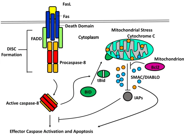
Caspases ... 48
The Extrinsic Pathway ... 48
The Intrinsic Pathway ... 49
Regulation of Remodeling ... 51
Autophagy and Skeletal Maintenance ... 55
Ubiquitin-Proteasome System ... 55
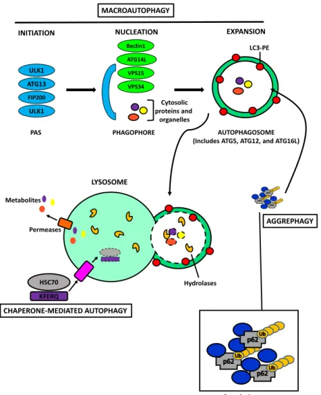
Autophagy ... 56
Selective Autophagy ... 59
The Role of p62 in Autophagosome Formation and Autophagy Regulation ... 60
Crosstalk Between Apoptosis and Autophagy ... 61
Autophagic Defects and Bone Diseases... 61
Paget’s Disease of Bone ... 64
Pathophysiology and Osteoclast Phenotype ... 64
Genetics ... 65
Nuclear Inclusions and Paramyxoviruses ... 67
Animal Models of Paget’s Disease of Bone... 68
Treatment ... 70
A Potential Role for p62 in pagetic resistance to Apoptosis and autophagy defects ... 70
Hypothesis ... 72
Objectives ... 73
2 Materials and Methods ... 74
Culture of Human Umbilical Cord Blood Derived-Monocytes ... 74
CBM cultures using Methocult Cell Amplification ... 75
Peripheral Blood-Derived Osteoclasts ... 76
Apoptosis Evaluation Via TUNEL ... 77
RNA Extraction and Real-Time PCR ... 81 Resorption Assay... 82 Kinase Assay ... 82 Immunofluorescence ... 83 Statistical Analysis ... 84 3 Results ... 85
PART I : The p62P392L Mutation and PKCζ Activation in PDB ... 85
PKCζ and p62 Interaction in NF-κB Activation ... 85
PKCζ May Act as a p65 Kinase Independently of IκBα ... 95
PART II: Kinase Cascades in Survival and Autophagy in Paget’s Disease of Bone ... 97
Kinome Activation in Normal Osteoclasts ... 97
PDK1, Akt, and RANKL Signaling in normal Osteoclasts ... 97
Kinome Regulation of Resorption ... 99
Kinome Activation in Pagetic Osteoclasts ... 99
Survival Pathways in Pagetic Osteoclasts ... 99
Impact of PDK1 Inhibition on Downstream Targets in Pagetic Osteoclasts ... 102
Autophagy in Pagetic Osteoclasts ... 104
Evaluation of Autophagy in Osteoclasts via LC3 Immunoblot ... 104
Autophagy-Related Gene Expression ... 105
The Dynamics of Autophagy in PDB as Observed by Immunofluorescence ... 106
The Regulation of Akt/mTORC1 Signaling in Pagetic Osteoclasts by PDK1 ... 109
The Impact of Kinase Inhibition on Osteoclast Apoptosis, Autophagy, and Activity ... 112
PDK1 Inhibition of Osteoclast Autophagy ... 112
Kinase Inhibition and Resorption in Pagetic osteoclasts ... 114
Pagetic Osteoclast Apoptotic Response to Kinase Inhibition ... 115
4 Discussion... 116
PKCζ and Osteoclast Activation Via NF-κB Pathways ... 117
Transcriptional Control of NF-κB by PKCζ Through p65 Phosphorylation ... 119
PKCζ and Non-Canonical NF-κB Signaling Pathways ... 121
The Role of P392L in Pagetic Osteoclast Activation ... 122
Osteoclast Apoptosis and Survival Pathways ... 123
PKC and Akt Regulatory Pathways in Survival ... 125
The Role of PDK1 in Osteoclast Signaling ... 128
ERK Activation and Targeting in Osteoclasts ... 130
Treatment of Invasive Cells Via PDK1 Targeting ... 132
Autophagic Defects in Paget’s Disease of Bone ... 134
Autophagy-associated Osteoclast Dysfunction ... 135
Autophagy and Non-Canonical NF-κB Signaling ... 137
p62 and Autophagic Regulation ... 138
Autophagy Inhibition and the Bone Microenvironment ... 139
PDK1 as an Autophagy Regulator in Osteoclasts ... 140
MAPK Regulation of Autophagy ... 140
Other Contributory Factors ... 141
Environmental Factors and Paget’s Disease of Bone ... 142
Alternative Splice Variants in Paget’s Disease of Bone ... 143
Conclusions ... 145
Perspectives ... 147
NF-κB Activation in Paget’s Disease of Bone ... 147
PDK1 Impact on Differentiation and Signaling ... 147
p62 Mutations and Autophagic Flux ... 148
Paget’s Disease and Pinpointing the Impact of Autophagy ... 149
Acknowledgements ... 151
Figure 1: Bone Remodeling ... 3
Figure 2 : RANKL/RANK Signaling in the Osteoclast ... 8
Figure 3 : Osteoclast Development and Lineage ... 14
Figure 4 : Resorption Mechanisms in the Mature Osteoclast ... 21
Figure 5 : Activation Pathways of NF-κB ... 29
Figure 6 : Interaction Motifs and Domains of p62, PKCζ, PDK1 and Akt ... 34
Figure 7 : The Intrinsic and Extrinsic Pathways of Apoptosis ... 50
Figure 8 : Mechanisms of Autophagy ... 58
Figure 9 : PKCζ and p62 Interactions in Human Osteoclasts ... 86
Figure 10 : Effect of a PKCζ Inhibitor on NF-κB Nuclear Translocation ... 88
Figure 11 : Study of IkB Expression ... 90
Figure 12 : Interactions between p62 and IB ... 92
Figure 13 : Expression of p-IKKβ in Osteoclasts Following 30-min RANKL Stimulation, With or Without PKCζ Inhibition ... 94
Figure 14 : p65 Phosphorylation and the Effects of PKCζ Inhibition ... 96
Figure 15 : PDK1 and Akt Signaling in Osteoclasts ... 98
Figure 16 : Resorption by CBMs Under PDK1 Inhibition ... 100
Figure 17 : Kinase Activation in Pagetic Osteoclasts ... 101
Figure 18 : Effects of PDK1 Inhibition on Survival Kinases in PDB ... 103
Figure 19 : Autophagy in Pagetic Osteoclasts ... 105
Figure 20 : Autophagy-Related Gene Expression Analysis ... 106
Figure 21 : LC3 in PDB Autophagic Flux ... 107
Figure 22 : p62 in PDB Autophagic Flux ... 108
Figure 23 : mTOR Signaling in Paget’s Disease ... 110
Figure 24 : ULK Regulation by PDK1 and in PDB ... 111
Figure 25: Impact of PDK1 and ERK inhibition on OC Autophagy ... 113
Figure 26 : Effects of Kinase Inhibition on Resorption in PDB ... 114
Figure 28 : p65 Modification Sites ... 120 Figure 29 : Regulatory Elements in Akt and aPKC Signaling Pathways ... 127 Figure 30 : PDK1-Regulated Pathways Altered in the Pagetic Osteoclast ... 146
Table 1 : PBMC-Donors by Subgroup ... 76 Table 2 : Antibodies ... 81 Table 3 : List of Primer Sets Used ... 82
AGC Family encompassing Protein Kinases A, G and C AKTIP Akt-interacting protein
AMP Adenosine monophosphate AMPK 5’ AMP-activated protein kinase ANOVA Analysis of variance
ARO Autosomal recessive osteopetrosis AS Alternative splicing
ATP Adenosine triphosphate
BAD Bcl-2-associated death promoter protein BMM Bone marrow-derived macrophages BMP Bone morphogenic proteins
BMU Basic multicellular unit BSA Bovine serum albumin BSP Bone sialoprotein CA Carbonic anhydrase
CARD Caspase recruitment domain CBM Cord-blood monocyte CBP CREB-binding protein CCL chemokine (c-c motif) ligand CDKI Cyclin-dependent kinase inhibitors CDV Canine distemper virus
CEF Chicken embryo fibroblast CRB (transmembrane protein) Crumbs CREB Cyclic AMP-response element binding CTMP Carboxyl-terminal modulator protein CTSK Cathepsin K
CYLD Cylindromatosis (turban tumor syndrome) DAG Diacyl-glycerol
DAPI 4',6-diamidino-2-phenylindole DISC Death inducing signal complex DUB De-ubiquitinating enzyme ECL Electrochemiluminescence ECM Extracellular matrix ER Endoplasmic reticulum
ERK Extracellular signal-regulated kinase
EV Empty vector
FADD Fas-associated death domain FasL Fas ligand
FBS Fetal bovine serum FGF Fibroblast growth factor
FoxO Forkhead box transcription factor HBSS Hanks’ balanced salt solution HDAC Histone deacetylase
HSC Hematopoietic stem cell IAP Inhibitor of apoptosis protein IB Inclusion body
IBMPFD Inclusion Body Myopathy, Paget's disease and Frontotemporal Dementia ICAD Inhibitor of caspase activated DNase
IGF Insulin-like growth factor IkB Inhibitor of κB
IKK IκB kinase
ITAM Immunoreceptor tyrosine-based activation motif JNK c-Jun N-terminal kinase
JNKK JNK kinase
KFERQ amino acid sequence Lys-Phe-Glu-Arg-Gln KIR Killer-cell immunoglobulin-like receptor LIR LC3-interacting region
MAP Mitogen-activated protein MAPK MAP kinase
MATH Meprin and TRAF homology domain MBP Myelin basic protein
MCPIP Monocyte chemotactic protein-induced protein M-CSF Macrophage colony-stimulating factor MEF ELF4 (E74-like factor 4)
MEK Mitogen-activated protein kinase kinase MGP Matrix gla protein
MITF Micropthalmia-associated transcription factor MKKK MAP kinase kinase kinases
MMP Matrix metalloproteinase MNC Mononuclear cell
MVNP Measles virus nucleocapsid protein
NADPH Nicotinamide adenine dinucleotide phosphate NEMO NF-kappa-B essential modulator
NFAT Nuclear factor of activated T cells
NF-κB Nuclear factor kappa-light-chain-enhancer of activated B-cells NIK NF-κB-inducing kinase
NT Non-treated
OC Osteoclast OCN Osteocalcin
OCP Osteoclast precursor OD Optical density
OPCA domain sequence containing octicosapeptide repeat motif, PC motif, and atypical PKC interaction domain motif
OPG Osteoprotegerin OPTN Optineurin
OSCAR Osteoclast-associated receptor PAS Pre-autophagosomal structure PBMC Peripheral blood mononuclear cell PBS Phosphate buffered saline
PDB Paget’s disease of bone PE Phosphatidyl-ethanolamine
PEST peptide sequence rich in Proline (P), Glutamic Acid (E), Serine (S), and Threonine (T)
PH Pleckstrin homology PI Phosphatidylinositol PIF PDK1-interacting fragment PKA Protein kinase A
PKB Protein kinase B PKC Protein kinase C PKN Protein kinase N1 PS Pseudosubstrate
PTEN Phosphatase and tensin homolog PTH Parathyroid hormone
PVDF Polyvinylidine fluoride
RANK Receptor activator of nuclear factor kappa-B RANKL RANK ligand
RANTES (CCL5) Regulated on activation, normal T-cell expressed and secreted RAPTOR Regulator-associated protein of mTOR
RelA (p65) v-rel avian reticuloendotheliosis viral oncogene homolog A RelB v-rel avian reticuloendotheliosis viral oncogene homolog B RGD Arginine (R), Glycine (G), Aspartic Acid (D) domain RHD Rel homology dimerization domain
RhoE Rho-related GTP-binding protein E
RICTOR Rapamycin-insensitive companion of mammalian target of rapamycin RIP Receptor interacting protein
RNA Ribonucleic acid RSK Ribosomal S6 kinase RSV Respiratory synctial virus SAPK Stress-activated protein kinase SD Standard deviation
SGK Serum glucocorticoid-dependent kinase SNARE Soluble NSF attachment protein receptor SNP Single nucleotide polymorphism
TFG TRK-fused gene TLR Toll-like receptor TNF Tumor necrosis factor TNFR TNF receptor
TRAF TNF receptor associated factor
TRAIL TNF-related apoptosis-inducing ligand TRAP Tartrate-resistant acid phosphatase
TUNEL Terminal deoxynucleotidyl transferase dUTP nick end labeling UBA Ubiquitin-associated domain
ULK Unc-51-like kinase
UPS Ubiquitin-proteasome system VCP Valoisin-containing protein WB Western blot
WIPI WD repeat domain phosphoinositide-interacting protein 1
WT Wild-type
XIAP X-linked inhibitor of apoptosis protein YAP Yes-associated protein
The Skeletal System
The skeletal system, while appearing inert at first glance, is a dynamic organ responsible for a number of vital functions in the body; including but not limited to providing protection and support to other organ systems, as well as permitting movement through collaboration with the muscular system. At the cellular level, bone provides a reservoir of growth factors and cytokines, maintains the acid-base balance and mineral homeostasis, and is the site of hematopoiesis. Like other connective tissue, bone has both a cellular and an extracellular matrix component. The matrix is made up of collagen fibers and collagenous proteins, with type I collagen accounting for ~90% of total protein, and the non-collagenous osteocalcin, osteopontin, and bone sialoprotein, and others making up the other 10%. In contrast with other connective tissue, the extracellular matrix of bone is mineralized physiologically, through the deposition of layers of carbonated hydroxyapatite. This mineral component, making up 50-70% of bone, provides bone's characteristic mechanical rigidity and strength (Clarke 2008). Elasticity and flexibility are due to the organic matrix, which makes up another 20-40%, lending bone incredible resilience without compromising its strength, and another 5-10% of bone is water.
There are two major types of bone; trabecular (also called cancellous or spongy), and cortical (also called compact). Cortical bone is denser and hard, whereas trabecular bone is a honeycomb network that is more mesh-like. Depending on their intended function, ratios of one type to the other at skeletal sites throughout the body will vary. For example, trabecular bone will more frequently be found within ribs, the skull, or the ends of long bones, while the denser cortical bone makes up the outer shell (or cortex) of most bones of the skeleton. Both of these bone types are typically formed in a lamellar pattern, where there is a highly ordered deposition of collagen fibers, with each layer in alternating orientation. This pattern is vital to providing bone with its mechanical strength and integrity (Ankersen et al. 1994). In disease, this pattern can be lost, leading to compromised structure.
Bone remodeling is the mechanism of bone renewal in the skeleton. This process involves continuous removal of small packets of old bone, the filling of these areas with newly synthesized collagen-rich matrix, and finally with mineralization of this matrix to form new bone. Remodeling sites develop primarily in a random manner, but are targeted to areas requiring repair as well (Clarke 2008). In addition to remodeling, according to Wolffs Law, the skeleton undergoes constant modeling in response to changes in biomechanical forces (Wolff 1892; Wolff 2010). The skeletal system, like any other, is in a state of controlled balance, dependent on cooperation between the mechanisms responsible for formation and those responsible for its counterpart, resorption.
The three major cell types forming the foundation of bone activity are the osteoclasts that break down bone, osteoblasts that build new bone, and osteocytes that maintain living bone. Bone remodeling relies on these activities to be carried out by the independent (yet ultimately synergistic) action of osteoblasts and osteoclasts in response to stimuli that can be biomechanical or strictly biological, depending on the circumstance.
The Remodeling Cycle
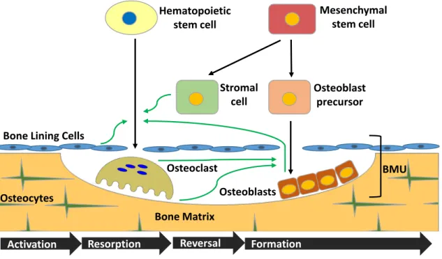
Bone replacement is initiated by osteoclastic resorption and immediately followed by osteoblastic formation. The remodeling cycle can be broken down into four sequential phases: resting/activation, followed by resorption/remodeling, then reversal and finally formation (Figure 1). Resorption and formation are closely linked within discrete temporary anatomic structures, described as "basic multicellular units," or BMUs, made up mainly of osteoclasts, osteoblasts, and bone-lining cells. A BMU is active in three dimensions; excavating and refilling a tunnel through cortical or compact bone, or a trench across the surface of cancellous or trabecular bone. At any given time it is estimated that there are approximately one million BMUs in action (Parfitt et al. 1996), and the lifetime of one of these units is estimated to be between six and nine months (Smith, Gardiner, and Dunstan 2012).
The activation of remodeling corresponds to a region of bone surface which converts from quiescence to remodeling activity, a process which requires recruitment of osteoclast
precursors from circulation, and changes in the lining cells that will close off the BMU (Parfitt 2002). The osteoclast precursors will develop, bind to the bone matrix, and eventually enact resorption as mature osteoclasts, as will be described in greater detail later. The characteristic resorbed areas left behind are referred to as Haversian canals on cortical bone, and Howship's lacunae on cancellous bone. This step is relatively short in the remodeling cycle, taking only two to four weeks.
Figure 1: Bone Remodeling
The process of remodeling is dynamic, but for one area to undergo all four stages of remodeling takes approximately four months. Following recruitment and activation of osteoclasts, they resorb the bone, then undergo apoptosis. The activity of the osteoclasts allows for the recruitment of mononuclear cells and preosteoblasts during the reversal phase. Finally, the rate limiting step of the process, bone formation takes place, carried out by the osteoblasts, followed by a number of them developing into osteocytes embedded in the bone, and the majority of the rest undergoing apoptosis.
Mesenchymal stem cell Osteoblast precursor Stromal cell Hematopoietic stem cell Osteoclast Osteoblasts Osteocytes
Bone Lining Cells
Activation Resorption Reversal Formation
Bone Matrix
The reversal stage begins following completion of the resorption phase, with initiation of apoptosis of the osteoclasts (Reddy 2004). Cytokines and growth factors are produced by active osteoclasts or released from the bone matrix during the resorption of bone, serving as recruitment and other stimulatory signals for other cells in the microenvironment (Martin and Sims 2005). Among these are transforming growth factor-β (TGF-β), released from the bone matrix. TGF-β activates osteoblasts, decreases osteoclast resorption by inhibiting RANKL production by osteoblasts and stromal cells in the bone microenvironment, and can also directly induce apoptosis in osteoclasts (Houde et al. 2009). This reversal phase allows for transition from resorption to formation, halting osteoclast activity and recruiting the bone-forming cells.
At this point, osteoblast precursors will proliferate locally, differentiating into osteoblasts in response to signals. During the formative phase, these cells deposit initially unmineralized bone matrix called osteoid, filling the lacunae produced by the osteoclasts. Once embedded in osteoid, osteoblasts can mature into terminally differentiated osteocytes, which produce and maintain a canalicular network permitting them to connect to other osteocytes as well as surface lining cells (Parfitt 1994). Other osteoblasts lying on the bone surface become quiescent lining cells (Clarke 2008), and with the completion of bone formation, approximately 70% of osteoblasts will undergo apoptosis ( Cohen 2006). The osteoblast-like bone-lining cells regulate the flow of mineral ions to the bone extracellular fluid, forming the blood-bone barrier, and can also re-differentiate to osteoblasts if called for (Seeman 2009).
In comparison with the relatively quick resorption phase, formation is a long process, taking 3 to 6 months to complete. This contrast provides an example to why precise regulation between these two phases is critical to balance in the skeletal system; different phases must succeed in maintaining homeostasis despite significant variation in the time required to complete their activities. During formation, osteoblasts synthesize new organic matrix, (which is primarily type-1 collagen), and regulate mineralization of that same matrix. The regulation of this mineralization step is carried out through the release of small membrane-bound vesicles containing concentrated calcium and phosphate, and enzymatically destroy inhibitors of mineralization like pyrophosphate or proteoglycans
(Anderson 2003). In a healthy system, these combined steps forming the process of bone remodeling help to preserve the mechanical strength of bone by replacing aged and microdamaged bone with healthier tissue; new bone that is more apt to maintain calcium and phosphate homeostasis, as well as a more stable supporting scaffold.
Regulation of Bone Remodeling
Hormones
Several hormones play important roles in maintenance of normal bone turnover. Calcium-regulating hormones 1,25-dihydroxy vitamin D and parathyroid hormone (PTH) lead to increases in osteoclast formation and resorptive activity (Teitelbaum 2000). One crucial systemic hormone whose presence varies drastically in more than half of the population is estrogen. Its function is complex and influences multiple cell lines and pathways still under study that inhibit bone resorption, as well as aid in bone formation (Weitzmann and Pacifici 2006). Studies in animal models have suggested that estrogen acts on both production and activity of local factors that regulate osteoblast and osteoclast precursors alike (Trivedi, Goswami, and Chattopadhyay 2010). Of the cytokines and growth factors with effects on bone cells that are released in response to estrogen, M-CSF, IL-1, -6, and -7 are included, as well as TNF, prostaglandins, and IGF-1 (Riggs, Khosla, and Melton 2002). While the majority of these are produced within the bone
microenvironment, some, like IGF, can be produced elsewhere, demonstrating that
estrogen-mediated bone regulation is para-, auto- and endocrine. This provides an example of the connectivity of the skeletal system that makes it a challenge to study in vivo.
Remodeling can be regulated by intestinal microbiota, inhaled pollutants, diet and physical activity, and much more, leading to great potential for variance even among a “control” population (Satarug et al. 2010; Rizzoli 2014; Sjögren et al. 2012).
The impact of hormones on remodeling are not limited to differentiation and activation of varying bone cells either. The pro-bone formation effect of estrogen is carried out in large part by regulation of programmed cell death, encouraging apoptosis in
osteoclasts, but preventing it in the mesenchymal stem cell derived osteoblasts and
as bisphosphonates is carried out primarily through activation of ERK, which regulates this pathway on two levels; the first of which being kinase-dependent activation of transcription factors and ultimately gene transcription (Kousteni et al. 2003). The second of these
mechanisms is dependent on the cytoplasmic ERK target p90RSK, which phosphorylates the pro-apoptotic protein BAD, as well as the CCAAT/enhancer binding protein (C/EBPβ) (Buck et al. 2001; Bellido and Plotkin 2011). The phosphorylation of C/EBPβ allows it to bind and inhibit pro-caspases, whereas the phosphorylation of BAD is directly inhibitory, rendering it inactive (Plotkin et al. 2005). Therefore apoptotic regulation by estrogen is carried out by pathways both dependent and independent of transcriptional activity. The exact mechanisms of increasing osteoclast sensitivity to apoptosis by hormones are still under investigation, but can act both directly on the osteoclasts in the form of interfering with receptor activator of nuclear factor kappa-B (RANK) signaling partners (Robinson et al. 2010), or indirectly through the induction of Fas ligand on osteoblasts (Krum et al. 2008).
Growth Factors
Bone is also the host of a great number of growth factors. Among the most abundant of these are the aforementioned IGFs, which are important modulators of local bone
remodeling. A murine IGF-1 knockout model showed an increase in bone volume that was primarily attributed to reduced osteoclast number (Wang et al. 2006), but IGF1R knockouts have also been observed to have significantly lowered bone formation rates (Yakar,
Courtland, and Clemmons 2010). Overexpression of IGF-1 in collagen-1 secreting cells (including but not necessarily limited to osteoblasts), led to increased bone width and length, indicating that IGF1is important to activity of both major cell types in the remodeling process (Jiang et al. 2006).
Transforming growth factor β (TGF-β) and its related family of bone morphogenic proteins (BMPs) are present and highly involved in the skeleton, functioning both in skeletal development and remodeling. TGF-β is an important player in bone biology, directly and indirectly inducing effects in both osteoclasts and osteoblasts; from inducing osteoclast formation (Nakamura et al. 2015), to the upregulation of Bim and eventual
osteoclast apoptosis in vitro (Houde et al. 2009), and by the coupling of resorption to formation by stimulating Wnt1 production (Weivoda et al. 2015). BMPs, as their name implies, are a group of signaling molecules originally characterized for their ability to induce bone formation (Wu, Shi, and Cao 2007). Many members of this family were first described in osteogenesis, but have recently been subjects of interest in the context of bone resorption as well. Most of the known BMPs are expressed in skeletal tissue, with a number detectable in osteoblasts (Anderson et al. 2000). The majority are recognized as promoters of bone formation, but there are exceptions like BMP-3; a negative regulator of this process (Daluiski et al. 2001). BMP-2 and -7 have been used following orthopedic surgery to improve bone repair in spinal fusion, for example (Senta et al. 2009). The potently
osteogenic BMP-9 also directs osteoclast activity and survival, as the addition of BMP-9 to mature osteoclasts in vitro significantly increased bone resorption while decreasing the rates of apoptosis in these cells (Fong et al. 2013). In this in vitro study, BMP-9 osteoclast activation involved phosphorylation of Smad-1/5/8 and ERK1, pathways common with TGF-β stimulation (Chen, Deng, and Li 2012).
Cytokines
In addition to the numerous hormones and growth factors influencing bone cell functions, there are also a number of cytokines of great importance to bone formation, balance, and remodeling. It has been established that the vast majority of all of these factors that influence bone resorption either play a role in or depend on a common final pathway involving the RANK and its ligand, RANKL. RANKL is the main stimulator of osteoclast differentiation and activation (Boyle, Simonet, and Lacey 2003). It is a TNF family member expressed by osteoblasts and stromal cells (as well as other tissues, including muscle, thymus, and intestinal cells) (Wada et al. 2006). In vivo as well as in vitro studies have demonstrated that by binding to the membrane-bound receptor RANK, RANKL is crucial to myriad pathways necessary for the formation, survival, and bone-resorbing capabilities of osteoclasts (Burgess et al. 1999; Hsu et al. 1999; Ikeda and Takeshita 2015). (Figure 2). Given its virtual omnipresence, the associated pathways of RANK will be further expanded in later sections.
Figure 2 : RANKL/RANK Signaling in the Osteoclast
RANKL is the principal cytokine in osteoclast differentiation, activation, and survival. It activates the TNF-related receptor RANK in a trimeric symmetric complex, recruiting TRAF6. Downstream signaling includes activation of Src, PI3K, MKK, TAK1 and others, leading to stimulation of numerous pathways including p38, MAPK, ERK, and JNK. Activation of the MAP kinases also leads to activation of the transcription factors Fos, c-Jun, and NFATc1. Among other cytoprotective effects, activation of Akt following PI3K activity results in the phosphorylation and inhibition of the pro-apoptotic protein BAD.
It is important to note in the context of the skeleton that there is a third partner in the RANKL/RANK axis, known as osteoprotegerin (OPG); another member of the TNF receptor family. OPG has no transmembrane domain, and is a secreted decoy receptor competing with RANK to bind with RANKL, thus inhibiting osteoclast differentiation and
ERK CYLD P P P JNK p38 cFos
Integrin ß3 NFAT2 TRAP CATK Myc Inf-ß Calcitonin receptor Src
bone-resorbing function (Simonet et al. 1997). This decoy activity is essential in the fine-tuning of bone resorption (Abrahamsen and Teng 2005; Wensel, Iranikhah, and Wilborn 2011).
Bone-resorbing factors like PTH, 1,25(OH)2D3, and TNF-α act principally through increasing the RANKL/OPG ratio in the bone microenvironment. Where the factors named all can act directly on the osteoclast as soluble factors in vitro, this effect is typically carried out by the osteoblast/stromal cells in vivo. Logically, inhibitors of bone resorption often function by lowering the RANKL/OPG ratio, or through other methods of blocking RANK signaling (Kong and Penninger 2000). The two sides of the coin in this model were
demonstrated decisively by Blair et al in 2005 when severe osteoporosis was shown in OPG knockout mice, as well as osteopetrosis in their RANKL knockout counterparts(Blair et al. 2006).
The Osteoblast
Development
The first major event in the bone microenvironment that must take place giving way to osteogenesis is the development of mesenchymal stem cells into differentiating and bone-forming osteoblasts. Their lineage separates the osteoblasts from their counterpart, the resorbing osteoclast, which arises from hematopoietic stem cells. While osteoclasts are thus more closely related to other myeloid-derived cells like the dendritic cell, osteoblasts are members of the same family tree that gives rise to bone, cartilage, fat, and fibrous connective tissue. Unlike the osteoclast, the osteoblast can dedifferentiate, and even re-differentiate into these other types under the right stimuli (Blum and Begemann 2015). However, this is uncommon and the vast majority of osteoblasts will either give rise to bone-lining cells and osteocytes, or die through apoptosis, depending on cytokine and biomechanical signals.
There are three major stages of osteoblastogenesis: proliferation, matrix maturation, and mineralization, with each stage determining the function of the osteoblast at that time.
Osteoblast differentiation is dependent on many factors and signaling pathways, including ATF4, transcription factor Runx2, and FGF, but the two best characterized pathways are those of the Wnt and BMP signaling families (Yamaguchi et al. 2008; Kobayashi et al. 2016).
The Wnt signaling pathway plays a particularly critical role in bone formation. The Wnt proteins bind to a receptor complex composed of a frizzled receptor coupled to a G protein and of the co-receptor LRP5/6. Activation of the Wnt-pathway induces a cascade of intracellular events that stabilize -catenin, which can then be more easily transferred to the nucleus, where it binds to transcription factors and modulates the expression of genes that promote osteoblast expansion and function (Baron and Kneissel 2013). Naturally occurring Wnt antagonists include Dickkopf (DKK1), secreted frizzled-related proteins (sFRP1/2) and sclerostin. These antagonists interfere with the activation of the complex, thereby inhibiting bone formation (Winkler et al. 2005).
The Wnt receptor LRP5 has been well characterized in osteoblasts, as human studies have shown that patients with loss-of-function mutations to LRP5 have low bone mass (Gong et al. 2001), and those with a mutation rendering LRP5 insensitive to inhibitors like DKK1 and Sclerostin were identified as having high bone mass (Little et al. 2002; Balemans et al. 2008). Conditional deletion of the gene coding β-catenin in osteoblasts or osteocytes resulted in severely low bone mass, and similarly, expression of a non-degradable form of β-catenin led to bone mass increase (Kramer et al. 2010). Both of these works demonstrated a dysregulation of the OPG/RANKL signaling axis, as OPG is a direct transcriptional target of β-catenin (Glass et al. 2005). Mice lacking Frizzled 9 have reduced bone mass resulting from altered β-catenin-independent Wnt signaling (Heilmann et al. 2013), and non-canonical Wnt signaling through PKCδ promotes bone formation as well (Tu et al. 2007).
Other cytokines and growth factors involved in modulating differentiation are numerous, including TGF-β and several BMPs and their respective inhibitors, like noggin, chordin, and others (Huang et al. 2007). Additionally, the function of osteoblasts is
regulated by a number of hormones as previously mentioned. Many of these cytokines, hormones, and growth factors have contrary effects on osteoblasts and osteoclasts, and often the effects of one stimulatory factor will indirectly regulate the other cell type through the first. Crosstalk among these bone cells and others in the microenvironment must thus
remain a consideration with any in vivo study, and likewise its absence under in vitro conditions (Sims and Walsh 2012). In the case of Wnt signaling, stabilized beta-catenin induces OPG expression, resulting in high bone mass by regulating osteoclast function (Glass et al. 2005). It has also been known to be involved in crosstalk with TGF-β, an osteoclast activity modulator (Mbalaviele et al. 2005).
Function
Excluding the contributions of resorption, bone mass is first determined by the number of mature osteoblasts as well as their bone-forming capability and activity. There are three bone-specific roles by which we can separate osteoblast function. The first of these is bone formation; the synthesis and subsequent secretion of proteins that make up the extracellular matrix (ECM) of bone. Continuing bone formation, following ECM synthesis, the osteoblast must express the genes responsible for the induction of mineralization of the ECM; this process will endow the bone its rigidity and hardness. The remainder of bone is an unmineralized, organic portion of the bone matrix referred to as osteoid, and it
eventually accounts for around 2% of bone volume. Osteoid is composed primarily of type 1 collagen, but also includes osteocalcin (OCN), chondroitin sulfate, matrix gla protein (MGP), bone sialoprotein (BSP), and growth factors like the aforementioned BMPs and TGF-B (Huang et al. 2007). As a regulatory mechanism, osteoblasts will only deposit osteoid on pre-existing mineralized matrix. While none of these molecules involved are unique to the osteoblast, it is the only cell type in the body found to co-express their genes, making it a unique and vital bone-depositing cell. Although less abundant, non collagenous proteins have crucial roles, particularly in bone mineralisation or cell attachment.
The second function of the mature osteoblast is to regulate stem cell population, involving hematopoietic stem cell (HSC) expansion in the bone marrow (Calvi et al. 2003). And the third of these functions is the role played by osteoblasts in the differentiation of osteoclasts, which will be further elaborated in the osteoclastogenesis section. Briefly, the two main cytokines necessary to trigger osteoclastogenic signaling pathways, M-CSF and RANKL, are expressed in and on osteoblasts, particularly active osteoblasts (Capulli, Paone, and Rucci 2014). Other factors affecting expression of these cytokines include the
surrounding environment as well as the maturity of the osteoblast. Likewise, expression of chemoattractants like those of the chemokine (c-c motif) ligand (CCL) family, many of which are osteoclast precursor recruiters, are modulated by calcineurin/NFAT signaling in osteoblasts (Winslow et al. 2006). In a healthy system, the osteoblast and osteoclast preserve a delicate balance, each helping to regulate the presence and activity of the other in order to maintain bone homeostasis, coupling bone formation and bone resorption.
The Osteoclast
The osteoclast is generally derived from hematopoietic cells of monocyte-macrophage lineage. Excluding a near negligible contribution from osteocytes, this multinuclear cell is uniquely responsible for the resorption of bone (Teitelbaum 2000). As large cells that are the product of fusion of monocytes, they are typically between 20-100 μm in diameter, with an average of 3-10 nuclei, though under certain pathologies they can grow much larger, and have been observed with up to 100 nuclei (Galson and Roodman 2014). Differentiation and eventual function of the osteoclast rely on a host of signals, both systemic and local, with the bone microenvironment alone generating possible
contributions from marrow stromal cells, T and B lymphocytes, osteoblasts, and osteocytes. In addition, as osteoclasts are secretory cells, they are responsible in part for their own stimulation or inhibition through feedback (Yavropoulou and Yovos 2008).
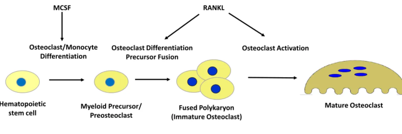
Osteoclastogenesis, the production and maturation of these cells, involves a complex development process that can be split into four steps; commitment, differentiation, multinucleation, and finally activation of immature osteoclasts.
Commitment
Excluding totipotent cells, the first cell type in the line giving way to osteoclast development is the pluripotent hematopoietic stem cell. This stem cell is still hardly limited in terms of variety, able to further differentiate into granulocytes, megakaryocytes,
monocytes, dendritic cells, and of course, the osteoclast (Figure 3). The earliest identifiable precursors with osteoclast-forming potential are granulocyte-macrophage colony forming units (GFU-GM) (Menaa, Kurihara, and Roodman 2000). The cytokines GM-CSF and later
M-CSF are required to stimulate proliferation and prevent apoptosis of the early osteoclast precursors. The principal transcription factors involved in these earliest stages of
development are PU.1, micropthalmia-associated transcription factor (MITF), and c-Fos. PU.1 is a member of the Ets family of transcription factors, more specifically the SP1 subfamily, which is involved in myeloid and b-lymphoid cell development. Ets factors can act as transcription activators, repressors, or both, depending on context and co-factors (Sharrocks 2001). As PU.1 is responsible for the earliest events in osteoclastogenesis, PU.1 null mice lack not only osteoclasts, but also macrophages, though they can still produce monocytic cells (Tondravi et al. 1997). Importantly, PU.1 regulates the transcription of the RANK gene in myeloid precursors, allowing for RANK ligand signaling (Kwon et al. 2005). As will be discussed in greater detail throughout this section, RANKL is the
preeminent osteoclastogenic cytokine, and thus this ability of precursors to respond to it is crucial.
MITF is a leucine zipper transcription factor that is implicated in the differentiation and survival of many cell types, including those of non-myeloid origin (Cheli et al. 2010). Like PU.1, MITF is expressed in macrophages and osteoclasts, as well as their precursors. Interaction with PU.1 allows MITF to regulate target genes such as cathepsin K, acid phosphatase, TRAP, and osteoclast-associated receptor (OSCAR) in early osteoclast differentiation (Hu et al. 2007). Other target genes include chloride channel 7 (Clcn7), necessary for bone-resorbing activity through acidification, and Ostm1, a membrane protein necessary for chloride channel stability in osteoclasts, whose knockout induces severe osteopetrosis in a murine model (Meadows et al. 2007; Pata, Héraud, and Vacher 2008).
Figure 3 : Osteoclast Development and Lineage
The hematopoietic stem cell, in response to M-CSF stimulation, gives rise to the myeloid stem cell. Depending on activation signals, this cell can further differentiate into
megakaryocytes, granulocytes, monocytes/macrophages, and osteoclasts, to name a few. It is through RANKL signaling that these myeloid precursors become preosteoclasts, which undergo fusion and form another multinucleated precursor. Further RANKL stimulation allows for continued differentiation and eventual activation of the mature osteoclast, capable of resorbing bone.
c-Fos is another key mediator of lineage commitment between osteoclasts and dendritic cells; another cell type derived from monocyte progenitors sensitive to GM-CSF, specifically MDPs (Monocyte and Dendritic cell Precursors) (Miyamoto et al. 2001; Liu and Nussenzweig 2010). However, differentiation is mediated by GM-CSF and M-CSF at the early phases of osteoclastogenesis. While RANKL and M-CSF are osteoclastogenic, RANKL in combination with GM-CSF becomes an activating factor of dendritic cells (Lee et al. 2009). However, after c-Fos expression following M-CSF signal transduction, these precursors are no longer competent to respond to GM-CSF, ensuring their path as
preosteoclasts (Miyamoto et al. 2001). Across cell types (and including osteoclasts), c-Fos is most heavily associated with proliferation, differentiation, and survival, and is necessary for NFATc1 transcription (Boyce et al. 2015). The murine c-Fos knockout results in an osteopetrotic phenotype (Matsuo et al. 2000).
Hematopoietic
stem cell Myeloid Precursor/Preosteoclast (Immature Osteoclast)Fused Polykaryon
Mature Osteoclast Osteoclast/Monocyte Differentiation Osteoclast Differentiation Precursor Fusion Osteoclast Activation MCSF RANKL
M-CSF, required for proliferation and osteoclast formation, has one receptor, transcribed by the macrophage c-FMS gene (Ross 2006), an activity that also depends on PU.1 (Tondravi et al. 1997). M-CSF can induce further expression of c-FMS by forming an autocrine loop to amplify M-CSF-mediated signals, and through stimulation of PU.1 (Yao et al. 2006). Loss of function of the M-CSF gene in mice led to decreases in macrophage numbers and an absence of osteoclasts, again producing an osteopetrotic phenotype (Dai et al. 2002). In cells deprived of M-CSF, the transcription factor MITF is sequestered to the cytoplasm through 14-3-3 protein interactions, thus inhibiting the translation of its
numerous target genes required for osteoclastogenesis (Bronisz et al. 2006). Production of this growth factor in the bone microenvironment is carried out constitutively by
T-lymphocytes, stromal cells, and osteoblasts. This production can be induced by elevated PTH levels, or inflammatory molecules like α or IL-10 (Agbanoma et al. 2012). TNF-α can also induce c-FMS expression (Yao et al. 2006).
RANKL/M-CSF signaling activates expression of osteoclastogenic genes via two mechanisms; the first being down-regulation at mRNA and protein levels of the DNA-binding protein Eos, a transcriptional repressor of the Ikaros family. This leads to a disassociation of co-repressors from PU.1 and MITF. The second is through
phosphorylation and activation of MITF through the ERK and p38 MAPK pathways, allowing for recruitment of co-activators (Mansky et al. 2002). These co-activators (BRG1 and CBP/p300) unwind chromatin, and recruit transcriptional machinery like RNA
polymerase II, respectively, in addition to acting as adaptor molecules (Bronisz et al. 2014; Asai, Funaba, and Murakami 2014). Downstream signaling from PI3K, p42/p44 ERK, and proto-oncogene c-Cbl are the key signal transducing mechanisms of M-CSF (Ross 2006). The PI3K/Akt cascades regulate (among other pathways) proliferation of osteoclast precursors through GSK3β and FoxO regulation. By phosphorylating these inhibitory factors, their ability to inhibit cell cycle entry is suppressed, allowing the cells to respond to proliferative stimuli (Manning and Cantley 2007).
Given that the majority of these transcription factors and associated cytokines are ubiquitous, regulating multiple cell lineages, none of these early osteoclastogenic pathways have been considered for therapeutic intervention. To block one or more of PU.1 or MITF,
for example, would have catastrophic side effects beyond regulation of osteoclastic selection. However, the further into osteoclast-specific pathways we venture, the more practical the consideration of particular targets becomes.
Differentiation
The aforementioned RANKL, member of the TNF superfamily, is produced by osteoblasts in the periosteum and stromal cells in the bone marrow under normal physiological conditions. In the case of skeletal inflammation, such as the conditions produced under rheumatoid arthritis, RANKL is also produced in large quantities by T-lymphocytes (Kong et al. 1999). In some situations, RANKL can be cleaved from the membrane of the cell and function as a soluble ligand (Kanamaru et al. 2004). Deletion of either RANKL or its receptor results in a complete absence of osteoclasts, arresting osteoclastogenesis immediately following M-CSF-induced expansion of osteoclast progenitor cells.
In addition to RANK, RANKL also has a soluble decoy receptor, osteoprotegerin (OPG), which is produced by osteoblasts and competitively binds RANKL so as to prevent RANK signaling (Yasuda et al. 1998). Deletion of the gene coding for OPG (TNFRSF11B) has been shown to increase osteoclast activity and numbers (Buckley and Fraser 2002). The expression of RANK is regulated by a variety of hormones including PTH, forskolin, and PGE2, all of which act on the cyclic AMP/PKA pathway, as well as vitamin D, which acts through its own pathway referred to as VDR-mediated (Takahashi et al. 2014). Upon binding RANKL, the receptor trimerizes, permitting its activation. One of the earliest steps of this activation is the recruitment of and interaction with the signal transducer TRAF6 (TNF receptor associated factor 6). The recruitment of TRAF6 is essential to
osteoclastogenesis, as TRAF6 deficient mice develop severe osteopetrosis stemming from an impairment of osteoclast differentiation, preventing eventual resorptive activity (T. Kobayashi et al. 2003). Likewise, osteoclast-related TRAF6 is RANKL-dependent, despite its association and signaling cascade participation with other receptors like CD40 and TLR family members (Ye et al. 2002). While the exact reasons for this unique RANKL/TRAF6 axis are not fully explained, some hypotheses include differing downstream MAPK
activation due to involvement of other RANKL signaling partners, and a higher recruitment of TRAF6 to the surface receptor (Kadono et al. 2005). Among these other downstream intracellular signaling pathways are activation of IκB kinases (IKK) α and β, MAP kinases p38 and ERK, and atypical PKCs. (Figure 2) Once TRAF6 activates the TAK1 kinase, TAK1 can phosphorylate IKKβ, which in turn phosphorylates the inhibitory protein IκB (Chen, Bhoj, and Seth 2006; Walsh et al. 2008). While IκB is ordinarily bound to the inactive form of NF-κB in the cytoplasm, this phosphorylation leads to the dissociation of the complex, and the shuttling off of IκB for degradation (May and Ghosh 1997). The release of NF-κB from IκB permits the former to translocate to the nucleus, where it binds DNA and induces transcription of a variety of genes necessary for osteoclast differentiation and survival. Studies have shown that mice lacking NF-κB subunits are osteopetrotic and missing osteoclasts in a similar manner to those lacking TRAF6 or RANKL (Iotsova et al. 1997), indeed without NF-κB there can be no transcription of a vast number of osteoclast-related genes, and thus no osteoclasts or resorption to follow (Boyce et al. 2015).
Another critical signaling pathway downstream of RANKL/TRAF6 is PI3K (Arron et al. 2001). This pathway is triggered by M-CSF to aid in cell proliferation, but it's also known for its cytoprotective effects. Briefly, PI3K activation leads to PDK1 activation, which subsequently phosphorylates Akt, which goes on to inactivate the pro-apoptotic BAD via phosphorylation (Toker and Cantley 1997). Overexpression studies of RANK demonstrated increases in intracellular Ca2+, regulated by PLC. This accelerated NF-κB translocation as well as the activation of JNK signaling (Komarova et al. 2003). The role of JNK in osteoclastogenesis is complicated, as complete blockade of its activity inhibits differentiation, but its activation is also accompanied by an increase in apoptotic cell death (Vaira et al. 2008). Generally, the activation of MAP kinases by RANKL leads to the activation and translocation of many transcription factors besides those already detailed, including ATF2, c-Jun, and members of the NFAT family, which is another factor leading to gene transcription vital to osteoclast differentiation and activation (Matsumoto et al. 2000; Sitara and Aliprantis 2010).
The NFAT family is necessary as a transcription regulator downstream of NF-κB; responsible for a number of genes crucial to osteoclast formation. There are five known
members, the most active of which being NFATc1, with in vitro studies demonstrating that induction of NFATc1 expression is sufficient for differentiation even in the absence of RANKL (Takayanagi et al. 2002). Correspondingly, deletion of the NFATc1 gene resulted in the cessation of osteoclast formation following RANKL stimulation. Nuclear
localization, and thus transcription of target genes by NFATc1 is regulated by intracellular calcium levels, and knockout of Ca2+-signaling modulator Inositol polyphosphate 4-phosphatase type IIa (Inpp4bα) resulted in non-RANKL-dependent NFATc1 nuclear localization and transcriptional activation (Ferron et al. 2011). In the early stages of osteoclastogenesis, NFATc2 is also recruited to the NFATc1 promoter, but it alone is not sufficient to activate the promoter. It is through cooperation with NF-κB that NFATc1 is induced, followed by an auto-amplification phase (Asagiri et al. 2005). Of the osteoclast-specific genes regulated by NFATc1 (and by extension, NF-κB), we observe cathepsin K, TRAP, CTR, OSCAR, and others (Matsumoto et al. 2004; Kim et al. 2005). To optimize transcription it can form complexes with the aforementioned PU.1, MITF, and c-FOS; however, these complexes are not required for osteoclastogenesis (Crotti et al. 2008). In addition to these effects, NFAT also negatively regulates osteoblast differentiation through the regulation of Fos-related protein (FRA)-2 (Zayzafoon 2005); the production of Fra-2 is a positive regulator of bone and matrix formation (Bozec et al. 2010). It has also been demonstrated that cell-cell contact between preosteoclasts and osteoblasts or stromal cells is important to RANK signaling (Garcia-Palacios et al. 2007). The importance of cell contact for osteoclastogenesis will be elaborated in greater detail in the following section.
Multinucleation
One of the most visibly characteristic activities of the developing osteoclast is cell fusion and resulting multinucleation. Despite there being other types of cell fusion in the body, the process that takes place during osteoclastogenesis is easily distinguishable (Ishii and Saeki 2008). The most obvious effect of multinucleation is the increase in cell size. This, along with a spreading of the cytoplasm, allows for a much larger surface area in contact with the bone, allowing for greater overall resorptive effect. This contact is vital, as unlike macrophages, osteoclasts do not primarily degrade their targets in lysosomes within the cell, but must create a seal at the bone surface, forming an “extracellular lysosome”
(Teitelbaum and Ross 2003). While the area of a cross-section of several non-fused cells and a fused multikaryon from these same cells is identical, the volume increases, as explained by their respective volume equations: 2
3𝜋𝑟
3× 4 versus 2 3𝜋(2𝑟)
3. Volume, in turn, is a limiting factor for determining resorbing capacity, dictating how far the osteoclast can spread. In addition, multinucleation has the positive effect of transferring the RANKL signaling cascade and its effects to the additional nuclei included in the cell, effectively multiplying the signal. Without this step of multinucleation, resorption is significantly reduced, as studies have proven mononuclear osteoclasts to be poor resorbers of bone (Vignery 2005).
In addition to the cascades already mentioned, RANKL stimulation also initiates gene expression of chemokines MCP-1 and RANTES, both of which are chemotactic signals for monocytes (Kim, Day, and Morrison 2005). Simultaneously, it induces MCP-1 receptors CCR2 and CCR4, which when activated stimulate the PI3K pathway (Hayashida et al. 2001). In addition to these monocyte-targeting chemoattractants, NFATc1 induces expression of cell fusion molecules such as vacuolar ATPase Vo domain d2 isoform, and the dendritic cell-specific transmembrane protein (DC-STAMP), by directly binding their promoter regions and initiating transcription (Kim et al. 2008). Murine models lacking functional v-ATPase Vod2 or DC-STAMP have been observed with impaired osteoclast fusion, and significantly reduced resorption (Lee et al. 2006).
RANKL has also been demonstrated to induce the translocation of membrane-bound CD9 from non-lipid raft microdomains to raft domains (Ishii et al. 2006). CD9 has been proposed to interact with other membrane-associated molecules responsible for cell fusion in osteoclastogenesis, bringing them to lipid raft microdomains (Claas, Stipp, and Hemler 2001). Once activated, CD9 also regulates osteoclast development through p42/p44 ERK (Yi et al. 2006). Additionally, CD9 expression is elevated in bone tissue in
osteoporosis, suggesting its dysregulation can be linked to bone resorptive disease (Iwai et al. 2008). The expression of the lipid raft component flotillin is markedly increased during osteoclast differentiation (Ha et al. 2003), as well as caveolin-1, a lipid raft stabilizer (Lee et al. 2015). Knockdown of Cav-1 reduces osteoclast formation and NFATc1 activation in response to RANKL. As the lipid composition of the plasma membrane plays a large role
in membrane fusion, the induction of a change in membrane composition is important (Chernomordik and Kozlov 2003). Disruption of lipid rafts has been shown to block RANK/TRAF6 interaction (Ha et al. 2003), as well as impairing v-ATPase activity in osteoclasts (Ryu et al. 2010). In short, the fusion mechanism is vital to healthy osteoclast function, enabling them to act as efficient remodelers of bone despite their relatively small numbers in the bone cell population.
Maturation and Actin Ring Formation
One of the final steps undertaken by an osteoclast on the way to becoming a mature, multinucleated, bone-resorbing cell is the polarization of the cell membrane. This is
necessary for the generation of resorptive pits (Saltel et al. 2004). The osteoclasts must therefore produce two structures; a villous organelle covering the bone surface referred to as the ruffled membrane, and an actin ring surrounding this contact area, forming the “sealing zone”, which isolates the resorptive microenvironment from the exterior. Without polarization and cytoskeletal re-organization resulting in the extracellular lysosome
formation, resorptive function is lost (Novack and Teitelbaum 2008). While all of the precise mechanisms necessary for this are not fully detailed, several key pathways and molecules of import have been identified (Ma et al. 2010).
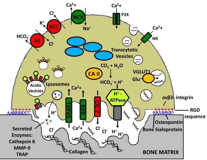
Prior to polarization, the osteoclast must receive confirmation that it is in contact with the bone surface. The integrin ανβ3 is the principal integrin expressed by osteoclasts, responsible for the recognition of mineralized matrix (Zou et al. 2007). (Figure 4) It does so by targeting the RGD sequence conserved in osteopontin and bone sialoprotein (Sharp et al. 1999). Integrins can transduce a variety of intracellular signaling pathways once activated; either directly mediated by the ligand itself, or through signaling downstream of growth factor receptors (Haas and Plow 1994). Deletion of the j83 integrin subunit was shown to induce osteopetrosis in mice (Cheng et al. 2000), and it has been shown to be necessary for motility, bone matrix adhesion, and polarization of the osteoclast (Novack and Teitelbaum 2008).
Figure 4 : Resorption Mechanisms in the Mature Osteoclast
Following αvβ3-integrin-mediated attachment of the cell to the surface of bone, the characteristic ruffled border of the osteoclast is formed by the transport of vesicles containing H+ATPase to the laminal membrane. Carbonic anhydrase generates H+ and HCO3 ions, acidifying the environment. This acidification is necessary for the activity of enzymes like Cathepsin K, an acid protease that degrades type 1 collagen, the primary component of organic bone matrix. H+ is transported out of the cell and into the resorption lacuna by electrogenic proton pumps (the aforementioned H+ATPase), and intracellular pH is maintained by a Cl-/HCO3 exchanger at the antiresorptive surface. Likewise,
electroneutrality is maintained by chloride channel activity (CLC7), which transports Cl- into the resorption lacuna. Finally, calcium balance within the cell is maintained by transporters both at the antiresorptive surface and the ruffled border (shown in green).
H+ -ATPase AE KCC NCX CA II Acidic Vesicles Transcytotic Vesicles VGLUT1 Glu NR P2X HCO3-+ H+ Ca2+ Ca2+ Ca2+ Ca2+ Cl -Cl -Ca2+ H+ H+ Cl -HCO3 -Na+ Secreted Enzymes: Cathepsin K MMP-9 TRAP CO2+ H2O Cl -K+ Lysosomes Cl -H+ Collagen VVVVVVV VVVVVVV BONE MATRIX avβ3- integrin Osteopontin Bone Sialoprotein RGD sequence TRP V5 R YR C lC -7
The mechanism of integrin contact is unique when compared with most other cells. Ordinarily, integrins mediate matrix contact through focal adhesions, containing signaling and cytoskeletal molecules, leading to the formation of stress fibers (contractile actin bundles providing force). With regards to the bone surface, however, the osteoclast organizes its fibrillar actin into sealing zones rather than forming stress fibers, and forms podosomes; ring structures of integrin receptors with an actin-rich core, rather than focal adhesions, which are made up of denser plaques of actin (Faccio et al. 2003). While somewhat tangential to the results later presented in this manuscript, and thus not further explained here, for interested parties a complete review summarizing the differences between different matrix contact structures and their roles in health and disease can be found in the Journal of Signal Transduction (Eleniste and Bruzzaniti 2012).
An effective resorbing osteoclast is constantly in motion, and thus in these motile cells the sealing zone is frequently being disassembled, and other, non-podosomal integrins move to lamellipodia (extensions of the membrane) at the leading edge of the cell. To efficiently resorb bone, the osteoclast must be able to detach, migrate, and re-attach in order to re-form the sealing zone. It is because of this movement that resorptive pits more
frequently resemble a chain of overlapping circular indentations, rather than single isolated holes, and the integrins, particularly ανβ3, are necessary for this process. c-Src, one of the best identified associated signaling molecules in osteoclastogenesis, was first studied in 1991 in a murine model, where c-Src -/- mice were severely osteopetrotic, yet expressed large numbers of osteoclast-like cells (Boyce et al. 1992). The problem developed by these mice was that their “osteoclasts” lacked ruffled membranes and actin rings. c-Src is known to regulate osteoclasts both through its role as a kinase and an adaptor molecule (Miyazaki et al. 2004), and it has since been established that one function of c-Src, as a re-organizer of the cytoskeleton, is to link ανβ3 to the cytoskeleton following RANKL stimulation (Izawa et al. 2012). Another notable interaction of the ανβ3 integrin is with the M-CSF/c-Fms pathways. Upon binding M-CSF, c-Fms activates the integrin by targeting its cytoplasmic domain, which alters the conformation of its extracellular, ligand binding region (Faccio et al. 2003). This conformational change can partially emulate ανβ3 activation.
Mechanisms of Resorption
The penultimate step of polarization and establishment of the sealing zone allows for the defining step of osteoclast activity to take place: the secretion of acid and acidic hydrolases onto the bone surface to degrade the extracellular matrix. The high amount of acid secreted by the osteoclast is necessary in order to dissolve the hydroxyapatite making up the bulk of the bone structure. This low pH is also necessary for the acidic hydrolases, notably cathepsin K, to be active, and degrade the remaining organic matrix (Teitelbaum and Ross 2003). Deletion of cathepsin K leads to decreased bone resorption coupled with an increase in formation (Saftig et al. 1998), driven by osteoclast-specific signaling (Lotinun et al. 2013). Similar to lysosomes, the engine driving acid secretion across the ruffled membrane are the v-type H+-ATPases. Their continued action would be impossible if the cytoplasm became alkalinized, however, so the protons and chloride ions being transported through the channels must be replenished. For this process, the osteoclast depends on the concerted activity of carbonic anhydrase, which is responsible for the conversion of CO2 to bicarbonate and protons, and an anion exchanger, such as anion exchange protein 2 (AE2) (Josephsen et al. 2009).
The resorbing osteoclast must also have mechanisms for the transportation of breakdown products that accumulate in the sealing zone. The acid and proteases it secretes solubilize large amounts of calcium (up to 40 mM) and phosphate, which will eventually be released to the bloodstream, as well as other organic breakdown products (Silver, Murrills, and Etherington 1988). While the majority of these products are moved via transcytosis via the osteoclast out through the other side of the cell, it is impossible to fully prevent some calcium and phosphate ions from entering the osteoclast (Salo et al. 1997). To maintain homeostasis, the osteoclast employs several calcium-transporting transmembrane proteins (Figure 4). Among these are included the voltage operated (opening at depolarized
membrane potentials) and ligand-gated Ca2+ channels, Na+-Ca2+ exchangers, and ryanodine receptor Ca2+ channels (Datta and Horrocks 2003). The development and utilization of ion channels and transporters is a hallmark of the adult osteoclast, the final manifestation of weeks of development in response to diverse signaling pathways.