ORIGINAL ARTICLE
Probe-based confocal laser endomicroscopy for pleural
malignancies diagnosis
OLIVIERBONHOMME,1 VINCENTHEINEN,1NANCYDETREMBLEUR,2JEAN-LOUISCORHAY,1 RENAUDLOUIS1AND BERNARDDUYSINX1
1Pneumology Department, CHU Liège, Domaine Universitaire du Sart-Tilman, Liège, Belgium;2Pathology Department, CHU
Liège, Domaine Universitaire du Sart-Tilman, Liège, Belgium
ABSTRACT
Background and objective: Probe based confocal laser endomicroscopy (pCLE) is an optical imaging technique allowing live tissue imaging at a cellular level. Currently, this tool remains experimental. Two studies regarding pleural disease have been published and suggest that pCLE could be valuable for pleural disease investigations. However, normal and malignant pleural pCLE features remain unknown. Therefore, we conducted a prospective trial of pCLE during medical thoracoscopy to study and describe the malignant and benign pleural pCLE features. Methods: Every patient >18 years referred to our department for medical thoracoscopy was eligible. Med-ical thoracoscopy was performed under sedation, all-owing spontaneous breathing. Five millilitres of fluorescein (10%) was intravenously administrated 5 min before image acquisition. The pCLE was intro-duced through the working channel of the thoracoscope and gently placed on the parietal pleura to record videos. Afterwards, biopsies were performed on the corresponding sites. Malignant and benign pleural pCLE features were precisely described and compared using 11 preselected criteria.
Results: A total of 62 patients were included in the anal-ysis including 36 benign and 26 malignant pleura. Among our preselected criteria,‘abnormal tissue archi-tecture’ and ‘dysplastic vessels’ were strongly associated with malignancies (100% and 85% ss, 721% and 74% sp, respectively) whereas, the‘full chia seeds sign’ and ‘cell shape homogeneity’ were associated with benignity (36% and 56% ss, 100% and 70% sp, respectively). No study-related adverse events occurred.
Conclusion: Benign and malignant pleural involvement have clearly distinct pCLE features.
Clinical trial registration:NCT03805971 at Clinicaltrial.org
Key words: medical thoracoscopy, pleural carcinomatosis, pleuroscopy, probe-based confocal laser endomicroscopy.
INTRODUCTION
Confocal laser endomicroscopy is an optical endo-scopic imaging tool.1This technique allows live in vivo imaging of tissues at a cellular level with a video frame of 12 images per second. In probe-based confocal laser endomicroscopy (pCLE), the device is miniaturized so that it can be introduced through the working channel of endoscopes providing live, dynamic and microscopic assessment of tissues.
pCLE remains an experimental technique and data are scarce in pulmonary medicine and thoracic
oncology.2–9 As for pleural diseases, Zirlik et al.
showed that, compared to pleural fluid cytology,
pCLE can detect malignant cells in pleural effusion
with 87% sensitivity and 99% specificity.9 Wijmans
et al. have recently published a prospective (n = 15) study assessing (p)CLE for guiding biopsies in the
specific indication of suspected malignant pleural
mesothelioma with different sampling methods. They showed that (p)CLE could distinguish malignant
mesothelioma from pleural fibrosis with high
preci-sion.8 However, only three thoracoscopies, which is
the gold standard procedure for malignant
mesothe-lioma management,10–12 were included in that study.
Otherwise, we have recently reported on three cases
of pCLE during medical thoracoscopy with the first
description of the parietal pleura during pCLE in a Correspondence: Olivier Bonhomme, Pneumology
Department, University of Liege, Sart-Tilman, B35, 4000 Liege, Belgium. Email: [email protected]
Received 11 February 2020; invited to revise 6 April and 20 July 2020; revised 9 May and 9 August 2020; accepted 26 August 2020 Associate Editor: Ioannis Kalomenidis; Senior Editor: Phan Nguyen
This is an open access article under the terms of the Creative Commons Attribution License, which permits use, distribution and reproduction in any medium, provided the original work is properly cited.
S U M M A R Y A T A G L A N C E
Probe based confocal laser endomicroscopy (pCLE) is an optical imaging tool allowing live imaging of tissues at a cellular level. It remains experimental but its clinical value as a diagnostic/guiding tool is apparent. To address the lack of data in thoracic oncology and pleural diseases, we show the ability of pCLE during medical thoracoscopy to distinguish benign from malignant pleural involvement.
healthy patient and two patients with malignant pleural involvement.13
Although these results suggest that pCLE could be valuable for pleural diseases investigation, large studies describing pleural pCLE in healthy subjects or in patho-logical conditions are still lacking and needed. Conse-quently, we conducted a prospective study to assess the pleural pCLE features obtained during thoracoscopy.
The main objectives were to confirm our previous
descriptions over a larger population, to assess the feasi-bility and safety of the technique and to identify specific criteria for benign or malignant pleural involvement.
METHODS
Study design and procedure
We performed a single-centre prospective cross
sec-tional study. Every patient ≥18 years referred for a
medical thoracoscopy between June 2018 and
September 2019 (pleural effusion work up, recurrent pneumothorax, talc pleurodesis, etc.) was eligible for the present study. Exclusion criteria were pregnancy
and known allergy to fluorescein. Thoracoscopy was
performed under sedation (allowing spontaneous
breathing) by two experienced investigators with a sys-tematic procedure. The patient was positioned in lat-eral decubitus during the intervention. After chest ultrasound examination, a pneumothorax was induced with the Boutin trocar and one entry port was
per-formed through the chest wall. Two different
thoracoscopes were used during this study: Wolf thoracoscope with an outer diameter of 7 mm (Richard Wolf GmbH, Knittlingen, Germany) and a Storz single puncture thoracoscope with an outer diameter of 10 mm (Karl Storz GmbH, Tuttlingen, Germany). Five
millilitres of fluorescein (10%) was intravenously
administrated 5 min before image acquisition. Thereaf-ter, the pleural cavity was examined macroscopically.
Then, the pCLE (Alveoflex, lateral resolution 3 μm,
optical area 1.13 mm2, depth of focus 0–50 μm)
(Cellvizio; Maunakea Technologies Paris, France) was gently placed on the parietal pleura and videos were recorded. If macroscopic abnormalities were noticed, pCLE was performed on the affected zones. In the absence of macroscopic abnormality, three random sites were selected. Finally, biopsies were systematically performed on the same sites. During the procedure, the thoracoscopists rated the quality of the pCLE acqui-sitions as good, acceptable or low.
We previously reported our first experience of pCLE
during medical thoracoscopy.13 For this publication,
we performed pCLE in seven patients but only three
were reported. This was the first step of our research.
Afterwards, in collaboration with the pathologists from our centre, the pCLE acquisitions and the histological sections of the seven patients were reassessed. The objective was to identify the potential discriminant criteria between benign and malignant pleura. Eleven criteria were selected (Table 1, Appendix S1, Fig. S1 in Supplementary Information) and were prospectively assessed in the present study which is the second step of our research. No training session was performed after thefirst step. The seven patients of step 1 are not
included in the present study to avoid the risk of bias. After video acquisition, a third blinded investigator
screened every video set to select the five best
repre-sentative images per patient for further analysis and assessment of the preselected criteria.
The criteria were not scored if assessable elements for their interpretation were missing. Our study was
approved by the CHU Liege ethics committee
(B707201837069). Every patient provided written
informed consent.
Statistical analysis
The Fisher’s exact test was used to analyse the link
between pCLE qualitative variables and thefinal
histo-logical diagnosis. For quantitative variables, the
unpaired t-test was used. If a criteria was not assess-able for a patient during the investigation, it was excluded from the statistical analysis.
RESULTS
Patients’ characteristics, feasibility and safety
A total of 64 patients (44 males and 20 females) were recruited. Two patients were excluded: pCLE images no more available for one patient and other one because of a lack of histological conclusion. Of the 62 remaining patients, 36 had benign pleura on
histo-logical analysis (7 normal and 29 inflammatory pleural
involvement) while 26 had a malignant pleural disease with variable histological findings (Table 2, Fig. S2 in Supplementary Information). A total of 310 images were available for the study and the 11 criteria analysis. Approximately 5 min were necessary after intravenous fluorescein injection for an optimal pleural staining.
Concerning the feasibility of pCLE during medical
thoracoscopy, the Alveoflex probe fitted easily through
the working channel of our thoracoscopes and the pari-etal pleura was easy to reach. However, based on thoracoscopists judgement, the quality of the acquisi-tion was inconstant from one patient to another. Indeed, 20 pCLE were of good quality, 22 of acceptable quality and 20 of low quality.
There were no adverse events related to the pCLE
procedure or to the intravenous fluorescein
administration.
Pleural pCLE descriptions
Eight patients were recruited for talc pleurodesis for recurrent spontaneous pneumothorax (with normal
or slightly inflammatory pleura on histological
sec-tion). The normal mesothelium appeared as a well-organized tissue with polyhedral cells of similar size,
shape, fluorescence intensity with well-delineated
intercellular gaps and cell borders. There was a fibroadipose connective tissue beneath the
mesothe-lium featuring well-delineated large (around 50μm)
round, dark adipocytes surrounded by blood vessels
and connective fibres. Sometimes, striated muscular
fibres could be imaged as large (100 μm) and long
(hundreds of μm) parallel dark bands (Fig. 1,
Twenty-six patients had malignant pleural diseases. With pCLE, the normal pleura was no more visible and severe architecture and tissue distortions were
identified. Indeed, the malignant pleural involvement
was characterized by the presence of heterogeneous and pleomorphic cells with distorted intercellular gaps and indistinct cell borders. Those cells could be orga-nized in clusters or nodules or take a papillar, a glan-dular or another aberrant tissue architecture (Fig. 2B, E, Appendix S2-Video 2 in Supplementary Informa-tion). Sometimes, vascular abnormalities were also identified with vascular leaks, tortuous or giant vessels (Fig. 2C,D).
Criteria assessment
Among the 11 assessed criteria, four were statistically significantly associated with the malignity or the benig-nity of the pleural involvement (Tables 3–5, Fig. 2).
The identification of the abnormal tissue architecture
was significantly associated with malignant pleural
involvement (P < 0.0001). It was present for all malig-nant diseases with a good specificity (sensitivity (ss): 100%, specificity (sp): 72.41%, positive predicted value (PPV): 75%, negative predicted value (NPV): 100%).
The ‘full chia seed sign’, which is a sign of normal
architecture, had a relatively low sensitivity for
benig-nity but was 100% specific, yielding a 100% PPV (ss:
Table 1 Criteria for pCLE pleural evaluation
Criteria Details
Abnormal tissue architecture No: Correct identification of the previously described normal pleura characteristics† • Mesothelial monolayer with a regular distribution of the cells
• Fibroadipose connective tissue with regular and homogeneous vascularization • Muscular fibres
Yes: Identification of cellular/tissue structures which are not known to belong to the normal pleura (cellular clusters or dark clumps, glands, cells cordons, dysmorphic cells, papillar distribution, etc.)
Not assessable Cellular homogeneity in size,
shape andfluorescence
Subjective description by the investigator with reference to the normal pleura† Yes
No
Not assessable
Mean cellular size Mean cellular size assessed on a full optical area (1.13 mm2)
Not assessable Mean cellular density (number
of cells/10.000μm2)
Mean of three measurements realized in areas where the cellular density was visually the highest
Not assessable Dysplastic vessels‡ Yes§:
• Presence of tortuous vessels • Vascular leaks of fluorescein • Giant vessels
No: No criteria for dysplastic vessel Not assessable
Max vascular diameter‡ Selection and measurement of the largest vascular diameter on the set of images Not assessable
Max vascular density‡ Number of assessable vessels/optical area (1.13 mm2) Not assessable
Connective tissuefibres organization
Anarchic: Coarsefibres, irregular in shape or direction, without well-defined architecture Organized: Regular in shape and direction, well-defined architecture
Not assessable
Full chia seed sign Yes: Full optical area with chia seed sign (Fig. 1C)
No: Absence or presence on only one portion of the optical area Not assessable
†Normal pleural pCLE description as previously reported by Bonhomme et al.13 ‡Vessels are defined as continuous tubular hyperfluorescent images of at least 50 μm. §As described by Cannizzaro et al.20
36%, sp: 100%, PPV: 100%, NPV: 53.06%, P = 0.0003). Likewise, the presence of cells homogeneously shaped was associated with benignity but with relatively low
sensitivity and specificity (ss: 56.%, sp: 70%, PPV: 72%, NPV: 53%, P = 0.0052).
Dysplastic vessels were significantly and strongly
associated with malignancy (P < 0.0001) with a good sensitivity and specificity (ss: 85%, sp: 74%, PPV: 68%, NPV: 88.46%).
The other assessed criteria (cellular size and
fluo-rescence homogeneity, cellular mean size and den-sity, vascular density and maximum diameter and
connective tissue fibre organization) did not show
significant association with the final histological
diagnosis (Tables 3,4).
DISCUSSION
Only two studies concerning pleural diseases pCLE investigation have been published so far and our trial is the largest to assess pCLE during medical thoracoscopy. Normal and malignant pleural pCLE fea-tures are precisely described. Furthermore, 11 criteria
were selected according to our previous experience13
and four of them proved to be associated with thefinal
histological diagnosis (tissue architecture, dysplastic
vessels, full chia seed sign and cellular shape
homogeneity).
As less invasive pleural investigation procedures such as thoracentesis or Abram’s needle biopsies lack sensi-tivity (less than 60% for repeated thoracentesis),14,15 thoracoscopy remains the gold standard for pleural
dis-eases management.15–17 Therefore, we chose the most
appropriate procedure to study and validate the pleural Table 2 Patients characteristics and histological
diagnosis (n = 62)
Sex 19 F/43 M
Mean age (SD) 64.4 (17.29) Thoracoscopy
indications
Talc pleurodesis for recurrent pneumothorax: n = 8 Pleural exudation management
(mainly lymphocytic): n = 46 Talc pleurodesis for recurrent
malignant effusion: n = 4 Pleural hypermetabolism on PET:
n = 2
Pleural nodularity on CT scan: n = 2 Histological diagnosis Normal pleura: n = 7 (Sub-)acute pleuritis: n = 15 Chronic pleuritis: n = 13 Pulmonary adenocarcinoma: n = 9 Malignant mesothelioma: n = 7 Large cell lymphoma: n = 2 Small cell lung carcinoma: n = 3 Squamous cell lung carcinoma: n = 3 Breast adenocarcinoma: n = 1 Urothelial carcinoma: n = 1 Sarcoidosis: n = 1
CT, computed tomography; PET, positon emission tomography.
Figure 1 Normal probe-based confocal laser endomicroscopy (pCLE) features of the parietal pleura (see the pleural pCLE description in the Results section for more details). (A) The pCLE image and the histological section (haematoxylin and eosin (HE)) show fibroadipose connective tissue of the pleura with adipocytes (crosses) and connective fibres (arrows). (B) The histological (HE) section and the pCLE image show striated muscularfibres (crosses). (C) The pCLE image shows the normal mesothelium (‘full chia seed sign’). The histological section (HE) shows a normal pleura. The arrow shows where the pCLE is positioned (perpendicularly to the mesothelial surface) to give the pCLE image. The triangle lies in the sub-mesothelial connective tissue and the cross highlights the adipocytes (which are not presented on this pCLE image). (D) The pCLE image shows the pleural vascularization and some meso-thelial cells. The histological (HE) section shows pleural vessels (arrows) lying in the pleural adipose tissue (cross).
pCLE features with their strictly corresponding histologi-cal samples, which is the strength of our work. In fact,
the clinical value of pCLE with fluorescein during
thoracoscopy remains to be determined, but we believe that pCLE could improve the diagnostic yield of less invasive pleural procedures (such as thoracentesis or
Abram’s needle biopsies) now that we have precisely
described and validated the normal and malignant pleu-ral pCLE features.8,18 This should be the third step of our research.
For our study, we used the Alveoflex probe. Because
of a confocal plane beginning at 0 μm (standing
between 0 and 50μm), this probe is probably the most
appropriate to study the pleural mesothelium, the most superficial layer of the pleura and associated
malignan-cies (e.g. the AQ-flex, Cellvizio; Maunakea
Technolo-gies Paris, France, probe has a confocal plane standing
between 40 and 70μm deep). In our study, the
pres-ence of the full chia seed sign is highly specific for
benignity with a 100% PPV. In fact, the mesothelium is a really thin, delicate and highly reactive monocellular
layer.19 Whenever involved in a pathological process,
alterations or distortions of the mesothelium can
pre-vent its identification during endomicroscopy. This
explains the relatively low sensitivity of this criterion
but, also its very high specificity for benignity.
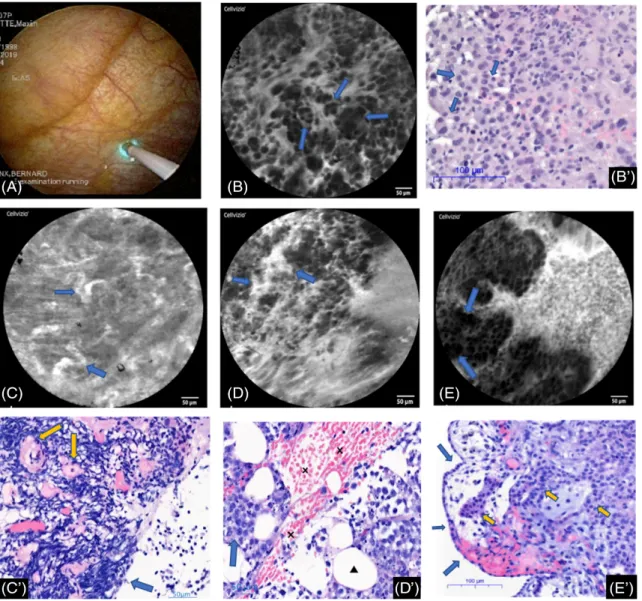
Figure 2 Illustrations of the significant probe-based confocal laser endomicroscopy (pCLE) features for pleural investigation. (A) pCLE gently applied on the parietal pleura (thoracoscopic view). (B, B0) Abnormal pleural architecture with cells of different size, shape and infiltration by glands (arrows) and cell clusters (lung metastatic adenocarcinoma). Haematoxylin and eosin (HE) staining for the histo-logical section. (C, C0) The pCLE image shows dysplastic tortuous vessels (arrows) in a patient with metastatic lung adenocarcinoma. The histological section (HE) shows tortuous pleural vessels (arrows) within the pleural connective tissue infiltrated by inflammatory cells. (D, D0) The pCLE image shows a dysplastic vessel with patentfluorescein leaks (arrows) in a patient with pleural infiltration by a large B cell lymphoma. The histological section (HE) shows a haemorrhagic suffusion (crosses) in the pleural connective tissue (trian-gle for adipocytes) infiltrated by malignant B cells (arrow). (E, E0) The histological section (HE) shows a pleura (blue arrows are for the mesothelial layer) infiltrated by malignant cells taking an epithelioid architecture (epithelioid mesothelioma) (yellow arrows). The pCLE image highlights the epithelioid (aberrant) architecture and identifies some cell clusters (arrows). The full chia seed sign is illustrated in Figure 1 with the normal pleural pCLE features.
Similarly, the cellular shape homogeneity was signi fi-cantly associated with benignity, although, in this case, with lower sensitivity and specificity.
As for the abnormal tissue architecture, we showed in the current study that the presence of pleomorphic cells, dark clumps and cell clusters was found in all pleural malignancies but only in 27% of the benign
pleura. Our finding is in keeping with previously
reported studies assessing pCLE in thoracic oncol-ogy.5,7,8,20Furthermore, as the Alveoflex probe, with an optic area of 1.13 mm2, is the probe with the largest field of view, it allows a better assessment of the gen-eral tissue architecture than other probes such as the
AQ-flex which has an optical area of only 0.33 mm2.
Thereby, the global cellular and tissue organization could be analysed allowing for identification of glands, papils, cordons and macronodules of cells in malignant pleura (Fig. 2).
Dysplastic vessels were significantly and strongly
associated with malignancy in our study. Fluorescein is a really good vascular staining agent. In fact, it has been used for decades in ophthalmology to stain retinal vessels.21 Our finding is not surprising as (neo)
angio-genesis is a hallmark of cancer and is identified as a
therapeutic target in malignancies management.22 The
new vessels are often tortuous, irregular (in size and
shape), disorganized and excessively leaky.23 In
endomicroscopy with fluorescein, the vessels appear
clearly as hyperfluorescent images and can be easily and precisely studied in a dynamic fashion. This live and dynamic evaluation of tissues at a cellular level is a hall-mark of pCLE (conversely to standard histology). In gas-troenterology, the pCLE study of the vascular network is already part of the diagnostic management of malignan-cies, mostly in pancreatic cystic diseases.24–27As a con-sequence, we believe that pCLE live imaging with Table 3 Qualitative criteria for pleural pCLE assessment
Benign pleura, n = 36 Malignant pleura, n = 26
Total not
assessable Fisher’s exact test
Yes No N.A. Yes No N.A. N.A. (%) P-value
Abnormal architecture 8 21 7 24 0 2 9 (14.5%) <0.0001
Cellular size homogeneity 18 14 4 7 16 3 7 (11.3%) 0.0987
Cellular shape homogeneity 18 14 4 4 19 3 7 (11.3%) 0.0052
Cellularfluorescence homogeneity 28 7 1 14 10 2 3 (4.83%) 0.0863
Blood vessels dysplasia 8 23 5 17 3 6 11 (17.7%) <0.0001
Organized connective tissue 11 20 5 5 12 9 14 (22.6%) 0.76
Chia seeds sign 13 23 0 0 26 0 0 (0%) 0.0003
N.A., not assessable; pCLE, probe-based confocal laser endomicroscopy. p < 0.05 is significant in our statistical analysis.
Table 5 Pleural pCLE significant criteria
Criteria Sensitivity (%) Specificity (%) Positive pred value (%) Negative pred value (%)
Full chia seed sign 36 100 100 53.06
Cell shape homogeneity 56.25 69.57 72 53.33
Dysplastic vessels 85 74 68 88.46
Abnormal tissue architecture 100 72.41 75 100
Abnormal tissue architecture, associated with malignity; cellular shape homogeneity, associated with benignity; dysplastic vessels, associated with malignity; full chia seed sign, associated with benignity; pCLE, probe-based confocal laser endomicroscopy; pred, predictive.
Table 4 Quantitative criteria for pleural pCLE assessment
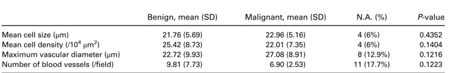
Benign, mean (SD) Malignant, mean (SD) N.A. (%) P-value
Mean cell size (μm) 21.76 (5.69) 22.96 (5.16) 4 (6%) 0.4352
Mean cell density (/104
μm2) 25.42 (8.73) 22.01 (7.35) 4 (6%) 0.1404
Maximum vascular diameter (μm) 22.72 (9.93) 27.08 (8.91) 8 (12.9%) 0.1216
Number of blood vessels (/field) 9.81 (7.73) 6.90 (2.53) 11 (17.7%) 0.1223
fluorescein is really suitable to study vascular abnormali-ties (static and dynamic) in thoracic malignancies.
None of the quantitative criteria were shown to be significantly associated with the histological diagnosis. This could be surprising because malignant involvement
is frequently associated with hypervascularization,
hypercellularity and tissue heterogeneity. However, because of the tissue heterogeneity, it was difficult to take reproducible and precise measurements with only five pictures per patient.
Regarding the qualitative criteria, it is worth
not-ing that those without significant association
with the histological diagnosis are the most subjec-tive criteria with probably the lowest reproducibil-ity. Furthermore, there might be no association between the connective tissue organization, the cell fluorescence homogeneity and the histological diagnosis.
Although promising, our results need to be validated prospectively in larger multicentre studies, with assess-ment of reproducibility of the results. A major limita-tion in our experience concerns the quality of the acquisition, as 20 out of 62 set of images were scored low by the two thoracoscopist investigators with some criteria not assessable and therefore had to be excluded from the statistical analysis. Although limited in propor-tion (Tables 3,4), this could lead to an overestimapropor-tion of our results. Several reasons can explain this low quality. First, it was sometimes difficult to stabilize the probe and the image acquisition because of the respiratory move-ments of the patient (depending, notably on the sedation depth), the curved shape of the thorax and the high flexi-bility of the probe. Second, when present, a pachypleuritis or a thickfibrinous pleuritis could prevent a good visuali-zation of the underlying mesothelial layer or other pleural structures either because of an increased tissue thickness
and/or a suboptimal fluorescein pleural staining.
How-ever, the quality of the acquisition has gradually improved along the study (learning curve). Therefore, we believe that experienced investigators could reach a more favourable proportion of interpretable pleural pCLE inves-tigation, as already shown by others.28,29
Another potential bias of our study is the selection of the‘most representative images’ by the same investiga-tors than the one who scored the criteria. However, we think that this potential bias is limited for qualitative criteria (the only ones to be significant in our study). In fact, those criteria are binary scored (yes or no) and the scoring investigator screened the entire pCLE
acquisition to find the best representative images. As
for quantitative criteria, which are not significant, we acknowledge a possible bias because of the high het-erogeneity of some acquisitions.
Finally, the criteria investigated in the present study are based on a small number of patients (n = 7) rec-ruited for the first step of our investigation. A larger prediction set would have strengthened the study.
In conclusion, this is thefirst study evaluating pCLE during medical thoracoscopy. Our results demonstrate the safety and the feasibility of the technique. We pro-vide a precise description of benign and malignant pleural pCLE features validated by the gold standard procedure for pleural diseases investigation. Discrimi-nating criteria between malignant and benign pleura
were identified. We believe that pCLE for pleural
dis-ease investigation and management could be a valu-able tool. Whether pCLE could become part of minimally invasive pleural investigation procedure or increase the diagnostic accuracy of thoracentesis needs to be investigated in further studies.
Data availability statement: The patients’ individual data will not be available.
Author contributions: Conceptualization: O.B., V.H., N.D. Formal analysis: O.B., J.-L.C., B.D., R.L. Funding acquisition: O.B., V.H. O.B., V.H., B.D., J.-L.C., R.L. Data curation: O.B., V.H., N.D. For-mal analysis: O.B., J.-L.C., B.D., R.L. Funding acquisition: O.B., V. H. Investigation: O.B., V.H., R.L., N.D., J.-L.C., B.D. Methodology: O.B. Project administration: O.B., V.H., J.-L.C., R.L., B.D. Resources: O.B., R.L. Software: O.B. Supervision: O.B., V.H., J.-L. C., R.L., N.D., B.D. Validation: O.B., J.-L.C., R.L., B.D. Writing— original draft: O.B., V.H. Writing—review and editing: V.H., J.-L. C., R.L., N.D., B.D.
Abbreviations: NPV, negative predictive value; pCLE, probe-based confocal laser endomicroscopy; PPV, positive predictive value; sp, specificity; ss, sensitivity
REFERENCES
1 Wijmans L, D’Hooghe JNS, Bonta PI, Annema JT. Optical coher-ence tomography and confocal laser endomicroscopy in pulmo-nary diseases. Curr. Opin. Pulm. Med. 2017; 23: 275–83.
2 Hassan T, Piton N, Lachkar S, Salaün M, Thiberville L. A novel method for in vivo imaging of solitary lung nodules using naviga-tional bronchoscopy and confocal laser microendoscopy. Lung 2015; 193: 773–8.
3 Hassan T, Thiberville L, Hermant C, Lachkar S, Piton N, Guisier F, Salaun M. Assessing the feasibility of confocal laser endomicroscopy in solitary pulmonary nodules for different part of the lungs, using either 0.6 or 1.4 mm probes. PLoS One 2017; 12: e0189846. 4 Wijmans L, de Bruin DM, Meijer SL, Annema JT. Needle based
confocal laser endomicroscopy for mediastinal lesions, an in vivo pilot-study. Eur. Respir. J. 2016; 48: OA3015.
5 Wijmans L, Yared J, de Bruin DM, Meijer SL, Baas P, Bonta PI, Annema JT. Needle-based confocal laser endomicroscopy (nCLE) for real-time diagnosing and staging of lung cancer. Eur. Respir. J. 2019; 53: 1801520.
6 Benias PC, D’Souza LS, Papafragkakis H, Kim J, Harshan M, Theise ND, Carr-Locke DL. Needle-based confocal endomicroscopy for evaluation of malignant lymph nodes– a feasibility study. Endoscopy 2016; 48: 923–8.
7 Fuchs FS, Zirlik S, Hildner K, Schubert J, Vieth M, Neurath MF. Confocal laser endomicroscopy for diagnosing lung cancer in vivo. Eur. Respir. J. 2013; 41: 1401–8.
8 Wijmans L, Baas P, Sieburgh TE, de Bruin DM, Ghuijs PM, van de Vijver MJ, Bonta PI, Annema JT. Confocal laser endomicroscopy as a guidance tool for pleural biopsies in malignant pleural mesothe-lioma. Chest 2019; 156: 754–63.
9 Zirlik S, Hildner K, Rieker RJ, Vieth M, Neurath MF, Fuchs FS. Confocal laser endomicroscopy for diagnosing malignant pleural effusions. Med. Sci. Monit. 2018; 24: 5437–47.
10 Galbis JM, Mata M, Guijarro R, Esturi R, Figueroa S, Arnau A. Clin-ical-therapeutic management of thoracoscopy in pleural effusion: a groundbreaking technique in the twenty-first century. Clin. Transl. Oncol. 2011; 13: 57–60.
11 Grossebner MW, Arifi AA, Goddard M, Ritchie AJ. Mesothelioma – VATS biopsy and lung mobilization improves diagnosis and pallia-tion. Eur. J. Cardiothorac. Surg. 1999; 16: 619–23.
12 Kindler HL, Ismaila N, Armato SG, Bueno R, Hesdorffer M, Jahan T, Jones CM, Miettinen M, Pass H, Rimner A et al. Treat-ment of malignant pleural mesothelioma: American Society of Clinical Oncology clinical practice guideline. J. Clin. Oncol. 2018; 36: 1343–73.
13 Bonhomme O, Duysinx B, Heinen V, Detrembleur N, Corhay J-L, Louis R. First report of probe based confocal laser endomicroscopy during medical thoracoscopy. Respir. Med. 2019; 147: 72–5.
14 Prakash UBS, Reiman HM. Comparison of needle biopsy with cytologic analysis for the evaluation of pleural effusion: analysis of 414 cases. Mayo Clin. Proc. 1985; 60: 158–64.
15 Loddenkemper R. Thoracoscopy– state of the art. Eur. Respir. J. 1998; 11: 213–21.
16 Skalski J, Astoul P, Maldonado F. Medical thoracoscopy. Semin. Respir. Crit. Care Med. 2014; 35: 732–43.
17 Xia H, Wang X-J, Zhou Q, Shi H-Z, Tong Z-H. Efficacy and safety of talc pleurodesis for malignant pleural effusion: a meta-analysis. PLoS One 2014; 9: e87060.
18 Fockens P, Chen Y, Dekker E, Sharma P, Lauwers G, Meining A, Wallace M. Miami classification for probe-based confocal laser endomicroscopy. Endoscopy 2011; 43: 882–91.
19 English JC, Leslie KO. Pathology of the pleura. Clin. Chest Med. 2006; 27: 157–80.
20 Cannizzaro R, Mongiat M, Canzonieri V, Fornasarig M, Maiero S, De Re V, Todaro F, De Paoli P, Spessotto P. Endomicroscopy and cancer: a new approach to the visualization of neoangiogenesis. Gastroenterol. Res. Pract. 2012; 2012: 1–5.
21 Wong TY, Sun J, Kawasaki R, Ruamviboonsuk P, Gupta N, Lansingh VC, Maia M, Mathenge W, Moreker S, Muqit MMK et al. Guidelines on diabetic eye care: the International Council of Ophthal-mology recommendations for screening, follow-up, referral, and treat-ment based on resource settings. Ophthalmology 2018; 125: 1608–22. 22 Dvorak HF. Vascular permeability factor/vascular endothelial growth
factor: a critical cytokine in tumor angiogenesis and a potential target for diagnosis and therapy. J. Clin. Oncol. 2002; 20: 4368–80. 23 Ramjiawan RR, Griffioen AW, Duda DG. Anti-angiogenesis for
can-cer revisited: is there a role for combinations with immunother-apy? Angiogenesis 2017; 20: 185–204.
24 Napoleon B, Palazzo M, Lemaistre A, Caillol F, Palazzo L, Aubert A, Buscail L, Maire F, Morellon BM, Pujol B et al.
Needle-based confocal laser endomicroscopy of pancreatic cystic lesions: a prospective multicenter validation study in patients with definite diagnosis. Endoscopy 2019; 51: 825–35.
25 Fumex F, Napoleon B, Lucidarme D, Filoche B, Palazzo L, Monges G, Giovannini M, Lepilliez V, Pujol B, Caillol F et al. A novel approach to the diagnosis of pancreatic serous cystadenoma: needle-based confo-cal laser endomicroscopy. Endoscopy 2015; 47: 26–32.
26 Konda VJ, Meining A, Jamil LH, Giovannini M, Hwang JH, Wallace MB, Chang KJ, Siddiqui UD, Hart J, Lo SK et al. A pilot study of in vivo identification of pancreatic cystic neoplasms with needle-based confocal laser endomicroscopy under endosonographic guid-ance. Endoscopy 2013; 45: 1006–13.
27 Krishna SG, Hart PA, Malli A, Kruger A, McCarthy ST, El-Dika S, Walker JP, Dillhoff ME, Manilchuk A, Schmidt CR et al. Endoscopic ultrasound-guided confocal laser endomicroscopy increases accu-racy of differentiation of pancreatic cystic lesions. Clin. Gastroenterol. Hepatol. 2020; 18: 432–40.e6.
28 Liu J, Li M, Li Z, Zuo X-L, Li C-Q, Dong Y-Y, Zhou C-J, Li Y-Q. Learning curve and interobserver agreement of confocal laser endomicroscopy for detecting precancerous or early-stage esopha-geal squamous cancer. PLoS One 2014; 9: e99089.
29 Buchner AM, Gomez V, Heckman MG, Shahid MW, Achem S, Gill KR, Laith J, Kahaleh M, Lo SK, Picco M et al. The learning curve of in vivo probe-based confocal laser endomicroscopy for prediction of colorectal neoplasia. Gastrointest. Endosc. 2011; 73: 556–60.
Supplementary Information
Additional supplementary information can be accessed via the html version of this article at the publisher’s website. Appendix S1 Preselected criteria for pCLE pleural evaluation. Appendix S2 Video descriptions.
Video 1: Normal pleura.
Video 2: Epithelioid mesothelioma Figure S1 Illustration of the chia seed sign.
Figure S2 Probe-based confocal laser endomicroscopy for pleural malignancies diagnosis: Flow chart.