AVIS
Ce document a été numérisé par la Division de la gestion des documents et des archives de l’Université de Montréal.
L’auteur a autorisé l’Université de Montréal à reproduire et diffuser, en totalité ou en partie, par quelque moyen que ce soit et sur quelque support que ce soit, et exclusivement à des fins non lucratives d’enseignement et de recherche, des copies de ce mémoire ou de cette thèse.
L’auteur et les coauteurs le cas échéant conservent la propriété du droit d’auteur et des droits moraux qui protègent ce document. Ni la thèse ou le mémoire, ni des extraits substantiels de ce document, ne doivent être imprimés ou autrement reproduits sans l’autorisation de l’auteur.
Afin de se conformer à la Loi canadienne sur la protection des renseignements personnels, quelques formulaires secondaires, coordonnées ou signatures intégrées au texte ont pu être enlevés de ce document. Bien que cela ait pu affecter la pagination, il n’y a aucun contenu manquant.
NOTICE
This document was digitized by the Records Management & Archives Division of Université de Montréal.
The author of this thesis or dissertation has granted a nonexclusive license allowing Université de Montréal to reproduce and publish the document, in part or in whole, and in any format, solely for noncommercial educational and research purposes.
The author and co-authors if applicable retain copyright ownership and moral rights in this document. Neither the whole thesis or dissertation, nor substantial extracts from it, may be printed or otherwise reproduced without the author’s permission.
In compliance with the Canadian Privacy Act some supporting forms, contact information or signatures may have been removed from the document. While this may affect the document page count, it does not represent any loss of content from the document.
THE INFLUENCE OF SLEEP ON MEMORY
par
Melodee A. Mograss Département de psychologie Faculté des Arts et des Sciencès
Thèse présentée à la Faculté des études supérieures en vue de l'obtention du grade de
Philisophire Doctor (Ph.D.) en psychologie
Octobre, 2007
© Melodee A. Mograss, 2007
Université de Montréal F acuité des études supérieures
Cette thèse intitulée :
L'INFLUENCE DU SOMMEIL SUR LA MÉMOIRE:
présentée par Melodee A. Mograss
a été évaluée par un jury composé des personnes suivantes:
Julie Carrier, Ph.D président -raporteur Francois Guillem, Ph.D directeur de recherche Roger Godbout, Ph.D codirecteur Marc Lavoie, Ph.D membre du jury Carlyle Smith, Ph.D examinateur externe Marie Dumont, Ph.D représentant du doyen de la FES
AVANT-PROPOS
Cette thèse de Doctorat est présentée sous forme d'articles et a été autorisée par le vice doyen de la faculté des études supérieures. Trois articles scientifiques composent cette thèse, deux sont publiés et le 3eme a été soumis. L'auteur de cette thèse est également le premier auteur des 3 articles. (Appendix 6: Déclaration des co-autheurs).
Le premier article intitulé « The ERP Old/New effect: a useful indicator in studying the effects of sleep on memory retrieval processes » est accepté dans le journal Sleep 2006; 29(11) 1401-1500.
Le deuxiemè article intitulé « Event-related potentials differentiates the processes involved in the effects of sleep on recognition memory » est sous presse dans le journal, Psychophysiology CPsyP-2006-0176.R2).
Le troisiéme article intitulé « The effects of sleep deprivation on memory retrieval processes: An event-related potential study » est soumis.
"Sleep appears to be ubiquitous and necessary .. .It is difficult to believe that it does not have an important function" (Empson, 1993)
"The existence of forgetting has never been proved: we only know that sorne things don't come to mind when we want them to." (Neitzsche)
"1 leamed this, at least, by my experiment; that if one advances confidently in the direction ofhis dreams, and endeavors to live the life which he has imagine d, he will meet with a success, unexpected in the common hours." (Henry David Thoreau, Walden)
Résumé
Les études de cette thèse visent à détenniner l'influence du sommeil sur la mémoire de reconnaissance. Lors de ces études comportant trois volets, l'effet du sommeil sur la mémoire a été détenniné à l'aide de la technique des potentiels évoqués cognitifs (PEC), en comparant une nuit de sommeil, à une journée de l'eveille ou une nuit de privation totale de sommeil (TSD). Lors de la première expérience, les résultats comportementaux ont démontré une perfonnance accrue après le sommeil comparativement à la veille. Les données PECs ont établi que l'effet de mémoire Ancienne/Récente (OldlNew effect), le déplacement positif qui se produit quand les stimuli sont répétés, est obtenu après un long délai, de façon identique a ce qui à été démontré par d'autres chercheurs après de plus courts délais, et la magnitude de l'effet de mémoire était plus grande après le sommeil qu'après la veille, ce qui indique un rôle du sommeil dans la consolidation.
La deuxième expérience approfondit davantage autour de cette découverte à l'aide d'un montage plus élaboré afin de mieux caractériser les effets frontaux associés au traitement de la mémoire par le sommeil. Les PEC ont été analysés pendant les sessions d'études afin de vérifier les effets possibles confondante qui résulteraient de la différence entre les moments d'études des deux sessions. Les résultats comportementaux ont démontré que le sommeil favorise ou agit davantage sur la précision que sur les temps de réaction, ce qui est en accord avec l'idée que la consolidation consiste en une réorganisation des associations faibles visant à renforcer les liens associatifs. L'analyse des PEC de la session d'étude a démontré que la qualité de l'apprentissage ne varie pas selon le moment de l'encodage, ce qui suggère que ce qui influence les composantes tardives ne peut être attribué aux variations cycliques au moment de l'encodage. Pour ce qui est de la session d'évaluation, les données PECs ont révélé que le sommeil influence non seulement l'intégration contextuelle mais aussi un processus frontal d'interférence.
La dernière étude compare la TSD au sommeil nonnal afin d'établir les effets de l'absence de la nuit de sommeil sur les processus de la mémoire. Contrairement aux études précédentes, nous avons contrôlé pour la vigilance dans un modèle
ANCOVA avec l'amplitude NIOO (reflète la vigilance). Nos données comportementales ont démontré que la TSD résulte en une performance moins précise avec les nouveaux items, ce qui suggère une incapacité de discriminer ce qui est et ce qui n'est pas en mémoire. Nous avons découvert une différence significative dans les réponses entre les items étudiés versus les nouveaux, mais aucune différence entre les sessions. Les résultats PEC ont révélé que les différences significatives reliées à l'effet Ancien/Récent étaient amoindries suite à la TSD. La TSD a affecté un processus postérieur hâtif de catégorisation et une composante frontale plus tardive représentant le traitement contextuel; cependant les deux étaient reliés à la vigilance amoindrie. Deux phénomènes inattendus, indépendants de la vigilance, ont été observés après TSD; 1) une amplitude N200 antérieure réduite de façon bilatérale et 2) une amplitude N200 postérieure réduite. Prises ensembles, les données suggèrent que la TSD résulte en un traitement moins structuré, amoindrit les processus alloués à l'indentification de détails et n'affecte pas l'accès à l'information sémantique. Cependant, l'information récupérée était moins élaborée, ce qui est cohérent avec un rôle du sommeil dans la consolidation.
Mots-clé: Sommeil, lent sommeil, PS". mémoire déclarative, épisodique, sémantique, reconnaissance, privation de sommeil, potentiels évoqués cognitif, PEC.
Abstract
The aim of this thesis was to detennine the influence of sleep on memory with an emphasis on recognition memory. These studies assess, using event':'related potentials (ERPs), the influence of nonnal sleep compared to daytime wake and compared to one night of sleep deprivation (TSD) on memory in three experiments, without recording sleep. In the first experiment, the behavioral results demonstrated enhanced perfonnance after sleep compared to wake. The electrophysiological data established that the "OldlNew" memory effect (Le.; positive shift that occurs when stimuli are repeated) was elicited after a long delay in the same way other researchers have shown this effect with shorter delays, which validates our protocol from an ERP perspective. More importantly, results revealed differences in the memory effect whereby the magnitude was larger after sleeping compared to wake, indicating a role for sleep in consolidation.
The second experiment expands on these findings by employing an extended montage to better characterize the frontal effects associated with memory processing by sleep. As a control for the possible confounding effects of the differences in the time of learning across the two sessions, the ERPs during the study session were analyzed. Our behavioral results showed that sleep favors or acts more on accuracy versus R Ts, which is in agreement with the idea that consolidation consists in restructuring or re-organizing weak associations in order to strengthen associative links. The analysis on the study session ERPs showed that the quality of learning do es not differ as a function of the time of encoding, suggesting that any effect on the later components cannot be attributed to cyclic variations at time of encoding. As for the test session, the electrophysiological data revealed that sleep influences not only contextual integration but also an early frontal process of interference inhibition.
The last study employs a TSD compared to nonnal sleep design to assess the effects of loss of a night of sleep on memory processes. Unlike previous studies, we controlled for vigilance across session in an ANCOV A model with the NIOO amplitude whose functional significance is thought to reflect vigilance. Our behavioral data showed that TSD resulted in less accurate perfonnance of the new
items, suggesting an inability to discrimination what is and is not in memory. We found a significant difference in the responses to the studied vs. new items but no difference across session. Electrophysiological results revealed significant differences related to the OldlNew effect that were reduced following TSD. Deprivation of sleep affected an eady posterior process of categorization and a later frontal component representing contextual processing, however both were related to lower vigilance. Two unexpected findings following TSD independent of vigilance were; 1) a reduced anterior N200 amplitude seen bilaterally, and 2) a reduced posterior N200 amplitude. Taken together the data suggests that TSD results in less structural processing, reduces processes allocated in identifying details, and does not affect access to semantic information. However, information retrieved was less elaborated, consistent with role of sleep in consolidation.
Keywords: Sleep, NonREM, REM, Memory, dec1arative memory, episodic, semantic, recognition, sleep deprivation, event related potentials, ERPs.
Table des Matières
Identification of the jury ... ii
AVANT-PROPOS ... iii
Résume/ Abstract ... v
Table of contents ... ix
List of abbreviations ... xiv
List of tables ... : ... xv
List of figures ... xvi
Acknowledgements ... xx
INTRODUCTION ... 2
1. Chapter 1: LONG-TERM MEMORY (LTM) SYSTEMS ... 4
1.1. The Principle of Double Dissociation: Partitioning L TM ... 4
1.1.1. Dissociating Procedural vs. Declarative L TM ... 6
1.1.2. Dissociating Episodic vs. Semantic LTM ... 8
1.2. Neuroantomical Basis for Multiple LTM Systems ... 14
1.2.1. Procedural Memory: Basal ganglion, Cerebellum & Motor Cortex ... 14
1.2.2. Episodic Memory: Medial Temporal Regions ... 15
1.2.2.1. Subcortical Structures ... 17
1.2.2.2. Frontal Structures: (Inferior Frontal & Superior Frontal) ... 17
1.2.3. Semantic Memory: Association Areas ... 18
1.2.3.1. Posterior Cerebral Cortex (Temporal, Parietal Regions) ... 19
2. Chapter 2: NORMAL SLEEP: The Structure ... 23
2.1. Electroencephalography, EEG ... 23
2.2. Sleep States: NonREM & REM ... 24
2.2.1. NonREM & REM Sleep Cycles ... 25
2.3. Neural Substrates & Pathways of Sleep & Wake ... 26
2.3.1. Wakefulness: Ascending Reticular Activating System ... 26
2.3.2. NonREM Sleep ... 27
2.3.3. REM Sleep ... 30
2.3.4. Reciprocal Interaction Model ... 31
2.4. Neuroimagery Studies during sleep ... 31
2.4.1. NonREM sleep Imagery Studies ... 32
2.4.2. REM Sleep Imagery Studies ... 32
3. Chapter 3: SLEEP & MEMORY STUDIES ... 35
3.1. Leaming Dependent Modifications on Sleep ... 35
3.2. Neuroimagery Studies involving Sleep & Memory ... 38
3.3. Sleep Deprivation Experiments ... 41
3.3.1. Total Sleep Deprivation (TSD) ... 41
3.3.1.1. General Effect on Cognition ... 41
3.3.1.2. TSD before Leaming (encoding) Studies ... 42
3.3.1.3. TSD after Leaming ... 44
3.3.1.4. Imagery Studies during & after TSD ... 45
3.3.2. Selective Sleep Deprivation ... 46
3.5. Theoretical Models: Dual vs. Sequential Hypothesis ofSleep Stages ... 52
4. Chapter 4: EEGs, EVOKED POTENTIALS & ERPs ... 55
4.1. ERPs in Simple Tasks during Wake & Sleep ... 56
4.1.1. NI00 & P200 Components: Fatigue, Arousal & Vigilance ... 56
4.1.2. N200 & P250 Components: Discrimination & Selection ... 57
4.1.3. P300 Component : Stimulus Evaluation ... 60
4.1.4. Effects ofTSD in Simple ERP Tasks ... 61
4.2. ERPs in Complex Tasks during Wake & Sleep ... 64
4.2.1. N400 Component: Semantic Integration ... 64
4.2.2. Late Positive Component, LPC: Episodic Memory ... 65
4.3. Cognitive Event-Related Potentials & Memory ... ; ... 67
4.3.1. ERPs in the Study Phase: Memory Encoding ... 67
4.3.2. Memory Retrieval: The'Old/New Effect' ... 69
4.3.2.1. Early Posterior Component, N2b: Categorization ... 70
4.3.2.2. Early Inferior Frontal Component: Interference & Saliency ... 72
4.3.2.3. Posterior N400 Component: Semantic Processing ... 73
4.3.2.4. Superior Frontal Component, F-N400: Familiarity ... 75
4.3.2.5. Late Positive Component, LPC: Elaboration & Episodic Memory ... 76
4.3.2.6. Late Frontal Component, LFC: Strategie & Contextual Processing .... 77
4.3.3. Summary Table; OldlNew ERP Components ... 79
5. OBJECTIVES OF THE THESIS ... 80
6. RA TIONALE & HYPOTHESES ... 81
7.1. Participants ... 82
7.2. Experimental Design ... 83
7.3. Questionnaires ... 84
7.4. Recording & Signal Extraction ... 85
7.5. Stimulus & Procedure ... 86
7.6. Data Analysis ... 88
7.6.1. ERP at Study: Difference in Memory (DM) effect Experiment 2 ... 88
7.6.2. ERP Measure of Vigilance (N100/P200 components) ... 89
7.6.3. ERP at Recognition ... 89
EXPERIMENT 1: The ERP Old/New effect: A useful indicator in studying the effects of sleep on memory retrieval processes ... 93
1. Abstract ... 94 2. Introduction ... 95 3. Method ... 98 4. Results ... 104 Discussion ... 112 Conclusion ... 117
EXPERIMENT 2: Event-related potentials differentiates the processes involved in the effects of sleep on recognition memory ... 128
1. Abstract ... 129
2. Introduction ... 130
4. Results ... 146
Discussion ... 158
Conclusion ... 166
EXPERIMENT 3: The effects of sleep deprivation on memory retrieval processes: An event-related potential study ... 183
1. Abstract... 184 2. Introduction ... 185 3. Method ... 191 4. Results ... 198 Discussion ... 209 Conclusion ... 216 8. GENERAL DISCUSSION ... 230
8.1. Limitations: Potential Effects of Circadian Rhythms ... 231
8.2. Stabilization (N400) vs. Enhancement (LPC/P600) ... 232
8.3. Interference Processing (N200) &Stabilization ... 234
8.4. Contextual Processing (LFC) & Enhancement ... 236
8.5. SUMMARY ... 238
9. FUTURE DIRECTIONS & CONCLUSIONS ... 242
REFERENCES ... 246
Ach ANOVA ANCOVA ARAS DR EEG EMG EOG ERP tMRI LC LDT ~V NE NonREM PPT REM 5HT SD TSD VLPO
List of AbbreviationslListe des Abreviations
Acetylcholine
Analysis of Variance Analysis of Covariate
Ascending Reticular Activating System Dorsal Raphe
Electroencephalogram Electromyogram Electro-oculogram
Cognitive Event Related Potentials functional Magnetic Resonance Imaging Locus Coeuleus
Lateral Dorsal Tegmental nucleus Microvolts
N orepinephrine
Non-Rapid Eye Movement Sleep Pedunculopontine nucleus
Rapid Eye Movement Sleep Serotonin
Sleep Deprivation Total Sleep Deprivation Ventrolateral Preoptic area
Table 1 Table 2 Table 3 Table 4 Table 1 Table 2. Table 3. Table 1. Table 1. Table 2. Table 3. Table 4.
LISTE DES TABLEAUX
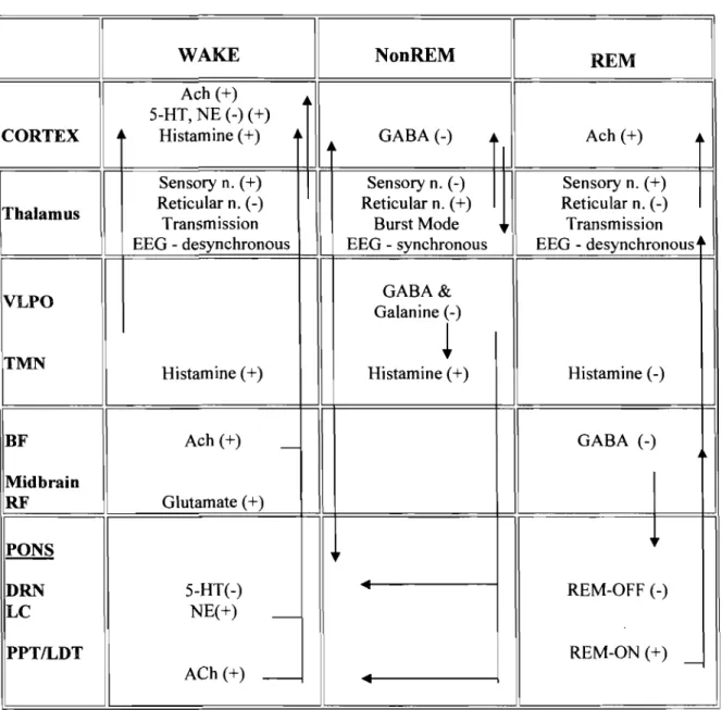
Mechanisms of Wakefulness & Sleep ... 29
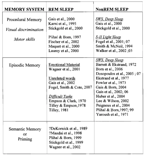
Relationship among Memory Systems, Sleep Studies & Sleep Stages ... 48
Summary Table: OldINew ERP Components ... 79
Protocol of the Experiments ... 83
Laboratory Sleep vs. Home Sleep Information ... 105
Nighttime Laboratory Sleep Information ... 106
Daytime Session Information (means+/-SD) ... 106
Participants Sleep History Data (mean ± S.E.M.) ... 147
Participants Sleep Agenda Information ... 199
Nighttime Laboratory & Sleep History Information (means+/-SD) ... 200
Behavioral Results from the Recognition Memory Task (means+/-SD) .. 201
Figure 1. Figure 2. Figure 3. Figure 4. Figure 5. Figure 6. Figure 7. Figure 8. Figure 9. Figure 10. Figure Il. Figure 12. Appendix 1 Appendix 2 Appendix 3 Appendix 4 Appendix 5 Appendix 6
LISTE DES FIGURES
Memory Deficits Corresponding to a Double Dissociation ... 6
Hierarchical Model for Familiar & Unfamiliar Face Recognition ... 10
Brain Regions involved in Multiple Memory Systems ... 22
Diagram ofEEG & Sleep Stages ... 24
Diagram of NonREM & REM Sleep Cycles ... 25
Brain Regions during NonREM and REM sleep ... 33
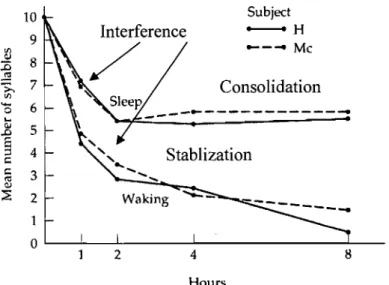
Jenkins & Dallenbach Diagram ... 50
Grand Average ERP during Study (encoding) Session ... 68
The Extended 10/20 International Classification System ... 85
Schematic Representation of the Stimulus Protocol.. ... 87
Regions oflnterest: Topographic Regions ... 90
Syntheses of the Relationships: ERPs, Sleep/Wake & 2-Stage Model ... 240
Screening Questionnaires ... 291
a. Sociodemographic & Medical History ... 292
b. Sleep (Habits) questionnaires ... 303
c. Inclusion/Exclusion Criteria ... 305
Informed Consent ... 309
Sleep Agenda ... 312
Description & Examples of the Questionnaires ... 314
a. Sleep & Evening Questionnaire ... 315
b. Morning Questionnaire ... 317
c. Daytime Questionnaire (Stanford Sleepiness Scale) ... 319
d. Visual Analog Scale (V AS) ... 320
Subjects Task Instructions ... 324
Declaration des co-auteurs ... 329 Pilot Study: The Influence ofSleep on Memory: An ERP study . ... 332
ARTICLE 1: Figure 1. Figure 2. Figure 3. Figure 4. Figure 5. ARTICLE 2: Figure 1. Figure 2. Figure 3. Figure 4. Figure 5. Figure 6.
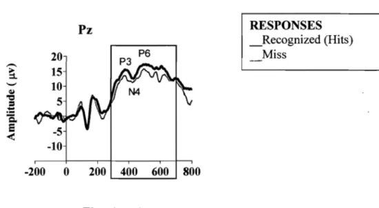
Protocol of the Experiment .. , ... , ... 99 Behavioral Performance (mean %correct +/-SEM) ... 107 Grand Average ERPs elicited by correctly recognized old or studied
stimuli (black line) & new (gray line) stimuli at the three midline (Fz,
Cz, Pz sites ... 108 ERP mean difference (old minus new) for the N200 & P250 complex
from the midline (Fz, Pz,Cz) electrodes corresponding to the early Old/New ERP memory effect in the normal Sleep (dotted tine) vs.
daytime Wake (solid line)session ... 110 ERP mean amplitude difference (old minus new) for the N400 and
LPC components from Fz, Cz & Pz electrode sites corresponding to the late Old/New memory effect in the normal sleep (dotted Hne) vs.
daytime wake (solid line) session ... 111
Behavioral Performance (mean % correct and % error ± S.E.M) for the old or previous studied stimuli after sleep and wake sessions ... 149 Behavioral Responses (mean RTs
±
S.E.M) for the old (studied) vs.new stimuli after sleep and wake sessions ... 150 Grand Average ERPs (hits & miss) elicited during the Study
(Learning) Session at the midIine sites. . ... 151 Grand Average ERPs (old & new) elicited by correctly recognized
stimuli during the Sleep & Wake Test (Recognition) Session at the
midIine sites ... 153 Mean amplitude difference (old minus new) for the N200-P250 time
window from midline (Fz, Cz, Pz) and lateral (leftlright) regions after
sleep and wake sessions ... 155 (top) ERP mean amplitude difference (old minus new) for the frontal
N400 and posterior N400 effect from midIine (Fz, Cz, Pz) and lateraI (leftlright) regions after sleep and wake sessions ... 157
ARTICLE 3:
Figure 1.
Figure 2.
Figure 3.
(bottom) ERP mean amplitude difference (old minus new) for the late frontal components (LFC) and late positive component (LPC) from midline (Fz, Cz, Pz) and lateral (leftlright) regions after sleep and wake sessions ... 157
Grand Average ERPs (old & new) eUcited by correctly recognized
stimuli during the Test (Recognition) Session at the midline sites ... 204
Mean amplitude difference (old minus new) for the early N200-P250 components from midline (Fz, Cz, Pz) and lateral (leftlright) regions
after sleep and TSD sessions (inserts, mean amplitude) ... 206 (top) ERP mean amplitude difference (old minus new) for the N400
memory component from midline (Fz, Cz, Pz) and lateral (leftlright)
regions after sleep and TSD sessions (inserts, mean amplitude) ... 208 (bottom) ERP mean amplitude difference (old minus new) for the late frontal component (LFC) and late positive component (LPC) from midline (Fz, Cz, Pz) and laterai (leftlright) regions after sleep and wake sessions (inserts, mean amplitude) ... 208
AcknowledgementslRemerciements
La réalisation de cette thèse de doctorat a été possible en grande partie grâce à l'enseignement et le soutien considérable de deux personnages extraordinaires, mon directeur de recherche, le Dr. François Guillem, et mon co-directeur, le Dr. Roger Godbout. Cette combinaison unique m'a permis de combiner à la fois le sommeil et la psychophysiologie cognitive lors de mes études doctorales. À François j'ajoute merci pour ta confiance, ta grande intelligence et tes compétences. À Roger, merci pour ta sagesse et ta perspicacité, ainsi que ton sens de l'humour indestructible et inimitable.
Je remercie également très sincèrement les chercheurs, les étudiants et le personnel du Centre de Recherche Fernand Séguin que j'ai côtoyés pendant mes travaux de maîtrise et de doctorat et, plus particulièrement, les collègues des autres laboratoires qui m'ont aidé avec les choses pratiques au cours de ces années. Aussi, je remercie également très sincèrement les étudiants du Laboratoire de recherche sur le sommeil, Hopital Riviere-des-Prairies.
Je ne peux éviter de mentionner à quel point je suis reconnaissante de l'aide précieuse que m'ont apporté le docteur Pierre Mayer du Centre Hospitalier de l'Université de Montréal et le personnel de son Laboratoire du Sommeil. J'adresse aussi un remerciement très spécial au docteur Robert Brouillette du Centre Universitaire de Santé McGill pour ses conseils et son soutien continu, ainsi qu'au personnel du Laboratoire du Sommeil de l'Hôpital pour Enfants de Montréal.
Finalement, je voudrais remercier les membres de ma famille pour l'appui qu'ils m'ont donné pendant mes années d'études. Je voudrais remercier tout particulièrement mon man, Richard, pour son amour indéfectible, ses encouragements et judicieux conseils, ainsi que sa patience qui fut sans doute occasionnellement testée pendant les mois derniers de l'écriture de cette thèse.
During the sleep, the brain continues to show a certain activity, although different from that during wakefulness. To understand this complex behavioral state, this thesis focuses on examining the influence of sleep on long-term episodic memory processes in humans. In the text that follows, 1 will present briefly the various'memory systems and structures of sleep, as weIl as the neuropsychological and neurophysiological support for the dissociation between memory systems and the distinction between the major sleep states. FoIlowing this, 1 shall de scribe in detail the results of research utilizing different experimental approaches (e.g. behavioral, total sleep deprivation and selective sleep deprivation) to study the relationship between the sleep and long-term memory. Two approaches were used for this investigation. The first approach measured cognitive event potentials and behavioral performance of a group of subjects following a night of normal sleep and after an equivalent period of daytime wake. The second approach measured the ERP neural activity and performance of another group of subjects after a sleepless night and after a period of nocturnal sleep.
INTRODUCTION
In 1885, Ebbinghaus performed a series of experiments of what are generally acknowledged to be the first study of human memory. In his studies, Ebbinghaus learned 169 separate lists of nonsense syllables, and charted the rate of forgetting. The final result of these investigations was the classic curve of forgetting. Ebbinghaus' forgetting curve revealed that most forgetting occurs within the first hour, after which forgetting slowed dramatically between the 8 to 24 hour interval. Although sleep occupied a large part of this interval, Ebbinghaus did not mention the possibility that sleep may somehow be responsible for the observed results. Subsequently, other investigators attempted to isolate the effects of sleep on memory.
Currently, memory is conceived as multiple interacting systems and processes represented by diverse networks of brain structures. In the same way that memory cannot be considered homogeneous, both sleep and memory consolidation are complex phenomena that appear equally diverse. Even the term consolidation originally referred to as the process of trace stabilization through which the representation become resistant to interference has been redefined. More recent studies suggest that upon recall of previous consolidated information, the memory or representation returns to an unstable state, once more requiring consolidation, or re-consolidation.
Following the discovery of discrete sleep stages in the 1950s, sleep was no longer considered a homogeneous state, and research investigating the influence of sleep on memory has become gradually more complex. Many experiments looked at specific effects of the newly described sleep stages on memory. Initially, most of the
research effort was on rapid-eye movement sleep (REM) sleep deprivation. Many of the animal studies supported the idea that post~training REM sleep was important for memory consolidation; however, negative results in humans were simultaneously published. Further, it was argued that the deleterious effect of sleep deprivation was due to nonspecific effects of the experimental design. Moving away from sleep deprivation paradigms, in the early 70s, a novel approach was employed where four-hour periods of undisturbed sleep covering either the first or the second half of the nighttime sleep cycle was used. The studies that followed looking at the specific effect of sleep stages on memory sometimes resulted in mixed and contradictory conclusions. Sorne that was in favor of sleep dependent memory processing, and others against it. Results seemed to suggest that each stage of sleep contribute to different memory processes. In addition, several studies indicated that the more difficult the task, the less convincing its sIeep-dependent consolidation.
Beginning about the 1980s, there was a marked reduction in the number of studies devoted to tbis area. The main reason for tbis decline was that a series of studies failed to convincingly demonstrate a relationsbip between sleep and declarative memory. In contrast, there has been robust support for the conclusion that REM sleep enhanced performance on tasks of procedural memory. As a consequence, procedural memory has been exclusively studied due to the consistent and strong support for sleep dependent processing. A comprehensive role of sleep in declarative memory and its subsystems remains to be established and represents a future challenge to researchers in the area of sleep and memory.
CHAPTER 1. LONG-TERM MEMORY (LTM) SYSTEMS
The tenn memory implies the capacity to encode, store and retrieve infonnation. Acquisition is a learning process that takes place at the time of exposure to the infonnation and involves the encoding (i.e. analysis, classification of infonnation and links it to past leaming). Storage is the maintenance of the infonnation across intervals of time between encoding and retrieval. Retrieval is the ability to recall or recognize the infonnation following a delay (Reeves & Wedding, 1994). Memory is defined as the retention of the learnt material. It should be highlighted, how well infonnation is encoded and stored in memory detennines how likely it is to be accessed or retrieved.
Memory lasting anywhere from an hour to lifetime is called long-tenn memory (L TM). The LTM répresents a system of storage of events, facts, procedures and skills that we accumulated over the years. The long-tenn system has an unlimited capacity and a relatively slow rate of acquisition of new material (Waugh & Nonnan, 1965). Research has given important insights on the nature of L TM. The most important is that L TM is made up multiple, interactive memory systems that are functionally independent (Squire, 1992)
1.1. The Principle of Double Dissociation: Partitioning L TM
The development of neuropsychological methods in the patients with cerebral lesions contributed to the reports of a distinction between different memory systems. The major argument of cognitive neuropsychologists in favor of a distinction between different memory systems, rested on revealing "functional dissociations" (McCarthy
& Warrington, 1990). In the mid-60s to early-70s, this logic was employed by cognitive psychologists to fractionate memory into separate systems (Glanzer &
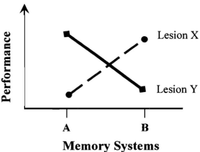
Cunitz, 1966). In this context, "functional dissociations" were used to support divisions of memory function but not necessarily to localize these functions in the brain. There are two main types of dissociations, single and double. A single dissociation corresponds to the observation that a localized brain lesion (X) comes along with a disturbance of a memory system A, without distorting another B system. Much stronger evidence for cornes from double dissociations. First introduced in the mid50s, a double dissociation is observed when in the single dissociation cornes to add to the fact that B system is disrupted by another insult (Y), which does not affect A system (Figure 1) (Teuber, 1955). For example, individuals with brain-Iesions who do well on task A but poody on task B, with another group showing the opposite pattern. This suggests that two tasks involve different processing mechanisms and implies that there is a memory system required by B but not A. A double dissociation allows one to infer that A and B systems are functionally independent.
In the 1990s, sorne suggested that dissociations were not weIl supported by the data but instead due to lack of statistical power to detect such impairments (Ostergaard, 1992). Stochastic or statistical dissociations involving the administration of one study (learning) test and subsequentIy two retrieval tests to the same subject were found to be more compelling. An item-by-item analysis is used to evaluate the probability that performance of a given item in one test is statistically unrelated to performance on the same item in another test, i.e., stochastic independence. And each task is thought to rely on a different memory system. Nevertheless, if different types
or multiple functional dissociations across different aspects of memory aU point to the same set of systems, then most likely that pattern of data is reliable (Schacter, 1992).
Lesion X
Lesion Y
A B
Memory Systems
Figure 1. Schematic representation of memory deficits corresponding to a double dissociation.
1.1.1. Dissociating Procedural vs. Declarative L TM
As early as 1970 a distinction was made that dissociates LTM into two systems (Squire, 2004). Subsequently, Squire (1987) proposes a model that dissociates L TM into procedural versus declarative memory systems (Squire, 1987). Procedural memory refers to skills or sequences of behavior and, is implicitly (unconsciously) learned and executed. Procedural leaming of perceptual and motor skills is usually achieved through periods of performance repetition. Declarative memory, instèad, consists of events that are usually explicitly (consciously) and intentionally leamed and retrieved (Cohen & Squire, 1980). Research has provided ample evidence that performance on tests of procedural memory and declarative memory can be dissociated (Schacter, 1992; Roediger, 1990).
Perhaps the most compelling evidence for dissociation between procedural and dec1arative memories is provided by studies of patients with amnesia after bilateral resection of the medial temporal region (inc1uding the hippocampus and the amygdale) to relieve severe epilepsy. First described by Scoville and Milner (1957) was a memory change in a patient (H.M.) and later eight other cases, with profound amnesia that were unable to form any new memories after surgery (anterograde amnesia). These authors, however, found that patient H.M. exhibited daily improvements in procedures and skills, suggesting that his procedural memory was intact (Scoville & Milner, 1957). A similar observation was confirmed in another study that indicated localized lesions in the hippocampus were sufficient to lead to this type of amnesia in humans. Along the same lines, Cermak and colleagues (1973) presented a1coholic Korsakoff patients to show the distinction between dec1arative and procedural memory (Cermak & Butters, 1973). Their study examined the leaming performance on a dec1arative task (finger maze1) vs. a procedural task (pursuit rotor2). The results showed that Korsakoff patients were able to learn the procedural task but were unable to leam the dec1arative task as weIl as the normal control group. Neuroimaging studies in the Korsakoff syndrome reveal general atrophy involving the frontal lobes (Shimamura, Jernigan, & Squire, 1988; Jacobson
& Lishman, 1990) and diencephalic structures (Jernigan, Schafer, Butters, & Cermak,
1991) inc1uding the medial thalamus and/or mamillary bodies (Colchester et al., 2001).
1 Finger maze task - (declarative task) the task consists oftrialleaming by tracing finger mazes while
The second part of the evidence of a double dissociation between declarative and procedural memory is found in neurological patients with Huntington's chorea characterized by the degeneration of cells in the basal ganglia (Martone, Butters, Payne, Becker, & Sax, 1984). The patients with Huntington's chorea are unimpaired on tasks of dec1arative memory but are often impaired in procedural skilllearning. A study by Martone et al. demonstrated impairments on procedural nonmotor skills task (i.e. mirror reading3) on which patients with temporal lobe lesions are unimpaired (Martone, Butters, Payne, Becker, & Sax, 1984). Neuroimaging studies using positron emission topography (PET) have provided evidence for the involvement of the basal ganglia in procedura1 skilllearning in normal individuals, e.g. (Grafton, Hazeltine, &
Ivry, 1998). Yet another distinction proposed within LTM is between subsystems of declarative memory.
1.1.2. Dissociating Episodic vs. Semantic LTM.
A previous distinction made by Tulving (1972) have been integrated into the long-term declarative memory model of Squire (1987). In his model Tulving (1972) argues that declarative memory is made of two different, nonetheless, interacting systems: semantic memory and episodic memory (Tulving, 1972). Others envisioned a similar distinction earlier, for example, Broad (1925) and Furlong (1948) distinguished "recollective" or episodic memory from what they referred to as "propositional" or semantic memory (Furlong, 1948; Broad, 1925).
2 Pursuit rotor task - (procedural task) the subject leams to track a metal dot on a tumtable platter with
Semantic memory relates to general world knowledge, concepts and facts (Tulving, 1972) not temporally dated. A concept is associated with the attributes for example, its function (a kitchen chair, office chair) or its form (round, square). Information is stored as semantic "nodes" which are interconnected to form a network of knowledge (Collin & Loftus, 1975; Quillian, 2007) such as, knowledge about the meaning of words (a chair is a type of fumiture), the properties of objects (chairs have four legs) and facts (we sit in chairs). This organization explains semantic associations (table, chair), categorizations (fumiture) (Morton, 1969; Anderson, 1983) and the implicit (i.e. unintentional) retrieval of information such as semantic priming (e.g. cue leaming) (Squire & Zola-Morgan, 1991).
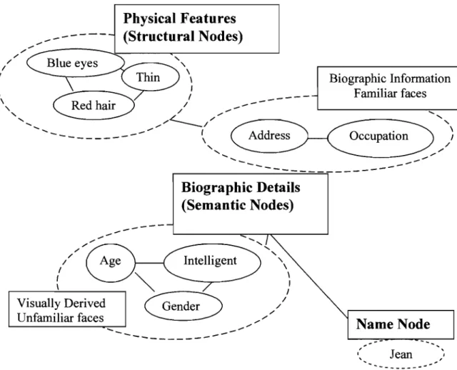
In addition, semantic memory is also affiliated with non-verbal knowledge (e.g. objects, faces). Early models of face recognition (Bruce & Young, 1986) propose that the identity of faces provides access to semantic knowledge and other identity information. Several different kinds of identity information exist and are clustered according to type (biographic details, physical features, name) , and may only be accessed in a particular order due to its proximity see Figure 2, below (Zeineh, Engel, Thompson, & Bookheimer, 2003). In addition, there are two entry points. One is the recovery of biographic details about a person without recognizing their face. A second entry point is via global information; an example of this would be when we vaguely recognize a face as familiar but are unable to recollect the biographic details (when, where) of the person.
; ' ; ' -...
-Physical Features
(Structural Nodes)
,,"'"
---~----~---~"---
'\ 1 1 1 \ \ \ \ 1 1 ---Biographie Information Fami1iar faces '\ "-,
... ... -1 1 ( \ /"
; ' ... -, 1 _ -/ ... "L ,,/, ----1
... ( \ Address Occupation--
---~
"-,
... ...--
- - -.==:...:==;
-Biographie Details
(Semantic Nodes)
...,
Intelligent,
"-'\ \ \ 1 1 1 / ; '-_
... ...,
"
, "-\ 1 / Visually Derived Unfamiliar faces~
... ' ,"
NameNode
--
---
--, 1 Jean ---Figure 2. Example of the hierarchical representation of information in semantic memory network during familiar and unfamiliar face recognition
On the other hand, episodic memory is memory for personal autobiographie events, and is usually acquired with relatively few exposures to the information. In episodic memory, information is encoded in relationship to their temporal and spatial context (i.e. when and where) (Clayton & Dickinson, 1998; Nyberg et al., 1996).
manner for the subjects to read.
According to Tulving (1972), every time we leam a piece of information, a new episode is stored with its new context. Subsequently, every rehearsal strengthens the trace of the episode. Thus, episodic memory "feeds" semantic memory, in that with iteration, the item and contextual association progressively consolidates as a semantic trace (Tulving, 1972).
Another distinction reminiscent to episodic and semantic memory is the relationship to the subjective experience of retrieva1 rather than on the type of information. Sorne have argued that recognition memory is not a unitary but is composed of two different processes (Yonelinas & Levy, 2002; Tulving, 1983; Mandler, 1980). One is an automatically or implicitly induced process (i.e. familiarity) at the re-exposure of an event, in the absence of any conscious knowledge about the context in which the event occurred (Jacoby, 1999; Knowlton & Squire, 1995). The other process refers to a conscious or exp1icit process of recollection, and involves the retrieval of contextual details (Knowlton & Squire, 1995; Jacoby, 1999). Research has shown that familiarity occurs earlier than recollecting detail in retrieval (Hintzman & Curran, 1995) and depends on the frontal structures (Rugg et al., 1998).
Evidence for dissociation between semantic and episodic memory cornes from the study of individuals with circumscribed amnesia, neurological problems and normal aging. Semantic dementia is characterized by a progressive. disorder of semantic knowledge while at the same time recalling a specifie episode is relatively preserved (Hodge s, Patterson, Oxbury, & Funnell, 1992; Snowden, Goulding, &
Nery, 1989). In this type of amnesia there is impaired ability to name objects, define words or in describing famous people. On imaging studies, semantic dementia
characteristically shows damage to the inferior and lateral temporal cortex (Hodges, Patterson, Oxbury, & Funnell, 1992b; Snowden et al., 1989).
Neurological patients with Alzheimer's disease show an initial impairment of episodic memory and, as the disease progresses, they become increasingly impaired on semantic memory tasks. This suggests that semantic memory is not dependent on the same MTL structures that are damaged in anterograde amnesia and believed to underlie the episodic memory deficits (Squire, Shimamura, & Graf, 1987) but rather may be stored in association areas4 presumed to be impaired in the later stages of Alzheimer's disease. These observations imply that lesions at the level of the association area of the posterior cerebral cortex provoke disturbances of the storage or the access of semantic knowledge.
In dementia patients with Alzheimer's disease (Warrington, 1975); (Martin &
Fedio, 1983), symptoms consist of a change in semantic knowledge (e.g. word knowledge and meaning of the objects). There are often selective deficits in stimulus modality (visual / verbal), of a specific category (concrete / abstracted) (Shallice &
Warrington, 1975) or from a domain (language / face) (Peretz, Belleville, & Fontaine, 1997). Similarly, Warrington and Shallice (1984) demonstrated that in herpes encephalitis, patient' s identification of living things and foods was severely impaired relative to their ability to identify inanimate objects, and that this was independent of modality of presentation (Warrington & Shallice, 1984). There is disagreement about the interpretation of these category specific impairments. One explanation for these
4 Association Areas: Parieto-temporal-occipital cortex receives input from the sensory modalities and
relays the information to the frontal association area that also receives input from the limbic association area.
selective deficits is that living things may be processed in terms of their sensory properties, whereas inanimate objects are process for the most part in terms of their function (e.g. (Warrington & Shallice, 1984; Borgo & Shallice, 2001). Other dissociations have been found in the processes involved in treatment (e.g. understanding words, identification of objects) versus those involved in the organization (e.g. reasoning, syntax) (Shallice, 1988).
In contrast, normal aging typically shows a mild graduaI impairment of episodic memory accompanied by a relative sparing of semantic memory(Craik, Anderson, Kerr, & Li, 1995). Jelicic et al. (1995) tested young and old adults on a
cued recall task5 under two different conditions: the first condition was to name the first word that cornes to mind, the other condition, was to recall a word from the word list (Jelicic, Craik, & Moscovitch, 1996). They reported that the younger participants performance was equal on each task, while the older participants found the word list task more difficult, pro vi ding support for the existence of separate memory subsystems. Sorne have attributed this age-related decline in memory to a decline in fronto-striatal function (Gabrieli, 1995). This idea is supported by neuroimaging studies that showed age-related differences in frontal but not medial temporal regions during episodic retrieval (Schacter, Savage, Alpert, Rauch, & Albert, 1996). More compelling evidence for double dissociation cornes from KC, a patient who lacked the ability to retrieve any specific personal events, who was able to learn semantic facts (Rosenbaum et al., 2000) due to bilateral MTL damage from a motorbike
5 Cued recall task - (declarative memory) subjects are shown a word list and following a delay are
presented with a cue (e.g. ftrst few letters ofa word or related word). The task is to recall a word from the studied list that matches the cue.
accident. When KC was presented with a series of pictures, with a short sentence, and then tested, he successfully completed the sentences in response to a cue. This occurred despite the fact that KC had no recollection of any specific episode on why he knew these facts.
Conversely, as mentioned above, patient HM initially demonstrated deficits in episodic and also in semantic memory (Gabrieli, Cohen, & Corkin, 1988; Postle &
Corkin, 1998). More recently it has been shown that HM was able to acquire new semantic but not episodic knowledge that suggests a distinction between episodic and semantic memory (O'Kane, Kensinger, & Corkin, 2004). In addition, it indicates that semantic memory is supported by structures beyond the MTL.
1.2 Neuroanatomical Basis for Multiple L TM Systems.
The observed dissociations in the memory performance of clinical and nonclinical populations support the functional dissociations of multiple memory processes. Further, the lesion studies and related selective deficits have prompted speculation about the cortical and the anatomical support for distinct brain regions associated with different types of L TM.
1.2.1. Procedural Memory: Basal ganglia, Cerebellum & Motor Cortex
Procedural memory is often intact in patients with declarative memory problems either due to amnesia (e.g. HM) or in the early stages of Alzheimer' s disease (Gabrieli, Corkin, Mickel, & Growdon, 1993). Procedural skills learning is often impaired in patients with basal ganglia diseases such as Huntington's disease
(Martone, Butters, Payne, Becker, & Sax, 1984). Grafton & coworkers scanned the brains of normal individuals during a procedural task (motor pursuit task) and found increases in cerebral blood flow in the motor cortex, basal ganglia and cerebellum, whereas the acquisition of the skill included a subset of structures (e.g. primary motor cortex, supplementary motor cortex and the pulvinar nucleus of the thalamus) (Grafton, Hazeltine, & Ivry, 1998). It appears that the neural structures involved in procedural leaming are diverse, involving both cortical and subcortical networks. While different perceptual-motor skills may share sorne anatomical commonalities, the networks modulating specific kinds of procedural leaming are defined by the sensory (input) and motor (output) demands of the task (Grafton, Hazeltine, & Ivry, 1998); (Jancke, Gaab, Wustenberg, Scheich, & Heinze, 2001; Kami et al., 1995; Schwartz, Maquet, & Frith, 2002).
1.2.2. Episodic Memory: Medial Temporal Regions
As mentioned above, the first lesions to appear in most cases of Alzheimer's disease occur in the medial temporal region (Hyman, Van Hoesen, & Damasio, 1987). However, Alzheimer patients also have early damage to cholinergie neurons in the basal forebrain (Arendt, Bigl, & Teanstedt, 1983), and lesions in this area cause impairments in declarative memory. Therefore, it is difficult to say that the deficits seen in Alzheimer's disease are exclusively to medial temporal injuries. Medial temporallesions (as in the case of HM) resulting from a bilaterallobectomy implicate the hippocampus and/or the amygdala in episodic memory. Later, studies showed that hippocampal damage alone was sufficient to lead to amnesia (Squire, 1992).
Further evidences come from numerous neuroimaging studies reporting medial temporal activations during memory retrieval (Squire et al., 1992; Schacter, Alpert, Savage, Rauch, & Albert, 1996; Schacter et al., 1995). One hypothesis regarding MTL lobe region is that it allows for the retrieval of contextual, spatial and temporal information with the relevant information in memory (Winocur, 1982; Hirsh, 1974). Thus, the hippocampus appears to be the key structure in the episodic system.
The amygdala is 10cated in the MTL lobe near the hippocampus, and patient HM underwent resection to both structures. By virtue of its location, the amygdala was thought to have a role in the formation of declarative memories. However, the role of the amygdala has been found to be separate from the "medial temporal lobe memory system" that comprises the hippocampus and adjacent cortex (Zola-Morgan, Squire, & Amaral, 1986; Squire & Zola-Morgan, 1991). This structure plays a more specific role in processing emotional context. Evidence for this cornes from numerous studies in normal individuals, where emotionally arousing stimuli are consistently remembered better than neutral stimuli (Heuer & Reisberg, 1990; Buchanan, Brechtel, Sollers, & Lovallo, 2001). In a recent fMRI study, Canli et al. showed that pictures rated as "extremely emotionally intense" led to increased amygdala activation on first exposure. In addition, the se same authors reported better memory for the "extremely emotionally intense" compared to "less emotionally intense" rated pictures, indicating a role of the amygdala in the enhancement of memory by emotion (Canli, Zhao,'Brewer, Gabrieli, & Cahill, 2000).
1.2.2.1. Subcortical Structures
Diencephalic lesions seen in Korsakoff s syndrome involve damage to the medial thalamus and often mamillary body. Press et al. (1989) reported that damage to these regions is sufficient to produce memory impairment even when the medial temporal regions are anatomically intact. However, these authors found the medial thalamic lesions to have a greater effect than the marnmillary body lesions on declarative memory (Press, Amaral, & Squire, 1989). At present, it is unclear what specifie aspect of the thalamic lesions account for this type of amnesia.
1.2.2.2. Frontal Structures (Inferior Frontal & Superior Frontal)
Lesions of frontal structures do not dramatically impair episodic memory, in contrast with damage to the medical temporal regions (Janowsky, Shimamura, Kritchevsky, & Squire, 1989; Millner, Corsi, & Leonard, 1991). This indicates that the frontal region is not necessary for episodic memory per se but helps in the processing of contextual information.
Studies in primates, where lesions are more precise, suggest that the prefrontal cortex can be divided into two main regions: the dorsolateral area (superior frontal) and the orbitofrontal area (inferior frontal) located on the medial and ventral surface of the brain. The superior frontal region is recruited when active manipulation or monitoring of information is required (Petrides, 1994). Evidence for this idea comes from recent study by Petrides (2000) reporting that lesioning the superior frontal region resulted in a mild impairment in the monitoring of information (Petri des, 2000). Similarly, functional neuroimaging studies have provided data showing
increased activity III the supenor frontal reglOn when information is monitored
(Tremblay & Schultz, 2000). There is sorne controversy whether the superior frontal region is involved in the maintenance, e.g. the process of keeping information in mind (Cohen, Porjesz, Begleiter, & Wang, 1997; Cohen, Porjesz, Begleiter, & Wang, 1997) or in the manipulation, e.g. the reorganization of the information that is being maintained (D'Esposito, Ballard, Aguirre, & Zarahn, 1998; Smith & Jonides, 1999) processes during retrieval. One explanation for the disagreement may be that with more complex tasks, the more likely it is to involve manipulation, and manipulation of the information probably requires increased maintenance (Raye, Johnson, Mitchell, Reeder, & Greene, 2002).
Neuropsychological investigations of individuals presenting with inferior frontal brain damage implicate the orbital frontal cortex in episodic memory (Wallesch, Kornhuber, Kollner, Haas, & Hufnagl, 1983). Since the famous case of Phineas Gage, it has been known that lesions of the inferior frontal cortex produce dramatic changes of personality (Damasio, Grabowski, Frank, Galaburda, &
Damasio, 1994). Patients with inferior frontal damage exhibit impulsive and disinhibited behaviors and fail to withstand interference from distraction. Recent work suggests that the inferior frontal region has a specific role in strategic control and/or inhibition of interfering information (Stuss et al., 1982; Fuster, 1980).
1.2.3 Semantic Memory: Association Areas
Around the mid-70's, data obtained in patients with focal cerebral lesions began to be analyzed within the theoretical framework of the semantic memory. This
was about the same time this subsystem was introduced by Tulving (1972). As mentioned previously, semantic memory is severely affected in disorders such as Alzheimer disease, herpes encephalitis and also semantic dementia. The first neuropsychological case study was applied by Warrington (1975) in dementia patients with Alzheimer's disease (Warrington, 1975) and short1y afterwards by others (Schwartz et al. 1980; Martin & Fedio, 1983).
1.2.3.1. Posterior Cerebral Cortex: Temporal, Parietal Regions
Semantic memory is strongly language-based and describes memory for general knowledge of facts, concepts and words. Language-related areas in the human brain are located in the left hemisphere. The processing of language involves a complex network of interacting brain regions. "Aphasia" is the term used to describe an acquired loss of language function, and manifest itself as impaired expression, comprehension, or both. For example, a common manifestation of aphasia is when a patient cannot find the right word. Snowden et al. (1989) coined the term "semantic dementia", a progressive disorder in naming and word comprehension (semantic knowledge) (for review see Murre, Graham, Hodges, 2001). Imagery studies in patients with semantic dementia show severe atrophy of the inferior temporal lobe (Hodges, Patterson, Oxbury & Funnell, 1992; Snowden, Goulding & Neary, 1989). Along the same lines, other studies show that the type of aphasia identified in 1873 by Karl Wernicke also affects the understanding of semantic contents. In this type of aphasia there is damage to the left, superior temporal gyrus that extends
into the inferior parietal reglOn (Coughan & Warrington, 1978; Basso et al. 1985).
Many similar examples can be derived from neurological disorders of apraxia, i.e. disorders of skilled movements and agnosia, i.e. inability to recognize or identify objects despite intact sens ory function. Jakobson et al (1991) observed a patient, V.K., with damage to the posterior parietal lobe that exhibited a deficit in her ability in how to reach for objects but was able to recognize and name objects (Jakobson, Archibald, Carey, & Goodale, 1991). V.K. was unable to coordinate reaching and hand postures for different objects that could not be ascribed to either motor or visual deficits.
On the other hand, visual agnosia is characterized by the inability to recognize familiar objects (Farah, 1994) and is confined to the vi suaI modality. Object recognition is the ability to place an object in a category of meaning. There are many subcategories of visual agnosia. Sorne patients lose their ability to recognize faces of friends and family members (Milner & Goodale, 1995) but are able to see and can recognize objects, while others retain only face recognition. In patients with facial agnosia, the anterior region of the temporal lobe (specifically the right fusiform gyrus) responsible for recognition and categorization of faces is damaged. Patients with this type of agnosia, referred to as prosoagnosia, often identify a person by other cues such as their voice, clothing, gait or their shape.
1.2.3.2. Frontal Structures: Hemispheric Lateralization
Neuroimaging studies distinguish separate roles of the right and left frontal cortex during retrieval. One proposaI is that the function of the left frontal cortex is
involved in semantic processing (Cabeza, Locantore, & Anderson, 2003; Poldrack et al., 1999) such as, the meaning of words. Others have shown that the function of the right frontal cortex is associated with monitoring during retrieval (Allan, Dolan, Fletcher, & Rugg, 2000; Henson, Rugg, Shallice, Josephs, & Dolan, 1999; Rugg, Fletcher, Chua, & Dolan, 1999). On the other hand, results from imaging studies using simple, memory retrieval tasks report a right frontal activation (Nolde et al., 1989) but more demanding tasks produce bilateral or left frontal activation (Rugg, Fletcher, Chua, & Dolan, 1999; Henson, Rugg, Shallice, Josephs, & Dolan, 1999). This is consistent with the idea that frontal activation reflects the degree of retrieval effort with increased activation when retrieval is difficult (Schacter, Alpert, Savage, Rauch, & Albert, 1996).
In summary, there IS evidence for several dissociations that suggests a distinction between the various types of memory systems. It should be mentioned that the above examples do not comprise a comprehensive list of the disorders that give rise to amnesia since it is beyond the scope of this thesis. What they do conclude, however, is a dysfunction or damage in medial temporalldiencephalic circuitry (or in the frontal regions) produces amnesia with a selective deficit in declarative memory and sparing of procedural memory. On the other hand, lesions of the association areas appear to have a greater impact on semantic memory processes. In the same way that memory cannot be considered unitary, the spectrum of sleep stages in the human brain, and the processes that create and sustain sleep, appear equally diverse.
Multiple Memory Systems
Explicit, Implicit (Graf & Schacter, 1985)
Procedural, Declarative (Squire, 1987) Episodic, Semantic (Tulving, 1972, 1983)
Encoding - - --+
Storage (Consolidation)
Long-Term
Memory
Explicit (direct, intentional)
Declarative Memory
Details, Events & Facts "Knowing what"
,/
~
Episodic Memory
Sem an tic Memory Autobiographic Events "When/Where" Facts General Knowledge 1 1 Medial Temporal Association Areas (Hippocampus & (Temporal, Parietal) Diencephalon) ... Frontal Cortex ~
Superior Frontal (Dorsolateral) Interior Frontal (Orbitofrontal)
--- - - - -~ Retrieval
Implicit (indirect, unintentional)
ProceduralMemory
Procedures & Skills "How to"
Striatum Basal ganglion
Cerebellum Motor Cortex
Figure 3. This figure shows the multiple memory systems associated with the brain regions and subcortical regions involved in long-term memory.
Chapter 2: NORMAL SLEEP: The Structure
For centuries, sleep was defined as an inactive or passive behavioral state. Prior to electroencephalography in 1929 by Hans Berger, it was impossible to demonstrate differences in the brain's electrical activity between sleep and wakefulness. Much ofwhat is known today about brain activity during human sleep is due to the discovery of electroencephalography, a method by which electrical activity in the cortex can be recorded by scalp electrodes.
2.1. Electroencephalography, EEG
The EEG displays fluctuations of electrical fields recorded at the scalp electrodes from neurotransmitter release and thalamocortical synapses. It is produced by the surnmation of transient excitatory (or depolarizing) and inhibitory (or hyperpolarizing) postsynaptic potentials in the pyramidal layer of the cortex (Lopes da Silva, Storm van Leeuwen, & Remond, 1986; Niedermeyer & Lopes da Silva, 1987). The brainwave patterns reflect voltage and time whereby the frequency, amplitude and waveform can be quantified. In addition to the EEG, sleep researchers also rely on electrophysiological muscle and eye potentials to determine sleep stages.
In 1937, sleep was classified into different stages (Loomis, Harvey, & Hobart, 1937). It was not, however, until the landmark 1953 discovery of REM sleep (Aserinsky & Kleitman, 1953), that sleep was considered as an active process. A few years later, it was discovered that REM sleep is associated with dreaming. Subsequently, the cyclic organization of sleep was described (Dement & Kleitman,
1957) and broadly defined as an alternation between distinctly different stages: Non-Rapid Eye Movement (NonREM) sleep and REM sleep.
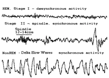
2.2. Sleep Stages: NonREM & REM sleep
N onREM sleep consists of several stages of light sleep (Stages l, II) and deep, slow wave sleep, SWS (Stages III, IV) (Rechtschaffen & Kales, 1968). The EEG during NonREM sleep begins with a dominance oftheta activity (4-8 Hz) in the early stages that progresses to synchronous intermittent spindling (12 to 14 Hz) to a predominance of high-voltage > .75 IlV, low frequency Delta slow activity (.5-3 Hz) (Figure 4), which provide a neural substrate to cognitive activity during sleep.
Human Sleep Stages
Awake - low voltage, fast activity
~'I1M~ .. ~_".f50
' F " "~uV 1 sec Drowsy - alpha 8 - 12
REM, Stage l - desynchronous activity
Stage II -. spindle, synchronous activity
NonREM . Delta Slow Waves synchronous activi ty
Figure 4. EEG Patterns associated with the stages of sleep in humans. [Adapted
from Hauri, P & Orr, W. C (1982) The Sleep Disorders, CUITent concepts, UpJohn Company.]
A rapid, low-voltage desynchronous theta, atonia of postural muscles and bursts of rapid eye movements characterize REM sleep. The different EEG activity during NonREM and REM sleep stages suggests that the two stages are governed by two different networks, each possibly associated with different cognitive functions involved in consolidation ofmemory.
2.2.1. NOnREM and REM Sleep Cycles
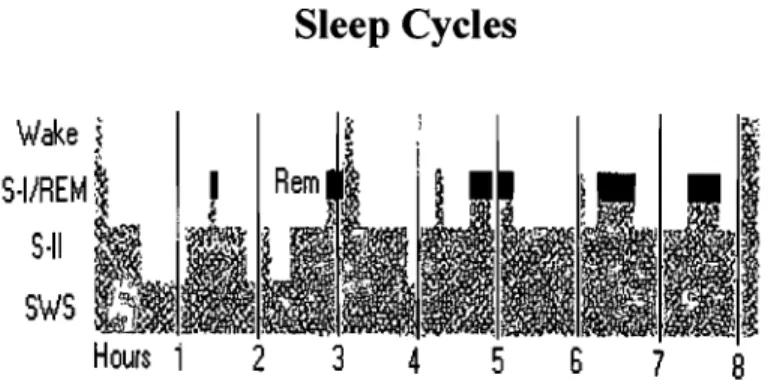
Sleep cycles show a periodicity of approximately 90 minutes in normal adults (see Figure 5). Overnight sleep often is divided into three time periods: the first third of the night, consisting of the highest percentage of deep NOnREM sleep; the middle third of the night; and the last third of the night, the majority of which is made up of REM sleep. The cycle repeats itself 4 to 6 times during a normal nights sleep (Kleitman, 1939). Knowing this provides a useful approach to study the interaction between sleep and memory by taking advantage of the naturally occurring fluctuations in REM and NonREM sleep stages that will be discuss in later chapters.
Wake < S·I/REM: 5·11 SWS 2
Sleep Cycles
3 45
6 7 8Figure 5. Example of the time course of sleep cycles arcoss a night of normal sleep in adults. [Adapted from Rauri, P & Orr, W. C (1982) The Sleep Disorders, Current concepts, UpJohn Company.]
2.3 Neural Substrates & Pathways of Sleep & Wake
As mentioned above, the electrical activity at the surface of the scalp picked up by the EEG reflects the firing patterns in the thalamocortical system. These firing patterns differentiate between wakefulness, NonREM and REM sleep. The thalamic neurons have two distinct stages (e.g. transmission mode and burst mode), which will be discussed below along with the neurotransmitters and neurocircuitry involved in sleep and wake. It is beyond the scope of this thesis to review the numerous neurotransmitter and neuropeptide systems implicated in sleep other than the basic regulatory role of aminergic and cholinergic systems in wake, NonREM and REM sleep.
2.3.1 Wakefulness: Ascending Reticular Activating System
The first evidence of specific brain regions responsible for the maintenance of wakefulness was by von Economo (1923) in patients with encephalitis resulting in coma and subsequent death. He reported a loss of cells in the posterior mesencephalic reticular formation and posterior hypothalamus (von Economo, 1923). A later study by von Economo performed in rats, demonstrated that lesions to the preoptic basal forebrain caused insomnia (von Economo, 1930). Following this was the classic study by Moruzzi & Magoun (1949) who showed that electrical stimulation of the mesencephalic reticular formation of the brainstem produced arousal (Moruzzi &
Magoun, 1949). The conclusion of the se early studies was that the reticular formation in the rostral region of the brainstem and the ascending neuronal pathways were critical for maintaining wakefulness and arousai. Subsequently, this region and