HAL Id: inserm-00545885
https://www.hal.inserm.fr/inserm-00545885
Submitted on 9 May 2011HAL is a multi-disciplinary open access
archive for the deposit and dissemination of sci-entific research documents, whether they are pub-lished or not. The documents may come from teaching and research institutions in France or abroad, or from public or private research centers.
L’archive ouverte pluridisciplinaire HAL, est destinée au dépôt et à la diffusion de documents scientifiques de niveau recherche, publiés ou non, émanant des établissements d’enseignement et de recherche français ou étrangers, des laboratoires publics ou privés.
monocrotaline- and hypoxia-induced pulmonary
hypertension in rats.
Guillaume Gary-Bobo, Amal Houssaini, Valerie Amsellem, Dominique Rideau,
Pierre Pacaud, Aline Perrin, Jérémy Brégeon, Elisabeth Marcos, Jean-Luc
Dubois-Randé, Olivier Sitbon, et al.
To cite this version:
Guillaume Gary-Bobo, Amal Houssaini, Valerie Amsellem, Dominique Rideau, Pierre Pacaud, et al.. Effects of HIV protease inhibitors on progression of monocrotaline- and hypoxia-induced pul-monary hypertension in rats.. Circulation, American Heart Association, 2010, 122 (19), pp.1937-47. �10.1161/CIRCULATIONAHA.110.973750�. �inserm-00545885�
Effects of HIV Protease Inhibitors on Progression of Monocrotaline- and
Hypoxia-Induced Pulmonary Hypertension in Rats
Gary-Bobo G : HIV Protease Inhibitors and Pulmonary Hypertension
Guillaume Gary-Bobo PhD*, Amal Houssaini MSC*, Valerie Amsellem PhD*, Dominique Rideau MSC*, Pierre Pacaud PhD††, Aline Perrin MSC*, Jérémy Brégeon MSC††, Elisabeth Marcos MSC*, Jean-Luc Dubois-Randé MD ‡, Olivier Sitbon MD†, Laurent Savale MD*, Serge Adnot MD-PhD*
G Gary-Bobo and A Houssaini contributed equally to the work. L Savale and S Adnot were both senior authors of this study.
*INSERM U955 and Département de Physiologie, Hôpital Henri Mondor, AP-HP, 94010, Créteil, France
‡ Service de Cardiologie, Hôpital Henri Mondor, AP-HP, 94010, Créteil, France † Service de Pneumologie, Hôpital A. Béclère, AP-HP, Clamart, France
†† Inserm U915, Faculté de médecine, 4 rue Gaston Veil, 44035 Nantes cedex 1, France
Correspondence:
Serge Adnot
Service de Physiologie-Explorations Fonctionnelles, CHU H Mondor, 94010, Créteil, France Tel: 33 1 49 81 26 77;
Fax: 33 1 48 98 17 77
E-mail: serge.adnot@inserm.fr
Word count: 6045
Abstract
Background: Pulmonary hypertension (PH) is among the complications of HIV infection.
Combination antiretroviral therapy may influence the progression of HIV-related PH. Because Akt signaling is a potential molecular target of HIV protease inhibitors (HPIs), we hypothesized that these drugs altered monocrotaline- and hypoxia-induced PH in rats by downregulating the Akt pathway, thereby inhibiting pulmonary-artery smooth-muscle-cell (PA-SMC) proliferation.
Methods and Results: Daily treatment with each of three first-generation HPIs (ritonavir,
30 mg/kg; amprenavir, 100 mg/kg; and nelfinavir, 500 mg/kg) started 3 weeks after a subcutaneous monocrotaline injection (60 mg/kg) substantially diminished pulmonary artery pressure, right ventricular hypertrophy, number of muscularized pulmonary vessels, pulmonary arterial wall thickness, and proliferating pulmonary vascular Ki67-labeled cells, without affecting vessel caspase 3 staining. HPI treatment partially prevented the development of hypoxia and monocrotaline-induced PH. Monocrotaline-induced PH was associated with marked activation of Akt signaling in the lungs and proximal pulmonary arteries, with increases in Akt, glycogen-synthase-kinase-3β (GSK3), and p-endothelial NO-synthase (eNOS), all of which decreased markedly after treatment with each HPI. In contrast, PH-associated increases in phosphorylated extracellular signal-related kinase 1/2 (Erk1/2) and myosin light-chain phosphatase (MYPT) were unaltered by the HPIs. The three HPIs and the phosphatidylinositol 3-kinase inhibitor LY294002 inhibited PDGF-induced phosphorylation of Akt and GSK3 in cultured PA-SMCs and blocked cell proliferation; this last effect was abolished by the GSK3 inhibitor SB216763.
Conclusions: These results support an effect of HPIs on pulmonary vascular remodeling
mediated by inhibition of Akt phosphorylation and, consequently, of PA-SMC proliferation.
INTRODUCTION
Pulmonary hypertension (PH) can occur as a life-threatening vascular complication of HIV infection 1. Survival gains in HIV-infected patients have translated into an increase in
the prevalence of PH, which has been estimated at 0.5% in this population, that is, about 1000-fold higher than the prevalence of idiopathic PH in the general population.2,3 HIV
infection may account for about 7% of all PH cases and 10% of cases accompanied with portal hypertension.2,3. The clinical presentation and underlying pathology of HIV-related
PH are similar to those of idiopathic or associated PH, with marked structural remodeling of the pulmonary vessels leading to an increase in pulmonary vascular resistance.4
The prevalence of HIV-related PH seems unchanged compared to the 1990s, before the introduction of highly active antiretroviral therapy (HAART).2,3 However, the potential
impact of combination antiretroviral therapy on the progression of HIV-related PH is still under investigation, and the effects of antiretroviral drugs on pulmonary function and hemodynamics remain controversial. HAART was associated with improved hemodynamics and survival in some studies5,6 and with improved exercise tolerance but unchanged
pulmonary hemodynamics in others.7,8 The effect of HAART in these studies showed
considerable interindividual variability, with some patients achieving near-normal pulmonary artery pressures and others experiencing either no change or a further pressure increase.7-9 Thus, an important and still unresolved question is whether antiretroviral
therapy, and more specifically HIV protease inhibitors (HPIs), interfere with pathophysiological processes underlying the pulmonary vascular remodeling that occurs during PH progression.
HPIs are peptidomimetics that inhibit the HIV enzyme aspartyl protease required to produce infectious viral particles.10 Interference of HPIs with host-cell functions leads to a
been shown to inhibit cancer cell growth in vitro and cancer growth in vivo in experimental animals.12-14 One possible mechanism of this growth-inhibiting effect, and perhaps also of
the insulin resistance-inducing effect, is inhibition of the Akt signaling pathway.12-14 In
pulmonary artery smooth muscle cells (PA-SMCs), Akt signaling is a downstream target of several factors such as PDGF15, 16and serotonin,17 which are important mediators of the
pulmonary vascular remodeling process.18,19 Because PH is primarily a proliferative disease,
and because the Akt pathway has been shown in vitro to mediate smooth muscle cell proliferation via phosphorylation and inactivation of its downstream effector glycogen-synthase-kinase-3 (GSK-3),20 we hypothesized that HPIs slowed PH progression by interfering with the Akt signaling pathway.
We therefore designed the present study to investigate whether three first-generation HPIs (ritonavir, amprenavir, and nelfinavir) altered monocrotaline- and hypoxia-induced PH in rats; whether alterations in the Akt signaling pathway occurred during PH progression; and whether effects of HPIs on the Akt signaling pathway decreased PA-SMC growth, thereby diminishing pulmonary vascular remodeling. In parallel, we investigated the potential effects of HPIs on the MAP kinase and RhoA signaling pathways by measuring the phosphorylation of extracellular signal-related kinase 1/2 (ERK1/2) and myosin light-chain phosphatase (MYPT).
METHODS
Animal model and experimental design
All experiments were performed in adult male Wistar rats (200-250 g) according to institutional guidelines complying with national and international regulations. PH was induced by administering 60 mg/kg of monocrotaline (Sigma-Aldrich, Lyon, France) subcutaneously or by exposing rats to 10% O2 in a ventilated chamber (Biospherix,
New-york, USA) for 21 days. To assess the potential curative effects of HPIs, rats given monocrotaline were left untreated for 21 days then randomly assigned to treatment with ritonavir (30 mg/kg), amprenavir (100 mg/kg), nelfinavir (500 mg/kg), or vehicle from day 21 to day 42 (10 animals in each group). All treatments were given by gavage, once a day except for nelfinavir, which was given twice daily. The ritonavir dose was close to that used in humans; the amprenavir dose was selected based on information from the manufacturer, to obtain in rats the plasma levels achieved in humans; and the nelfinavir dose was selected based on preliminary studies showing decreased effectiveness with lower doses. Rats in each group were sacrificed on days 28 and 42 for assessments of treatment effects after 1 and 3 weeks. In another series of experiments, rats were treated with HPIs for 3 weeks either after monocrotaline injection or during hypoxia exposure then sacrificed for assessment of PH. Additional studies were also performed at various times after monocrotaline injection for assessments of Akt signaling during PH development.
Assessment of pulmonary hypertension
Rats were anesthetized with ketamine (60 mg/kg i.m.) and xylazine (3 mg/kg i.m.). A polyvinyl catheter was introduced into the right jugular vein and pushed through the right ventricle into the pulmonary artery.21 After measurement of pulmonary (Pap) and systemic
arteries were immediately removed and frozen in liquid nitrogen for protein immunoblotting. The heart was dissected and weighed for calculation of the right ventricular hypertrophy index (ratio of right ventricular free wall weight over sum of septum plus left ventricular free wall weight: RV/LV+S). The right lung was fixed in the distended state with formalin buffer. After paraffin embedding, 5-m thick lung sections were stained with hematoxylin-phloxin-saffron. In each rat, the distribution and degree of artery muscularization were assessed by categorizing 40-60 intraacinar arteries as muscular, partially muscular, or nonmuscular. Normalized arterial wall thickness was calculated as the ratio of the difference between external and internal diameter over external diameter of the pulmonary artery.
Evaluation of in situ PA-SMC death and proliferation
Proliferation and apoptosis of PA-SMCs were assessed using Ki67 and caspase 3 immunostaining. Tissue sections were deparaffinized in xylene, treated with a graded series of alcohol washes, rehydrated, and incubated in citrate buffer (0.01 M, pH 6) at 90°C for 20 minutes. Endogenous peroxidase activity was blocked with 3% H2O2 and 10% methanol in
PBS for 10 minutes. Slides were first incubated for 60 minutes in 1% bovine serum albumin and 5% goat serum in PBS then incubated overnight with anti-Ki67 rabbit antibody (1:500, Abcam, Cambridge, MA, USA) and anti-caspase 3 active rabbit antibody (1:200, R&D Systems, Minneapolis, MN, USA). The slides were incubated with biotinylated goat anti-rabbit antibody (1:200, Vector Labs, Burlingame, CA, USA) and the signal was then developed using an immunoperoxidase reagent (ABC-HRP, Vector Labs) and DAB (Sigma-Aldrich) as the substrate.
Effects of HPIs on isolated PA-SMC proliferation and apoptosis
PA-SMCs from rat pulmonary arteries were cultured and characterized as previously described 21. Before treatment, PA-SMCs were placed for 24 hours in DMEM with 0.1%
serum. The cells were exposed to ritonavir, amprenavir, nelfinavir, the selective phosphatidylinositol 3-kinase (PI3K) inhibitor LY294002, or vehicle in serum-free medium for 24 hours then treated with PDGF-BB (10 ng/ml). After 48 hours, tetrazolium salt (MTT), (Sigma, Lyon, France) was added to each well (0.2 mg/ml). After 4 hours’ incubation at 37°C, the culture medium was removed and formazan crystals were solubilized by adding 100 L of DMSO. Tetrazolium salt reduction to formazan within the cells was quantified by spectrophotometry at 520 nm and taken as an indicator of the number of cells.
Annexin V flow cytometry was performedusing a commercially available annexin V fluorescein isothiocyanate (FITC) assay (Sigma-Aldrich). Cells were trypsinized and resuspended in media at 1x10⁶cells/mL then incubated with annexin V-FITC–conjugated
antibody and stained with propidium iodide according to the manufacturer’s instructions. Annexin V staining and propidium iodide staining were detected by FACS (Becton Dickinson, Franklin Lakes, NJ, USA). Apoptotic cells were propidium iodide-positive cells and annexin V/ propidium iodide-positive cells.
Western Blot analysis
Akt, GSK3, ERK 1/2, MYPT, and endothelial nitric oxide synthase (eNOS) proteins were detected and measured in lung tissues and/or cells using Western blotting. Total protein from lung tissue (60 g) or cell sample (30 g) was subjected to 10% SDS-PAGE, and the separated proteins were transferred onto PVDF membranes (Millipore, Molsheim, France). After incubation in blocking solution (TBS/5% milk), the membranes were incubated sequentially with the following antibodies: anti-p-Akt, anti-Akt, anti-p-GSK3, anti-GSK3,
anti-ERK 1/2, and anti-p-ERK 1/2 (Cell Signaling Technology, Boston, USA); anti eNOS, and anti p-eNOSS1177 (BD Transduction Laboratories, Franklin Lakes, USA); anti MYPT1
and anti p-MYPT1Thr696 (Upstate, Euromedex, Munolsheim, France); and anti–β-actin
(Sigma, Saint-Quentin-Fallavier, France). Densitometric quantification was normalized for the beta-actin level in each sample (Gene Tools, Ozyme, Montigny le Bretonneux, France).
Drugs
Amprenavir was a gift from GlaxoSmithKline (Les Ulis, France) and was supplied as a powder to be dissolved in DMSO. Ritonavir and nelfinavir were obtained from the pharmacy of the Henri Mondor Teaching Hospital, Créteil, France. Ritonavir came as gelatin capsules, which were punctured to enable recovery of the content, a viscous liquid that was dissolved in 100% ethanol to produce a concentrated stock solution for use in the experiments. Nelfinavir was available as solid caplets that were ground into a fine powder then dissolved in water or PBS. The phosphatidylinositol 3-kinase (PI3K) inhibitor LY924002 was obtained from Calbiochem (San Diego, USA) and the glycogen-synthase-kinase-3 (GSK3) inhibitor SB216763 from Sigma (Lyon, France).
Statistical analyses
The data are described as mean±SEM. Parametric tests were used after verification that the variables in each group were normally distributed. Two-way analysis of variance (ANOVA) was performed to compare HPI treatment effects at various times after monocrotaline administration. Comparisons of treatments (three HPIs and vehicle) at a given time point were performed using one-way ANOVA. One-way ANOVA was also used to evaluate the time-dependent effects of monocrotaline administration on Akt signaling and to compare data from isolated PA-SMCs treated with various HPI doses. When a significant
difference was found, group means were compared using the modified t test. P values lower than 0.05 were considered significant.
RESULTS
Effects of HIV protease inhibitors on progression of monocrotaline-induced pulmonary hypertension
Rats examined 21 days after monocrotaline administration had severe PH with marked increases in Pap, RV/LV+S, and pulmonary-artery muscularization compared to control rats injected with saline instead of monocrotaline (Fig 1-2). Pulmonary artery pressure (Pap), right ventricular hypertrophy, and pulmonary vessel wall thickness increased further between day 21 and day 42 in the monocrotaline-treated rats given the vehicle instead of HPI (Fig 1-2). Daily administration of ritonavir (30 mg/kg), amprenavir (100 mg/kg), or nelfinavir (500 mg/kg) from day 21 to day 42 after monocrotaline injection led to marked decreases in Pap, right ventricular hypertrophy, number of muscularized pulmonary vessels, and pulmonary arterial wall thickness, compared to vehicle-treated rats (Fig 1-2). Treatment with any of the three HPIs also markedly reduced the number of Ki67-stained cells in the media of remodeled pulmonary vessels, without affecting caspase 3 staining (Fig 3). The decrease in Ki67-stained cells was already apparent on day 28, after only 1 week of HPI treatment, compared to the vehicle-treated animals. Rats treated with the three HPIs did not differ from vehicle-treated rats with respect to systemic arterial pressure and heart rate (data not shown). Studies were also performed to assess the preventive effects of HPIs on PH development. Treatment of rats with HPIs during exposure to chronic hypoxia or after monocrotaline injection attenuated the development of PH as judged on Pap, right ventricular hypertrophy, and pulmonary-artery muscularization (Fig. 4).
Inhibition of Akt signaling by HIV protease inhibitors in lungs and proximal pulmonary arteries from monocrotaline-treated rats
Although large amounts of Akt and GSK3 protein were found in the lungs and proximal pulmonary arteries of the control animals, only traces of p-Akt and p-GSK3 were detected. Monocrotaline administration was followed by a gradual p-Akt increase in the lungs and proximal pulmonary arteries, with no change in total Akt (Fig 5A). Similarly, p-GSK3 levels increased gradually in the proximal pulmonary arteries, reaching a peak on day 42 (Fig 5A). Lung p-Akt levels were also increased in chronically hypoxic rats compared to normoxic rats (data not shown). Treatment with ritonavir, amprenavir, or nelfinavir from day 21 to day 42 after monocrotaline injection was associated with a large decrease in lung p-Akt and with considerably lower p-Akt and p-GSK3 levels in the proximal pulmonary arteries compared to the monocrotaline-injected vehicle-treated controls (Fig 5B). Compared to the monocrotaline-injected rats treated with vehicle instead of an HPI, the rats given 1 week of HPI treatment (day 28 after the monocrotaline injection) had lower p-Akt levels in the lungs and proximal pulmonary arteries, despite only small changes in pulmonary vessel muscularization. Similarly, lung p-eNOS levels, which increased in monocrotaline-treated rats compared to control rats, were markedly reduced by treatment with the three HPIs (Fig. 6A). In contrast, p-ERK 1/2 and p-MYPT levels, which increased in the pulmonary arteries and lungs, respectively, from monocrotaline-treated rats, showed no significant changes in response to HPI treatment (Fig 6B, 6C).
Inhibition of Akt signaling by HIV protease inhibitors in cultured pulmonary artery smooth muscle cells (PA-SMC)
Pretreatment of PA-SMCs with ritonavir, amprenavir, or nelfinavir for 24 h led to dose-dependent inhibition of PDGF-induced cell proliferation. Inhibition was complete with 20 M of ritonavir, 10 M of amprenavir, 50 M of nelfinavir, or 30 M of LY924002 (Fig. 7A). Increasing the ritonavir concentration resulted in cell toxicity, as shown by the decrease
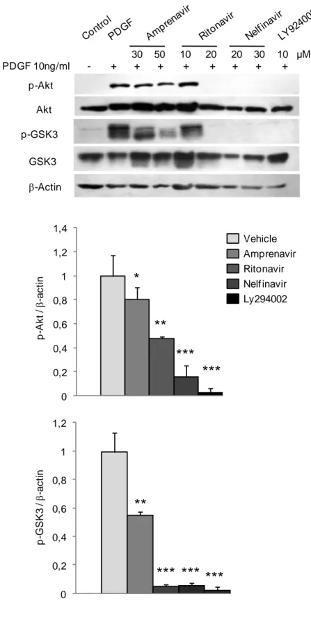
in MTT-labeled cells below the pre-PDGF count with a simultaneous increase in the apoptotic cell count. At doses below the toxic level, ritonavir, amprenavir, and LY924002 inhibited growth without increasing apoptosis; whereas nelfinavir at the highest concentration of 50 M increased the apoptotic cell count (Fig 7A). Cell pretreatment with amprenavir, nelfinavir, ritonavir, or LY924002 dose-dependently inhibited PDGF-induced phosphorylation of Akt and GSK3 (Fig 7B). Phosphorylation of Akt or GSK3 was completely inhibited by ritonavir, nelfinavir, or LY924002; in contrast, even the highest amprenavir dose only partially inhibited the phosphorylation of Akt (by 20%) and GSK3 (by 50%). These HPI-induced changes in p-Akt and p-GSK3 were not accompanied with changes in nonphosphorylated Akt or GSK3 (Fig 7B). Because p-GSK3 is inactive, we investigated whether inhibition of the active (nonphosphorylated) form of GSK3 abolished the growth-inhibiting effects of HPIs or LY924002 on PA-SMCs stimulated with PDGF. Treatment of the cells with the GSK3 inhibitor SB216763 completely abolished the inhibitory effect of amprenavir, nelfinavir, and LY924002 and almost completely abolished the inhibitory effect of ritonavir (Fig. 8).
DISCUSSION
Our main finding is that the HIV protease inhibitors ritonavir, amprenavir, and nelfinavir reversed or attenuated the progression of PH induced in rats by monocrotaline injection or hypoxia exposure. In both models, PH development was associated with marked activation of the Akt signaling pathway in the lungs and proximal pulmonary arteries. Ritonavir, amprenavir, or nelfinavir given to monocrotaline-injected rats inhibited Akt phosphorylation in the lungs and pulmonary arteries and decreased the number of muscularized pulmonary vessels without altering p-ERK or p-MYPT levels. In addition, the three HPIs inhibited PDGF-induced phosphorylation of Akt and GSK3 in cultured PA-SMCs and blocked PA-SMC proliferation. Taken together, these results support the ability of HPIs to interfere with pulmonary vascular remodeling by inhibiting Akt phosphorylation and, consequently, PA-SMC proliferation.
That HPIs may interfere with the PI3K-Akt axis was suggested by several previous studies of tumor cells12,13,14. The PI3K-Akt axis is a major pathway for cell proliferation and
cancer. This pathway is often activated in tumor cells but not in normal host cells and may therefore hold considerable promise as a target for future treatments against cancer22. PH is
primarily a proliferative disorder that shares several features with cancer23. We reasoned that
the Akt pathway may be activated in PH and that HPIs may interact with the pulmonary vascular cell proliferation that underlies PH progression. We found that the development of monocrotaline- and hypoxia-induced PH in rats was associated with marked activation of the Akt-GSK3 axis, as shown by increased phosphorylation of Akt and GSK3 in the lungs and proximal pulmonary arteries. Akt activation occurred early during PH development, within the first week after the monocrotaline injection, concomitantly with induction of the PA-SMC proliferation that underlies the structural remodeling of pulmonary vessels. Cultured
rat PA-SMCs in the quiescent state showed no Akt pathway activation but exhibited marked PDGF-induced phosphorylation of Akt and GSK3. Among the various intracellular downstream effectors of Akt, GSK3 phosphorylation and inactivation of is considered the main mechanism controlling cell proliferation20. Our data are consistent with this concept,
since PA-SMC treatment with LY294002, a specific PI3K inhibitor that blocked PDGF-induced phosphorylation of Akt and GSK3, completely abolished the growth-promoting effect of PDGF. Furthermore, studies of human PA-SMCs showed that PI3K activity was both necessary and sufficient to mediate mitogen-induced cell proliferation and migration 17.
Interestingly, the LY294002 doses used in our study to block Akt and to inhibit PA-SMC growth did not induce apoptosis. Taken together, these findings indicate clearly that activation of the Akt/GSK3 signaling pathway is a major contributor to PA-SMC growth and that rats given monocrotaline or exposed to hypoxia exhibit marked activation of this pathway.
The main goal of this study was to investigate the ability of three HPIs to affect pulmonary vascular remodeling in vivo. In rats with established monocrotaline-induced PH, daily HPI treatment started 3 weeks after the monocrotaline injection produced large decreases in Pap, right ventricular hypertrophy, number of muscularized pulmonary vessels, and pulmonary arterial wall thickness compared to monocrotaline-injected vehicle-treated rats. Similar reductions in these parameters were obtained when the drugs were given preventively, before the onset of PH, to rats exposed to monocrotaline or hypoxia. In monocrotaline-treated rats with established PH, ritonavir, amprenavir, or nelfinavir given for only 1 week significantly decreased pulmonary arterial wall thickness and the number of Ki67-labeled cells in the pulmonary vascular wall without inducing major changes in caspase 3 staining. These in vivo results are consistent with a main effect of HPIs mediated by cell proliferation inhibition rather than by apoptosis induction. The absence of a
pro-apoptotic effect probably explains why none of the HPIs tested induced complete PH reversal. Although the partial nature of the reversal seen with HPI treatment might be ascribable to the use of insufficient dosages, increasing the dosage was limited by poor tolerance, especially with ritonavir. Moreover, the dosages used in our study replicate those given to human patients and may therefore reflect the effects of the serum concentrations achieved clinically.12,14 The effect on PH progression was not markedly different across the
three HPIs used in our study, and inhibition of pulmonary vascular remodeling by HPIs may therefore constitute a class effect. Moreover, our results suggest that the three drugs may act on pulmonary vascular remodeling via a common mechanism.
Because regulation of cell proliferation and motility is a critical step in pulmonary vascular remodeling, researchers are focusing on the development of treatments that specifically target PA-SMC proliferation. Drugs targeting the PIK3-Akt pathway have not been evaluated as possible treatments for PH. All three HPIs evaluated in our study inhibited the phosphorylation of Akt and of its downstream effector GSK3. The effects of the HPIs were observed both in vitro and in vivo. The in vitro effects of the three HPIs were very similar to those of the selective PI3K inhibitor LY294002, with a marked decrease in cell proliferation contrasting with a small effect on cell apoptosis. The HPI doses required in vitro to block PDGF-induced Akt phosphorylation and PA-SMC proliferation were also similar to those of the specific PI3K inhibitor LY294002 and to those of the HPIs shown in previous studies to inhibit tumor cell growth and to block Akt signaling12-14. Thus, our
finding that the HPIs inhibited Akt suggests mediation of HPI effects by inhibition of GSK3 phosphorylation, i.e., GSK3 activation. Accordingly, cell treatment with a GSK3 inhibitor completely abolished the growth-inhibiting effects of amprenavir, nelfinavir, and LY294002 and markedly decreased the inhibitory effect of ritonavir. We also found that each of the three HPIs decreased p-Akt in the lungs and pulmonary vessels. To determine whether this
effect preceded or was secondary to the changes in pulmonary vascular remodeling, we studied rats after only 1 week of HPI treatment, when only minor changes in pulmonary artery muscularization had occurred. The decreases in p-Akt and p-GSK3 in the pulmonary arteries at this time point, together with the decrease in Ki67-labeled cells in the vascular walls, are consistent with a primary effect of HPI treatment on the Akt signaling pathway, with secondary inhibition of PA-SMC proliferation. At this time point, HPI treatment did not affect the phosphorylation of Erk or MYPT, indicating that HPIs did not interfere with these signaling pathways.
Our results in an experimental model of monocrotaline-induced PH support a protective role of HPIs against HIV-related PH. However, several HPIs including ritonavir, have been reported to induce endothelial dysfunction in systemic arteries of HIV-infected patients and to diminish endothelium-dependent relaxation and endothelial nitric oxide expression in porcine pulmonary arteries and human pulmonary endothelial cells24. Because
the expression and activity of endothelial nitric oxide synthase (eNOS) are dependent on Akt signaling, the same pharmacological effects of HPIs may lead to two apparently opposite vascular effects, one exerted on endothelial cells and responsible for a decrease in eNOS activation with subsequent endothelial dysfunction, and the other exerted on PA-SMCs and responsible for inhibition of cell proliferation. Our results are consistent with this possibility. We found that eNOS protein levels remained unchanged in lungs from monocrotaline-treated rats, in keeping with previous studies showing either unchanged or decreased lung eNOS protein levels in this model 25 26. However, lung p-eNOS, which increased markedly
in monocrotaline-treated rats, decreased to basal levels after HPI treatment. Because PA-SMC proliferation is the most prominent abnormality in monocrotaline-induced PH in rats, the antiproliferative effect of HPIs probably played a major role in slowing PH progression in our HPI-treated monocrotaline-injected rats.
The relevance of our findings to patients with HIV-related PH must be considered with circumspection. Although the HPI doses used in our study animals were chosen based on the plasma levels achieved in humans, the nelfinavir dosage needed to obtain an effect was higher than the dosage used clinically. Thus, our results are consistent with a pharmacological action of HPIs in humans, but other parameters may interfere with HPI effects on the Akt pathway in vivo, including drug pharmacokinetics, concomitant drugs, and side effects, as well as patient characteristics. Prominent side effects of HPI treatment include insulin resistance; diabetes; and dyslipidemia with increases in low-density lipoprotein (LDL)-cholesterol and triglycerides, body fat accumulation, and lipodystrophy.11
Previous studies also suggest that diminished Akt activity in response to insulin may lead to insulin resistance.27 Thus, insulin resistance, a cause of glucose intolerance and
dyslipidemia, may be one of the consequences of Akt inhibition by HPIs in HIV-infected patients.28 Whether the ability of HPIs to inhibit Akt in vivo is useful for treating patients
Acknowledgments
We are grateful to the pharmaceutical company GSK for generously donating the amprenavir used in this study.
Funding sources
This study was supported by grants from the INSERM, Ministère de la Recherche, and Fondation Carvsen. Financial support was also received from the European Commission under the 6th Framework Program (Contract No: LSHM-CT-2005-018725,
PULMOTENSION). This publication reflects only the authors´ views, and under no circumstances is the European Community liable for any use that may be made of the information it contains.
Disclosures
References
1. Lederman MM, Sereni D, Simonneau G, Voelkel NF. Pulmonary arterial hypertension and its association with HIV infection: an overview. AIDS. 2008;22 Suppl 3:S1-6.
2. Sitbon O, Lascoux-Combe C, Delfraissy JF, Yeni PG, Raffi F, De Zuttere D, Gressin V, Clerson P, Sereni D, Simonneau G. Prevalence of HIV-related pulmonary arterial hypertension in the current antiretroviral therapy era. Am J Respir Crit Care Med. 2008;177(1):108-113.
3. Nunes H, Humbert M, Sitbon O, Morse JH, Deng Z, Knowles JA, Le Gall C, Parent F, Garcia G, Herve P, Barst RJ, Simonneau G. Prognostic factors for survival in human immunodeficiency virus-associated pulmonary arterial hypertension. Am J
Respir Crit Care Med. 2003;167(10):1433-1439.
4. Sitbon O. HIV-related pulmonary arterial hypertension: clinical presentation and management. AIDS. 2008;22 Suppl 3:S55-62.
5. Opravil M, Pechere M, Speich R, Joller-Jemelka HI, Jenni R, Russi EW, Hirschel B, Luthy R. HIV-associated primary pulmonary hypertension. A case control study. Swiss HIV Cohort Study. Am J Respir Crit Care Med. 1997;155(3):990-995.
6. Zuber JP, Calmy A, Evison JM, Hasse B, Schiffer V, Wagels T, Nuesch R, Magenta L, Ledergerber B, Jenni R, Speich R, Opravil M. Pulmonary arterial hypertension related to HIV infection: improved hemodynamics and survival associated with antiretroviral therapy. Clin Infect Dis. 2004;38(8):1178-1185.
7. Barbaro G, Lucchini A, Pellicelli AM, Grisorio B, Giancaspro G, Barbarini G. Highly active antiretroviral therapy compared with HAART and bosentan in combination in patients with HIV-associated pulmonary hypertension. Heart. 2006;92(8):1164-1166.
8. Degano B, Guillaume M, Savale L, Montani D, Jais X, Yaici A, Le Pavec J, Humbert M, Simonneau G, Sitbon O. HIV-associated pulmonary arterial hypertension: survival and prognostic factors in the modern therapeutic era. AIDS.24(1):67-75.
9. Barnier A, Frachon I, Dewilde J, Gut-Gobert C, Jobic Y, Leroyer C. Improvement of HIV-related pulmonary hypertension after the introduction of an antiretroviral therapy. Eur Respir J. 2009;34(1):277-278.
10. Deeks SG, Smith M, Holodniy M, Kahn JO. HIV-1 protease inhibitors. A review for clinicians. JAMA. 1997;277(2):145-153.
11. Carr A, Samaras K, Thorisdottir A, Kaufmann GR, Chisholm DJ, Cooper DA. Diagnosis, prediction, and natural course of HIV-1 protease-inhibitor-associated lipodystrophy, hyperlipidaemia, and diabetes mellitus: a cohort study. Lancet. 1999;353(9170):2093-2099.
12. Gupta AK, Cerniglia GJ, Mick R, McKenna WG, Muschel RJ. HIV protease inhibitors block Akt signaling and radiosensitize tumor cells both in vitro and in vivo. Cancer Res. 2005;65(18):8256-8265.
13. Yang Y, Ikezoe T, Nishioka C, Bandobashi K, Takeuchi T, Adachi Y, Kobayashi M, Takeuchi S, Koeffler HP, Taguchi H. NFV, an HIV-1 protease inhibitor, induces growth arrest, reduced Akt signalling, apoptosis and docetaxel sensitisation in NSCLC cell lines. Br J Cancer. 2006;95(12):1653-1662.
14. Srirangam A, Mitra R, Wang M, Gorski JC, Badve S, Baldridge L, Hamilton J, Kishimoto H, Hawes J, Li L, Orschell CM, Srour EF, Blum JS, Donner D, Sledge GW, Nakshatri H, Potter DA. Effects of HIV protease inhibitor ritonavir on Akt-regulated cell proliferation in breast cancer. Clin Cancer Res. 2006;12(6):1883-1896.
15. Goncharova EA, Ammit AJ, Irani C, Carroll RG, Eszterhas AJ, Panettieri RA, Krymskaya VP. PI3K is required for proliferation and migration of human
pulmonary vascular smooth muscle cells. Am J Physiol Lung Cell Mol Physiol. 2002;283(2):L354-363.
16. Garat CV, Fankell D, Erickson PF, Reusch JE, Bauer NN, McMurtry IF, Klemm DJ. Platelet-derived growth factor BB induces nuclear export and proteasomal degradation of CREB via phosphatidylinositol 3-kinase/Akt signaling in pulmonary artery smooth muscle cells. Mol Cell Biol. 2006;26(13):4934-4948.
17. Liu Y, Fanburg BL. Serotonin-induced growth of pulmonary artery smooth muscle requires activation of phosphatidylinositol 3-kinase/serine-threonine protein kinase B/mammalian target of rapamycin/p70 ribosomal S6 kinase 1. Am J Respir Cell Mol
Biol. 2006;34(2):182-191.
18. Eddahibi S, Humbert M, Fadel E, Raffestin B, Darmon M, Capron F, Simonneau G, Dartevelle P, Hamon M, Adnot S. Serotonin transporter overexpression is responsible for pulmonary artery smooth muscle hyperplasia in primary pulmonary hypertension. J Clin Invest. 2001;108(8):1141-1150.
19. Schermuly RT, Dony E, Ghofrani HA, Pullamsetti S, Savai R, Roth M, Sydykov A, Lai YJ, Weissmann N, Seeger W, Grimminger F. Reversal of experimental pulmonary hypertension by PDGF inhibition. J Clin Invest. 2005;115(10):2811-2821.
20. Park KW, Yang HM, Youn SW, Yang HJ, Chae IH, Oh BH, Lee MM, Park YB, Choi YS, Kim HS, Walsh K. Constitutively active glycogen synthase kinase-3beta gene transfer sustains apoptosis, inhibits proliferation of vascular smooth muscle cells, and reduces neointima formation after balloon injury in rats. Arterioscler
Thromb Vasc Biol. 2003;23(8):1364-1369.
21. Guignabert C, Raffestin B, Benferhat R, Raoul W, Zadigue P, Rideau D, Hamon M, Adnot S, Eddahibi S. Serotonin transporter inhibition prevents and reverses
monocrotaline-induced pulmonary hypertension in rats. Circulation.
2005;111(21):2812-2819.
22. Vivanco I, Sawyers CL. The phosphatidylinositol 3-Kinase AKT pathway in human cancer. Nat Rev Cancer. 2002;2(7):489-501.
23. Adnot S. Lessons learned from cancer may help in the treatment of pulmonary hypertension. J Clin Invest. 2005;115(6):1461-1463.
24. Wang X, Chai H, Lin PH, Yao Q, Chen C. Roles and mechanisms of human immunodeficiency virus protease inhibitor ritonavir and other anti-human immunodeficiency virus drugs in endothelial dysfunction of porcine pulmonary arteries and human pulmonary artery endothelial cells. Am J Pathol. 2009;174(3):771-781.
25. Jasmin JF, Mercier I, Dupuis J, Tanowitz HB, Lisanti MP. Short-term administration of a cell-permeable caveolin-1 peptide prevents the development of monocrotaline-induced pulmonary hypertension and right ventricular hypertrophy. Circulation. 2006;114(9):912-920.
26. Zhao YD, Campbell AI, Robb M, Ng D, Stewart DJ. Protective role of angiopoietin-1 in experimental pulmonary hypertension. Circ Res. 2003;92(9):984-99angiopoietin-1.
27. Bae SS, Cho H, Mu J, Birnbaum MJ. Isoform-specific regulation of insulin-dependent glucose uptake by Akt/protein kinase B. J Biol Chem.
2003;278(49):49530-49536.
28. Ben-Romano R, Rudich A, Tirosh A, Potashnik R, Sasaoka T, Riesenberg K, Schlaeffer F, Bashan N. Nelfinavir-induced insulin resistance is associated with impaired plasma membrane recruitment of the PI 3-kinase effectors Akt/PKB and PKC-zeta. Diabetologia. 2004;47(6):1107-1117.
Figure legends
Figure 1. Pulmonary artery pressure (Pap), right ventricular hypertrophy index
[(RV/(LV+S) weight ratio], and muscularization of pulmonary vessels in rats studied at various times after administration of monocrotaline (MCT) or saline (controls). Muscularization of pulmonary vessels is reported as the percentages of nonmuscularized, partially muscularized, and fully muscularized distal vessels. Treatment with amprenavir (A), ritonavir (B), nelfinavir (C), or vehicle was given from day 21 to day 42 (n=10 in each group), in separate experiments for each drug including different controls. *P<0.05, **P<0.01, and ***P<0.001 compared with corresponding values recorded on day 28 or 42 in the monocrotaline-injected vehicle-treated rats.
Figure 2. Pulmonary vascular remodeling. Representative photomicrographs of pulmonary
vessels (A) and pulmonary artery wall thickness (B) from rats on day 21, 28, and 42 after saline (controls) or monocrotaline (MCT) administration. Each value is mean±SEM of 10 independent determinations. * P<0.05 and ** P<0.01 compared with corresponding values recorded on day 28 or 42 in monocrotaline-injected vehicle-treated rats.
Figure 3. A. Rat pulmonary-artery smooth-muscle-cell (PA-SMC) proliferation (Ki67
staining, upper sections) on days 28 and 42 after monocrotaline (MCT) injection. Brown staining indicates Ki-67-positive cells. The number of proliferating vascular cells expressed as the percentage of Ki-67 labeled cells over the total number of cells counted in the media of at least 20 muscularized vessels per rat was markedly reduced by treatment with each of the HPIs, *P<0.05 and **P<0.01 compared with corresponding values recorded on day 28 or 42 in the monocrotaline-injected vehicle-treated rats (n=10 in each group). B. Rat PA-SMC apoptosis (Caspase 3 staining, lower sections) on days 28 and 42 after monocrotaline
injection. The number of apoptotic cells (caspase 3-positive cells) was not affected by HPI treatment (n=10 in each group). Bar=50 µm.
Figure 4. Pulmonary artery pressure, right ventricular hypertrophy index [(RV/(LV+S)
weight ratio], muscularization of pulmonary vessels (percentage of partially and fully muscularized pulmonary vessels), and pulmonary artery wall thickness in rats studied on day 21 after administration of monocrotaline or exposure to hypoxia. Control rats were studied after saline administration and kept in normoxic conditions. Treatment with amprenavir, ritonavir, nelfinavir, or vehicle was given from day 0 to day 21 (n=10 in each group). *
P<0.05 and ** P<0.01 compared with values in vehicle-treated rats after monocrotaline
administration or exposure to hypoxia.
Figure 5. A. Time-course of Akt expression and Akt phosphorylation in lung tissue and
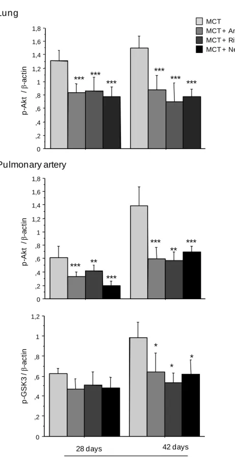
proximal pulmonary arteries after monocrotaline (MCT) injection. Protein expression and phosphorylation were evaluated by western blot in rat lung and pulmonary artery extracts 7, 21, 28, and 42 days after MCT administration. The column graph shows the mean±SEM values obtained in pulmonary arteries (n=10 in each group) * P<0.05; ** P<0.01; and ***P<0.001 compared with values obtained before MCT administration. B. Changes in pAkt levels in lungs and changes in p-Akt and p-GSK3 levels in proximal pulmonary arteries measured 28 and 42 days after monocrotaline injection. Treatment with ritonavir, amprenavir, nelfinavir, or vehicle was given from day 21 to day 42 (n=10 in each group). *P<0.05; **P<0.01; and ***P<0.001 compared with values obtained in monocrotaline-injected vehicle-treated rats.
Figure 6. Changes in p-eNOS and eNOS levels in lung (A), changes in p-Erk and Erk levels
in proximal pulmonary arteries (B) and changes in p-MYPT and MYPT levels in lung (C) in rats injected with saline (controls) or monocrotaline, measured 28 days after monocrotaline injection. Treatment with ritonavir, amprenavir, nelfinavir, or vehicle was started at day 21 (n=10 in each group). * P<0.05; ** P<0.01; and ***P<0.001 compared with values obtained in monocrotaline-injected vehicle-treated rats.
Figure 7. A. Effects of ritonavir, amprenavir, nelfinavir, and LY924002 on
pulmonary-artery smooth-muscle-cell (PA-SMC) proliferation (column graph) and apoptosis (diamond graph). The cells were starved of fetal calf serum for 48 h and exposed to the HPIs or LY924002 at increasing concentrations during 24 hours. Cellular proliferation was measured using the tetrazolium assay 48 h after stimulation by 10 ng/ml of PDGF-BB. Apoptotic cells were determined at the same time. Values are mean±SEM of 12 values obtained from four independent experiments. * P<0.05; ** P<0.01; and ***P<0.001 compared with values obtained with PDGF alone. The apoptotic cell number increased in response to the 50µM concentration of ritonavir and nelfinavir (* P<0.05); B. Effects of treatment with amprenavir, ritonavir, nelfinavir, or LY924002 on p-Akt and p-GSK3 protein levels in cultured PA-SMC stimulated with 10 ng/ml of PDGF for 48 hours. Values are mean±SEM of 12 values obtained from four independent experiments. *P<0.05; **P<0.01; ***P<0.001 compared to values obtained with PDGF alone.
Figure 8. The GSK3 inhibitor SB216763 (10 µM) abolished the inhibitory effect of ritonavir
(20 µM), amprenavir (30 µM), nelfinavir (50 µM), and LY924002 (30 µM) on PA-SMC proliferation. Values represent the percentage of cell numbers measured after stimulation with PDGF and are the mean±SEM of 12 values obtained from four independent
experiments. *P<0.05; **P<0.01; ***P<0.001 compared to values obtained with PDGF alone; †P<0.05 compared to values obtained with PDGF plus SB216763 (10 µM).
Figure 2
Ritonavir Amprenavir
Nelfinavir
Controls Monocrotaline
21 days 28 days 42 days
MCT
A
B
0 20 40 60 ** ** W all th ic kn e ss (% ) 0 20 40 60 ** W all th ic kn e ss (% ) 0 20 40 60 80 ** * W all th ic kn e ss (% )Figure 3
0 30 60
% Ki67 positive cells
** ** **
0 30 60
% Ki67 positive cells
** ** * 2 8 d a y s 4 2 d a y s 0 20 40
% Caspase 3 positive cells
0 25 50
% Caspase 3 positive cells
4 2 d a y s 2 8 d a y s MCT MCT + Amprenavir MCT + Ritonavir MCT + Nelf inavir
A
B
MCT MCT + Amprenavir MCT + Ritonavir MCT + Nelfinavir MCT MCT + Amprenavir MCT + Ritonavir MCT + Nelf inavirFigure 4 P u lm o n a ry a rt e ry p re s s u re (m m H g ) R V /L V + S r a ti o (% ) P u lm o n a ry v e s s e ls (% ) W a ll th ic k n e s s in d e x (% )
Control Monocrotaline Hypoxia
0 10 20 30 40 50 60 0 10 20 30 40 50 60 0 10 20 30 40 50 0 5 10 15 20 25 30 35 40 45
*
*
*
*
*
*
** **
**
** ** **
*
*
*
*
*
*
*
*
*
*
*
vehicle Amprenavir Ritonavir NelfinavirFigure 5 A Lung Pulmonary artery p-Akt Akt β-Actin p-GSK3 GSK3 p-Akt Akt β-Actin p -Ak t / β -a c ti n p -G SK3 / β -a c ti n
Figure 5 B 0 ,2 ,4 ,6 ,8 1 1,2 1,4 1,6 1,8 *** *** *** *** *** *** 0 ,2 ,4 ,6 ,8 1 1,2 1,4 1,6 1,8 p -A k t / β-a c ti n *** *** ** 0 ,2 ,4 ,6 ,8 1 1,2 42 days Monocrotaline 28 days * * * p -G S K 3 / β-a c ti n *** *** ** p -A k t / β -a c ti n Lung Pulmonary artery MCT MCT + Amprenavir MCT + Ritonavir MCT + Nelfinavir
Figure 6 A p-eNOS eNOS -actin p -e N O S / –a c ti n 0 ,1 ,2 ,3 ,4
*
*
*
*
Controls Monocrotaline e N O S / –a c ti n 0 ,1 ,2 ,3 ,4 ,5 ,6 ,7 Controls Monocrotaline MCT MCT + Amprenavir MCT + Ritonavir MCT + NelfinavirA
Figure 6 B, C Erk β-Actin p-Erk 1/2 0 ,4 ,8 1,2 1,6 2 Controls p -E rk / –a c ti n Monocrotaline 0 ,2 ,4 ,6 ,8 1 1,2 E rk / –a c ti n Controls Monocrotaline MCT MCT + Amprenavir MCT + Ritonavir MCT + Nelfinavir 0 ,2 ,4 ,6 ,8 1 M Y P T / –a c ti n 0 ,6 1,2 1,8 p -M Y P T / –a c ti n
Controls Monocrotaline Controls Monocrotaline
MCT MCT + Amprenavir MCT + Ritonavir MCT + Nelfinavir β-Actin p-MYPT1 MYPT1
B
C
Figure 7 B Vehicle Amprenavir Ritonavir Nelf inavir Ly294002 0 0,2 0,4 0,6 0,8 1 1,2