HAL Id: hal-01053314
https://hal.archives-ouvertes.fr/hal-01053314
Submitted on 15 Sep 2014HAL is a multi-disciplinary open access
archive for the deposit and dissemination of sci-entific research documents, whether they are pub-lished or not. The documents may come from teaching and research institutions in France or abroad, or from public or private research centers.
L’archive ouverte pluridisciplinaire HAL, est destinée au dépôt et à la diffusion de documents scientifiques de niveau recherche, publiés ou non, émanant des établissements d’enseignement et de recherche français ou étrangers, des laboratoires publics ou privés.
cements
Alexandre Govin, Iolanda Albuquerque, Philippe Grosseau
To cite this version:
Alexandre Govin, Iolanda Albuquerque, Philippe Grosseau. Development of an accelerated test of fungal biodeterioration. Application to calcium aluminate cements. International Conference on Calcium Aluminates, May 2014, Palais des Papes, Avignon, France. pp. 511-522. �hal-01053314�
Calcium Aluminates: Proceedings of International Conference, Avignon, 18 –21 May 2014. Fentiman CH, Mangabhai RJ and Scrivener KL (Editors). IHS BRE Press, 2014, EP104. ISBN 978-1-84806-316-7.
DEVELOPMENT OF AN ACCELERATED
TEST OF FUNGAL BIODETERIORATION.
APPLICATION TO CALCIUM
ALUMINATE CEMENTS
A. GOVIN, I. ALBUQUERQUE and P. GROSSEAU
Ecole Nationale Supérieure des Mines de Saint Etienne, SPIN-EMSE, CNRS:UMR5307, LGF, 158 cours Fauriel, 42023 St-Etienne, France
govin@emse.fr and grosseau@emse.fr
SUMMARY: Microorganisms, such as bacteria, cyanobacteria, fungi, algae, and lichens, are liable to grow on building materials. Biological activity contributes to deterioration of building material. Fungi are among the most harmful organisms associated to biodeterioration of organic and inorganic materials. Cement-based materials are porous and contain organic admixtures. Thus they possess significant bioreceptivity. The objectives of the study were firstly to develop a methodology and an accelerated test in order to evaluate the bioreceptivity of cement-based materials to fungal colonization and secondly to evaluate the ability to resist of Calcium Aluminate Cements compared to that of Portland Cements. Three specific fungi (Aspergillus niger, Coniosporium uncinatum and Alternaria
alternata), often identified on monuments or degraded buildings, were
tested. The biocolonization of mortar samples was evaluated by quantifying the amount of ergosterol which is a predominant and endogenous sterol met only in the cell membrane of fungi. The results show that the resistance ability of CACs to fungal colonization seems to be higher than that of Portland based mortars.
Keywords: Accelerated laboratory test, Biodeterioration, Calcium Aluminate Cement, Fungi, Mortars.
INTRODUCTION
Biodeterioration can be defined as an irreversible transformation of all types of material (organic or inorganic) possessing an economic, artistic, historic or archaeological value, due to organisms in natural environments [1, 2]. An example of biodeterioration is given on Fig. 1[3]. In 1995, Guillitte defines the concept of the bioreceptivity as “the ability of a material to be colonized by one or more groups of living organisms without
necessarily inducing biodeterioration” [4]
. The bioreceptivity can also be defined as, the set of material properties that contribute to installation, adhesion and the development of organisms. It depends on the intrinsic properties of the matrix, such as its chemical composition, porosity, permeability, or roughness, etc.
Fig. 1: Influence of biodeterioration process on an angel statue of the cathedral of Cologne (Germany),
(a) original object in 1880 (photograph by Anselm Schmitz, Cologne) and (b) the respective weathered statue in 1993 (photograph by A. Wolff, Cologne)
In fact, many microorganisms such as; bacteria, cyanobacteria, algae, and fungi, etc. are able to colonize and degrade materials [5]. The biodeterioration is often the result of complex microbial interactions [3]. Microorganisms are naturally selected depending on pH and available nutrients. The development of the first colonizers modifies the physico-chemical parameters of the surface, thus facilitating colonization of new species, the second colonizers, and so forth, defining thereby the biological succession. Bacteria are usually the first colonizers, because they can grow on any type of support and in extended pH ranges [6].
Fungi are eukaryotic organisms (cells with a nucleus). They are devoid of chlorophyll and therefore incapable of photosynthesis. Thus they are heterotrophic organisms and need an external source of carbon [6,7]. Wastes or dead cells, from algae and bacteria, dead leaves and bird droppings are sources of carbon [7]. Fungi can be regarded as the best armed organisms for the biodeterioration of organic and inorganic materials [8]. Indeed, they are ubiquitous and colonize a wide variety of substrates, such as textile, leather, paper, stones, wood, plastic, paint and construction materials [9]. Moreover, they are able to penetrate into a material by hyphal growth and by biocorrosive activity due to the excretion of organic acids or by the oxidation of compounds of the matrix and to discolor material surfaces by excretion of melanins or carotenoids and to induce mechanical stress into materials [3].
The biodeterioration process begins as soon as the fungi settle and colonize the surface of the material. This process can be summed up as follows for the biodeterioration of stones:
Production of organic acids (such as oxalic, citric, propionic, etc...) which leads to the dissolution of carbonates and the de-cohesion of the grains.
Aesthetical damage due to the excretion of fungal pigment such as melanin and carotenoid [10,11].
Inter-crystalline growth of the fungi within the faults of the matrix.
Biopitting which corresponds to the formation of small craters due to the de-cohesion of grains induced by the penetration of hyphae within the stones [12]. As fungal or biological stains clearly affect the aesthetics of a façade and represent a significant economic loss, due to the costs of maintenance and repair, a
focus on building materials is necessary. The objectives of this study were firstly to develop an accelerated, reliable and reproducible method in order to evaluate the bioreceptivity of cement-based materials to fungal colonization, and secondly to highlight the possible effect of the chemical composition of cements on their susceptibility to fungal growth. To achieve this last objective, Calcium Aluminate and Portland Cement-based materials were studied with three specific fungi (Aspergillus
niger, Coniosporium uncinatum and Alternaria alternata).
MATERIALS AND METHODS Fungal Strains
In order to evaluate the bioreceptivity of the cementitious materials, 3 fungal strains have been tested (Fig. 2).
Aspergillus niger was chosen because this fungi is often identified from samples
on monuments or degraded buildings [3,13] and grows on various substrates, such as decaying organic matters, soils, foodstuffs, grains, minerals and building materials, and is ubiquitous in the human environment. A. niger is an acidogenic fungus i.e. that it produces organic acids, such as citric, ascorbic, gluconic, acetic acid, etc. Thus, they are responsible for the biodeterioration of the materials by chemical route.
Coniosporium uncinatum is commonly isolated from monuments located in the
Mediterranean basin. In many cases it is the predominant microflora [14]. C.
uncinatum is a meristematic fungus, which is almost all isolated from the surface
of stones. This fungus degrades materials by physical process (by deep penetration into the matrix).
Alternaria alternata is ubiquist (indicating that it is found in different biotopes)
and its habitat is very diverse ranging from very wet areas to dry and arid regions. This species has already been isolated on various substrates and materials, such as marble, paintings, plants, food, textiles and floors and is considered as main cause for brown and black coloring of limestone, marble, and sandstone of European and African monuments [15]. As with C. uncinatum,
A. alternata implies a physical degradation of materials.
Strains Alternaria alternata Fries von Keissler, MUCL 30967 and Aspergillus
niger van Tieghem, MUCL 19002, came from the collection of microorganisms of the
Mycothèque at the Catholic University of Louvain (MUCL), Belgium. Coniosporium
uncinatum CBS 100212 strains, originated in Italy, was isolated by De Leo et al (1999)
and provided by the Centraal Bureau voor Schimmel cultures (CBS - Holland).
Aspergillus niger and Alternaria alternata strains have been cultivated in Petri
box on Potatoes Dextrose Agar (PDA, Difco) solid media. Coniosporium uncinatum stain has been cultivated in Potatoes Dextrose Broth (PDB, Sigma) liquid media. All the media have been previously sterilized. After inoculation of the culture media, the cultures were incubated at 80±5% RH et 24±1°C until sufficient growth occurred.
Fungal cell suspensions have been prepared from the cultures. After centrifugation at 3000 rpm for 15 min (Multifuge 3SR+ - Thermo Fisher), the pellet was suspended in a nutritive media composed of Yeast Nitrogen Broth (YNB) 1wt% and Glucose 0.01wt% in water, in order to obtain a fungal cell concentration in the suspension, of 106 fungal units/ml.
Fig. 2: Fungal strains used: (a) Aspergillus niger, (b) Alternaria alternata, (c) Coniosporium uncinatum
Materials:
The ability to resist to fungal biodeterioration was investigated by working on cement based mortars. Indeed, rendering is a common construction technique for masonry walls and façades. In order to study the potential effect of the chemical composition of mortars on the fungal biodeterioration a panel of eight cementitious matrices was prepared. Among this panel, five were mortars and three were pure pastes (without sand) in order to emphasize the potential effect of the chemical composition.
Table 1. Nomenclature and formulation of mortars and pure pastes.
Nomenclature Denomination Binder (wt%)
WP-M White Portland-Mortar
100% White Portland CEMII/A 42.5
WP-PP White Portland-Pure Paste
PL-M Portland Lime-Mortar 80% White Portland CEMII/A 42.5 /
20% Lime
TSA-M Ettringite-Mortar 70%Ternal White / 30% Anhydrite
MCA-M Medium Content of Aluminate-Mortar
100% Calcoat RG MCA-PP Medium Content of Aluminate-Pure
Paste
HCA-M High Content of Aluminate-Mortar
100% Ternal White HCA-PP High Content of Aluminate-Pure Paste
A white Portland cement (CEMII/A 42.5, Lafarge) and two Calcium Aluminate Cements (CAC) (Kerneos) with different aluminate content were tested (Table 1 and Table 2). Moreover, two binary binders made of Portland with lime, and CAC with anhydrite, completed the panel. The white Portland cement was used as a reference for the evaluation of the biocolonization. Oxide composition of all cements and admixtures were determined by X-ray fluorescence spectroscopy (SRS3400, Bruker-AXS) and are given by Table 2.
The specimens were prepared according to the standard EN196-1, i.e., a water-to-cement ratio (w/c) equal to 0.5 and proportions of binder and sand (Sibelco CV32) respectively of 25% and 75% in weight. However, for the preparation of pure pastes, the w/c was lowered to 0.3 in order to have a correct consistency.
The fresh mortars or pure pastes were cast into 8 × 2 × 1 cm PVC molds. The specimens were striped after 24 hours of hydration under 100% of relative humidity (RH) and 21°C and then kept 28 days under the same conditions of humidity and temperature.
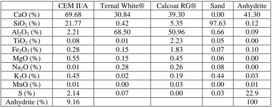
Table 2. Chemical compositions of the investigated cements and admixtures determined by X-ray
fluorescence
CEM II/A Ternal White® Calcoat RG® Sand Anhydrite
CaO (%) 69.68 30.84 39.30 0.00 41.30 SiO2 (%) 21.77 0.42 5.35 97.63 0.12 Al2O3 (%) 2.21 68.50 50.96 0.66 0.09 TiO2 (%) 0.08 0.01 2.23 0.05 0.00 Fe2O3 (%) 0.28 0.15 1.83 0.07 0.10 MgO (%) 0.55 0.15 0.45 0.06 0.00 Na2O (%) 0.01 0.28 0.26 0.08 0.00 K2O (%) 0.45 0.02 0.19 0.44 0.03 MnO (%) 0.01 0.00 0.03 0.00 0.01 S (%) 2.14 0.07 0.00 0.03 22.9 Anhydrite (%) 9.16 100
In order to decrease the surface pH, an accelerated weathering was applied to the specimens. In the case of Aspergillus niger and Coniosporium uncinatum, the weathering of samples consisted of a combination of a carbonation step, under pure CO2
(21 ± 1°C, 65 ± 5% RH for 7 days) and a lixiviation step by water (25 days). For pure pastes, a second carbonation step (14 days) was imposed to intensify the decrease of surface pH. A simple carbonation step (21 ± 1°C, 65 ± 5% RH for 40 days) was applied to specimens in the case of Alternaria alternata. The surface pH was measured by a surface electrode (WTW Sentix Sur). To ensure contact between the substrate and the pH-electrode, the surface was moistened.
All the specimens were sterilized by gamma ray (30kGy) to avoid contaminations and to quantify only the fungal colonization.
Accelerated test
The accelerated test was previously described by Wiktor et al.[16]. It was performed in polypropylene and autoclavable boxes (Fig. 3). All the materials used have been previously sterilized. The bottom of the box was filled with a thin layer (about 1cm) of vermiculite. Then 70mL of deionized water was spread on the vermiculite in order to keep the necessary moisture for the fungal growth into the box. Two cementitious specimens were placed into the box. To avoid direct contact between the specimens and the vermiculite, a paper sheet was introduced between the both.
In sterile conditions, the sterilized samples were inoculated by spreading 1.5mL of the fungal suspension on the surface of each specimen. Then the boxes were closed and stored at 80±5% RH and 24±1 °C.
Fig. 3: Scheme of the experimental set-up used for the accelerated test
Carded cotton Specimens Wet vermiculite Paper sheet Fungal suspension
Evaluation of the fungal colonization
The quantification of the biocolonization by fungi was carried out by ergosterol assay. Indeed, the ergosterol is a predominant and endogenous sterol, and specific to the cell membrane of fungi and some microalgae. The method for the determination of ergosterol used in this study was based on the protocol proposed by Ruzicka et al.[17]. At the end of the test, the ergosterol was extracted from the samples by immersion of a whole specimen in 20mL of methanol. The whole was subjected to ultrasound for 45 min and then stored in methanol at 4°C for 12 h. The supernatant was then filtered at 0.22 µm before being analyzed by HPLC (pump Waters 616, 600S controller and PDA detector Waters 996). The column used was a C18 Novapack and its temperature was fixed at 30°C. The mobile phase was pure methanol and the flow was 0.5 mL.min-1. The peak area at the absorption of 282 nm was used for quantification with an ergosterol external standard (Sigma-Aldrich). All the quantifications were conducted in triplicate.
RESULTS AND DISCUSSION Characterization of weathering
The objective of the accelerated weathering method was to decrease the surface pH of the samples. The initial surface pH was around 12.0-12.5 for the Portland-based samples (WP-M, PL-M, WP-PP). Concerning the Aluminate-based samples (HCA-M, MCA-M, TSA-M, HCA-PP and MCA-PP), the pH was lower, i.e. in the range of 10.8 to 11.4. At the end of the weathering process (carbonation and lixiviation), all the mortars exhibited similar surface pH. Indeed, the pH ranged from 9.3 to 9.6 and from 9.5 to 9.7 respectively for Portland-based samples and for Aluminate-based samples. The carbonation of the Portland-based materials mainly led to the conversion of the portlandite into calcite ( ). For the Aluminate-based materials, the hydrates such as C3AH6 and CAH10 were converted into and AH3. For TSA-M, the high amount of
Aft and AFm, such as ettringite and monosulfoaluminate, totally disappeared and led to the formation of and AH3. Thanks to a second carbonation step, the surface pH of
the pure pastes was lower than for mortars, and was between 7.7 and 8.0. The second weathering method (carbonation step alone) was more efficient for the mortars. Indeed, the final pH was around 8.0-8.9. However, the carbonation was less effective in the case of the pure pastes. Indeed, the surface pH reached was higher (8.9-9.3) than for mortars. This result could be due to the lower porosity of the pure pastes compared to that of mortars.
Biodeterioration test Aspergillus niger
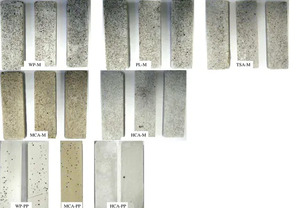
The samples inoculated with Aspergillus niger were incubated for 72 days at 80% RH and 24°C. The visual observation indicates a total absence of fungal development on the surface of all specimens (Fig. 4).
While Aspergillus niger is often identified from samples of monuments or buildings degraded, its growth on the cementitious materials was not observed. Similar results were obtained by Wiktor [18]. However, a significant growth of Aspergillus niger on the paper sheet was observed, confirming the good conditions of development.
_ C C _ C C _ C C
Fig. 4: Samples inoculated with Aspergillus niger after 72 days of incubation.
Coniosporium uncinatum
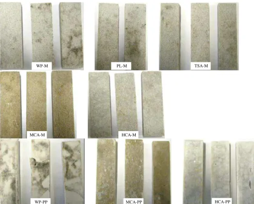
Fig. 5 represents the specimens inoculated with Coniosporium uncinatum after 52 days of incubation.
Fig. 5: Samples inoculated with Coniosporium uncinatum after 52 days of incubation.
With the exception of the High Content of Aluminate Mortars (HCA-M), C.
uncinatum colonies developed on all specimens (black dots). However, for Portland
cement-based mortars (WP-m and PL-M) these colonies of C. uncinatum were the most developed, and with the largest diameter. The fungal growth seems to be lower for MCA-M and TSA-M. No fungal colonization was noted on HCA-M. The same trend was obtained with pure pastes. The advantage of these samples was that they were smoother, and therefore making the colonization more visible. The results of the quantification of ergosterol are shown in the fig. 6 and confirm the visual observation. It is clear that Portland-based mortars (WP-M and PL-M) are the most colonized by fungi. Moreover, these results suggest that the higher the aluminate content, the lower is the fungal colonization. Indeed, for TSA-M and MCA-M, the amount of ergosterol is halved compared to the Portland-based materials and no ergosterol was detected for HAC-M. Furthermore the amount of ergosterol is the same in TSA-M and MCA-M as is the initial amount of aluminate.
HCA-M MCA-M
TSA-M MCA-PP HCA-PP PL-M WP-M WP-PP HCA-M TSA-M HCA-PP MCA-PP PL-M WP-M WP-PP MCA-M
Fig. 6: Quantification of ergosterol for samples inoculated with Coniosporium uncinatum after 52 days of
incubation.
For pure pastes, with the exception of CACs, the results are not discriminating, as expected. Indeed, the evolution of the amount of ergosterol is consistent with the aluminate content of the formulation. However, the difference between WP-PP and MCA-PP is very low or even non-existent.
Alternaria alternata
The results of the biodeterioration test carried out with Alternaria alternata after 52 days of incubation, are shown in fig. 7. This fungus strongly colonized all specimens with the production of spores and the development of black mycelium specific to this species on the surface of the specimens. However, the fungal development was observed on the surface of the specimen in contact with paper sheet (i.e. the surface opposite the inoculated surface with the fungal suspension). The paper sheet was also strongly colonized by Alternaria alternata. As in the case of C. uncinatum, matrices made of high aluminate content in the cement (HCA-M and HCA-PP) were the least covered by fungi. Compared to this the Portland-based samples (WP-M, WP-PP and PL-M) were the most biodegraded.
The results of the assay of ergosterol are shown in the Fig. 8 and confirm the strong fungal development mentioned shown by visual observation of the samples. Indeed, the amount of ergosterol is around ten times higher than in the case of C.
uncinatum, and ranges from 1 to 15 µg per sample. Moreover, contrary to C. uncinatum,
a fungal growth was detected for the binders with high aluminate content (HCA-M and HCA-PP). The increase in the fungal growth could be due to the lower surface pH of these specimens, compared to those used for C. uncinatum. Indeed, the surface pH reached after weathering, was around 8.0-9.3 and 9.3-9.7 respectively in the case of A.
alternata and C. uncinatum.
For mortars, WP-M and PL-M are the most colonized by A. alternata, followed by TSA-M and MCA-M and then HCA-M. As previously, the order of magnitude of the colonization intensity is inversely proportional to the aluminate content of the cement. A net difference in the ergosterol content was measured between WP-PP and HCA-PP, suggesting, again, a good resistance of aluminous cement to the development of fungi. However, the standard deviation obtained for MCA-PP is very high; therefore it is difficult to conclude on this formulation.
0.0 0.2 0.4 0.6 0.8 1.0 1.2 1.4 1.6 1.8 2.0
WP-M PL-M TSA-M MCA-M HCA-M WP-PP MCA-PP HCA-PP
E rg o st e ro l (µ g /S a mp le )
Fig. 7: Samples inoculated with Alternaria alternata after 52 days of incubation.
Fig. 8: Quantification of ergosterol for samples inoculated with Alternaria alternata after 52 days of
incubation.
Fig. 9 shows the relationship between the amount of aluminium present in the mortars and the amount of ergosterol. Due to the complexity of the composition in aluminate hydrates and in aluminate anhydrous phases of the mortars, it was not possible to quantify the amount of aluminate phases present. Therefore a measure of the amount of aluminate phases was represented by the total amount of aluminium contained in the initial cement (as determined by X-ray fluorescence and expressed as aluminium oxide). Despite not knowing the nature and the actual quantity of the aluminate phases present, it seems that there is a clear relationship between the amount of ergosterol, and thus the fungal colonization of the mortars, with the aluminium oxide content. Thus, the higher is the aluminium oxide content; the lower is the fungal growth.
0 2 4 6 8 10 12 14 16 18 20
WP-M PL-M TSA-M MCA-M HCA-M WP-PP MCA-PP HCA-PP
Er g o s te ro l (µ g /Sa mp le ) HCA-M MCA-M TSA-M HCA-PP MCA-PP PL-M WP-M WP-PP
Fig. 9: Relationship between aluminium oxide content in the mortars and the amount of ergosterol.
Despite many researches devoted to study the effect of aluminium on cells or microorganisms, the mechanism is still not fully understood. Since many years, aluminium is described as having bacteriostatic [19], fungicide [20], parasiticide [21] and phytotoxic [22] properties. According to the authors, aluminium adsorbs onto the wall cells or is absorbed into the cells, resulting in deleterious metabolic effect which induces stop or delay in cell division. However, for all the studies, the experiments were carried out in acidic media, which highlights only the effect of aqueous aluminium (Al3+). In our case, the pH of the medium and of the substrate surface was around 7.5 and higher than 8.0 respectively, thus, the expected concentration of Al3+ should be very low. To improve the knowledge on the bioreceptivity of CACs, it would be necessary to study the toxicity of the stable aluminate phases (AH3, C3AH6), unstable aluminate phases
(CAH10, C2AH8) and anhydrous aluminate phases (CA, CA2, C2AS).
CONCLUSION
The accelerated biodeterioration test proposed in this study is reliable and reproducible for the evaluation of the bioreceptivity of cement-based materials to fungal colonization. Among the three fungi tested, no fungal growth was only detected for Aspergillus niger. However, Coniosporium uncinatum and Alternaria alternata both showed a strong development on cement-based specimens. Moreover, the development of A. alternata was higher than that of C. uncinatum. Thus A. alternata could be a good candidate to evaluate the bioreceptivity of these building materials.
The ergosterol assay allows the fungal development to be quantified and gives an estimation of the extent of biodeterioration. The results showed that, for mortars and pure pastes, the specimens composed of Portland cement were the most vulnerable to the fungal colonization. Furthermore, as amount of aluminium increases, the fungal biodeterioration decreases. Hence, TSA-M and MCA-M were more colonized than HCA-M. These results seem to highlight that the bioreceptivity of cement-based materials to fungi increases with the decrease in the aluminium (and therefore aluminate) content. Despite the high standard deviation, the same tendency was noted for pure pastes.
y = -0.56x + 11.31 R² = 0.80 y = -0.07x + 1.35 R² = 0.80 0 2 4 6 8 10 12 14 16 0 5 10 15 20 E rgo ste rol (µg /S a mp le )
Aluminium oxide content (%)
A. alternata C. uncinatum
ACKNOWLEDGEMENT
The authors would like to acknowledge Kerneos for their financial and technical support.
REFERENCES
[1] Allsopp D, Seal K J, Gaylarde C C. Introduction to biodeterioration. Second Edition, Cambridge University Press, Cambridge, 2004.
[2] Urzì C. Biodeterioration of stone mortar and wallpainting. Presentation for Sciences of Materials of Cultural Heritage. European Doctoral Theorectical and Practical Course, October 2006, Ravello – Italy, 2006.
[3] Warscheid T, Braams J. Biodeterioration of stone: a review. International Biodeterioration & Biodegradation, 2000; 46(4):343-368.
[4] Guillitte O. Bioreceptivity: a new concept for building ecology studies. The Science of the Total Environment, 1995; 167:215-220.
[5] Sand W. Microbial mechanisms of deterioration of inorganic substrates: A
general mechanistic overview. International Biodeterioration & Biodegradation,
1997; 40(2):183-190.
[6] Dubosc A. Etude du développement de salissures biologiques sur les parements
en béton: Mise au point d’essais accélérés de vieillissement. PhD thesis of Institut
National des Sciences Appliquées de Toulouse, 2000.
[7] Kumar R. Kumar A V. Biodeterioration of stone in tropical environments: An
Overview. Oxford University Press, Oxford, 1999.
[8] De Leo F, Urzì C. Fungi in the biodeterioration of Cultural Heritage. In: Roger-Alexandre Lefèvre (Eds), The Materials of the Cultural Heritage in their Environment 8, Edipuligia, Bari, 2006.
[9] Khandelwal A. Sampling and Estimate of fungal biodeteriogens of Lucknow,
India. The Australian Institute for the Conservation of Cultural Material Bulletin,
2003; 28:76-81. Papers from the fifth International Conference on Biodeterioration of Cultural Property, Sydney, November 2001.
[10] Sterfingler K. Fungi as geologic agents. Geomicrobiology Journal, 2000; 17: 97-124.
[11] Urzì C, De Leo F, de Hoog G S, Sterflinger K. Recent Advances in the molecular
biology and ecophysiology of meristematic stone-inhabiting fungi. In: Ciferri O,
Tiano P, Mastromei G (Eds.), Proceedings of the International Congress on Microbes and Art. Plenum Publishing Co. Ltd., New York, 2000.
[12] Sterflinger K, Krumbein W E. Dematiaceous fungi as a major agent for biopitting
Mediterranean marbles and limestones. Geomicrobiology Journal, 1997;
14(3):219-230.
[13] De Leo F, Urzì C, de Hoog G S. Two Coniosporium species from rock surfaces. Studies in Mycology, 1999; 43:70-79.
[14] Garcia Vallès M, Vandrell-Saz M, Krumbein W E, Urzì C. Coloured mineral
coatings on monument surfaces as a result of biomineralization: the case of Tarragona cathedral (Catalonia), Applied Geochemistry, 1997; 12 (3):255-266.
[15] Diakumaku E, Gorbushina A A, Krumbein W E, Panina L, Soukharjevski S.
problem for the conservation of monuments. The Science of the Total
Environment, 1995; 167:295-304.
[16] Wiktor V, De Leo F, Urzì C, Guyonnet R, Grosseau P, Garcia-Diaz E.
Accelerated laboratory test to study fungal biodeterioration of cementitious matrix, International Biodeterioration & Biodegradation, 2009; 63(8):1061-1065.
[17] Ruzicka S, Edgerton D, Norman M, Hill T. The utility of ergosterol as a
bioindicator of fungi in temperate soils. Soil Biology & Biochemistry, 2000;
32(7):989-1005.
[18] Wiktor V. Biodétérioration d’une matrice aire par les champignons : Mise au
point d’un test accéléré de laboratoire. PhD thesis of Ecole Nationale Supérieure
des Mines de Saint Etienne, 2008.
[19] Munns D N, Keyser H N. Response of Rhizobium strains to acid and aluminium
stress. Soil Biology and Biochemistry, 1981; 13:115-118.
[20] Chamier A C, Tipping E. Effects of aluminium in acid streams on growth and
sporulation of aquatic hyphomycetes. Environmental Pollution, 1997;
96(3):289-298.
[21] Pettersen R A, Vøllestad L A, Flodmark L E W, Poleo A B S. Effects of aqueous
aluminium on four fish ectoparasites. Science of the Total Environment, 2006;
369: 129-138.
[22] Poschenrieder C, Gunsé B, Corrales I, Barceló J. A glance into aluminum toxicity