This Provisional PDF corresponds to the article as it appeared upon acceptance. Fully formatted PDF and full text (HTML) versions will be made available soon.
Sequencing of Bovine herpesvirus 4 V.test strain reveals important genome
features
Virology Journal 2011, 8:406 doi:10.1186/1743-422X-8-406
Leonor Palmeira ([email protected]) Benedicte Machiels ([email protected])
Celine Lete ([email protected])
Alain Vanderplasschen ([email protected]) Laurent Gillet ([email protected])
ISSN 1743-422X
Article type Research
Submission date 17 June 2011
Acceptance date 16 August 2011
Publication date 16 August 2011
Article URL http://www.virologyj.com/content/8/1/406
This peer-reviewed article was published immediately upon acceptance. It can be downloaded, printed and distributed freely for any purposes (see copyright notice below).
Articles in Virology Journal are listed in PubMed and archived at PubMed Central.
For information about publishing your research in Virology Journal or any BioMed Central journal, go to
http://www.virologyj.com/authors/instructions/
For information about other BioMed Central publications go to
http://www.biomedcentral.com/
Virology Journal
© 2011 Palmeira et al. ; licensee BioMed Central Ltd.
This is an open access article distributed under the terms of the Creative Commons Attribution License (http://creativecommons.org/licenses/by/2.0), which permits unrestricted use, distribution, and reproduction in any medium, provided the original work is properly cited.
Sequencing of Bovine herpesvirus 4 V.test strain
reveals important genome features
Leonor Palmeira1, Bénédicte Machiels1, Céline Lété1, Alain Vanderplasschen1, Laurent Gillet1§
1
Immunology-Vaccinology (B43b), Department of Infectious and Parasitic Diseases (B43b), Faculty of Veterinary Medicine, University of Liège, B-4000 Liège, Belgium
§ Corresponding author Email addresses: LP: [email protected] BM: [email protected] CL: [email protected] AV: [email protected] LG: [email protected]
Abstract
Background
Bovine herpesvirus 4 (BoHV-4) is a useful model for the human pathogenic gammaherpesviruses Epstein-Barr virus and Kaposi's Sarcoma-associated Herpesvirus. Although genome manipulations of this virus have been greatly facilitated by the cloning of the BoHV-4 V.test strain as a Bacterial Artificial
Chromosome (BAC), the lack of a complete genome sequence for this strain limits its experimental use.
Methods
In this study, we have determined the complete sequence of BoHV-4 V.test strain by a pyrosequencing approach.
Results
The long unique coding region (LUR) consists of 108,241 bp encoding at least 79 open reading frames and is flanked by several polyrepetitive DNA units (prDNA). As previously suggested, we showed that the prDNA unit located at the left prDNA-LUR junction (prDNA-G) differs from the other prDNA units (prDNA-inner). Namely, the prDNA-G unit lacks the conserved pac-2 cleavage and packaging signal in its right terminal region. Based on the mechanisms of cleavage and packaging of herpesvirus genomes, this feature implies that only genomes bearing left and right end prDNA units are encapsulated into virions.
Conclusions
In this study, we have determined the complete genome sequence of the BAC-cloned BoHV-4 V.test strain and identified genome organization features that could be important in other herpesviruses.
Background
Gammaherpesviruses are archetypal persistent viruses which are ubiquitous in both human and animal populations. The human gammaherpesviruses, Epstein-Barr virus (EBV) and Kaposi's Sarcoma-associated Herpesvirus (KSHV), infect respectively some 90% [1] and 30% [2] of human populations and cause several cancers [2-3]. Although much effort has been invested on these viruses, studies of EBV or KSHV are difficult to perform directly because these viruses show limited lytic growth in vitro and have no well-established in vivo infection model. Related animal
gammaherpesviruses are therefore an important source of information.
Bovine herpesvirus 4 (BoHV-4) belongs to the Gammaherpesvirinae subfamily, and to the Rhadinovirus genus [4]. Similarly to its human counterparts, BoHV-4 was found to be widespread in all bovine populations and to persist in the vast majority of individuals as a lifelong, asymptomatic infection [5].However, in some
circumstances, BoHV-4 has been associated with various clinical symptoms such as skin lesions, respiratory diseases, metritis, malignant catarrhal fever or tumors [5]. The virus was first isolated in Europe by Bartha et al. from calves with respiratory diseases [6] and later in North America by Mohanty et al. [7]. Besides cattle, BoHV-4 has also been detected in a variety of ruminants. In particular, BoHV-4 seems to be highly prevalent among wild African buffalo (Syncerus caffer) which could be
considered as the natural reservoir of the virus [8-10]. Overall, more than 40 BoHV-4 strains have been isolated across the world. These strains can be classified in three groups: the European strains (or Movar 33/63-like strains), the American strains (or DN 599-like strains) and the African buffalo strains [9].
It is estimated that the taurine and buffalo strains diverged around 730,000 years ago [9] and that the European and North American clades diverged around 260,000 years ago [9]. The genome of the BoHV-4 66-p-347 North American strain has entirely
been sequenced [11]. However, the BAC-cloned reference strain V.test [12-13] belongs to the European clade [9, 14]. Previous studies suggested that the BoH4 V-test strain contains regions of high dissimilarity compared to the BoHV-4 66-p-347 strain. Indeed, the nucleotide identity between the two strains has been previously measured to be as low as 88% on the BORFB2 region [11]. However, the lack of a complete genomic sequence for the V.test strain prevents from drawing a general view concerning this divergence level. Therefore, the low quality of the genomic information hampers the use of the BAC-cloned BoHV-4 V.test strain as a good model for studying gammaherpesvirus biology. In this study, we have determined the genomic sequence of the BoHV-4 V.test strain and analyzed its overall differences with the available sequence of the BoHV-4 66-p-347 strain [11, 15]. The results obtained highlighted important differences between BoHV-4 66-p-347 and V.test strains. Moreover complete sequencing of the BoHV-4 V.test strain also revealed genome features potentially important in other herpesviruses.
Methods
BAC sequencing
BAC DNA was purified using Qiagen large-construct kit as described by the
manufacturer. The complete BAC cloned viral genome of BoHV-4 V.test strain was determined by pyrosequencing using the 454 GS FLX Titanium (Roche) high-throughput sequencer and resulted in 48,967 reads of an average read length of 265 nucleotides and a total of 12,997,275 bases. A targeted ABI-Sanger sequencing of fragments of the prDNA region was also conducted using the primers listed in Table 1. The raw 454 data has been deposited in the NCBI Sequence Read Archive (SRA) database with accession number SRA037246.
BoHV-4 genome LUR assembly
The reads were de novo assembled with gsAssembler (Roche), where the E. coli genome was used as a contaminant to filter out cellular reads [16]. The filtering removed 1,167 contaminant cellular reads. The de novo assembly yielded 11 contigs which were subsequently BLASTed against 66-p-347’s long unique region (LUR) and polyrepetitive DNA (prDNA) -accession numbers NC_002665 and AF092919- to define their relative positions [17]. Contigs were assembled into a large scaffold using two previously published V.test sequences (accession numbers Z46380 and Z46385 [18]) overlapping contig borders. A careful comparison of the bordering contigs with the previously sequenced fragments showed a high percent identity (>99.99%). After verification of the quality of the assembly, the BAC sequence was removed and the genome sequence was annotated as detailed hereunder.
BoHV-4 genome prDNA assembly
The prDNA was determined by a hybrid 454/Sanger strategy where 17 ABI-Sanger fragments of prDNA were de novo assembled with the 454 reads. Briefly, in order to correctly assemble the prDNA and to disentangle different prDNA units, this second de novo assembly was optimized for highly repetitive segments using MIRA
[19]. 454 reads and quality information were extracted from the raw .sff file with 'sff_extract'. The base-calling and quality-calling for Sanger sequences were inferred from the .ab1 raw chromatogram files using 'phred' [20-21] and the sequences were quality-trimmed using 'lucy' [22]. MIRA assembler (v 3.2.0) was used to build an assembly of the V.test genome with the following flags and options:
"-job=denovo,genome,accurate,sanger,454 -highlyrepetitive -AS:klrs=no
454_SETTINGS -AS:urdcm=1.1:ardml=100''. This assembly yielded a very large contig containing a complete prDNA unit, and a
second contig containing an incomplete unit bearing the prDNA/prDNA junction. The complete prDNA unit was extracted from the first contig and identified as being the last prDNA unit before the LUR junction and noted prDNA-G following Bublot et al. [14]. By analysing the contig bearing the prDNA/prDNA junction in GAP4 [23], we determined a 518bp fragment of the prDNA-inner unit (as named by Bublot et al. [14]) bordered on the left by lower read qualities and coverage, and on the right by the beginning of a new prDNA unit. This end was joined to the beginning (2,089bp) of the prDNA-G unit in order to obtain a complete prDNA-inner unit (2,607bp). We verified that this complete unit was compatible with previously published information [14].
BoHV-4 genome annotation
All Open Reading Frames (ORFs) from all 6 frames were retrieved from the complete genomic sequence and matched against the Conserved Domain Database [24] using the position-specific scoring matrices (PSSM) based Reverse PSI-BLAST [25]. For all ORFs sharing the same STOP and containing a PSSM match, the smallest ORF containing the largest PSSM match was retained. 59 ORFs were thus considered evolutionarily conserved and were annotated with the corresponding matching
all 59 ORFs matched previously annotated 66-p-347 ORFs. The 20 remaining CDS were added by similarity to this strain and were annotated as such. Repeat segments and special features were annotated according to 66-p-347 if they were present in V.test. The complete genome sequence containing the LUR, G and prDNA-inner were annotated and submitted to GenBank with respective accession numbers: JN133502, JN133503 and JN133504.
Comparative genomics analysis of 66-p-347 and V.test
The LUR and prDNA sequences of the 66-p-347 strain were joined into a complete genome (accession numbers NC_002665 and AF092919) and aligned against the joined LUR and prDNA-inner V.test sequences with ClustalW 2.0.10 [26]. Percent divergence, percent insertions and deletions, and percent G+C content were computed (i) along the alignment on a 100bp sliding window of step 3bp and (ii) on all
individually aligned proteins. Analyses and figures were conducted using R [27] and the seqinr [28] package in combination with ad hoc programs written in Python and using the Biopython libraries [29-30].
RT-PCR analysis
These experiments were performed as described elsewhere [31]. Briefly,
subconfluent monolayers of MDBK cells were infected with BoHV4 V.test strain at a m.o.i. of 1 PFU/cell. 18 hours after infection, cytoplasmic RNA was extracted,
purified and treated for RT-PCR. The cDNA products were amplified by PCR using specific primers listed in Table 1.
Results and discussion
BAC sequencing and genome assembly
Pyrosequencing of herpesviral genomes is often limited by the high concentration of contaminating cellular DNA [32]. We therefore prepared the BoHV-4 V.test strain DNA from BAC maintained genomes and sequenced it using a high-throughput pyrosequencing approach [16]. This yielded 48,967 reads among which 47,800 were BoHV-4 specific (>97% of the reads). After assembly, the mean genome coverage was of the order of 96x. In comparison to the whole genome sequencing of another herpesvirus based on DNA isolated from virus particles, which exhibited a 13x average base pair coverage [32], our strategy showed a more than 7-fold increase. This is probably mainly due to the high proportion of viral to cellular reads present in our dataset. Indeed, only 1,167 Escherichia coli contaminant reads had to be
discarded from the data, indicating less than 2.38% of contaminated reads, compared to the previously reported 62.72% contaminating cellular reads in [32]. Our
sequencing strategy based on a BAC cloning approach, thus revealed itself very powerful in terms of contamination and subsequent coverage.
V.test genome analysis and comparison to other BoHV-4 strains
The BoHV-4 genome has a B-type structure consisting of a long unique region (LUR) flanked by several polyrepetitive DNA units (prDNA). We assembled the complete LUR of the V.test strain BoHV-4 genome into a 108,241 bp sequence. The average G+C content is of 41.21%. This value as well as the G+C% variation observed on Fig. 1 is in agreement with previously reported results on the 66-p-347 strain, namely on the high G+C content of R2a region [11]. The observed-to-expected CpG ratio is of 0.225 on the LUR and is compatible with the value measured on Bos taurus (0.234) [33] suggesting (i) a high degree of methylation of CpG nucleotides and (ii) similar methylation mechanisms acting on the viral and cellular genome.
As expected, the nucleotide identity between our assembled genome and previously published V.test strain sequence data was of 99.55% in average, falling into the ranges of comparison between 454 and Sanger sequencing [34].
Compared to the 66-p-347 strain, the V.test strain had previously shown divergence up to 12% on the region surrounding BORFB2 (ORF 16, v-Bcl-2) [11]. However, the lack of a complete genomic sequence for the V.test strain prevented from drawing a general conclusion concerning this divergence level. Compared to 66-p-347 strain, the overall V.test nucleotide identity is high (99.1%), but shows a large variability at the genome level (Fig. 1). As expected, the repetitive regions contained in the LUR (R1, R2a and R2b) exhibit a high nucleotide divergence, up to more than 40%, as well as large gaps (Fig. 1). This indicates that the very high divergence levels seem confined to specific repetitive genomic regions. However, some rather high divergence levels were also identified in other regions (Fig. 1) and namely in ORF-containing regions such as ORF 10, Bo5, ORF 57, and ORF 68 region. We also note a large deletion and a high divergence at the beginning of the LUR compared to the 66-p-347 strain. Overall, these differences in protein-coding region as well as in repetitive regions that bear predicted microRNA coding sequences [35] will require specific experiments to identify possible links with observed phenotypic differences between strains.
Conserved protein-coding genes
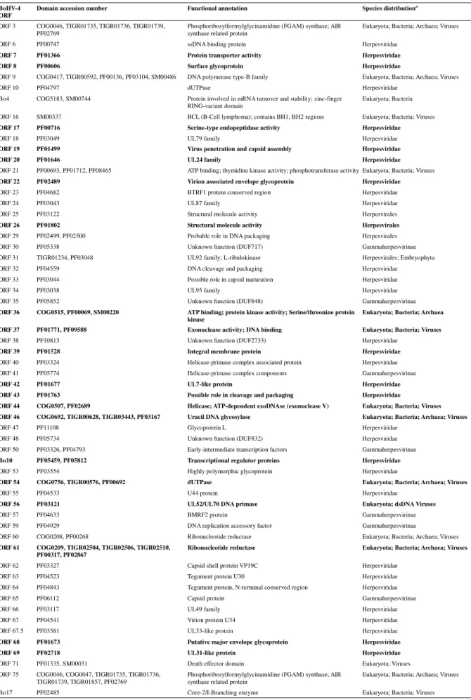
In order to develop an ab initio approach of gene annotation, we extracted all possible ORFs in all 6 frames from the complete genomic sequence of the BoHV-4 V.test strain. On each of these ORFs, we ran a Reverse PSI-BLAST [25] against all protein domains from the Conserved Domain Database [24]. ORFs containing an
evolutionarily conserved domain were defined as the smallest ORF containing the longest CDD match (see Methods). This approach revealed 59 ORFs containing a conserved CDD domain (Table 2). All 59 detected ORFs corresponded to ORFs
previously annotated in the 66-p-347 strain (on a total of 79 ORFs listed in Table 3), indicating that 75% of BoHV-4 ORFs contain conserved domains. Most of these ORFs (37/59) contain domains that are either conserved at different levels in the Herpesvirales (either gammaherpesvirinae, herpesviridae or herpesvirales), or at a much larger scale that include Eukaryota, Bacteria and Archaea (22/59) (Table 2). This second set of genes might bear good candidates for genes having been the stage of lateral gene transfer events as observed for several herpesvirus genes [36] such as the BoHV-4 Bo17 gene that encodes a homologue of the cellular core 2 beta -1,6-N-acetylglucosaminyl-transferase M [37]. These results will deserve further studies to identify the evolutionary history responsible for these observations.
Non-conserved protein-coding genes
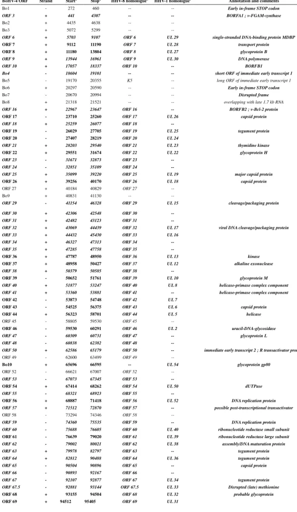
The remaining 20 annotated ORFs were determined by similarity to the 66-p-347 strain, and correspond for most of them to ORFs unique to BoHV-4 as described previously (Table 3) [11]. Some of these ORFs, however, contain odd characteristics that needed to be investigated (Fig. 2, Additional file 1 Figures S1-9). Indeed Bo1, Bo6, Bo7, Bo12 and Bo13 genes of the BoHV-4 V.test strain present in-frame STOP codons. Bo5 presents rather high divergency levels and large insertions/deletions (>5 % of its coding sequence as shown in Fig. 2) compared to the genomic sequence of the 66-p-347 strain. Moreover, ORFs 36, 67.5 and 75, which bear an evolutionary conserved domain, present late methionines compared to the 66-p-347 annotation. Indeed, in ORF 36 (see Additional file 1 Figures S5), the smallest ORF containing an evolutionary conserved domain is slightly shorter than the one annotated in 66-p-347 and there is no evidence that the previously annotated methionine is the correct one. However, comparison with homologous genes in other rhadinoviruses suggests that the start codon proposed in the 66-p-347 annotated sequence is the most likely. In ORF 67.5 (see Additional file 1 Figures S6), there is a point substitution in the
66-p-347 annotated ATG leading to the identification of a subsequent ATG as the V.test methionine. Finally, ORF 75 presents a small phase-disrupting indel in its 5' end (see Additional file 1 Figures S9), leading to the absence of the 66-p-347 annotated methionine in the V.test strain. All these annotated genes requested therefore an investigation of their transcription in mRNA products.
As these sequence properties could be specific to the BAC clone of the BoHV-4 V.test strain, we investigated the transcription of these genes on MDBK cells infected with the BoHV-4 V.test WT strain as described in the methods. The primers used are described in Table 1 and highlighted in Additional file 1. For all couple of primers, cDNA from BoHV-4-infected MDBK cells gave rise to the expected PCR products (Fig. 3). The absence of contaminant viral DNA in the mRNA preparations was confirmed by a lack of PCR product without reverse transcriptase. The size of the Bo5 RT-PCR product was also consistent with its known mRNA splicing (868 bp rather than 1140 bp). Moreover, the sequences of these RT-PCR products were in agreement with the BoHV-4 V.test sequence derived from our BAC cloned genome (data not shown). Therefore, we can conclude that all these coding sequences are transcribed during BoHV-4 infection of MDBK cells. However, further investigation is needed to determine the presence of proteins and ensure their accurate annotation.
BoHV-4 V.test replication origin
A large region containing the potential lytic replication origin (oriLyt) of the BoHV-4 66-p-347strain was determined by Zimmermann et al [11]. Based on this information, we mapped this region on the V.test genome (Fig. 1). This regioncontains Bo12, the R2b region and partially overlaps with Bo11. Compared to the 66-p-347 strain sequence, the corresponding region in the V.test genome is highly divergent (Fig. 4). Although this region shows high divergence rates, we expected the replication origin to be conserved between the two BoHV-4 strains. Previous work on other
herpesviruses has identified in oriLyt the presence of palindromic motifs essential for viral replication [38-40]. When we compared the potential region containing oriLyt in the two strains, a single conserved palindromic region was observed
(AATCCAGGCCCCTGATTGGTAGATTGCTGAAAGCCAATCAGGGGCCTGG ATT, Fig. 4). Interestingly, this region forms a perfect hairpin structure (Fig. 4B) that resembles DNA structures formed at other herpesvirus origins [41-42] and may therefore represent a common secondary structure used by all herpesvirus family members during the initiation of DNA replication. In the future, this structure will be tested as a candidate for an essential oriLyt replication motif.
BoHV-4 V.test polyrepetitive DNA
In the BAC clone, previous restriction profiles had determined a hypermolar prDNA band indicating that the BAC contained several prDNA units [12]. Therefore, the major pitfall in the assembly of the BoHV-4 V.test strain was the determination of the prDNA sequence. Indeed, (i) the higher per base coverage on this region due to repetition of prDNA units, (ii) the high GC content, along with (iii) the presence of several long repeats within the prDNA and (iv) the variability observed between prDNA units [14] made it extremely difficult to resolve and assemble with
pyrosequencing data alone. Interestingly, it has been shown for several rhadinoviruses that the left junction between the prDNA and the LUR is the site of genome
rearrangements and that sequences of the prDNA are found within the first base pairs of the LUR. These properties make this region very difficult to sequence [43-47]. Therefore, we adopted a hybrid strategy consisting in adding some ABI-Sanger reads (with the primers described in Table 1) to guide the 454 assembly on the prDNA region (see methods).
Bublot, et al. [14] described the different prDNA unit variants present in BoHV-4 V.test, and namely the differences between prDNA units. Firstly, the prDNA units
vary according to the number of repetitions of a ~200bp Pst-I bordered fragment. Secondly, the last prDNA before the prDNA/LUR junction (prDNA-G) displays a different ending than the inner prDNA units [14]. Our method allowed us to disentangle the repeats and to assemble a contig containing a whole prDNA unit (2,440bp) along with the left prDNA-LUR junction. This prDNA unit, corresponding to prDNA-G following Bublot et al. [14], was extracted from the contig and
annotated. A second contig from this hybrid assembly yielded the prDNA/prDNA junction. The presence of the prDNA/prDNA junction in our assembly confirmed the presence of at least two prDNA units in our BAC clone and allowed us to build a complete prDNA-inner unit (see Methods). The assembled prDNA-G and inner prDNA units have sizes of 2,440bp and 2,607bp respectively. Both these units are in agreement with their previously published restriction maps [14].
Specifically, we showed that, comparatively to the 66-p-347 strain, the V.test prDNA-inner unit presents several indels including two large indels in the repetitive PstI region (Fig. 5). This PstI-rich repetitive region seems to be the one presenting the most variation as it also presents comparatively large differences between prDNA units within the same strain. Indeed, Bublot et al. [14] roughly determined the size of the V.test major prDNA-inner unit to be around 2,650bp due to the presence of 4 repetitions of the two small PstI bordered fragments.
In the prDNA-G unit, we established that these two small PstI-bordered fragments make up a fragment of 186bp and that these are indeed repeated 4 times (Fig. 6). In the prDNA-inner unit, we determined that the last PstI-bordered fragment is actually a variation of the 186bp fragment where the inner Pst-I site is slightly modified (Fig. 6). Therefore, the rough 200bp size discripancy between the prDNA-G (2,440bp) and the prDNA-inner units (2,607bp) is due to the presence of a slightly modified repetition
of the previous segment. These results are compatible with the restriction profiles presented in Bublot et al. [14] as detailed by the positions of several restriction sites on Figure 6.
In addition to the variations in the PstI-bordered repetitions, one of the major differences between the prDNA-inner units and the prDNA-G lies in their 5' end. Indeed, the prDNA-inner contains a conserved pac-2 cleavage/packaging signal in its right terminal region, which is not the case of prDNA-G (Fig. 6). Both units however, possess a conserved pac-1 cleavage/packaging signal in their left terminal
region. Interestingly, the pac-1 and pac-2 cleavage and packaging signals show a good conservation between 66-p-347 and V.test's inner units, despite the presence of these signals in a repeated region bearing high divergence levels. Broll et al. [15] have determined, by transient cleavage/packaging assay, that a single prDNA unit is
sufficient for cleavage and packaging. However, from the absence of a conserved pac-2 motif in the prDNA-G, we suggest that, even if a single inner prDNA unit is indeed sufficient for cleavage and packaging, the prDNA-G alone would not suffice. This would therefore indicate that two prDNA units at least are necessary in the context of naturally occurring BoHV-4 genomes for correct cleavage and packaging.
The packaging of herpesvirus genomes is still not fully understood, however, detailed studies in herpes simplex virus type 1 (HSV-1), human and murine cytomegaloviruses (HCMV and MCMV) have highlighted the roles of the major conserved motifs and suggested the following general mechanism by which concatemers are cleaved and packaged [48-50]. Firstly, the T-box of the pac-2 signal is essential for the cleavage that initiates DNA packaging. Cleavage occurs at a fixed distance from the pac-2 T-box, and the resulting end that contains the pac-2 GC-box and other cis acting elements is inserted into the procapsid. Packaging is therefore directional and
proceeds from pac-2 towards the pac-1 terminus [48]. A second cleavage event, directed by pac-1, then terminates DNA packaging. If we apply this model to BoHV-4, the divergence of the pac-2 signal in prDNA-G, namely the absence of a T-box, indicates that it is not a functional pac-2 initiation signal. As the genome packaging is directional from pac-2 to pac-1 (therefore, from the right to the left end of the
genome), the lack of a pac-2 initiation signal in prDNA-G ensures that no packaging would lead to a remaining concatemer lacking a left end prDNA. This would therefore guarantee that genomes bearing at least one left and one right end prDNA unit are encapsulated into virions. This model and its implications will require further investigations in the future.
Conclusions
BAC-cloning of the BoHV-4 V.test strain has greatly facilitated the use of this virus as a model for human pathogenic gammaherpesviruses. However, until now, the complete genome sequence of this strain was unavailable. In this study, we have determined the complete genome sequence of the BoHV-4 V.test strain. In
comparison with the previously sequenced 66-p-347 strain, we identified important differences in 9 potential open reading frames. Moreover, sequence analyses allowed us to identify genome features that are potentially important for viral replication. All together, these results should have implications for the study of BoHV-4 and
Competing interests
The authors declare that they have no competing interests.
Authors' contributions
LP analyzed the data in silico, participated in the RT-PCR assay and drafted the manuscript. BM prepared the viral DNA, participated in data analysis and performed the RT-PCR assay. AV and CL participated in data analysis. LG analyzed the data and drafted the manuscript. All authors read and approved the final manuscript.
Acknowledgements
LP is supported by a post-doctoral fellowship from the University of Liège. BM, CL and LG are Research Fellows and Research Associate of the “Fonds de la Recherche Scientifique - Fonds National Belge de la Recherche Scientifique” (F.R.S. - FNRS), respectively. This work was supported by the following grants: starting grant (D-09/11) and GLYVIR ARC of the University of Liège and scientific impulse grant of the F.R.S. – FNRS n° F.4510.10.
References
1. Henle G, Henle W, Clifford P, Diehl V, Kafuko GW, Kirya BG, Klein G, Morrow RH, Munube GM, Pike P, et al: Antibodies to Epstein-Barr virus in
Burkitt's lymphoma and control groups. J Natl Cancer Inst 1969,
43:1147-1157.
2. Verma SC, Robertson ES: Molecular biology and pathogenesis of Kaposi
sarcoma-associated herpesvirus. FEMS Microbiol Lett 2003, 222:155-163.
3. Thorley-Lawson DA, Gross A: Persistence of the Epstein-Barr virus and
the origins of associated lymphomas. N Engl J Med 2004, 350:1328-1337.
4. Davison AJ, Eberle R, Ehlers B, Hayward GS, McGeoch DJ, Minson AC, Pellett PE, Roizman B, Studdert MJ, Thiry E: The order Herpesvirales. Arch Virol 2009, 154:171-177.
5. Thiry E, Bublot M, Dubuisson J, Van Bressem MF, Lequarre AS, Lomonte P, Vanderplasschen A, Pastoret PP: Molecular biology of bovine herpesvirus
type 4. Vet Microbiol 1992, 33:79-92.
6. Bartha A, Juhasz M, Liebermann H: Isolation of a bovine herpesvirus from
calves with respiratory disease and keratoconjunctivitis. A preliminary report. Acta Vet Acad Sci Hung 1966, 16:357-358.
7. Mohanty SB, Hammond RC, Lillie MG: A new bovine herpesvirus and its
effect on experimentally infected calves. Brief report. Arch Gesamte
Virusforsch 1971, 33:394-395.
8. Rossiter PB, Gumm ID, Stagg DA, Conrad PA, Mukolwe S, Davies FG, White H: Isolation of bovine herpesvirus-3 from African buffaloes
(Syncerus caffer). Res Vet Sci 1989, 46:337-343.
9. Dewals B, Thirion M, Markine-Goriaynoff N, Gillet L, de Fays K, Minner F, Daix V, Sharp PM, Vanderplasschen A: Evolution of Bovine herpesvirus 4:
recombination and transmission between African buffalo and cattle. J
Gen Virol 2006, 87:1509-1519.
10. Dewals B, Gillet L, Gerdes T, Taracha EL, Thiry E, Vanderplasschen A:
Antibodies against bovine herpesvirus 4 are highly prevalent in wild African buffaloes throughout eastern and southern Africa. Vet Microbiol
2005, 110:209-220.
11. Zimmermann W, Broll H, Ehlers B, Buhk HJ, Rosenthal A, Goltz M: Genome
sequence of bovine herpesvirus 4, a bovine Rhadinovirus, and
identification of an origin of DNA replication. J Virol 2001, 75:1186-1194.
12. Gillet L, Daix V, Donofrio G, Wagner M, Koszinowski UH, China B,
Ackermann M, Markine-Goriaynoff N, Vanderplasschen A: Development of
bovine herpesvirus 4 as an expression vector using bacterial artificial chromosome cloning. J Gen Virol 2005, 86:907-917.
13. Thiry E, Pastoret PP, Dessy-Doizé C, Hanzen C, Calberg-Bacq CM:
Herpesvirus in infertile bull's testicle. Vet rec 1981, 108:426.
14. Bublot M, Van Bressem MF, Thiry E, Dubuisson J, Pastoret PP: Bovine
herpesvirus 4 genome: cloning, mapping and strain variation analysis. J
Gen Virol 1990, 71 ( Pt 1):133-142.
15. Broll H, Buhk HJ, Zimmermann W, Goltz M: Structure and function of the
prDNA and the genomic termini of the gamma2-herpesvirus bovine herpesvirus type 4. J Gen Virol 1999, 80 ( Pt 4):979-986.
16. Margulies M, Egholm M, Altman WE, Attiya S, Bader JS, Bemben LA, Berka J, Braverman MS, Chen YJ, Chen Z, et al: Genome sequencing in
microfabricated high-density picolitre reactors. Nature 2005, 437:376-380.
17. Altschul SF, Gish W, Miller W, Myers EW, Lipman DJ: Basic local
alignment search tool. J Mol Biol 1990, 215:403-410.
18. Lomonte P, Bublot M, van Santen V, Keil GM, Pastoret PP, Thiry E: Analysis
of bovine herpesvirus 4 genomic regions located outside the conserved gammaherpesvirus gene blocks. J Gen Virol 1995, 76 ( Pt 7):1835-1841.
19. Chevreux B, Pfisterer T, Drescher B, Driesel AJ, Muller WE, Wetter T, Suhai S: Using the miraEST assembler for reliable and automated mRNA
transcript assembly and SNP detection in sequenced ESTs. Genome Res
2004, 14:1147-1159.
20. Ewing B, Green P: Base-calling of automated sequencer traces using
phred. II. Error probabilities. Genome Res 1998, 8:186-194.
21. Ewing B, Hillier L, Wendl MC, Green P: Base-calling of automated
sequencer traces using phred. I. Accuracy assessment. Genome Res 1998, 8:175-185.
22. Chou HH, Holmes MH: DNA sequence quality trimming and vector
removal. Bioinformatics 2001, 17:1093-1104.
23. Staden R, Beal KF, Bonfield JK: The Staden package, 1998. Methods Mol Biol 2000, 132:115-130.
24. Marchler-Bauer A, Anderson JB, Chitsaz F, Derbyshire MK, DeWeese-Scott C, Fong JH, Geer LY, Geer RC, Gonzales NR, Gwadz M, et al: CDD: specific
functional annotation with the Conserved Domain Database. Nucleic Acids
Res 2009, 37:D205-210.
25. Schaffer AA, Wolf YI, Ponting CP, Koonin EV, Aravind L, Altschul SF:
IMPALA: matching a protein sequence against a collection of
PSI-BLAST-constructed position-specific score matrices. Bioinformatics 1999, 15:1000-1011.
26. Larkin MA, Blackshields G, Brown NP, Chenna R, McGettigan PA,
McWilliam H, Valentin F, Wallace IM, Wilm A, Lopez R, et al: Clustal W
and Clustal X version 2.0. Bioinformatics 2007, 23:2947-2948.
27. Team RDC: R: A Language and Environment for Statistical Computing Vienna: R Foundation for Statistical Computing; 2010.
28. Charif D, Lobry J: SeqinR 1.0-2: a contributed package to the R project for statistical computing devoted to biological sequences retrieval and analysis. New York: Springer Verlag; 2007.
29. Cock PJ, Antao T, Chang JT, Chapman BA, Cox CJ, Dalke A, Friedberg I, Hamelryck T, Kauff F, Wilczynski B, de Hoon MJ: Biopython: freely
available Python tools for computational molecular biology and bioinformatics. Bioinformatics 2009, 25:1422-1423.
30. van Rossum G, Drake Jr F: The Python Language Reference Manual (version 2.5). Network Theory Ltd; 2006.
31. Machiels B, Lete C, de Fays K, Mast J, Dewals B, Stevenson PG,
Vanderplasschen A, Gillet L: Bovine Herpesvirus-4 Bo10 gene encodes a
non-essential viral envelope protein that regulates viral tropism through both positive and negative effects. J Virol 2010.
32. Spatz SJ, Rue CA: Sequence determination of a mildly virulent strain
(CU-2) of Gallid herpesvirus type 2 using 454 pyrosequencing. Virus Genes
33. Elsik CG, Tellam RL, Worley KC, Gibbs RA, Muzny DM, Weinstock GM, Adelson DL, Eichler EE, Elnitski L, Guigo R, et al: The genome sequence of
taurine cattle: a window to ruminant biology and evolution. Science 2009, 324:522-528.
34. Wicker T, Schlagenhauf E, Graner A, Close TJ, Keller B, Stein N: 454
sequencing put to the test using the complex genome of barley. BMC
Genomics 2006, 7:275.
35. Walz N, Christalla T, Tessmer U, Grundhoff A: A global analysis of
evolutionary conservation among known and predicted gammaherpesvirus microRNAs. J Virol 2010, 84:716-728.
36. Fu M, Deng R, Wang J, Wang X: Detection and analysis of horizontal gene
transfer in herpesvirus. Virus Res 2008, 131:65-76.
37. Vanderplasschen A, Markine-Goriaynoff N, Lomonte P, Suzuki M, Hiraoka N, Yeh JC, Bureau F, Willems L, Thiry E, Fukuda M, Pastoret PP: A
multipotential beta -1,6-N-acetylglucosaminyl-transferase is encoded by bovine herpesvirus type 4. Proc Natl Acad Sci U S A 2000, 97:5756-5761.
38. Portes-Sentis S, Sergeant A, Gruffat H: A particular DNA structure is
required for the function of a cis-acting component of the Epstein-Barr virus OriLyt origin of replication. Nucleic Acids Res 1997, 25:1347-1354.
39. Zhu Y, Huang L, Anders DG: Human cytomegalovirus oriLyt sequence
requirements. J Virol 1998, 72:4989-4996.
40. AuCoin DP, Colletti KS, Xu Y, Cei SA, Pari GS: Kaposi's
sarcoma-associated herpesvirus (human herpesvirus 8) contains two functional lytic origins of DNA replication. J Virol 2002, 76:7890-7896.
41. Rennekamp AJ, Wang P, Lieberman PM: Evidence for DNA hairpin
recognition by Zta at the Epstein-Barr virus origin of lytic replication. J
Virol 2010, 84:7073-7082.
42. Aslani A, Macao B, Simonsson S, Elias P: Complementary intrastrand base
pairing during initiation of Herpes simplex virus type 1 DNA replication.
Proc Natl Acad Sci U S A 2001, 98:7194-7199.
43. Russo JJ, Bohenzky RA, Chien MC, Chen J, Yan M, Maddalena D, Parry JP, Peruzzi D, Edelman IS, Chang Y, Moore PS: Nucleotide sequence of the
Kaposi sarcoma-associated herpesvirus (HHV8). Proc Natl Acad Sci U S A
1996, 93:14862-14867.
44. Searles RP, Bergquam EP, Axthelm MK, Wong SW: Sequence and genomic
analysis of a Rhesus macaque rhadinovirus with similarity to Kaposi's sarcoma-associated herpesvirus/human herpesvirus 8. J Virol 1999, 73:3040-3053.
45. Stamminger T, Honess RW, Young DF, Bodemer W, Blair ED, Fleckenstein B: Organization of terminal reiterations in the virion DNA of herpesvirus
saimiri. J Gen Virol 1987, 68 ( Pt 4):1049-1066.
46. Lagunoff M, Ganem D: The structure and coding organization of the
genomic termini of Kaposi's sarcoma-associated herpesvirus. Virology
1997, 236:147-154.
47. Albrecht JC, Nicholas J, Biller D, Cameron KR, Biesinger B, Newman C, Wittmann S, Craxton MA, Coleman H, Fleckenstein B, et al.: Primary
structure of the herpesvirus saimiri genome. J Virol 1992, 66:5047-5058.
48. Tong L, Stow ND: Analysis of herpes simplex virus type 1 DNA packaging
signal mutations in the context of the viral genome. J Virol 2010,
49. Wang JB, McVoy MA: A 128-Base-Pair Sequence Containing the pac1 and
a Presumed Cryptic pac2 Sequence Includes cis Elements Sufficient To Mediate Efficient Genome Maturation of Human Cytomegalovirus. J
Virol 2011, 85:4432-4439.
50. McVoy MA, Nixon DE, Hur JK, Adler SP: The ends on herpesvirus DNA
replicative concatemers contain pac2 cis cleavage/packaging elements and their formation is controlled by terminal cis sequences. J Virol 2000, 74:1587-1592.
Figures
Figure 1 - Map of the BoHV-4 V.test strain genome and divergence with the 66-p-347 strain sequence
The LUR of both strains have been aligned. Genome features are represented in the upper part as grey and red oriented arrows. Red arrows represent genes with an in-frame STOP codon, an early Methionine or a high divergence level in the V.test strain compared to 66-p-347. Dark (resp. light) grey arrows represent genes with (resp. without) an evolutionarily conserved domain (see Methods). The exons of spliced genes are indicated under the given gene as thin light-blue lines. Percent divergence is shown as a black-filled curve, percent insertions and deletions are shown as a blue-filled curve. Percent G+C content is shown as a thin green curve, with the mean G+C content drawn as a thin horizontal green line. These percentages are measured in a 100bp window sliding 3bp. Repeat regions (R1, R2a, R2b) are depicted as hatched areas. The oriLyt region is mapped as a light-grey area within R2b and the conserved quasi-palindromic motif in the oriLyt region is indicated by a small vertical arrow.
Figure 2 - Proteins in V.test : divergence with the previously published 66-p-347 strain
Percentage divergence and percentages indels on the aligned amino-acid sequences are represented as, respectively, black and light-blue bars. The dotted black line represents a 5% threshold. Genes containing an evolutionarily conserved domain (see Methods) are represented on a light-grey background. Previously annotated genes presenting in the V.test strain an in-frame stop codon, a late Methionine or large divergence levels compared to the 66-p-347 strain are indicated in red.
Figure 3 - RT-PCR amplification of the coding regions of the genes Bo1, Bo5, Bo6, Bo7, ORF67.5, Bo12, Bo13 and ORF75 of the BoHV-4 V.test strain
Subconfluent monolayers of MDBK cells were infected with BoHV4 V.test strain at a m.o.i. of 1 PFU/cell. 18 hours after infection, cytoplasmic RNA was extracted,
purified and treated for RT-PCR. The cDNA products were amplified by PCR using specific primers listed in Table 1
Figure 4 - Prediction of BoHV-4 V.test OriLyt.
A. Alignment of the V.test strain (above) and 66-p-347 strain (below) regions
predicted to contain the OriLyt in the 66-p-347 strain. The differences observed in the alignment are highlighted in light grey. The predicted potential OriLyt is highlighted in dark grey. B. The predicted secondary structures of the top (+) and bottom (–) strands of the predicted BoHV-4 OriLyt sequence were analyzed using the Vienna RNA website program RNAfold with DNA parameters. The predicted free energy ( G) of each structure is given, as well as the positional entropy of each nucleotide.
Figure 5 - The BoHV-4 inner prDNA units contain conserved cleavage/packaging signals
Alignment of the prDNA-inner units from V.test strain (above) and 66-p-347 strain (below). The differences observed in the alignment are highlighted in grey. The cleavage/packaging signals pac-1 and pac-2 are represented in boxes, and their composing C-rich, G-rich, GC-rich and T-rich units are indicated. PstI, EcoRI, SstII, BamHI restriction sites are represented in coloured font.
Figure 6 - The prDNA-G unit does not present a complete pac-2 cleavage/packaging signal
Alignment of the prDNA units from V.test strain (prDNA-inner above and prDNA-G below). The differences observed in the alignment are highlighted in grey. The cleavage/packaging signals pac-1 and pac-2 are represented in boxes, and their composing C-rich, G-rich, GC-rich and T-rich units are indicated. PstI, EcoRI, SstII, BamHI restriction sites (here PstI) are represented in coloured font. One modified PstI restriction site in the prDNA-inner is also highlighted to indicate the divergence between the fragments composing both units.
Additional file 1 - Alignments of the nucleotide and predicted amino acid sequences of Bo1 (Figure S1), Bo5 (Figure S2), Bo6 (Figure S3), Bo7 (Figure S4), ORF36 (Figure S5), ORF67.5 (Figure S6), Bo12 (Figure S7), Bo13 (Figure S8) and ORF75 (Figure S9) of BoHV-4 V.test and 66-p-347 strains
Nucleotide sequences aligned at the amino-acid level are represented for BoHV-4 V.test (red) and 66-p-347 strains (blue). Mismatching residues are highlighted in a shaded grey box. The predicted amino-acid sequences are respectively drawn for V.test and for 66-p-347 above and below the nucleotide sequences. The STOP codons are highlighted by small colored boxes. The annotated Methionine are highlighted in bold font. In the Bo5 sequence, introns are represented by boxes. Positions of the specific primers used in Fig. 3 are underlined.
Tables
Table 1. Primers used in this study
a
according to Genbank JN133502 sequence (BoHV-4 V.test strain long unique region)
b
according to Genbank JN133504 sequence (BoHV-4 V.test strain prDNA inner region)
c
according to Genbank AY665170.1 sequence (pBeloBAC modified)
name Sequence Coordinates according to
Genbank
Bo1 Fwd 5’- ATGGAGGGTGATGGATTCATG-3’ 460-440 a
Bo1 Rev 5’- TTAAGGCCTCATTCCAGGAAG-3’ 272-292 a
Bo5 Fwd 5’- GCTACAGAAAATGGCCAGTAAAG-3’ 20366-20342 a Bo5 Rev 5’- TCATGTCCTGAGTGGGTCTATG-3’ 19170-19191 a Bo6 Fwd 5’- ATGGTCATCCTAAATGCTCAAG -3’ 20297-20318 a Bo6 Rev 5’- TCACCTAGTGTTGCAACCCC -3’ 20497-20478 a Bo7 Fwd 5’- ATGGAGACAATTTCCATAAACTG -3’ 20994-20972 a Bo7 Rev 5’- CTAGCTGGGGTAGAGTGATC -3’ 20671-20690 a ORF67.5 Fwd 5’- ATGGCTGATGGTGATGTTTTAG -3’ 93144-93123 a ORF67.5 Rev 5’- TCAATGTTTGTCCAGAGCACT -3’ 92881-92901 a Bo12 Fwd 5’- ATGGGGGCGCTATTTGGGC -3’ 97442-97460 a Bo12 Rev 5’- TCAACTGATGAAACCCACCC -3’ 97525-97506 a Bo13 Fwd 5’- ATGCGTCTCGATGGCAAGC -3’ 98838-98856 a Bo13 Rev 5’- CTATGGTTGTTTTTTAAAGAAAATC -3’ 98981-98957 a ORF75 Fwd 5’- ATGTATCCCAGATACAGTAACA -3’ 103606-103585 a ORF75 Rev 5’- TTACATTTTATTTTTCAGACACCA -3’ 100274-100297 a prDNA Fwd 1 5’- GGAGCCCAAAACCAAAAGAG -3’ 870-889 b prDNA Rev 1 5’- CTCTTTTGGTTTTGGGCTCC -3’ 889-870 b prDNA Fwd 2 5’- CGTAGGCCTCACATTCAGC -3’ 908-926 b prDNA Rev 2 5’- GCTGAATGTGAGGCCTACG -3’ 926-908 b prDNA Fwd 3 5’- CGAGAGATGGTTCTTGCACA -3’ 940-959 b prDNA Rev 3 5’- TGTGCAAGAACCATCTCTCG -3’ 959-940 b BAC Rev 5’- TTGCCAATCCCAAAAAGAAG -3’ 9859-9878 c
Table 2 - Potential BoHV-4 V.test ORFs presenting conserved functional domains
BoHV-4 ORF
Domain accession number Functional annotation Species distributiona
ORF 3 COG0046, TIGR01735, TIGR01736, TIGR01739, PF02769
Phosphoribosylformylglycinamidine (FGAM) synthase; AIR synthase related protein
Eukaryota; Bacteria; Archaea; Viruses
ORF 6 PF00747 ssDNA binding protein Herpesviridae
ORF 7 PF01366 Protein transporter activity Herpesviridae ORF 8 PF00606 Surface glycoprotein Herpesviridae
ORF 9 COG0417, TIGR00592, PF00136, PF03104, SM00486 DNA polymerase type-B family Eukaryota; Bacteria; Archaea; Viruses
ORF 10 PF04797 dUTPase Herpesviridae
Bo4 COG5183, SM00744 Protein involved in mRNA turnover and stability; zinc-finger RING-variant domain
Eukaryota; Bacteria ORF 16 SM00337 BCL (B-Cell lymphoma); contains BH1, BH2 regions Eukaryota; Bacteria; Viruses
ORF 17 PF00716 Serine-type endopeptidase activity Herpesviridae
ORF 18 PF03049 UL79 family Herpesviridae
ORF 19 PF01499 Virus penetration and capsid assembly Herpesviridae
ORF 20 PF01646 UL24 family Herpesviridae
ORF 21 PF00693, PF01712, PF08465 ATP binding; thymidine kinase activity; phosphotransferase activity Eukaryota; Bacteria; Viruses
ORF 22 PF02489 Virion associated envelope glycoprotein Herpesviridae
ORF 23 PF04682 BTRF1 protein conserved region Herpesviridae
ORF 24 PF03043 UL87 family Herpesviridae
ORF 25 PF03122 Structural molecule activity Herpesvirales
ORF 26 PF01802 Structural molecule activity Herpesvirales
ORF 29 PF02499, PF02500 Probable role in DNA packaging Herpesvirales
ORF 30 PF05338 Unknown function (DUF717) Gammaherpesvirinae
ORF 31 TIGR01234, PF03048 UL92 family; L-ribulokinase Herpesvirales; Embryophyta
ORF 32 PF04559 DNA cleavage and packaging Herpesviridae
ORF 33 PF03044 Possible role in capsid maturation Herpesviridae
ORF 34 PF03038 UL95 family Herpesviridae
ORF 35 PF05852 Unknown function (DUF848) Gammaherpesvirinae
ORF 36 COG0515, PF00069, SM00220 ATP binding; protein kinase activity; Serine/threonine protein kinase
Eukaryota; Bacteria; Archaea
ORF 37 PF01771, PF09588 Exonuclease activity; DNA binding Eukaryota; Bacteria; Viruses
ORF 38 PF10813 Unknown function (DUF2733) Herpesviridae
ORF 39 PF01528 Integral membrane protein Herpesviridae
ORF 40 PF03324 Helicase-primase complex associated protein Herpesviridae ORF 41 PF05774 Helicase-primase complex components Gammaherpesvirinae
ORF 42 PF01677 UL7-like protein Herpesviridae ORF 43 PF01763 Possible role in cleavage and packaging Herpesviridae
ORF 44 COG0507, PF02689 Helicase; ATP-dependent exoDNAse (exonuclease V) Eukaryota; Bacteria; Viruses ORF 46 COG0692, TIGR00628, TIGR03443, PF03167 Uracil DNA glycosylase Eukaryota; Bacteria; Archaea; Viruses
ORF 47 PF11108 Glycoprotein L Herpesviridae
ORF 48 PF05734 Unknown function (DUF832) Herpesviridae
ORF 50 PF03326, PF04793 Early-intermediate transcription factors Gammaherpesvirinae
Bo10 PF05459, PF05812 Transcriptional regulator proteins Herpesviridae
ORF 53 PF03554 Highly polymorphic glycoprotein Herpesviridae
ORF 54 COG0756, TIGR00576, PF00692 dUTPase Eukaryota; Bacteria; Archaea; Viruses
ORF 55 PF04533 U44 protein Herpesviridae
ORF 56 PF03121 UL52/UL70 DNA primase Eukaryota; dsDNA Viruses
ORF 57 PF04633 BMRF2 protein Gammaherpesvirinae
ORF 59 PF04929 DNA replication accessory factor Gammaherpesvirinae
ORF 60 COG0208, PF00268 Ribonucleotide reductase Eukaryota; Bacteria; Archaea; Viruses
ORF 61 COG0209, TIGR02504, TIGR02506, TIGR02510, PF00317, PF02867
Ribonucleotide reductase Eukaryota; Bacteria; Archaea; Viruses
ORF 62 PF03327 Capsid shell protein VP19C Herpesviridae
ORF 63 PF04523 Tegument protein U30 Herpesviridae
ORF 64 PF04843 Tegument protein, N-terminal conserved region Herpesviridae
ORF 65 PF06112 Capsid protein Gammaherpesvirinae
ORF 66 PF03117 UL49 family Herpesviridae
ORF 67 PF04541 Virion protein U34 Herpesviridae
ORF 67.5 PF03581 UL33-like protein Herpesviridae
ORF 68 PF01673 Putative major envelope glycoprotein Herpesviridae ORF 69 PF02718 UL31-like protein Herpesviridae
ORF 71 PF01335, SM00031 Death effector domain Eukaryota; Viruses
ORF 75 COG0046, COG0047, TIGR01735, TIGR01736, TIGR01739, TIGR01857, PF02769
Phosphoribosylformylglycinamidine (FGAM) synthase; AIR synthase related protein
Eukaryota; Bacteria; Archaea; Viruses
Conserved functional domains were determined for each BoHV-4 V.test ORF using METHOD and the domains accession numbers, functional annotation and the species distribution were listed. When several domains were conserved, the annotations were either merged when possible or juxtaposed. Domains present in herpesviridae conserved gene families are highlighted in bold (from Fu, 2008). (PFxxx: Pfam Accession Number; SMxxx: SMART Accession Number; COGxxx: COG Accession Number; TIGRxxx: TIGRFam Accession Number).
a
Table 3 - Potential BoHV-4 V.test ORFs and homologues to HHV-8 and HHV-1 BoHV-4 ORF Strand Starta
Stopa
HHV-8 homologueb
HHV-1 homologuec
Annotation and comments
Bo1 - 272 460 -- -- Early in-frame STOP codon
ORF 3 + 441 4307 -- -- BORFA1 ; v-FGAM-synthase
Bo2 + 4435 4638 -- --
Bo3 + 5072 5299 -- --
ORF 6 + 5703 9107 ORF 6 UL 29 single-stranded DNA-binding protein MDBP ORF 7 + 9112 11190 ORF 7 UL 28 transport protein
ORF 8 + 11180 13804 ORF 8 UL 27 glycoprotein B
ORF 9 + 13944 16961 ORF 9 UL 30 DNA polymerase
ORF 10 + 17057 18337 ORF 10 -- BORFB1
Bo4 - 18604 19101 -- -- short ORF of immediate early transcript 1
Bo5 - 19170 20355 K5 -- long ORF of immediate early transcript 1
Bo6 + 20297 20590 -- -- Early in-frame STOP codon
Bo7 - 20670 20994 -- -- Disrupted frame
Bo8 + 21318 21521 -- -- overlapping with late 1.7 kb RNA
ORF 16 + 22967 23647 ORF 16 -- BORFB2 ; v-Bcl-2 protein
ORF 17 - 23710 25260 ORF 17 UL 26 capsid protein
ORF 18 + 25259 26077 ORF 18 --
ORF 19 - 26029 27705 ORF 19 UL 25 tegument protein ORF 20 - 27407 28219 ORF 20 UL 24
ORF 21 + 28203 29540 ORF 21 UL 23 thymidine kinase
ORF 22 + 29551 31674 ORF 22 UL 22 glycoprotein H
ORF 23 - 31671 32873 ORF 23 --
ORF 24 - 32851 35109 ORF 24 --
ORF 25 + 35099 39220 ORF 25 UL 19 major capsid protein
ORF 26 + 39256 40170 ORF 26 UL 18 capsid protein
ORF 27 + 40184 40829 ORF 27 --
Bo9 + 40831 41130 -- --
ORF 29 - 41154 46328 ORF 29 UL 15 cleavage/packaging protein
ORF 30 + 42306 42548 ORF 30 --
ORF 31 + 42482 43123 ORF 31 --
ORF 32 + 43069 44439 ORF 32 UL 17 viral DNA cleavage/packaging protein
ORF 33 + 44432 45430 ORF 33 UL 16
ORF 34 + 46327 47313 ORF 34 --
ORF 35 + 47285 47758 ORF 35 --
ORF 36 + 47787 48950 ORF 36 UL 13 kinase ORF 37 + 48958 50427 ORF 37 UL 12 alkaline exonuclease
ORF 38 + 50379 50585 ORF 38 --
ORF 39 - 50652 51761 ORF 39 UL 10 glycoprotein M
ORF 40 + 51877 53247 ORF 40 UL 8 helicase-primase complex component
ORF 41 + 53360 53881 ORF 41 -- helicase-primase complex component
ORF 42 - 53873 54748 ORF 42 UL 7
ORF 43 - 54525 56375 ORF 43 UL 6 capsid protein ORF 44 + 56323 58701 ORF 44 UL 5 helicase
ORF 45 - 58805 59530 ORF 45 --
ORF 46 - 59530 60291 ORF 46 UL 2 uracil-DNA-glycosidase
ORF 47 - 60309 60731 ORF 47 -- glycoprotein L
ORF 48 - 60838 62382 ORF 48 --
ORF 50 + 62586 65179 ORF 50 -- immediate early transcript 2 ; R transactivator protein
ORF 49 - 62600 63499 ORF 49 --
Bo10 + 65696 66595 -- UL 54 glycoprotein gp80
ORF 52 - 66621 67007 ORF 52 --
ORF 53 - 67073 67345 ORF 53 --
ORF 54 + 67414 68262 ORF 54 UL 50 dUTPase
ORF 55 - 68321 68923 ORF 55 --
ORF 56 + 68887 71418 ORF 56 UL 52 DNA replication protein ORF 57 + 71512 72870 ORF 57 -- possible post-transcriptional transactivator
ORF 58 - 73294 74346 ORF 58 --
ORF 59 - 74360 75535 ORF 59 -- DNA replication protein
ORF 60 - 75688 76605 ORF 60 UL 40 ribonucleotide reductase small subunit ORF 61 - 76639 79020 ORF 61 UL 39 ribonucleotide reductase large subunit
ORF 62 - 79002 80021 ORF 62 UL 38 assembly/DNA maturation protein
ORF 63 + 79978 82797 ORF 63 -- tegument protein
ORF 64 + 82812 90488 ORF 64 UL 36 tegument protein
ORF 65 - 90504 90896 ORF 65 -- capsid protein
ORF 66 - 90893 92167 ORF 66 --
ORF 67 - 92107 92877 ORF 67 UL 34 tegument protein
ORF 67.5 - 92881 93144 ORF 67.5 UL 33 Disrupted (late) methionine
ORF 68 + 93155 94504 ORF 68 UL 32 probable glycoprotein ORF 69 + 94512 95405 ORF 69 UL 31
Bo11 - 96158 96703 -- --
Bo12 + 97442 97684 -- -- Early in-frame STOP codon
ORF 71 - 98328 98876 K13/ORF 71 -- BORFE2 ; v-FLIP
Bo13 + 98838 98983 -- -- Disrupted frame
ORF 73 - 99022 99783 ORF 73 -- BORFE3
ORF 75 - 100274 103606 ORF 75 -- Disrupted (late) methionine ; tegument protein/v-FGAM-synthetase
Bo14 - 104273 104785 -- --
Bo15 - 105724 106038 -- --
Bo16 + 106225 106494 -- --
Bo17 + 106681 108003 -- -- viral beta-1,6-N-acetylglucosaminyltransferase
a
Positions of the respective ORFs of BoHV-4 on the LUR sequence. These are given from the first nucleotide of the start codon ATG to the last nucleotide of the stop codon.
b
Correspondance to HHV-8 genes is according to Zimmermann (2001).
c
Correspondance to HHV-1 genes was based on the presence of conserved domain with a BoHV-4 gene. Genes containing evolutionary conserved domains are highlighted in bold italic. The genes conserved in Herpesviridae are highlighted in bold (from Fu, 2008).
Additional files provided with this submission:
Additional file 1: Additional file 1.pdf, 7440K