Molecular Dynamics Laboratory – Professor B. LEYH
Center for Education and Research on Macromolecules
CERM – Professor C. JEROME
Stimuli-responsive self-assembled
macromolecular systems with potential drug
delivery applications: structural characterization
by Small-Angle Neutron Scattering
Academic year 2015-2016
Dissertation presented by
Arnaud Joset
To obtain the grade of
Doctor in Sciences
UNIVERSITY OF LIÈGE
Faculty of Sciences
stimuli à applications biomédicales potentielles: caractérisation
structurale par diffusion neutronique aux petits angles
Par Arnaud Joset
Résumé : Les colloïdes sont connus depuis le début du 19ème siècle mais ils ont suscité un regain d’intérêt durant les vingt dernières années, notamment grâce à leur utilisation dans des applications biomédicales ou la création de nouveaux matériaux.
L’objectif de cette thèse est l’étude d’échantillons colloïdaux sensibles à un ou plusieurs stimuli (pH, température, ligand, ions métalliques) par diffusion neutronique aux petits angles (SANS). Cette technique permet d’obtenir des informations sur la structure interne des objets mais aussi, dans certaines conditions, sur l’organisation des nano-objets en solution. À cette fin, plusieurs échantillons, formés par l’auto-assemblage de copolymères séquencés et de liposomes, ont été préparés. Une modélisation des sections efficaces a été menée afin de calculer l’évolution des principaux paramètres structuraux : taille, polydispersité, structure et volumes occupés par les différents composants hydrophiles et hydrophobes au sein des nano-objets. Les mesures SANS ont été réalisées en fonction de l’intensité du stimulus afin de quantifier l’évolution de ces paramètres.
La première partie expérimentale de ce travail porte sur l’analyse d’échantillons micellaires formés à partir de copolymères séquencés. Les séquences peuvent être biodégradables (ex. poly(ε-caprolactone)), biocompatibles (ex. poly(oxyde d’éthylène)), sensibles au pH (ex. poly(2-vinylpyridine), poly(acide acrylique)) ou à la température (ex. poly(N-isopropylacrylamide)). L’influence de la concentration et du pontage entre micelles a été étudiée via l’analyse de gels formés à partir de polystyrene-b-poly(tert-butylacrylate) PS-b-PtBA-tpy (-tpy: terpyridine) en présence d’ions métalliques.
La seconde partie expérimentale consiste en l’analyse de liposomes en présence d’une quantité croissante de β-cyclodextrine méthylée aléatoirement (RAMEB). Cette partie est divisée en deux chapitres. Le premier traite du cas de liposomes majoritairement composés de dimyristoylphosphatidylcholine (DMPC) et le deuxième traite de liposomes de DMPC dopés à l’aide de 30% de cholestérol.
potential drug delivery applications: structural characterization by
Small-Angle Neutron Scattering
By Arnaud Joset
Abstract: Colloids are known since the early 19th century but they have mainly sparked interest since the last few decades thanks to their use in biomedical applications or in the design of new materials.
The aim of this thesis is the study of stimuli-responsive colloids by Small-Angle Neutron Scattering (SANS). The stimuli may be the pH, the temperature, the addition of a ligand or of metallic ions or a combination of them. SANS is a useful technique which provides information about the internal structure of nano-objects but also, if appropriate conditions are met, about the organization of the objects in solution. Several samples built from the auto-assembly of block copolymers and liposomes have been prepared. The macroscopic cross sections have been modeled with the aim to infer the main structural parameters of the samples: the global size, the polydispersity, the structure and volume occupied by the hydrophilic and hydrophobic components inside the nano-objects. The SANS measurements have been performed as a function of the intensity of the stimulus, in order to quantify the evolution of the structural parameters.
The first experimental part focuses on micellar samples built from sequenced block copolymers. The blocks may be biodegradable (e.g., poly(ε-caprolactone)), biocompatible (e.g., poly(ethylene oxide)), pH-sensitive (e.g., poly(2 vinylpyridine), poly(acrylic acid)) or temperature-sensitive (e.g., poly(N-isopropylacrylamide)). The influence of the concentration and the formation of bridges between micelles have been investigated through the analysis of metallo-supramolecular micellar gels resulting from the self-assembling of polystyrene-block-poly(tert-butylacrylate) PS-b-PtBA-tpy (tpy stands for terpyridine) block copolymers in the presence of transition metal ions.
The second part focuses on the analysis of liposomes interacting with an increasing concentration of Randomly Methylated β-cyclodextrins (RAMEB). This part is divided into two chapters. The first one deals with liposomes mainly composed of dimyristoylphosphatidylcholine (DMPC) and the second one investigates the effects of cholesterol doping on the same DMPC liposomes interacting with RAMEB.
Bernard Leyh qui a permis la réalisation de ce travail. Sans sa confiance et ses conseils, ce travail n’aurait pas pu aboutir. Merci pour sa disponibilité, la justesse de ses remarques, ainsi que l’attention qu’il porte aux détails.
Ce travail est le fruit de la collaboration entre deux services et je tiens également à remercier le Professeur Christine Jérôme qui m’a accueilli chaleureusement au sein de son service. Malgré un emploi du temps surchargé, elle a pris le temps d’écouter mes remarques et questions et d’y répondre de manière décontractée.
Mes plus vifs remerciements vont également au Professeur Jean-François Gohy, au docteur Charles André Fustin ainsi qu’au Docteur Clément Mugemana de l’Université Catholique de Louvain pour notre collaboration sur les gels micellaires supramoléculaires.
Merci au Docteur Julian Oberdisse, au Professeur Jean-François Gohy, ainsi qu’au Docteur Cédric Gommes, de me faire l’honneur d’être membre de mon jury de thèse mais également au Professeur Anne-Sophie Duwez d’avoir accepté la présidence de ce jury.
Un volet important de ce travail consistait à analyser des échantillons de liposomes. Cette analyse a été réalisée conjointement avec Angeliki Grammenos. Merci pour tes conseils, les moments de franche rigolade lorsque ces lipos nous rendaient fous, ton humour ainsi que les nombreuses découvertes musicales. Tu m'as aussi présenté ton formidable mari et comme tu le disais dans tes propres remerciements, c’est plus des amis que des collègues que j’ai rencontrés.
beaucoup de temps à analyser les échantillons de triblocs. Les expériences de diffusion à Saclay ainsi que les réunions tardives dans les inondations resteront d’excellents souvenirs.
Cette thèse a été réalisée en coopération avec le CERM. Je tiens également à remercier les personnes qui font que ce service fonctionne aussi bien. Merci à Philippe, Christophe, Antoine, Valérie, Charlotte, Martine, Florence, Stéphanie, Bruno, Raphaël et à tous les autres pour leur bonne humeur, leur dynamisme et leur aide.
Je remercie également le Professeur Jean-François Focant ainsi que les assistants et préparateurs avec qui j’ai eu l’occasion de travailler dans le cadre du volet pédagogique de mon mandat : Grégory, Perrine, Armélinda, Thierry, Nicolas, Marie, Antoine Pierre-Hugues et Romain. Merci à vous pour les nombreux bons moments durant les réunions pour examens et les TP.
Toute ma sympathie va à Kathy et Véronique, mes chère voisines de bureau. Je suis très heureux que le hasard nous ai placé dans le même local.
Toute ma gratitude va à Enza, Stéphanie, Marie-Noëlle et Sophie pour leur disponibilité et de l’aide qu’elles m’ont apportée.
Je tiens également à remercier chaleureusement le professeur Robert Locht. Il est vrai que nos domaines de prédilection sont bien différents mais sa bonne humeur, ses conseils avisés et sa (longue rajouterait-il) expérience m’ont été utiles à plus d’une reprise. Nous nous soutenions dans les
un spectromètre muet pour l’un et des ajustements numériques capricieux pour l’autre. Néanmoins, les difficultés ont passé. Il m’a également été d’un grand secours lors de la correction de mes soumissions en anglais. Son aimable contribution m’a grandement aidé à l’avancement et je ne saurais le remercier suffisamment.
Merci également à ma famille pour son soutien, ses marques d’attention et sa confiance qu’elle m’a manifesté tout au long de mes études et de ce travail. Je n’oublie pas non plus pas mes amis qui m’ont aidé à relativiser (en général autour d’un verre) lorsque les résultats n’étaient pas au rendez-vous.
Je remercie également Lalita pour ses encouragements, ses attentions, son optimisme à mon égard et son amour. Merci d’être là et de me renforcer par tes mots et tes attentions.
“The only real failure is the failure to try, and the measure of success is how we cope with disappointment.” ― Deborah Moggach, The Best Exotic Marigold Hotel
Chapter I: Introduction ... 1
Context and aim ... 5
References ... 8
Chapter II: Small-angle neutron scattering ... 13
1 Introduction to neutron properties ... 15
2 Why using small angle scattering? ... 16
3 Scattering of X-rays or neutrons by a target ... 17
3.1 Incoming and outgoing wave functions ... 17
3.2 Differential scattering cross section ... 20
4 Small Angle Neutron Scattering (SANS) ... 24
5 Form and Structure factors for micellar and liposomes solutions ... 25
5.1 Scattering by one-component (quasi) continuous sample ... 27
5.2 Scattering from incompressible multi-component systems ... 30
5.3 Contribution of the interactions between the nano-objects ... 33
6 Influence of the polydispersity of the self-assembled objects ... 37
7 Experiment ... 39
8 Form factors of common nano-objects ... 43
9 A few examples from the literature ... 46
10 Molecular Dynamics Simulation ... 51
11 Summary... 52
1 IntroductionChapitre d'équation 3 Section 1 ... 59
2 Materials and methods ... 62
2.1 Copolymer synthesis ... 62
2.2 Micellization of diblock micelles ... 62
2.3 Small-Angle Neutron Scattering ... 63
3 Models ... 64
3.1 Model 1: PCL core, P2VP homogenous shell and PEO Gaussian chains ... 64
3.2 Model 2: PCL core,mixed P2VP and PEO Gaussian chains ... 66
3.3 Model 3: PCL core, P2VP rods, and PEO Gaussian chains ... 67
3.4 Fitting procedures ... 68
4 Comparison of SANS and DLS size distributions ... 68
5 Results and discussion ... 69
6 Conclusions ... 78
7 References ... 80
Chapter IV: SANS study of pH- and thermo-responsive micelles: from block-copolymers to aggregates ... 85
1 Introduction ... 87
2 Materials and methods ... 90
2.1 Materials ... 90
2.2 Synthesis ... 90
2.2.1 Synthesis of PEO-b-PNIPAM-b-PAA block copolymer ... 91
2.2.2 Preparation of Triblock Copolymer solutions ... 91
2.3 Small-Angle Neutron Scattering (SANS) ... 92
3 Results and discussion ... 101
3.1 Acidic conditions (pH < pKa,PAA) ... 103
3.2 Neutral and slightly basic conditions (pH > pKaPAA) ... 107
4 Conclusions ... 109
5 References ... 111
Chapter V: Structure of Metallo-Supramolecular Micellar Gels ... 115
1 Introduction ... 117
2 Experimental Section ... 119
2.1 Instrumentation ... 119
2.2 Synthesis of terpyridine end-functionalized poly(tert-butylacrylate) macro-initiator PtBA180-tpy ... 120
2.3 Preparation of micellar gels ... 121
3 Results and Discussion ... 122
4 Conclusion ... 136
5 References ... 138
Chapter VI: Investigation of the interaction between a cyclodextrin and DMPC liposomes: a small angle neutron scattering study. ... 141
1 Introduction ... 143
2 Materials and methods ... 145
2.1 Liposome preparation ... 145
2.2 Surface Tension Measurements... 146
2.3 Dynamic Light Scattering (DLS) ... 146
2.4 Small-Angle Neutron Scattering (SANS) ... 146
2.5 SANS Data handling ... 147
3.1 Fraction of DMPC molecules included in the unilamellar liposomes ... 153
3.2 Liposome Radius, aggregation Number, and polydispersity ... 156
3.2.1 Below the DMPC transition temperature ... 159
3.2.2 Above the DMPC transition temperature... 159
3.2.3 Comparison between the behaviors below and above the transition temperature ……….160
3.2.4 Coverage of the liposome external layer by RAMEB ... 161
3.3 Bilayer thickness ... 162
4 Conclusions ... 165
5 References ... 166
Chapter VII: Small-Angle Neutron Scattering investigation of cholesterol-doped DMPC liposomes interacting with β-cyclodextrin ... 171
1 Introduction ... 173
2 Materials and methods ... 175
2.1 Liposome preparation ... 175
2.2 SANS measurements... 175
2.3 SANS data analysis ... 176
2.4 Dynamic Light Scattering Measurements (DLS) ... 177
3 Results and discussion ... 178
3.1 Fraction of molecules included into unilamellar liposomes ... 179
3.2 Liposome Radius and Aggregation Number ... 180
3.3 Polydispersity of the liposomes ... 183
3.4 Coverage of the outer liposome interface by RAMEB ... 185
3.5 Bilayer thickness ... 186
Chapter VIII: General conclusions and perspectives ... 193
Chapter IX: Appendices ... 203
Appendix I: Supporting Information of chapter IV ... i
1.1 Contribution of the core ... iii
1.2 Contribution of the Gaussian chains of the corona ... iv
1.3 Contribution of the rods ... iv
1.4 Interference term between the core and the Gaussian chains ... v
1.5 Interference terms between two Gaussian chains ... vi
1.6 Interference term between the core and the rods... vi
1.7 Interference terms between the rods ... vii
1.8 Interference terms between a Gaussian chains and a rod ... viii
1.9 Scattering cross section of a micelle population ... ix
Appendix II : Contribution of the aggregates... xi
Ac Area per chain at the core-corona interface
b Kuhn length of a polymer
j Excess scattering length of the scatterer j
bj Scattering length of the scatterer j
CD Cyclodextrin CHOL Cholesterol
D Average thickness of the hydrophobic part of a liposome membrane
d Average thickness of the hydrophilic part of a liposome membrane DLS Dynamic Light Scattering
DMF Dimethylformamide
DMPC Dimyristoylphosphatidylcholine dΣ(q)/dΩ Macroscopic scattering cross section dσ(q)/dΩ Differential scattering cross section
⃗ Wave vector
L Effective length of a chain considered as a rigid rod LCST Lower Critical Solution Temperature
m Fraction of the phosphatidylcholine molecules included in liposomes.
Nch Number of chains
Nm Aggregation number
P(q) Form Factor
P2VP poly(2-vinylpyridine) PAA Poly(acrylic acid) PCL poly(ε-caprolactone) PEO Poly(ethylene oxide)
PNIPAM Poly(N-isopropylacrylamide) PPO Poly(propy1ene oxide) PS Polystyrene
PtBA Poly(tert-butylacrylate) ⃗ Scattering vector
RAMEB Randomly Methylated β-Cyclodextrin Rc Radius of the core (micelles)
Rg Radius of gyration
⃗j Position vector of the scatterer j
S(q) Inter-particle structure factor SA Stearylamine
V Volume of the sample vj Volume of the scatterer j
z Degree of polymerization Γ Nano-object size distribution κT Isothermal compressibility
ρj Scattering length density j Excess scattering length density
CHAPTER I
3
Even if colloids have been used for many centuries, their scientific investigation started in the early 19th century. The word “colloid” is derived from the Greek word “kolla” which means glue with the suffix “oid” meaning “of similar form to”. Before 1830, Brown [1] observed them using a microscope and described their random motion which was later named in his honor. Between 1850 and 1852, Sobrero [2] and Selmi [3,4] studied inorganic colloids made of silver chloride and Prussian blue, a pigment consisting of iron cyanide complexes. Later, Graham [5,6] managed to isolate some colloids with a dialysis system and published his results during the 1860s. He showed that colloid systems cannot diffuse through semi-permeable membranes. He discovered the ripening process which explains the growth of droplets through the continuous phase. In the beginning of the 20th century, the progress in chemistry and physics about atomic structure, diffusion processes, electrophoresis and light scattering favored the development of the colloid studies. Ostwald [7], Buzagh [8] and Hauser [9] worked for the promotion of colloid science while most of their colleagues still considered this field as a side branch of physical chemistry. In this context Ostwald wrote [10,9] in 1914 “Die Welt der vernachlässigten Dimensionen” (The world of neglected dimensions) and fought for the use of “colloid science” against “colloid chemistry”. Today colloidal solutions are well known and extremely common in everyday life applications like paintings, inks, drugs, gelatin or food. Furthermore, many “smart” stimuli-responsive colloids have been developed during the last decades. The improvement of analysis and optical instruments has contributed to the development of this quite recent field. Colloidal dispersions are composed of nanoparticles and their building bricks have sizes of a few nanometers up to the micrometer range for the aggregates. Figure 1 shows three colloidal nanostructures architectures which will be considered in the present work i.e. polymeric micelles, nanogels and liposomes.
Micelles are spherical self-assemblies of amphiphilic molecules formed to decrease the surface tension. A common example is the self-assembly of fatty acids whose properties have been known for centuries. The first evidence of soap use is from Babylonian times and oil, soap and scrappers were commonly used in ancient Greek and Roman times. Surfactants have been used for ages to stabilize emulsions like Hollandaise Sauce for which the first references can be found already in 1593 and 1651 [11,12]. The cleaning properties of surfactants are also known for ages but the advent of nanotechnology has promoted them to the forefront during the two last decades. The understanding of the nature of micellar solutions has only started in the beginning of the 20th century. One of the precursors is McBain [13,14] who played a role in the study of surfactants, micellisation processes, their composition, charge and mobility.
4
Figure 1 a) micelle formation from block-copolymers b) micellar gel c) liposome
When the surfactant concentration is higher than the critical micelle concentration (or CMC), the micellisation process takes place. In a polar solvent, the hydrophobic tails assemble to form the core and the hydrophilic tails form the corona toward the solvent. Nowadays micelles, liposomes and other soft matter material are extensively used in many fields [15] like thickening agents in water-based paints [16], cleaning agents in the restauration of art pieces [17] or to produce new mesoporous materials [18,19]. But the largest number of publications are devoted to possible nano-medicine applications. They are investigated to solubilize poorly soluble drugs in aqueous medium. [20-26]. The drug is encapsulated into the hydrophobic core of micelles which are designed to release it near the targeted sick tissues. The corona composition is designed to allow biocompatibility with plasma and to increase the selectivity while decreasing the drawback effects [27,28,20,29-34]. Such systems have complex structures in order to achieve their task e.g. access a tumor without being caught by the immune system [35-38,25,39-41,24,26].
Liposomes are spherical vesicles which have at least one lipid bilayer. They were discovered during the 1960’s by Bangham et al [42]. When liposomes are made of several concentric lipid bilayers, they are called Multilamellar Large Vesicles (MLV). Their size may vary from 100 nm up to several micrometers. On the other side, the Small Unilamellar Vesicle (SUVs) are smaller than 100 nm.
5
Liposomes are used in a large variety of applications like drug delivering structure [43-45,38] and as models of biological membranes because of their similar composition but without organelles,which simplifies the data interpretation. [46].
In 2012, 53 systems based on liposomes were investigated as therapeutic treatment and 7 were already commercially available. Moreover, 8 systems based on micelles were investigated and 3 were commercially available [26]. Nowadays, the nano-objects are extensively studied but their size range and atomic content make it difficult to access their inner structure with the help of usual technique widely available like dynamic light scattering (DLS), atomic force microscopy or electron microscopy [28,35-37,39,47-49,25,50-52]. Nowadays the nano-object are expected to undergo structural changes under controlled stimuli i.e., modification of the temperature, pH change, or the presence of a triggering molecule. The stimuli-responsive behavior may allow the nano-object to target sick tissues. The structural change may result in the disruption of the objects, their aggregation or their reorganization. The study of the inner structure is challenging and the common techniques to study soft matter i.e. transmission microscopy or light scattering does not allow to isolate each component of the colloid. Small Angle Neutron Scattering is a helpful technique in this context because, coupled to a tailored modelling, it gives information on the organization of amphiphilic molecules inside the objects. Furthermore the measurements are performed in solution and dynamic processes can be studied over periods of several hours or days. The aim of this thesis is to study several stimuli-responsive colloids and the evolution of their internal structure when the intensity of the stimulus is tuned.
Context and aim
The synthesis of smart nanomaterials and their analysis with the help of SANS are tasks requiring distinctive but complementary skills. The birth of this project with the partnership of the Molecular Dynamics Laboratory and the Center for Education and Research on Macromolecules (CERM) was an excellent opportunity to study samples which have already potential use or which are good candidates in their area but lack some structural information.
This manuscript is divided into three parts. The first part (see Chapter 2) introduces SANS in order to (i) describe the general experimental and theoretical framework, (ii) enlighten the advantages of this technique in the study of soft matter samples and outline its peculiarities and (iii) introduce some models which are instrumental to our work.
6
In a second part (chapters 3 to 7) the experimental results are set out and discussed. In chapter 3, the effect of pH stimuli is assessed on polymeric micelles bearing polybase sequences. The studied objects are built from mixtures of two diblock copolymers, a polycaprolactone-b-poly(ethylene oxide) one and a polycaprolactone-b-poly(2-vinylpyridine) one. The hydrophobic core is built from polycaprolactone (PCL). The corona of hydrophilic poly(ethylene oxide) (PEO) stabilizes the nanocarriers. Thanks to a pH-sensitive sequence of poly(2-vinylpyridine) (P2VP), the micelles can adopt different structures in acidic and basic conditions. When the pH is acidic, the P2VP is protonated and the chains are repulsive. The diffusion of a hydrophobic drug would be facilitated in acidic conditions because the core is smaller than at basic pH when the P2VP collapses on the PCL core. SANS helped us to understand the behavior of the P2VP which is correlated with the micelle size evolution, a fact that DLS experiment could not explain.
In chapter 4, the stimuli are both the pH and the temperature. This section presents the investigation of micelles built from triblocks copolymers of polyacrylic acid (PAA), poly(N-isopropylacrylamide) (PNIPAM) and poly(ethylene oxide) in several conditions. At low pH, the PAA is protonated and expected to be hydrophobic, while at high pH, deprotonation leads to a hydrophilic polyelectrolyte. The PNIPAM is hydrophilic at temperatures below the transition temperature (32°C) and hydrophobic at higher temperature [53]. SANS measurements have been performed at pH equal to 2, 6 and 8 and at at 20, 30, 40 and 50°C. Dynamic light scattering and SANS revealed a bimodal distribution for micelles and aggregates. The presence of free chains at pH 6 and 8 that could not be detected by other common techniques is proved and quantitatively estimated by the analysis of the experimental data.
The three next chapters aim to study structural perturbation by addition of a triggering molecule.
The formation of metallo-supramolecular micellar gels is investigated by SANS in chapter 5. The micelles consist of polystyrene-b-poly(tert-butylacrylate), PS-b-PtBA-[, (-[ represents terpyridine) block copolymers, dissolved in deuterared ethanol. The hydrophobic polystyrene core is stabilized by the poly(tert-butylacrylate) corona. The influence of the copolymer concentration on the micelle structure and on its space organization has been investigated, as well as the significant rheological impact of the subsequent addition of three metal ions (Fe(II), Ni(II) and Zn(II)) [54].
7
In chapters 6 and 7, the action of cyclodextrins (CDs) on liposomes (doped or not with cholesterol) is investigated. CDs are known to interact with the constituents of the membrane. The global size and the polydispersity of the liposomes, the thickness of both the hydrophilic and the hydrophobic parts of the membrane, and the coverage by the cyclodextrins of the liposome-water interface are inferred from the SANS data in the presence of an increasing concentration of CDs. The effect of CDs on the liposome structure was investigated at two temperatures bracketing the phospholipidic phase transition of the DMPC.
Finally, in a third part (Chapter 8) the general conclusions which summarize and encompass all our experimental and modelling results as well as possible perspectives for future work are presented.
8
References
1. Brown, R.: A brief account of microscopical observations made on the particles contained in the pollen of plants, and on the general existence of active molecules in organic bodies. 17 (1828).
2. Ieluzzi, G.: Ascanio Sobrero. Il contributo della Chimica per una strategia formativa e … 37, 41-47 (2006).
3. Bary, P.-. Les origines de la chimie colloïdale : A. Baudrimont (1806-1880). In. L'Expansion scientifique française (Paris)
4. Guareschi, I.: Die Pseudosolutionen oder Scheinlösungen nach Francesco Selmi. Zeitschrift für Chemie und Industrie der Kolloide 8, 113-123 (1911). doi:10.1007/BF01502930
5. Zsigmondy., R.: Colloids And The Ultramicroscope. 282 (1914).
6. Graham, T.: Liquid Diffusion Applied to Analysis. Philosophical Transactions of the Royal Society of London 151, 183-224 (1861). doi:10.1098/rstl.1861.0011
7. Ostwald, W.: Ueber die Geschwindigkeitsfunktion der Viskosität disperser Systeme. I. Kolloid-Zeitschrift 36, 99-117 (1925). doi:10.1007/BF01431449
8. Buzagh, A., Clayton, W., Darbishire, O.B.: Kolloidik. Colloid Systems. A Survey of the Phenomena of Modern Colloid Physics and Chemistry ... Translated by Otto B. Darbishire ... Edited by William Clayton, Etc. (1937).
9. Hauser, E.A.: The history of colloid science: In memory of Wolfgang Ostwald. Journal of Chemical Education 32, 2 (1955). doi:10.1021/ed032p2
10. Deichmann, U.: “Molecular” versus “Colloidal”: Controversies in Biology and Biochemistry, 1900–1940. Bulletin for the History of Chemistry 32, 105-118 (2007). 11. Battus, C.: Eenen seer schoonen ende excellenten Cocboeck. (1593).
12. Varenne, F.d.L.: Le vrai cuisinier françois. 383 (1651).
13. Rideal, E.K.: Obituary Notices of Fellows of the Royal Society. The Royal Society 8, 529-547 (1953).
14. McBain, J.W., Sierichs, W.C.: The solubility of sodium and potassium soaps and the phase diagrams of aqueous potassium soaps. Journal of the American Oil Chemists Society 25, 221-225 (1948). doi:10.1007/BF02645899
15. Stuart, M.a.C., Huck, W.T.S., Genzer, J., Müller, M., Ober, C., Stamm, M., Sukhorukov, G.B., Szleifer, I., Tsukruk, V.V., Urban, M., Winnik, F., Zauscher, S., Luzinov, I., Minko, S.: Emerging applications of stimuli-responsive polymer materials. Nature materials 9, 101-113 (2010). doi:10.1038/nmat2614
16. Maestro, A., González, C., Gutiérrez, J.M.: Interaction of surfactants with thickeners used in waterborne paints: a rheological study. Journal of colloid and interface science 288, 597-605 (2005). doi:10.1016/j.jcis.2005.03.034
17. Grassi, S., Carretti, E., Pecorelli, P., Iacopini, F., Baglioni, P., Dei, L.: The conservation of the Vecchietta's wall paintings in the Old Sacristy of Santa Maria della Scala in Siena: The use of nanotechnological cleaning agents. Journal of Cultural Heritage 8, 119-125 (2007). doi:10.1016/j.culher.2006.10.008
18. Caes, S., Malherbe, C., Krins, N., Arrebola, J.C., Henrist, C., Cloots, R., Vertruyen, B.: Lithium transition metal (Ti, Nb, V) oxide mesoporous thin films: Contrasting results when attempting direct synthesis by evaporation-induced self assembly. Microporous and Mesoporous Materials 172, 87-94 (2013). doi:10.1016/j.micromeso.2013.01.015 19. Borlaf, M., Caes, S., Dewalque, J., Colomer, M.T., Moreno, R., Cloots, R., Boschini, F.:
Effect of the RE (RE=Eu, Er) doping on the structural and textural properties of mesoporous TiO2 thin films obtained by evaporation induced self-assembly method. Thin Solid Films 558, 140-148 (2014). doi:10.1016/j.tsf.2014.03.002
20. Danhier, F., Vroman, B., Lecouturier, N., Crokart, N., Pourcelle, V., Freichels, H., Jérôme, C., Marchand-Brynaert, J., Feron, O., Préat, V.: Targeting of tumor endothelium by RGD-grafted PLGA-nanoparticles loaded with paclitaxel. Journal of controlled release : official journal of the Controlled Release Society 140, 166-173 (2009). doi:10.1016/j.jconrel.2009.08.011
9
21. Freichels, H., Alaimo, D., Auzély-Velty, R., Jérôme, C.: α-Acetal, ω-alkyne poly(ethylene oxide) as a versatile building block for the synthesis of glycoconjugated graft-copolymers suited for targeted drug delivery. Bioconjugate chemistry 23, 1740-1752 (2012). doi:10.1021/bc200650n
22. Liu, J., Detrembleur, C., De Pauw-Gillet, M.-C., Mornet, S., Elst, L.V., Laurent, S., Jérôme, C., Duguet, E.: Heat-triggered drug release systems based on mesoporous silica nanoparticles filled with a maghemite core and phase-change molecules as gatekeepers. Journal of Materials Chemistry B 2, 59 (2014). doi:10.1039/c3tb21229g 23. Liu, J., Detrembleur, C., Debuigne, A., De Pauw-Gillet, M.-C., Mornet, S., Vander Elst, L.,
Laurent, S., Duguet, E., Jérôme, C.: Glucose-, pH- and thermo-responsive nanogels crosslinked by functional superparamagnetic maghemite nanoparticles as innovative drug delivery systems. Journal of Materials Chemistry B 2, 1009 (2014). doi:10.1039/c3tb21272f
24. Schleich, N., Sibret, P., Danhier, P., Ucakar, B., Laurent, S., Muller, R.N., Jérôme, C., Gallez, B., Préat, V., Danhier, F.: Dual anticancer drug/superparamagnetic iron oxide-loaded PLGA-based nanoparticles for cancer therapy and magnetic resonance imaging. International journal of pharmaceutics 447, 94-101 (2013). doi:10.1016/j.ijpharm.2013.02.042
25. Gong, J., Chen, M., Zheng, Y., Wang, S., Wang, Y.: Polymeric micelles drug delivery system in oncology. Journal of controlled release : official journal of the Controlled Release Society 159, 312-323 (2012). doi:10.1016/j.jconrel.2011.12.012
26. Etheridge, M.L., Campbell, S.a., Erdman, A.G., Haynes, C.L., Wolf, S.M., McCullough, J.: The big picture on nanomedicine: the state of investigational and approved nanomedicine products. Nanomedicine : nanotechnology, biology, and medicine 9, 1-14 (2013). doi:10.1016/j.nano.2012.05.013
27. Cajot, S.: Tailor-made degradable copolymers for the design of advanced drug delivery systems. (2012).
28. Cajot, S., Van Butsele, K., Paillard, a., Passirani, C., Garcion, E., Benoit, J.P., Varshney, S.K., Jérôme, C.: Smart nanocarriers for pH-triggered targeting and release of
hydrophobic drugs. Acta biomaterialia 8, 4215-4223 (2012).
doi:10.1016/j.actbio.2012.08.049
29. Kwon, G.S., Okano, T.: Polymeric micelles as new drug carriers. Advanced Drug Delivery Reviews 21, 107-116 (1996). doi:http://dx.doi.org/10.1016/S0169-409X(96)00401-2
30. Lavasanifar, A., Samuel, J., Kwon, G.S.: Poly(ethylene oxide)-block-poly(L-amino acid) micelles for drug delivery. Advanced drug delivery reviews 54, 169-190 (2002). 31. Hamaguchi, T., Matsumura, Y., Suzuki, M., Shimizu, K., Goda, R., Nakamura, I.,
Nakatomi, I., Yokoyama, M., Kataoka, K., Kakizoe, T.: NK105, a paclitaxel-incorporating micellar nanoparticle formulation, can extend in vivo antitumour activity and reduce the neurotoxicity of paclitaxel. British journal of cancer 92, 1240-1246 (2005). doi:10.1038/sj.bjc.6602479
32. Kim, D.-W., Kim, S.-Y., Kim, H.-K., Kim, S.-W., Shin, S.W., Kim, J.S., Park, K., Lee, M.Y., Heo, D.S.: Multicenter phase II trial of Genexol-PM, a novel Cremophor-free, polymeric micelle formulation of paclitaxel, with cisplatin in patients with advanced non-small-cell lung cancer. Annals of oncology : official journal of the European
Society for Medical Oncology / ESMO 18, 2009-2014 (2007).
doi:10.1093/annonc/mdm374
33. Matsumura, Y., Hamaguchi, T., Ura, T., Muro, K., Yamada, Y., Shimada, Y., Shirao, K., Okusaka, T., Ueno, H., Ikeda, M., Watanabe, N.: Phase I clinical trial and pharmacokinetic evaluation of NK911, a micelle-encapsulated doxorubicin. British journal of cancer 91, 1775-1781 (2004). doi:10.1038/sj.bjc.6602204
34. Saif, M.W., Podoltsev, N.a., Rubin, M.S., Figueroa, J.a., Lee, M.Y., Kwon, J., Rowen, E., Yu, J., Kerr, R.O.: Phase II clinical trial of paclitaxel loaded polymeric micelle in patients with advanced pancreatic cancer. Cancer investigation 28, 186-194 (2010). doi:10.3109/07357900903179591
10
35. Van Butsele, K., Cajot, S., Van Vlierberghe, S., Dubruel, P., Passirani, C., Benoit, J.-P., Jérôme, R., Jérôme, C.: pH-Responsive Flower-Type Micelles Formed by a Biotinylated Poly(2vinylpyridine) block poly(ethylene oxide) block poly( ε -caprolactone) Triblock Copolymer. Advanced Functional Materials 19, 1416-1425 (2009). doi:10.1002/adfm.200801117
36. Cajot, S., Riva, R., Billiet, L., Du Prez, F., Alexandre, M., Lecomte, P., Jérôme, C.: Novel Amphiphilic Mikto-Arm Star-Shaped Copolymers for the Preparation of PLA-Based
Nanocarriers. Macromolecular Symposia 309-310, 111-122 (2011).
doi:10.1002/masy.201100044
37. Cajot, S., Lautram, N., Passirani, C., Jérôme, C.: Design of reversibly core cross-linked micelles sensitive to reductive environment. Journal of controlled release : official
journal of the Controlled Release Society 152, 30-36 (2011).
doi:10.1016/j.jconrel.2011.03.026
38. Ganta, S., Devalapally, H., Shahiwala, A., Amiji, M.: A review of stimuli-responsive nanocarriers for drug and gene delivery. Journal of controlled release : official journal
of the Controlled Release Society 126, 187-204 (2008).
doi:10.1016/j.jconrel.2007.12.017
39. Rapoport, N.: Physical stimuli-responsive polymeric micelles for anti-cancer drug
delivery. Progress in Polymer Science 32, 962-990 (2007).
doi:10.1016/j.progpolymsci.2007.05.009
40. Gravel, E., Ogier, J., Arnauld, T., Mackiewicz, N., Ducongé, F., Doris, E.: Drug delivery and imaging with polydiacetylene micelles. Chemistry (Weinheim an der Bergstrasse, Germany) 18, 400-408 (2012). doi:10.1002/chem.201102769
41. Gohy, J.-f., Willet, N., Varshney, S.K., Zhang, J.-x., Jérôme, R.: pH Dependence of the morphology of aqueous micelles poly ( ethylene oxide ) copolymers. e-Polymers(035), 1-10 (2002).
42. Bangham, A.D., Horne, R.W.: Negative staining of phospholipids and their structural modification by surface-active agents as observed in the electron microscope. Journal of Molecular Biology 8, 660-IN610 (1964). doi:10.1016/S0022-2836(64)80115-7
43. Joset, A., Grammenos, A., Hoebeke, M., Leyh, B.: Investigation of the interaction between a β-cyclodextrin and DMPC liposomes: a small angle neutron scattering study. Journal of Inclusion Phenomena and Macrocyclic Chemistry 83(3), 227-238 (2015). doi:10.1007/s10847-015-0558-z
44. Gharib, R., Greige-Gerges, H., Fourmentin, S., Charcosset, C., Auezova, L.: Liposomes incorporating cyclodextrin-drug inclusion complexes: current state of knowledge. Carbohydrate Polymers 129, 175-186 (2015). doi:10.1016/j.carbpol.2015.04.048 45. Belička, M., Devínsky, F., Balgavý, P.: Neutrons in studies of phospholipid bilayers and
bilayer–drug interaction. II. Small-angle scattering. Acta Facultatis Pharmaceuticae Universitatis Comenianae 61, 12-20 (2014). doi:10.2478/afpuc-2014-0011
46. Oberholzer, T., Luisi, P.L.: The Use of Liposomes for Constructing Cell Models. Journal of Biological Physics 28, 733-744 (2002). doi:10.1023/A:1021267512805
47. Hoo, C.M., Starostin, N., West, P., Mecartney, M.L.: A comparison of atomic force microscopy (AFM) and dynamic light scattering (DLS) methods to characterize nanoparticle size distributions. Journal of Nanoparticle Research 10, 89-96 (2008). doi:10.1007/s11051-008-9435-7
48. Mata, J.P., Majhi, P.R., Kubota, O., Khanal, a., Nakashima, K., Bahadur, P.: Effect of phenol on the aggregation characteristics of an ethylene oxide-propylene oxide triblock copolymer P65 in aqueous solution. Journal of colloid and interface science 320, 275-282 (2008). doi:10.1016/j.jcis.2007.12.033
49. Jensen, G.V., Shi, Q., Hernansanz, M.J., Oliveira, C.L.P., Deen, G.R., Almdal, K., Pedersen, J.S.: Structure of PEP–PEO block copolymer micelles: exploiting the complementarity of small-angle X-ray scattering and static light scattering. Journal of Applied Crystallography 44, 473-482 (2011). doi:10.1107/S0021889811013343
11
50. Patterson, J.P., Kelley, E.G., Murphy, R.P., Moughton, A.O., Robin, M., Lu, A., Colombani, O., Chassenieux, C., Cheung, D., Sullivan, M.O., Epps, T.H., O'Reilly, R.K.: Structural characterization of amphiphilic homopolymer micelles using light scattering, SANS, and cryo-TEM. Macromolecules 46, 6319-6325 (2013). doi:10.1021/ma4007544
51. Yu, Y., Wu, G., Liu, K., Zhang, X.: Force Required to Disassemble Block Copolymer Micelles in Water. Langmuir 26, 9183-9186 (2010). doi:10.1021/la101235e
52. Duwez, A.S., Willet, N.: Molecular Manipulation with Atomic Force Microscopy. 287 (2011).
53. Hsu, S.-h., Yu, T.-L.: Dynamic viscoelasticity study of the phase transition of poly(N-isopropylacrylamide). Macromolecular Rapid Communications 21, 476-480 (2000). doi:10.1002/(SICI)1521-3927(20000501)21:8<476::AID-MARC476>3.0.CO;2-O 54. Mugemana, C., Joset, A., Guillet, P., Appavou, M.-S., De Souza, N., Fustin, C.-A., Leyh,
B., Gohy, J.-F.: Structure of Metallo‐Supramolecular Micellar Gels. Macromolecular Chemistry and Physics 214, 1699-1709 (2013). doi:10.1002/macp.201300288
CHAPTER II
1 Introduction to neutron properties ... 15 2 Why using small angle scattering? ... 16 3 Scattering of X-rays or neutrons by a target ... 17 3.1 Incoming and outgoing wave functions ... 17 3.2 Differential scattering cross section ... 20 4 Small Angle Neutron Scattering (SANS) ... 24 5 Form and Structure factors for micellar and liposomes solutions ... 25 5.1 Scattering by one-component (quasi) continuous sample ... 27 5.2 Scattering from incompressible multi-component systems ... 30 5.3 Contribution of the interactions between the nano-objects ... 33 6 Influence of the polydispersity of the self-assembled objects ... 37 7 Experiment ... 39 8 Form factors of common nano-objects ... 43 9 A few examples from the literature ... 46 10 Molecular Dynamics Simulation... 51 11 Summary ... 52 12 References ... 53
15
The aim of this chapter is to introduce the small angle scattering (SAS) techniques, their advantages and limitations. The first section describes briefly some useful neutrons properties. The second section explains the reasons for choosing SANS to analyze our samples. The principles of SANS theory are presented in Sections 3 to 6. The production of neutrons and concerns about the experimental resolution are presented in Section 7. Sections 8 and 9 deal with examples of soft matter samples analyzed by SANS. Molecular Dynamics Simulations, a complementary theoretical method, is briefly discussed in Section 10.
1 Introduction to neutron properties
Neutrons show useful and amazing properties. This section will illustrate several of them. In order to simplify the account of these properties and the benefits of neutrons, a radiography experiment of an old camera is discussed. Figure 1 shows two radiographies of a usual camera. Figure 1a is a neutron radiography and Figure 1b shows an X-ray radiography. The camera is not digital as the photographic films holder is visible. The camera absorbing a few neutrons, the first image appears to be transparent: the inner parts are visible. The lenses, mirrors and film holder absorb more neutrons and the internal mechanism is clearly visible. At the time, camera housings were typically made of light metal (e.g. magnesium alloy) or thermosets. This part appears almost transparent on the X-ray radiography because the X-rays are weakly absorbed by light atoms which substantiates the hypothesis of a plastic housing frame. Furthermore the inner parts made of metal absorb the X-rays and appears black. The contrasts are strong and the inner structure of the camera is not as well defined on the X-ray radiography as on the neutron radiography and some parts are only visible with the help of neutrons.
16
The study of micelles with the help of neutron scattering is motivated by the above-explained advantages. While dynamic and static light scattering give information on the size of objects, both small angle scattering (using neutrons or X-rays) techniques allows determining the internal structure of self-assembled polymeric materials in solution, like the radiographies shown in Figure 1.
2 Why using small angle scattering?
Most micellar sample characterizations are performed using dynamic light scattering (DLS), static light scattering (SLS) and transmission electron microscopy (TEM). These techniques have their well-known advantages but they give only limited information about the inner structure of samples. Light scattering, for example, gives access to the size distribution and hydrodynamic radius but all the information on the size and composition of the core and corona is lost. Electronic microscopic pictures provide average radius of the coreR and size c distribution. Unfortunately, most block copolymers only contain light atoms like carbon and oxygen which give poor contrast with the background. Sometimes, information can however be obtained on the core or corona by using a contrast agent [2]. Moreover the measurements have to be made in dry conditions, leading to potential artefacts [3]. Contrarily, small angle neutron scattering (SANS) and X-ray small angle scattering (SAXS) do not suffer from this drawback because they can be directly performed on micellar solutions.
Small-angle scattering of neutrons (SANS) or X-rays (SAXS) techniques were originally developed by Guinier [4,5]. Both techniques are contemporary. The sources differ: a synchrotron or an X-Ray laboratory source for the SAXS and a nuclear reactor for SANS but these two techniques are based on common principles. In 1955 Guinier & Fournet [4] published “Small-Angle scattering of X-rays” to summarize the state of the art in the SAS field at the time. Since the first scattering experiments in the 1930’s, numerous applications were found already in the 1950’s in fields like chemistry, biology, and metallurgy.
In the following sections 3 to 6, we aim to give the necessary information to understand the main principles of SAS (Section 3 and 4) and the features of SAS study of soft matter samples (Section 5 and 6).
17
3 Scattering of X-rays or neutrons by a target
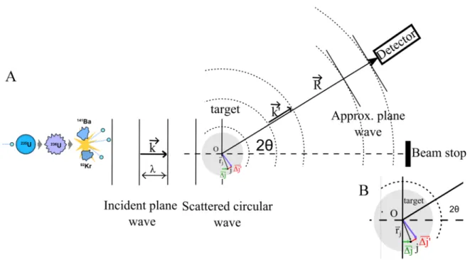
A neutron beam is focused on the sample and part of it is scattered by its atoms. The velocity of the neutrons and therefore the wavelength of the beam must be well-defined (see Section 7). The scattered neutrons undergo a change of direction which is characterized by the diffusion vector q⃗, which is also called scattering vector.
3.1 Incoming and outgoing wave functions
We shall focus here on elastic scattering and consider how the scattered wave can be written as a function of the scattering vector.
Figure 2 A : Neutron scattering by an isotropic sample. The production of neutrons is described in section 7. B: enlargment of the neutron path difference between the scatterer at the origin of the target O and the scatterer j.
The incoming plane wave of wavelength can be written as
( , ) exp in r t B i k r t (1) where k is the wave vector whose modulus is equal to k = 2π/, B is a normalization factor and is the angular frequency. Some dephasing will appear between the waves
18
scattered by the various scatterers of the target. Let’s consider two of them: one at the origin and one at the positionrj. The wave scattered by j will be dephased with respect to that scattered by 0 by a quantity equal to the product of the wave vector and of the optical path difference.
' ' ' ' j j j j j j k k k r k r k k r q r (2) where the scattering vector qk'k has been defined.Figure 3 Definition of the scattering vector ⃗.
The modulus of ⃗ is given by the following formula:
4 sin q (3)For N scatterers, the outgoing wave is therefore equal to
1 exp ' , exp N out j j j i k r t r t B b iq r r
(4)if the sample-detector distance is much larger than the sample size. As the scatterers act as a source of scattered spherical waves, the last factor is divided by r. We can re-write (4) as
,
( )exp
'
out i k r t r t B A q r (5)with the scattering amplitude defined as ( ⃗):
1 ( ) exp N j j j A q b iq r
(6)19
The coefficient bj accounts for the particle-scatterer interactions and is called the
scattering length because it has the dimension of a length. It can be shown that, within the
so-called Born Approximation valid for weak target-particle interactions, bj is given by the
following equation
2 ( ) exp 2 j j space m b V r iq r dr
(7)bj is therefore proportional to the Fourier transform of the particle-scatterer interaction
potential Vj( )r . For neutrons, the potential is short-range with respect to the usual q-1domain
(~1Å-1) so that exp
iq r
1 and 2 ( ) 2 j j space m b V r dr
(8)which does not depend on q. bj varies relatively randomly through the periodic table as displayed in Figure 4. For X-rays, the range of the electromagnetic interaction is close to q-1 so that bj depends on q
. The Cromer-Mann formula reproduces empirically this dependency. In SAXS, for q < 0.5Å-1, however, bj can be considered as constant. It is given by the following formula at high X-ray energy:
2 2 0 4 j j e e b Z m c (9)
where Zj is the atomic number of atom j and me is electron mass at rest. For X-rays, bj increases therefore linearly with the atomic number.
20
Figure 4 Scattering lengths b of neutrons with respect to the atomic number Z. b varies relatively randomly through the periodic table.[6]
3.2 Differential scattering cross section
If N is the number of neutrons scattered per unit time into the solid angle d around direction q, the differential scattering cross section (dσ(q)/dΩ) is defined as [7]
density of flux of incoming neutrons
d q d d N (10)
where the density of flux is the number of incident neutrons per unit area per second. The measured intensity during SANS experiments is thus related to the cross section of the sample (see Section 7).
21
Figure 5 Scattering geometry for an incident neutron beam ( ⃗) scattered by a target (green cube).
If we denote the incoming and outgoing densities of flux as Jin and Jout, we have
2 2 ( ) ( ) ( ) out out in in J q r d J q r d q d J d J (11)
In quantum mechanics, the density of flux is given by
* *
2 i i i i
i m
J (12)
where is the gradient operator. Inserting (1) and (5) into (12) leads to 2 in J B k m (13) and 2 2 2 ( ) ' out A q k J B ù r (14)
Inserting now (13) and (14) into (11), noting also that k k' for elastic scattering, we can write that
22
2 1 1 ( ) ( ) exp N N j l l j j l d q A q b b i q r r d
(15)This relationship is valid for both neutrons and X-Rays scattering. For neutron scattering an additional subtlety arises due to the fact that isotopes of the same element usually have quite different scattering lengths and that the different isotopes are distributed randomly in the sample. In addition, the neutron possesses a spin (I=1/2) which can interact with magnetic nuclei (that is, those that also have a non-zero spin). The resulting coupling can lead to different value of the total angular momentum, each situation being characterized by a different value of the associated scattering length. As a result, an ensemble average has to be performed so that (15) now becomes:
1 1 exp N N j l l j j l d q b b i q r r d
(16)Separating the diagonal and the non-diagonal contributions
2
1 1 exp N N N j j l l j j j l j d q b b b i q r r d
(17)If we consider that all nuclei of the sample correspond to the same element (but with a distribution of isotopes), we can write
b2j b2 when l = j
b bj l b b b 2 when l because the isotopes are randomly distributed. j As the variance
b
2 is equal to
2 2 2b b b
, we can also write that
2 2 2 b b b . So that (17) becomes
2 exp
2 N N l j j l d q b i q r r N b d
(18)The cross section is now the sum of two terms called, respectively, the coherent ( ( ⃗)/ ) and the incoherent ( ( ⃗)/ ) cross sections.
23
2
exp N N l j j l coh d q b i q r r d
(19) This contribution depends on the spatial organization of the scatterers within the sample and, as a consequence, it is a function of q . From this q variation, information can be inferred about the sample structure.
2 inc d q N b d (20) This contribution represents a constant background which provides no structural information. Incoherent scattering can represent a problem for polymer samples and for aqueous polymeric solutions because these samples contain a large fraction of H-scatterers, for which
3.74 25.27 h h b fm b fm
As a consequence, coherent scattering by H nuclei represents only a small contribution on a large incoherent background. For 2H (D) nuclei, however, the coherent contribution is larger than the incoherent one:
6.674 4.041 d d b fm b fm
This is the reason why most of our experiments were performed in D2O solutions.
In the following, we shall focus on the coherent scattering contribution. The incoherent background will always be subtracted during the experimental data handling procedure. For modelling purposes, we shall use, according to eq(19), the average b for each kind of nuclei of our polymer samples. In practice, only H, C, N and O need to be considered.
24
4 Small Angle Neutron Scattering (SANS)
Most of what will be presented in this section is valid for both X-Rays and Neutrons and for various kinds of samples. However, as we shall be interested mostly in polymer samples investigated by neutron scattering, we shall focus on self-assembled polymeric nano-objects like micelles or liposomes and neutron scattering.
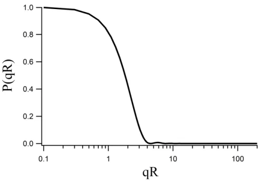
Micelles of amphiphilic copolymers have usually a size of a few tens of nanometers. As a rule, they adopt a spherical shape so that we consider first the scattering cross section by a homogenous sphere. According to equations (15) or (19), and adopting the approximation of continuum, we can write that
2 2 exp coh space d q b r i q r dr d
(21) where b has been simply denoted as b.
r is the constant particle density:
3 4 3 N r R
where R is the radius of the
sphere. It can be easily shown that
2 2 2 6 9 sin( ) cos( ) coh d N b q qr qR qR d qR (22)Because of the isotropic nature of the problem, the cross section depends only on the modulus of the scattering vector.
This relationship is graphically displayed in Figure 6 which shows that that the cross section reaches the zero line for qR ~ 4.5 and remains small afterwards (although it continues to oscillate). The q-domain where relevant information can be obtained is thus defined by
typical size 1.
q For polymer micelles, the typical size is located between: the monomer lengths:~3Å
25 so that the useful q-range is
3 1 1
10 Å q 0.3Å (23)
For thermal neutrons, is in the Å range, i.e. 5Å. Converting the q range in a 2 range using (3) leads to:
0.04214 (24)
This means that, only the small angle neutron scattering angle range needs to be sampled, hence the denomination “Small Angle Neutron Scattering” (SANS). The same is of course also valid for X-Rays (SAXS).
Figure 6 Form factor of a sphere (Eq (22)) The cross section reaches the zero line for qR ~ 4.5 and remains small afterwards.
5 Form and Structure factors for micellar and liposomes solutions
We shall focus here on what represents the nucleus of our research that is the investigation of aqueous solutions of self-organized copolymer micelles or phospholipid liposomes. We consider only the coherent scattering contribution. In this section we limit ourselves to monodisperse nanosized objects. The problems linked with the influence of polydispersity will be dealt with in the next section. The problem at hand is sketched in the figure below.26
Figure 7 Micellar solution. The micelles are built from a polymer block forming the core P (red), a polymer block forming the corona P' (blue) and solvent molecules S (not displayed).
We are confronted to a multicomponent system: the polymer blocks forming the core (P) the polymer block forming the corona (P’) the solvent molecules (S)
Because of the large density of particles in a liquid, we shall adopt a continuum formulation of eq(19). To simplify the notations, we replace b by b. We shall also use the so-called “macroscopic cross section” obtained by dividing the cross section by the sample volume, and denoted as d
d
/ . Equation(19) becomes then, for a one-component system,
2 exp ' ' ' V V d b i q r r r r dr dr d V
(25)where V is the volume of the entire sample.
r is, as before (equation(21)) the particle (scatterer) density. Because of the fluctuating nature of polymer samples (and in general of27
soft matter samples) and of liquid solvents, an ensemble average has to be performed, which is denoted by the brackets.
5.1 Scattering by one-component (quasi) continuous sample
Because of the already mentioned fluctuating nature of the systems considered in this work, when we focus on the situation at a given position r, we observe density fluctuations,
r
, so that we can write, in general,
r
r (26)
where is the average density around which fluctuations take place. Eq (25) becomes now
2 exp ' ' ' V V d b q i q r r r r dr dr d V
(27)Expanding the expression between brackets gives
2
exp exp ' '
exp exp ' ' ' exp exp '
exp exp ' ' ' V V V V V V V V i q r dr i q r dr i q r dr i q r r dr i q r r dr i q r dr i q r r dr i q r r dr
(28)An alternativeway of writing (28) is:
2
exp exp ' '
exp exp ' ' ' exp ' exp
exp ' ' ' V V V V V V V V i q r dr i q r dr i q r dr i q r r dr i q r i q r r dr i q r r r r dr dr
(28 bis) The average of the fluctuations of the number of scatterers in volume V, N, is equal to28
V V V N N N r dr dr r dr
It is clear that N N N N N . Taking into account that 0
3 exp 2 V i q r dr q q q
where is the Dirac distribution. Denoting n q
the Fourier transform of
r :
exp
V
n q
i q r r dr we can write (28) and (28 bis) as:
2 6
2 q q n q n q
(29)
This is the sum of two contributions:
an infinitely narrow contribution at q 0 which will be in any practical situation suppressed by the beam stop
the
exp
'
' 'V V
n q n q
i q r r r r dr dr contribution originating from the density fluctuations.The final result for the cross section becomes:
2 2 exp ' ' ' V V d b b q n q n q i q r r r r dr dr d V V
(30)It is interesting to look at the situation when ⃗ approaches 0.
2 0 ' ' V d b r r dr dr d V
(31)29
Because the average of the square of the fluctuations of the number of scatterers in volume V,
N2
is equal to
2 2 ' ' V V V N r dr r r dr dr
(32) it follows that
2 2 0 d b N d V (33)A well-established relationship for the grand canonical ensemble which applies here for density fluctuating systems is
2 T 2kT
N N
V
(34)
where k is the Boltzmann constant, T is the temperature and
1 T T V V P (35)
is the isothermal compressibility. As a result
2 2 2 2 2 0 T T N b kT d b kT d V v (36)where v = V/<N> is the volume of the elementary scatterers.
Equations (30) and (36) are essential because they show that, for a one-component system, no scattering will be detected in the absence of any density fluctuation, that is, if
![Figure 4 Scattering lengths b of neutrons with respect to the atomic number Z. b varies relatively randomly through the periodic table.[6]](https://thumb-eu.123doks.com/thumbv2/123doknet/6088414.153982/40.892.118.765.114.537/figure-scattering-lengths-neutrons-respect-relatively-randomly-periodic.webp)


![Figure 12 a) BF3 detection tube. [19] b) Paxy’s detector at Laboratoire Leon Brillouin, Saclay](https://thumb-eu.123doks.com/thumbv2/123doknet/6088414.153982/63.892.140.753.110.404/figure-detection-paxy-detector-laboratoire-leon-brillouin-saclay.webp)