RESEARCH
The association between FABP7
serum levels with survival and neurological
complications in acetaminophen-induced acute
liver failure: a nested case–control study
Constantine J. Karvellas
1*, Jaime L. Speiser
2, Mélanie Tremblay
3, William M. Lee
4, Christopher F. Rose
3and For
the US Acute Liver Failure Study Group
Abstract
Background: Acetaminophen (APAP)-induced acute liver failure (ALF) is associated with significant mortality due to
intracranial hypertension (ICH), a result of cerebral edema (CE) and astrocyte swelling. Brain-type fatty acid-binding protein (FABP7) is a small (15 kDa) cytoplasmic protein abundantly expressed in astrocytes. The aim of this study was to determine whether serum FABP7 levels early (day 1) or late (days 3–5) level were associated with 21-day mortality and/or the presence of ICH/CE in APAP-ALF patients.
Methods: Serum samples from 198 APAP-ALF patients (nested case–control study with 99 survivors and 99
non-sur-vivors) were analyzed by ELISA methods and assessed with clinical data from the US Acute Liver Failure Study Group (ALFSG) Registry (1998–2014).
Results: APAP-ALF survivors had significantly lower serum FABP7 levels on admission (147.9 vs. 316.5 ng/ml,
p = 0.0002) and late (87.3 vs. 286.2 ng/ml, p < 0.0001) compared with non-survivors. However, a significant association between 21-day mortality and increased serum FABP7 early [log FABP7 odds ratio (OR) 1.16, p = 0.32] and late (log FABP7 ~ OR 1.34, p = 0.21) was not detected after adjusting for significant covariates (MELD, vasopressor use). Areas under the receiver-operating curve for early and late multivariable models were 0.760 and 0.892, respectively. In a second analysis, patients were grouped based on the presence (n = 46) or absence (n = 104) of ICH/CE. A significant difference in FABP7 levels between patients with or without ICH/CE at early (259.7 vs. 228.2 ng/ml, p = 0.61) and late (223.8 vs. 192.0 ng/ml, p = 0.19) time points was not identified.
Conclusion: Serum FABP7 levels were significantly elevated at early and late time points in APAP-ALF non-survivors
compared to survivors. However, significant differences in FABP7 levels by 21-day mortality were not ascertained after adjusting for significant covariates (reflecting severity of illness). Our study suggests that FABP7 may not discriminate between patients with or without intracranial complications.
Keywords: Liver-type fatty acid-binding protein, Multiorgan failure, Prognosis, ALFSG index
© The Author(s) 2017. This article is distributed under the terms of the Creative Commons Attribution 4.0 International License (http://creativecommons.org/licenses/by/4.0/), which permits unrestricted use, distribution, and reproduction in any medium, provided you give appropriate credit to the original author(s) and the source, provide a link to the Creative Commons license, and indicate if changes were made.
Background
Acute liver failure (ALF) is defined by the occurrence of hepatic encephalopathy (HE) and hepatic synthetic
dysfunction within 26 weeks of the first symptoms of liver disease [1]. Severe coagulopathy, encephalopathy and hemodynamic instability contribute to a picture of multiorgan failure. Currently, the most common cause of ALF in North America is acetaminophen (APAP) [2]. Particularly in APAP-induced ALF, cerebral edema (CE) and intracranial hypertension (ICH) are major causes of morbidity and mortality [3]. The pathogenesis for ICH
Open Access
*Correspondence: dean.karvellas@ualberta.ca
1 Division of Gastroenterology (Liver Unit), Department of Critical Care
Medicine, University of Alberta, 1-40 Zeidler Ledcor Building, Edmonton, AB T6G-2X8, Canada
and CE in ALF is not fully understood, but astrocyte swelling causing cellular dysfunction as well as increased cerebral blood flow is believed to be implicated [4]. The degree of hyperammonemia has been demonstrated to be associated with ICH [5]. Ammonia, as a gas (NH3) and
ion (NH4+), freely crosses the blood–brain barrier and is
primarily removed by glutamine synthetase, an enzyme solely found in astrocytes within the brain [6]. Glutamine synthetase catalyzes the amidation of glutamate to glu-tamine that subsequently leads to hyperosmotic changes and astrocyte swelling. However, studies have shown that hyperammonemia alone does not predict ICH [7].
Given the challenges presented in managing critically ill ALF patients with potential CE/ICH including the consideration for liver transplant (LT), the development of a noninvasive biomarker with the potential to predict ICH would be of great value, especially given the signifi-cant bleeding risks of invasive intracranial pressure mon-itoring in these coagulopathic patients [8].
Fatty acid-binding proteins (FABP) are small (15 kDa) cytoplasmic proteins that are abundantly expressed in tis-sues with active fatty acid metabolism, such as brain and liver. The primary function of FABPs is the intracellular transport of long-chain fatty acids [9]. The cellular expres-sion of FABPs is responsive to changes in lipid metabolism, which can be induced during pathophysiological condi-tions, such as ischemia/inflammation or pharmacological stimuli [10]. Brain-type FABP (FABP7) is solely expressed in brain, exclusively in astrocytes [11]. Previous investiga-tions have shown serum levels of FABP7 to be elevated in patients with various neurological diseases including stroke [12] and dementia [13]. While our group recently demon-strated the prognostic value of serum levels of liver-type FABP (FABP1) in ALF, to date FABP7 as a biomarker for the risk of ICH in ALF has not been investigated [14].
This nested case–control study of randomly selected sam-ples from prospectively enrolled patients from the US Acute Liver Failure Study Group (ALFSG) registry aimed to exam-ine levels of FABP7 in APAP-ALF patients. Specifically, our primary objectives were to test the following hypotheses
(a) Higher FABP7 serum levels are significantly associ-ated with 21-day transplant-free mortality (in the absence of transplant) after adjusting for other signifi-cant covariates (Analysis 1).
(b) Elevated serum levels of FABP7 in APAP-ALF are sig-nificantly associated with ICH/CE after adjusting for other significant covariates (Analysis 2).
Methods
This study is a nested case–control study of prospectively collected data and biosamples of 198 patients enrolled in the US ALFSG registry/biorepository and is outlined
in detail in Additional file 1: Figure S1. Between Janu-ary 1998 and December 2014, 1027 APAP-ALF patients were enrolled in the registry from which 704 patients were alive at day 21 in the absence of LT. We identified 124 survivors with early and late serum samples from which 99 were randomly selected for analysis. Of 224 patients who died in the absence of LT, 87 patients with early and late samples were also included in this analy-sis. A further 12 patients with exclusively an early sample (of a possible 92) were randomly selected for inclusion in this analysis. Personnel not involved in the analysis of the samples or statistical analysis for the paper per-formed random selection of patients. All enrolling cent-ers were tertiary academic centcent-ers, and all but one were LT centers. The authors’ Institutional Review Board (IRB)/Health research ethics boards of all enrolling US ALFSG sites have approved all research, and all clinical investigation has been conducted according to the princi-ples expressed in the 1975 Declaration of Helsinki. Given patients were unable to provide written consent (critical illness, HE), written assent was obtained from the next of kin from each patient. Each center implemented moni-toring and therapeutic interventions according to insti-tutional standards of care. Reporting of the analysis of this study followed the STROBE Guidelines for reporting case–control studies [15]. Consistent with ALFSG stud-ies [16], the primary outcome (Analysis 1) was 21-day LT-free survival (no patients included in the analysis received LT). Secondary outcome (Analysis 2) was the development of ICH/CE.
Participants
Inclusion criteria were: (1) evidence of ALF according to
the enrollment criteria for the ALFSG (see operational definitions); (2) age ≥ 18 years; (3) HE during the first seven days of study admission (West Haven Criteria) [17]; and (4) patients within the ALFSG registry with primary diagnoses of APAP determined by the site inves-tigator. Exclusion criteria were: (1) cirrhosis/acute-on chronic liver failure; (2) patients without a primary diag-nosis of APAP; and (3) patients who received a LT. Serum samples were analyzed on study admission (early; day 1) and late (either day 3, 4 or 5) where available. Patients who received a LT were excluded from our study because listing for transplant is a clinical decision, which is not standardized among ALFSG sites. A further 51 healthy controls were analyzed (University of Alberta) for FABP7 only.
Operational definitions
For the purposes of this study, ALF was defined as INR ≥ 1.5 and HE within the first 26 weeks of liver dis-ease in a patient with an acute hepatic insult [18]. HE
coma grade was defined by the West Haven Criteria (sim-plified) as follows: grade 1 ~ any alteration in mentation, grade 2 being somnolent or obtunded but easily rousable or presence of asterixis, grade 3 being rousable with dif-ficulty, and grade 4 being unresponsive to deep pain [17]. In this study, we defined ‘low coma grade’ as grade 1 or 2 and ‘high coma grade’ as grade 3 or 4. The KCC [19] predicts poor outcome (death/transplant) if: (a) pH is less than 7.3 or (b) if INR is greater than 6.5, creatinine is greater than 3.4 mg/dl, and coma grade is high (3 or 4). The model for end-stage liver disease (MELD) is defined as 10*(0.957*log(4) + 0.378*log(bilirubin) + 1.12*log(IN R)) for dialyzed patients and 10*(0.957*log(creatinine) + 0.378*log(bilirubin) + 1.12*log(INR)) for patients not dialyzed [20].
Laboratory Assays of FABP7
FABP7 was measured in serum samples with a solid-phase enzyme-linked immunosorbent assay (ELISA) following manufacturer’s instructions (Biomatik, USA). Briefly, samples were incubated 2 h on a monoclonal anti-FABP7 pre-coated plate. A specific FABP7 biotin-conjugated polyclonal antibody solution was added for 2 h. After washing plates, avidin conjugated to horserad-ish peroxidase was added for 30 min. Finally, substrate tetramethylbenzidine was added for 15 min. Reactions were stopped by addition of sulfuric acid, and absorbance was read at 450 nm. Standard curve ranges from 0.47 to 30 ng/ml. Samples were performed in duplicate and accepted valid with a variation coefficient less than 25%.
Statistical methods aim one: FABP7 and 21‑day survival in APAP‑ALF
For differences between outcome groups (APAP-ALF survivors, n = 99, APAP-ALF non-survivors, n = 99), categorical variables were compared using the Chi-squared test or Fisher’s exact test (if n < 10 in any cell of the two-by-two table). FABP7 was treated as a con-tinuous variable. Concon-tinuous variables were reported as medians with interquartile range (IQR) and compared using the Wilcoxon rank-sum test. Survival was defined as the dichotomous outcome, alive or dead at 21 days after enrollment into the registry (no patients received a LT in this analysis). A two-sided p value of < 0.05 was considered statistically significant for all comparisons (Additional file 2).
In order to control for variables that may confound the effect of FABP7 on 21-day mortality, logistic regres-sion analysis was performed [21]. Aside from FABP7, covariates considered in multivariable modeling included MELD, lactate, vasopressors use, RRT, MV and high coma grade. Separate multivariable (logistic) regression models were derived for FABP7 early (day 1) and late
(days 3–5) by including variables, which were significant on univariate analysis and performing backward elimina-tion with a p value threshold of 0.05.
Statistical methods aim two: FABP7 and ICH in APAP‑ALF
In this secondary analysis, the outcome of interest exam-ined was intracranial hypertension (ICH) either based on (a) intracranial pressure monitoring with ICP > 25 mm Hg or based on (b) computed tomography (CT) imaging of the brain. CT evidence of cerebral edema was defined as a hypodense signal, effacement of the gray white matter junction, loss of differentiation of the lenticular nucleus and decreased visualization of the sulci, insula and cisterns [22]. Out of the 150 APAP-ALF patients where data were available to determine the presence or absence of ICH, 46 deceased patients had evidence of ICH based on these criteria. Statistical methods for this analysis will be similar to the first analysis except the primary outcome (ICH). Multivariable logistic regres-sion analysis (as described above) was performed [21] to assess independent variables associated with ICH including FABP7. The pre-specified prognostic vari-ables were based on previous publications [5] included at admission into the registry; age, lactate value, MELD [20] score (admission) and other variables with statistical significance on univariable analysis. Model performance for both Aim 1 and Aim 2 was assessed using area under the receiver-operating curve (AUROC) and the Hosmer– Lemeshow test for goodness of fit. SAS software version 9.3 was used for univariate comparisons and multivari-able logistic regression modeling.
Results
Analysis one: comparative analysis of 198 APAP‑ALF patients
Demographic and clinical outcomes stratified by mortal-ity (alive at day 21, n = 99; deceased, n = 99) are listed in Table 1. No patients in this analysis received LT. Com-paring APAP-ALF survivors and non-survivors at day 21, survivors required significantly less organ support during the 7 days of inpatient study (MV 65 vs. 93%; vasopres-sors 12 vs. 70%; RRT 27 vs. 45%; p < 0.008 for all). Sur-vivors were less likely to achieve high (3 or 4) HE coma grade (62 vs. 93%, p < 0.0001) and less likely to receive mannitol for intracranial hypertension (22 vs. 46%,
p = 0.0003). APAP-ALF survivors were less likely to have
complications during the first 7 days of study includ-ing seizures (3 vs. 21%, p < 0.0001), arrhythmias (25 vs. 38%, p = 0.047) or gastrointestinal bleeding (8 vs. 19%,
p = 0.037). On admission, 7% of APAP-ALF survivors
and 16% of non-survivors met KCC (p = 0.13). Among the 99 APAP-ALF non-survivors, the most common causes of death reported were multiorgan failure (53%)
and neurological complications (38%). Cause of death was unknown in 9% of cases.
Clinical parameters in 198 APAP‑ALF patients: admission (early)
Comparisons of clinical parameters on study admission are listed in Table 2. APAP-ALF survivors demonstrated significantly lower MELD scores (23 vs. 29, p < 0.0001) than non-survivors on admission. Survivors were
significantly less likely to be on organ support (MV 58 vs. 80%, p = 0.0007; vasopressors, 9 vs. 42%, p < 0.0001) or achieve high HE grade (57 vs. 71%, p = 0.034) on admission.
FABP7 levels at admission (early) are listed in Table 2 and graphically shown in Fig. 1. APAP-ALF survivors had significantly lower admission serum FABP7 levels (147.9 vs. 316.5 ng/ml) compared with non-survivors (p = 0.0002). In comparison, 52 healthy
Table 1 Demographic, clinical and biochemical parameters in 198 APAP-ALF patients by outcome
N frequency, IQR interquartile range, ARDS acute respiratory syndrome, CT computed tomography, CXR chest x-ray
APAP alive day 21 (n = 99) APAP dead day 21 (n = 99) p value
N Number (%) or median (IQR) N Number (%) or median (IQR)
Age 99 35 (28–43) 99 40 (30–48) 0.084 Sex (female) 99 75 (76%) 99 72 (73%) 0.63 Race 0.23 White 99 83 (84%) 99 79 (80%) African-American 99 8 (8%) 99 15 (15%) Other 99 8 (8%) 99 5 (5%)
Organ support (days 1–7)
Mechanical ventilation 99 64 (65%) 99 93 (93%) < 0.0001
Vasopressors 99 12 (12%) 99 69 (70%) < 0.0001
Renal replacement therapy 99 27 (27%) 99 45 (45%) 0.0078
KCC 87 7 (7%) 87 16 (16%) 0.13
Coma grade 3/4 (worst days 1–7) 99 61 (62%) 98 91 (93%) < 0.0001
ICP-directed therapies (days 1–7)
ICP monitor 99 12 (12%) 99 21 (21%) 0.086 Mannitol 99 22 (22%) 99 46 (46%) 0.0003 Hypertonic saline 99 11 (11%) 99 14 (14%) 0.52 Barbiturates 99 9 (9%) 99 20 (20%) 0.043 Hypothermia 99 17 (17%) 99 14 (14%) 0.56 Sedatives 99 70 (71%) 99 88 (89%) 0.0014
Blood products (days 1–7)
Red blood cells 99 34 (34%) 99 50 (51%) 0.021
Fresh-frozen plasma 99 50 (51%) 99 76 (77%) 0.0001
Recombinant VIIA 99 3 (3%) 99 5 (5%) 0.72
Platelets 99 17 (17%) 99 36 (36%) 0.0023
ICU complications (days 1–7)
Seizures 99 3 (3%) 99 21 (21%) < 0.0001 Arrhythmias 99 25 (25%) 99 38 (38%) 0.047 GI bleeding 99 8 (8%) 99 19 (19%) 0.037 ARDS 99 0 (0%) 99 3 (3%) 0.25 CT (cerebral edema) 55 7 (13%) 72 32 (44%) < 0.001 Abnormal CXR 99 88 (89%) 99 83 (84%) 0.30
Bacteremia/blood stream infection 99 7 (7%) 99 10 (10%) 0.61
Cause of death
Multiorgan failure 99 52 (53%)
Cerebral edema 99 38 (38%)
Table 2 Demographic, clinical and biochemical parameters in 198 APAP-ALF patients by outcome (admission)
Early (admission) APAP alive day 21 (n = 99) APAP dead day 21 (n = 99) p value
N Number (%) or median (IQR) N Number (%) or median (IQR)
Biochemistry
Hemoglobin (g/dl) 99 10.4 (9.2–12.5) 97 10.9 (9.5–12.2) 0.52
White blood count (109/l) 98 8.6 (6.4–11.2) 97 10.9 (7.3–17.5) 0.0008
Platelet count (109/l) 98 132.5 (90.0–195.0) 97 110.0 (67.0–160.0) 0.0045
INR 99 2.7 (1.8–4.1) 96 3.4 (2.3–4.8) 0.0023
ALT (IU/l) 98 3380 (1949–6576) 99 3235 (1483–5716) 0.37
Bilirubin (mg/dl) 98 4.1 (2.5–5.6) 99 5.0 (3.6–7.8) < 0.0001
pH 88 7.4 (7.4–7.5) 88 7.4 (7.3–7.5) 0.22
Ammonia (venous) (μmol/l) 51 92 (73–140) 32 139 (72–205) 0.068
Creatinine (mg/dl) 98 1.4 (0.8–3.0) 98 2.6 (1.2–3.8) 0.0007
Lactate (mmol/l) 71 2.8 (1.7–5.5) 68 7.0 (4.8–11.8) < 0.0001
Phosphate (mg/dl) 88 2.3 (1.7–3.4) 76 3.2 (2.1–4.5) 0.0061
MELD 98 23.4 (12.7–27.7) 96 29.1 (23.8–34.5) < 0.0001
High coma grade (3 or 4) 99 56 (57%) 97 69 (71%) 0.034
Organ support
Mechanical ventilation 99 57(58%) 99 79 (80%) 0.0007
Vasopressors 99 9 (9%) 99 42 (42%) < 0.0001
Renal replacement therapy 99 19 (19%) 99 24 (24%) 0.39
FABP7 (ng/ml) 99 147.9 (66.6–296.2) 99 316.5 (119.8–562.2) 0.0002
Late (n = 186) APAP alive day 21 (n = 99) APAP dead day 21 (n = 87) p value
Late (days 3–5) N Number (%) or median (IQR) N Number (%) or median (IQR)
Biochemistry
Hemoglobin (g/dl) 94 9.9 (9.0–11.1) 81 10.2 (9.2–10.9) 0.63
White blood count (109/L) 94 8.1 (5.9–11.6) 81 11.3 (7.1–15.0) 0.0029
Platelet count (109/L) 95 111.0 (68.0–153.0) 81 66.0 (47.0–100.0) < 0.0001
INR 94 1.5 (1.3–1.8) 75 2.5 (1.8–4.4) < 0.0001
ALT (IU/L) 94 1172 (612–2007) 78 938 (383–1995) 0.31
Bilirubin (mg/dl) 92 5.5 (2.7–8.2) 78 9.8 (6.7–13.7) < 0.0001
pH 55 7.4 (7.4–7.5) 76 7.4 (7.3–7.5) 0.042
Ammonia (venous) (μmol/L) 25 62 (44–84) 17 119 (78–133) 0.0038
Creatinine (mg/dl) 94 1.2 (0.7–2.5) 82 2.4 (1.3–4.0) < 0.0001
Lactate (mmol/L) 31 1.7 (1.0–2.2) 41 3.8 (2.6–6.7) < 0.0001
Phosphate (mg/dl) 73 2.8 (2.3–3.6) 35 3.3 (2.5–4.5) 0.054
PO2/FiO2 ratio 48 3.3 (2.1–4.5) 71 2.5 (1.4–3.9) 0.0087
MELD 87 14.2 (5.4–24.6) 73 29.7 (23.5–35.4) < 0.0001
High coma grade (3 or 4)a 59 35 (59%) 82 72 (88%) < 0.0001
Organ support
Mechanical ventilation 99 49 (49%) 88 74 (85%) < 0.0001
Vasopressors 99 5 (5%) 88 45 (52%) < 0.0001
Renal replacement therapy 99 10 (19%) 88 27 (31%) 0.062
FABP7 (ng/ml) 99 87.3 (48.0–261.5) 87 286.2 (146.7–536.9) < 0.0001
N frequency, IQR interquartile range, INR international normalized ratio, AST aspartate aminotransferase, ALT alanine aminotransferase, MELD model for end-stage liver disease
controls had median serum levels of 13.5 (8.7–20.2) ng/ml.
Clinical parameters in 186 APAP‑ALF patients: days 3–5 (late)
Comparisons of clinical parameters on days 3–5 (late) are listed in Table 2. Of the 99 APAP-ALF non-survivors, samples of late time points were available in 87 patients as 12 died before days 3–5. APAP-ALF survivors (n = 99)
were significantly less likely to be on MV (49 vs. 85%, p < 0.0001) and vasopressors, (5 vs. 52%, p < 0.0001) or achieve high HE grade (59 vs. 88%, p < 0.0001) than non-survivors.
Late (days 3–5) FABP7 are listed in Table 2 and graphi-cally shown in Fig. 1. APAP-ALF survivors had signifi-cantly lower late serum FABP7 levels (87.3 vs. 286.2 ng/ ml) compared with non-survivors (p < 0.0001). FABP7 levels were significantly higher in all ALF patients (survi-vors and non-survi(survi-vors) compared to healthy controls for both early and late time points (p < 0.0001).
Multivariable analysis: associations with 21‑day mortality
In order to adjust for covariates, multivariable logistic regression for 198 APAP-ALF patients to determine asso-ciations (adjusted) with 21-day mortality was performed (Table 3). Two models were derived: one on admis-sion (early) and one at days 3–5 (late). Values of serum FABP7 were transformed to their natural logarithm (log FABP1) to comply with the linearity assumption in logis-tic regression.
Early (admission) model
FABP7 was not associated with 21-day mortality [odds ratio OR 1.001 per increment, 95% CI (1.000, 1.001),
p = 0.18] after adjusting for significant covariates
Fig. 1 Serum levels of FABP7 (ng/ml) in healthy controls,
non-survi-vors (early ~ admission), survinon-survi-vors (early), non-survinon-survi-vors (late ~ days 3–5), survivors (late)
Table 3 Early (day 1) and late (days 3–5) predictors of 21-day mortality in 198 APAP-ALF patients
Early: lactate (p = 0.52), high coma grade (p = 0.46), and mechanical ventilator (p = 0.084) were not significant on multivariable analysis so not included in the final early model
Late: mechanical ventilation (p = 0.69) and high coma grade (p = 0.53) were not significant on multivariable analysis so not included in the final late model. Lactate was not included due to missing data
Early Unadjusted Multivariable model (N = 194), AUROC = 0.766
N OR 95% OR CI p value Included in Model OR 95% CI p value
FABP7 198 1.001 (1.000, 1.002) 0.0078 Yes 1.001 (1.000, 1.001) 0.18
MELD 194 1.083 (1.047, 1.119) < 0.0001 Yes 1.056 (1.020, 1.093) 0.0021
Lactate 132 1.205 (1.095, 1.327) 0.0001 No
Vasopressors 198 7.368 (3.335, 16.287) < 0.0001 Yes 4.138 (1.769, 9.677) 0.0011 Renal replacement therapy 198 1.347 (0.683, 2.658) 0.390 No
Mechanical ventilation 198 2.910 (1.547, 5.475) 0.0009 No High coma grade (3 or 4) 196 1.892 (1.047, 3.421) 0.0348 No
Late (days 3–5) Unadjusted Multivariable model (N = 160), AUROC = 0.891
N OR 95% OR CI p value Included in model OR 95% CI p value
FABP7 186 1.003 (1.001, 1.004) 0.0001 Yes 1.001 (0.999, 1.003) 0.40
MELD 160 1.115 (1.075, 1.157) < 0.0001 Yes 1.084 (1.038, 1.132) 0.0003
Lactate 71 6.908 (2.592, 18.406) 0.0001 No
Vasopressors 186 20.143 (7.462, 54.370) < 0.0001 Yes 20.419 (6.221, 67.021) <0.0001 Renal replacement therapy 186 1.895 (0.964, 3.724) 0.0638 No
Mechanical ventilation 186 5.808 (2.859, 11.802) < 0.0001 No High coma grade (3 or 4) 141 4.936 (2.129, 11.446) 0.0002 No
including MELD [OR 1.056 (1.020, 1.093) per increment,
p = 0.0021] and requirement for vasopressors [OR 4.14
(1.77, 9.07), p = 0.0011]. This early model demonstrated AUROC of 0.766.
Late (days 3–5) model
FABP7 was not associated with 21-day mortality [OR 1.001 (0.999, 1.003) per increment, p = 0.40] after adjust-ing for significant covariates includadjust-ing MELD [OR 1.084 (1.038, 1.132) per increment, p = 0.0003] and require-ment for vasopressors [OR 20.42 (6.22, 67.02), p < 0.0001]. This late model demonstrated AUROC of 0.891.
Analysis two: comparative analysis of 150 APAP‑ALF patients
Demographic and clinical outcomes of 150 patients strat-ified by the presence (n = 46) and absence (n = 104) of ICH/CE based on review of subject data (ICP measure-ments, CT brain, cause of death) are shown in Additional file 3: Table S1. (In 48 patients, the presence or absence of ICH/CE could not be determined.) There were no sig-nificant differences in age (36 vs. 39, p = 0.11) or gender (female 74 vs. 69%, p = 0.56). During the 7 days of inpa-tient study, APAP-ALF painpa-tients with ICH/CE had higher requirements for ventilation (MV 100 vs. 75%, p < 0.0001) and were more likely to achieve high (3 or 4) HE coma grade (100 vs. 72%, p < 0.0001). APAP-ALF patients with evidence of ICH/CE were less likely to be alive at day 21 (17 vs. 41%, p = 0.0049) but were more likely to be listed for LT (33 vs. 13%, p = 0.0062).
Clinical parameters in 150 APAP‑ALF patients: admission (early)
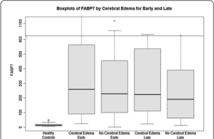
Comparisons of clinical parameters on study admis-sion are shown in Additional file 4: Table S2. APAP-ALF patients with ICH/CE had significantly higher serum INR (3.6 vs. 2.9) compared to patients without ICH/CE (p = 0.024). On study admission, patients who went on to develop ICH/CE were significantly more likely to be on mechanical ventilation (MV 85 vs. 65%, p = 0.019) and achieve high HE grade (76 vs. 59%, p = 0.043). Admis-sion (early) levels of FABP7 are listed in Additional file 4: Table S2 and graphically shown in Fig. 2. There were no significant differences in FABP7 levels on admission between APAP-ALF patients with or without ICH/CE (259.7 vs. 228.2 ng/ml, p = 0.61).
Clinical parameters in 186 APAP‑ALF patients: days 3–5 (late)
Comparisons of clinical parameters on days 3–5 (late) are shown in Additional file 4: Table S2. Patients who developed ICH/CE were significantly more likely to be on mechanical ventilation (MV 95 vs. 63%, p < 0.0001) and
achieve higher grades of HE (100 vs. 75%, p = 0.0004). Days 3–5 (late) levels of FABP7 are shown in Additional file 4: Table S2 and graphically in Fig. 2. There were no significant differences in late FABP7 levels between APAP-ALF patients with or without ICH/CE (223.8 vs. 192.0 ng/ml, p = 0.19).
Multivariable analysis: associations with 21‑day mortality
Multivariable logistic regression for 198 APAP-ALF to determine associations (adjusted) with the development of ICH/CE was performed (Table 4). Two models were derived; one on admission (early) and one at days 3–5 (late). Values of serum FABP7 were transformed to their natural logarithm (log FABP1) to comply with the linear-ity assumption in logistic regression.
Early (admission) model
FABP7 was not associated with the development of ICH/ CE [OR 1.000 per increment, 95% CI (1.000, 1.001),
p = 0.65] after adjusting for the only significant
covari-ate, mechanical ventilation [OR 2.880 (1.166, 7.111),
p = 0.022]. This early model demonstrated AUROC of
0.590.
Late (days 3–5) model
FABP7 was not associated with the development of ICH/ CE [OR 1.000 per increment, 95% CI (0.999, 1.001),
p = 0.57] after adjusting for the only significant
covari-ate, high hepatic coma grade [OR 25.76 (1.40, 472.5),
p = 0.029]. This late model demonstrated AUROC of 0.641. Discussion
Key results
In this nested case–control study, we report the first published analysis of FABP7 in a large series of 198
Fig. 2 Serum levels of FABP7 (ng/ml) in healthy controls, cerebral
edema (early ~ admission), no cerebral edema (early), cerebral edema (late ~ days 3–5), no cerebral edema (late)
well-characterized APAP-ALF patients. Compared with survivors, serum FABP7 levels were significantly higher at serial time points (early and late) in APAP-ALF non-survivors. However, significant differences in FABP7 levels by 21-day mortality were not ascertained after adjusting for significant covariates reflecting severity of illness (MELD, vasopressor dependence). No differ-ences in the FABP7 levels were detected for APAP-ALF patients with and without evidence of ICH/CE.
Comparison with literature
In our study, ICH/CE was the cause of death in 39% of patients, similar to what has been previously reported [16]. ICH/CE arises due to astrocyte swelling, cerebral vasodilatation, dilated cerebral arterioles and altered cer-ebral blood flow [23, 24]. Furthermore, it has been shown that patients with signs of cerebral edema and ICH have increased cerebral blood flow compared to patients with-out brain edema [25, 26]. Given the bleeding risks associ-ated with direct intracranial pressure monitoring [8], there is an unmet need for noninvasive markers of brain edema and ICH to help inform medical decisions. In the setting of ALF, astrocyte swelling/injury leads to astrocyte dysfunc-tion and consequently impairs neuronal funcdysfunc-tion leading to HE. However, in parallel, swollen astrocytes release small proteins, molecules and osmolytes in response to
astrocyte hypertonicity to reduce swelling. In the past 20 years, several biochemical biomarkers have been inves-tigated for the detection of cerebral injury, including pro-tein S100b [27], neuron-specific enolase (NSE) [27] and glial fibrillary protein (GFAP) [9]. Studies by Strauss et al. [27], as well as Vaquero et al. [28], which included 35 and 54 ALF patients, respectively, concluded that S-100b was not a useful marker of neurological outcome in ALF. Fur-thermore, despite a consistent increase of S-100b in serum, levels did not correlate with severity of HE, development of brain herniation or outcome. In the same patients, Strauss et al. found that serum levels of NSE were higher in ALF patients with ICH than those who survived with-out ICH [27]. However, this univariate comparison did not adjust for significant confounding factors/covariates, an important limitation to the study.
Major components of the brain are lipids with brain cells having a high cell membrane/cytoplasm ratio. Cell membranes are formed of lipid bilayers consisting of sat-urated and unsatsat-urated fatty acids, which can also be oxi-dized for generating ATP. The primary function of FABPs is to facilitate the transport of intracellular long-chain fatty acids. In the brain, FABP7 and heart FABP (FABP3) are expressed with FABP7 primarily found in astrocytes [29] and FABP3 in neurons [30]. With astrocyte swelling being a neuropathological landmark of ALF along with
Table 4 Early (admission) and late (days 3–5) predictors of cerebral edema in 150 APAP-ALF patients
Early: high coma grade (p = 0.69) was not significant on multivariable analysis so not included in the final early model Late: mechanical ventilation (p = 0.65) was not significant on multivariable analysis so not included in the final late model *Statistically significant on multivariable analysis
Early Unadjusted Multivariable model (N = 148), AUROC = 0.590
N OR 95% OR CI p value Included in model OR 95% CI p value
FABP7 150 1.000 (1.000, 1.001) 0.58 Yes 1.000 (1.000, 1.001) 0.65
MELD 146 1.036 (0.997, 1.078) 0.072 No
Lactate 100 1.000 (0.997, 1.002) 0.80 No
Vasopressors 150 0.777 (0.363, 1.660) 0.51 No Renal replacement therapy 150 1.313 (0.598, 2.886) 0.50 No
Mechanical ventilation 150 2.950 (1.199, 7.257) 0.019 Yes 2.880 (1.166, 7.111) 0.022 High coma grade (3 or 4) 148 2.227 (1.017, 4.878) 0.045 No
Late (days 3–5) Unadjusted Multivariable model (N = 113), AUROC = 0.641
N OR 95% OR CI p value Included in model OR 95% CI p value
FABP7 138 1.000 (0.999, 1.001) 0.96 Yes 1.000 (0.999, 1.001) 0.57
MELD 118 1.030 (0.994, 1.066) 0.10 No
Lactate 53 1.031 (0.979, 1.086) 0.25 No
Vasopressors 138 2.043 (0.947, 4.408) 0.069 No Renal replacement therapy 138 1.756 (0.789, 3.909) 0.17 No Mechanical ventilation 138 10.115 (2.300, 44.491) 0.0022 No
a hyperdynamic circulation frequently occurring in ALF leading to an increased myocardial demand, FABP3 may be confounded with myocardial injury. Therefore, FABP7 is more specific to brain injury in ALF.
FABP7 has physiological properties that render this protein advantageous as a prognostic biomarker in ALF: (i) it is abundantly present in astrocytes (between approximately 0.8 and 3.1 μg/g of brain tissue), (ii) it has a lower molecular mass (14 kDa); it is smaller than S-100b (21 kDa), enolase (47 kDa) and GFAP (50 kDa) with a much shorter plasma half-life (11 min) [31–33]. Smaller proteins such as FABP7 diffuse more rapidly (via transcy-tosis) than larger proteins though the interstitial space and cross the blood–brain barrier (BBB).
The release of cerebrovascular proteins into blood plasma is dependent on the status (breakdown) of the BBB, which in ALF is dependent on the underlying mech-anisms of cerebral edema, cytotoxic and vasogenic [34]. Astrocyte swelling plays a definitive role in the develop-ment of cytotoxic brain edema. In cytotoxic edema, the BBB is intact in the presence of intracellular swelling [35], whereas in vasogenic edema there is breakdown of the BBB and water and plasma constituents accumulate in the extracellular space [36]. Although a complete break-down of the blood–brain barrier is not evident in ALF, increased permeation to water and other small molecules such as ammonia has been demonstrated resulting from subtle alterations in the protein composition of paracel-lular tight junctions [37].
Despite elevated levels in ALF patients (survivors and non-survivors) in this analysis, FABP7 did not discrimi-nate between patients who went on to develop significant signs of ICH/CE either on imaging, ICP measurements or at death. One explanation is that variability in BBB permeability during ICH/CE could impact the diffusion rate of FABP7 into the peripheral circulation. In this study, we speculate that heterogeneity in the permeability of the BBB in ALF patients likely impacted the discrimi-natory ability of serum FABP7 measurements and impor-tant neurological outcomes in ALF. While FABP7 in cerebrospinal fluid may be more discriminatory between patients with and without astrocyte swelling/injury, this would not be feasible as a noninvasive biomarker as it would require an interventricular drain.
Limitations
The following limitations of this study warrant con-sideration. It is a nested case–control study, and as such the event rate of the primary outcome (21-day mortality) was 50%, higher than published in cohort series. Although patients were enrolled and samples were collected prospectively, analysis was performed
retrospectively and therefore can comment on asso-ciation and discrimination (between survivors and non-survivors) and not on the absolute risks of death and intracranial complications according to serum FABP7 levels. To account for potential confounding in the study design, we performed multivariable analy-sis to adjust for other significant covariates reflecting severity of illness (MELD, vasopressors, mechanical ventilation, hepatic coma grade). To avoid confound-ing related to LT since transplant listconfound-ing decisions for APAP-ALF and the organ availability were not con-sistent between study centers (Simmons et al., ALFSG unpublished data), samples from patients who received a LT were not evaluated in this study. The case–con-trol design of the study may have introduced selection bias, as the primary outcome of survival is automati-cally unbalanced within the clinical profile of the groups. However, in an attempt to reduce observa-tion bias, data were collected prospectively and within this specific study design, researchers measuring FABP7 were blinded to the clinical and outcome data of patients at the time of patient selection and sam-ple analysis. Finally, we acknowledge that determina-tions of ICH were done retrospectively using available data from death summaries: cranial imaging and ICP measurements (if a monitor was used), and this may have introduced further bias. The decision to order computed tomography of the brain and the use of ICP monitors were individual decisions made by the prac-ticing clinician, and these were not standardized across the ALFSG registry and may have varied between centers. While renal function may have impacted serum FABP7 levels, we attempted to adjust for this by including renal function (MELD) in multivariable analysis. Nonetheless despite these limitations, we believe these results are robust as they include APAP-ALF cases from across 16 tertiary liver transplant cent-ers comprising the US ALFSG and are the first report of FABP7 in acute liver failure.
Conclusions
Brain FABP levels were elevated in APAP-ALF patients with significantly higher serum levels at early and late time points in APAP-ALF non-survivors. However, sig-nificant differences in FABP7 levels by 21-day mortal-ity were not ascertained after adjusting for significant covariates reflecting severity of illness (MELD, vaso-pressor dependence). No differences in the FABP7 levels were detected for APAP-ALF patients with and without evidence of ICH/CE. FABP7 does not appear to discrimi-nate between patients who did and did not have signifi-cant intracranial complications of APAP-ALF.
Abbreviations
ALF: acute liver failure; ALFSG: Acute Liver Failure Study Group; APAP: acetami-nophen; FABP7: brain-type fatty acid-binding protein; HE: hepatic encepha-lopathy; ICU: intensive care unit; INR: international normalized ratio; IQR: interquartile range; KCC: King’s College criteria; LT: liver transplantation; MELD: model for end-stage liver disease score; MV: mechanical ventilation; OR: odds ratio; RRT: renal replacement therapy.
Authors’ contributions
CJK conceived the study concept and design, performed analysis and interpretation of the data, and drafted the final manuscript. JLS performed statistical analysis and interpretation of data and critically revised the final manuscript. MT performed laboratory analysis and revised the final manu-script. WML supervised the entire US Acute Liver Failure Study Group (U-01 Grant) and critically revised the manuscript for important intellectual content. CFR conceived the idea of the study, assisted in developing study design and interpretation of data and critically revised the final manuscript for important intellectual content. All authors read and approved the final manuscript. Author details
1 Division of Gastroenterology (Liver Unit), Department of Critical Care
Medicine, University of Alberta, 1-40 Zeidler Ledcor Building, Edmonton, AB T6G-2X8, Canada. 2 Department of Public Health Sciences, Medical University
of South Carolina, Charleston, SC, USA. 3 Hepato-Neuro Laboratory, CRCHUM,
Université de Montréal, Montreal, Canada. 4 Division of Digestive and Liver
Diseases, Department of Internal Medicine, University of Texas Southwestern Medical Center, Dallas, TX, USA.
Acknowledgements
Current members and institutions participating in the Acute Liver Failure Study Group are as follows: W.M. Lee, M.D. (Principal Investigator); Anne M. Larson, M.D., Iris Liou, M.D., University of Washington, Seattle, WA; Michael Schilsky, M.D., Yale University, New Haven, CT; Daniel Ganger, M.D., Northwest-ern University, Chicago, IL; Robert Fontana, M.D., University of Michigan, Ann Arbor, MI; Brendan McGuire, M.D., University of Alabama, Birmingham, AL; David Koch MD, Medical University of South Carolina, Charleston, SC; R. Todd Stravitz, M.D., Virginia Commonwealth University, Richmond, VA; Constantine J. Karvellas MD, University of Alberta, Edmonton, AB; Jody Olson MD, University of Kansas, Kansas City, KA; Ram Subramanian MD, Emory, Atlanta, GA; James Hanje MD, Ohio State University, Columbus, OH; Bilal Hameed MD, University of California San Francisco, CA.
The University of Texas Southwestern Administrative Group included Grace Samuel, Ezmina Lalani, Carla Pezzia, and Corron Sanders, Ph.D., Nahid Attar, Linda S. Hynan, Ph.D., and the Medical University of South Carolina Data Coordination Unit included Valerie Durkalski, Ph.D., Wenle Zhao, Ph.D., Jaime Speiser, Catherine Dillon, Holly Battenhouse and Michelle Gottfried. Competing interests
The authors declare that they have no competing interests. Availability of data and materials
Requests for data may be made directly through the US Acute Liver Failure Study (Dr. William M Lee, PI) at http://www.acuteliverfailure.org.
Additional files
Additional file 1. Figure S1. APAP-ALF patients in ALFSG registry as of January 1, 2015.
Additional file 2. Receiver operator curve (ROC) for independent predic-tors of 21-day mortality in APAP-ALF patients.
Additional file 3. Table S1. Demographic and clinical parameters in 150 APAP-ALF patients stratified by cerebral edema.
Additional file 4. Table S2. Biochemical and organ support parameters early (admission) and late (days 3–5) stratified cerebral edema.
Additional file 5. STROBE Statement—Checklist of items that should be included in reports of case–control studies.
Consent for publication
Not applicable (no individual persons’ data). Ethics approval and consent to participate
This is a nested case–control study of the prospective observational study: A Multi-Center Trial to Study Acute Liver Failure in Adults (ALFSG) (NCT00518440). This study is observational on not interventional. This study was approved by Institutional Review Boards (IRBs)/Health Research Ethics Boards* at all participating sites in US Acute Liver Failure Study Group Registry. Assent was provided at the time of enrollment by the next of kin (subjects unable to provide consent at enrollment due to hepatic encephalopathy). No individual or identifying data were presented in this analysis.
University of Alabama
University of Alberta* (Pro00041365) University of California San Francisco University of Kansas
University of Washington University of Texas Southwestern Emory University
Medical University of South Carolina The Ohio State University Virginia Commonwealth University Yale University.
Format
This paper followed the STROBE guideline for reporting cohort studies (BMJ 2007): See Additional file 5.
Funding
The study was sponsored by NIH Grant U-01 58369 (from NIDDK) and a Grant from the University of Alberta Hospital Foundation (UHF).
Publisher’s Note
Springer Nature remains neutral with regard to jurisdictional claims in pub-lished maps and institutional affiliations.
Received: 13 April 2017 Accepted: 19 September 2017
References
1. O’Grady JG, Williams R. Classification of acute liver failure. Lancet. 1993;342(8873):743.
2. Larson AM, Polson J, Fontana RJ, Davern TJ, Lalani E, Hynan LS, et al. Acetaminophen-induced acute liver failure: results of a United States multicenter, prospective study. Hepatology. 2005;42(6):1364–72. 3. Bernal W, Wendon J. Acute liver failure; clinical features and management.
Eur J Gastroenterol Hepatol. 1999;11(9):977–84.
4. Blei AT, Larsen FS. Pathophysiology of cerebral edema in fulminant hepatic failure. J Hepatol. 1999;31(4):771–6.
5. Bernal W, Hall C, Karvellas CJ, Auzinger G, Sizer E, Wendon J. Arterial ammonia and clinical risk factors for encephalopathy and intracranial hypertension in acute liver failure. Hepatology. 2007;46(6):1844–52.
6. Martinez-Hernandez A, Bell KP, Norenberg MD. Glutamine synthetase: glial localization in brain. Science. 1977;195(4284):1356–8.
7. Davern TJ. Predicting prognosis in acute liver failure: ammonia and the risk of cerebral edema. Hepatology. 2007;46(6):1679–81.
8. Karvellas CJ, Fix OK, Battenhouse H, Durkalski V, Sanders C, Lee WM, et al. Outcomes and complications of intracranial pressure monitor-ing in acute liver failure: a retrospective cohort study. Crit Care Med. 2014;42(5):1157–67.
9. Pelsers MM, Glatz JF. Detection of brain injury by fatty acid-binding proteins. Clin Chem Lab Med. 2005;43(8):802–9.
10. Bass NM, Barker ME, Manning JA, Jones AL, Ockner RK. Acinar heteroge-neity of fatty acid binding protein expression in the livers of male, female and clofibrate-treated rats. Hepatology. 1989;9(1):12–21.
11. Glatz JF, van der Vusse GJ. Cellular fatty acid-binding proteins: their func-tion and physiological significance. Prog Lipid Res. 1996;35(3):243–82. 12. Wunderlich MT, Hanhoff T, Goertler M, Spener F, Glatz JF, Wallesch CW,
et al. Release of brain-type and heart-type fatty acid-binding proteins in serum after acute ischaemic stroke. J Neurol. 2005;252(6):718–24. 13. Teunissen CE, Veerhuis R, De Vente J, Verhey FR, Vreeling F, van Boxtel
MP, et al. Brain-specific fatty acid-binding protein is elevated in serum of patients with dementia-related diseases. Eur J Neurol. 2011;18(6):865–71. 14. Karvellas CJ, Speiser JL, Tremblay M, Lee WM, Rose CF, Group USALFS.
Elevated FABP1 serum levels are associated with poorer survival in aceta-minophen-induced acute liver failure. Hepatology 2017;65(3):938–49. 15. von Elm E, Altman DG, Egger M, Pocock SJ, Gotzsche PC, Vandenbroucke
JP. Strengthening the Reporting of Observational Studies in Epidemiol-ogy (STROBE) statement: guidelines for reporting observational studies. BMJ. 2007;335(7624):806–8.
16. Reuben A, Tillman H, Fontana RJ, Davern T, McGuire B, Stravitz RT, et al. Outcomes in adults with acute liver failure between 1998 and 2013: an observational cohort study. Ann Intern Med. 2016;164(11):724–32. 17. Conn HO, Lieberthal MM, editors. The hepatic coma syndromes and
lactulose. Baltimore: Williams & Wilkins; 1979.
18. O’Grady JG, Schalm SW, Williams R. Acute liver failure: redefining the syndromes. Lancet. 1993;342(8866):273–5.
19. O’Grady JG, Alexander GJ, Hayllar KM, Williams R. Early indicators of prog-nosis in fulminant hepatic failure. Gastroenterology. 1989;97(2):439–45. 20. Kamath PS, Wiesner RH, Malinchoc M, Kremers W, Therneau TM, Kosberg
CL, et al. A model to predict survival in patients with end-stage liver disease. Hepatology. 2001;33(2):464–70.
21. Li X, Song X, Gray RH. Comparison of the missing-indicator method and conditional logistic regression in 1:m matched case-control studies with missing exposure values. Am J Epidemiol. 2004;159(6):603–10. 22. Shawcross DL, Wendon JA. The neurological manifestations of acute liver
failure. Neurochem Int. 2012;60(7):662–71.
23. Larsen FS, Ejlersen E, Hansen BA, Knudsen GM, Tygstrup N, Secher NH. Functional loss of cerebral blood flow autoregulation in patients with fulminant hepatic failure. J Hepatol. 1995;23(2):212–7.
24. Strauss G, Hansen BA, Kirkegaard P, Rasmussen A, Hjortrup A, Larsen FS. Liver function, cerebral blood flow autoregulation, and hepatic encepha-lopathy in fulminant hepatic failure. Hepatology. 1997;25(4):837–9. 25. Wendon JA, Harrison PM, Keays R, Williams R. Cerebral blood flow and
metabolism in fulminant liver failure. Hepatology. 1994;19(6):1407–13.
26. Aggarwal S, Kramer D, Yonas H, Obrist W, Kang Y, Martin M, et al. Cerebral hemodynamic and metabolic changes in fulminant hepatic failure: a retrospective study. Hepatology. 1994;19(1):80–7.
27. Strauss GI, Christiansen M, Moller K, Clemmesen JO, Larsen FS, Knudsen GM. S-100b and neuron-specific enolase in patients with fulminant hepatic failure. Liver Transpl. 2001;7(11):964–70.
28. Vaquero J, Jordano Q, Lee WM, Blei AT. Group USALFS: serum protein S-100b in acute liver failure: results of the US Acute Liver Failure Study group. Liver Transpl. 2003;9(8):887–8.
29. Owada Y, Abdelwahab SA, Kitanaka N, Sakagami H, Takano H, Sugitani Y, et al. Altered emotional behavioral responses in mice lacking brain-type fatty acid-binding protein gene. Eur J Neurosci. 2006;24(1):175–87. 30. Myers-Payne SC, Hubbell T, Pu L, Schnutgen F, Borchers T, Wood WG, et al.
Isolation and characterization of two fatty acid binding proteins from mouse brain. J Neurochem. 1996;66(4):1648–56.
31. Ghanem G, Loir B, Morandini R, Sales F, Lienard D, Eggermont A, et al. On the release and half-life of S100B protein in the peripheral blood of melanoma patients. Int J Cancer. 2001;94(4):586–90.
32. Mrozek S, Dumurgier J, Citerio G, Mebazaa A, Geeraerts T. Biomarkers and acute brain injuries: interest and limits. Crit Care. 2014;18(2):220. 33. Wunderlich MT, Ebert AD, Kratz T, Goertler M, Jost S, Herrmann M. Early
neurobehavioral outcome after stroke is related to release of neurobio-chemical markers of brain damage. Stroke. 1999;30(6):1190–5. 34. Scott TR, Kronsten VT, Hughes RD, Shawcross DL.
Pathophysiol-ogy of cerebral oedema in acute liver failure. World J Gastroenterol. 2013;19(48):9240–55.
35. Traber PG, Dal Canto M, Ganger DR, Blei AT. Electron microscopic evalu-ation of brain edema in rabbits with galactosamine-induced fulminant hepatic failure: ultrastructure and integrity of the blood-brain barrier. Hepatology. 1987;7(6):1272–7.
36. Cui W, Sun CM, Liu P. Alterations of blood-brain barrier and associated factors in acute liver failure. Gastroenterol Res Pract. 2013;2013:841707. 37. Nguyen JH, Yamamoto S, Steers J, Sevlever D, Lin W, Shimojima N, et al.
Matrix metalloproteinase-9 contributes to brain extravasation and edema in fulminant hepatic failure mice. J Hepatol. 2006;44(6):1105–14.