Comparison of automatic segmentation versus visual assessment of white matter hyperintensities and parenchymal atrophy in Alzheimer’s disease
Texte intégral
Figure
Documents relatifs
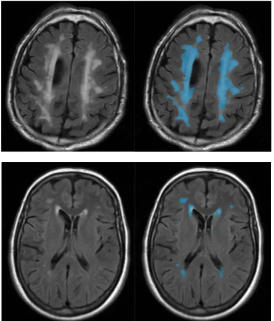
Volumetric overlap between each automatic segmentation method (i.e., FreeSurfer, ANIMAL with label fusion, patch-based, nonlinear patch- based, and nonlinear patch-based with
ةيلك رييستلا مولعو ةيراجتلاو ةيداصتقلاا مولعلا مسق : ةبساحملاو ةيلاملا لخدت ةركذم ةبساحملاو ةيلاملا يف رتساملا ةداهش لين تابلطتم نمض صصخت : ةسسؤم ةيلام دادعإ نم نيبلاطلا
In particular, the interplay of stochasticity and spatial degrees of freedom leads to spatial demixing of different species or genotypes in the population [ 3 , 4 ], which
( ); adjustmen t of in ter - ann ual v ariability for both no bias correction, model uses onl tem per ature and pre- cipita tion anomalies downscaling spa tiall y v arying tem
Data presented herein, based on engineered primary HMEC models, show that transformation induced by distinct oncogenes resulted in different and reproducible patterns of genetic
The comparison between localized flame wounding vs localized bending treatments brought first insights into poplar primary stem elongation with growth and molecular responses to
Objectives: To assess the ability of 3D amide proton transfer weighted (APTw) imaging based on magnetization transfer analysis to discriminate between multiple sclerosis lesions
Keywords : Medical Imaging, Image Analysis, Image Segmentation, multi- atlas based segmentation, Shape Analysis, Statistical Shape Model, Hippocampus, Alzheimer’s
