A
NTIGENIC DIVERSITY OF BOVINE VIRAL DIARRHOEA VIRAL
ISOLATES CONTRADICTS THE CONCEPT OF HERD SPECIFIC
STRAIN
C. Hamers, M. Lambot, M. Onclin, C. Lecomte, P.-P. Pastoret
Department of Veterinary Immunology-Vaccinology, Faculty of Veterinary Medicine, University of Liège, Bld de Colonster, 20, Bat B 43 b, 4000 Liège, Belgium
ABSTRACT
In the epidemiology of bovine viral diarrhoea (BVD), immunotolerant – persistently infected animals (IPI) appear to be major sources of contamination. These animals produce large quantities of replicating virus and have therefore been proposed as being responsible for generating antigenic variability. However, limited studies have failed to detect antigenic or genetic changes in viruses isolated at different times from IPI. An hypothesis is that the immunotolerance of IPI against their homologous strain is accompanied by immune elimination of antigenic variants. The presence of an IPI in a herd could therefore limit antigenic variation, eventually leading to the existence of herd specific strains. To verify this hypothesis we characterized, against a panel of monoclonal antibodies, 37 BVD virus strains isolated from IPI of 12 herds in Eastern Belgium. Intra-herd antigenic variation was compared to inter-herd variation. Antigenic variation within herds was found to be surprisingly high but, nevertheless, significantly lower than variation between herds.
INTRODUCTION
Bovine viral diarrhoea virus (BVDV) is currently classified in the genus pestivirus, family Flaviviridae, which includes two other common viruses of livestock: classical swine fever virus (CSFV) and border disease virus (BDV). Pestiviruses are antigenically heterogeneous, even within a virus species, yet there is a considerable cross-reactivity within the genus [1]. Two contrasting forms of disease are attributed to BVDV depending upon whether infection is post-natal or fetal. Post-natal infections produce a very common, usually mild or inapparent disease of cattle called bovine viral diarrhoea. When it occurs in early fetal development, BVDV infection may lead to the generation of animals that are immunotolerant to BVDV and permanent virus excretors [2, 3]. The permanent infection of these animals is often inapparent. Many of them will ultimately develop a specific lethal illness called mucosal disease [4, 5].
As permanent virus excretors, immunotolerant – persistently infected (IPI) animals play undoubtedly a key role in the perpetuation of BVDV infections [6]. These animals multiply BVDV at a high rate for months or years [7] and have therefore been sometimes compared to BVDV producing factories. However this abundant proliferation does not appear to generate extensive diversity; limited studies have failed to detect antigenic or genetic changes in viruses isolated at different times from IPI [8, 9]. This is consistent with the hypothesis of immunological elimination of antigenic variants which arise in the otherwise in the immunotolerant animal [1, 9]. On the contrary, acute infections would favour variant BVDV that can escape the immune response [10].
The contamination of pregnant females by a permanent excretor may result in the generation of new IPI. According to the hypothesis of strain stability in IPI, the strains isolated from these newly generated IPI should be antigenically close, or even identical to the strain of the contaminator. Authors have accordingly suggested the existence of herd-specific strains [11]. In order to verify the hypothesis of herd specific strains, 37 BVDV strains were isolated from IPI in 12 herds of Eastern Belgium presenting more than one IPI. Isolates were characterized against a panel of monoclonal antibodies and grouped according to epitope similarities. Based on this grouping, intraherd antigenic variations were compared to inter-herd variations.
MATERIALS AND METHODS
IDENTIFICATION OF THE IPI
This previously reported identification [12] was carried out in herds that had been diagnosed positive for a BVDV infection. Out of 3267 blood samples, 49 animals, in 24 herds, were found viraemic after 2 consecutive tests done at 3–4 weeks intervals. These animals were considered as IPI. The study presented here involved 37 IPI from 12 herds harbouring more than one IPI. Moreover, these animals were all born in the herd where the diagnostics was made. The history of each animal was recorded, blood samples were taken and identified by a letter (A–L) representing the herd and a digit [1–6] for their relative order of birth in the herd.
CELLS
All isolates were grown on calf testicular cell cultures (CT). Cells were cultured in opti-MEM® (Gibco) supplemented with 5 % foetal calf serum (Gibco). Prior to inoculation, CT cells were washed with opti-MEM®. After inoculation, cells were grown in opti-MEM® supplemented with 4 % horse serum, 100 IU/100 µg penicillin/streptomycin per ml. All cultures and media were screened for the presence of pestivirus contaminants.
BIOLOGICAL CLONING AND PRODUCTION OF BVDV ISOLATES
Whole blood samples were centrifuged. Buffy coat was collected and frozen at -80 °C. After thawing, 100 µl of these cell suspensions were seeded on subconfluent CT cells, grown in 4-well multidish (Nunc), and cultured for 7 days. Isolates underwent three successive clonings by limiting dilution. Cloned isolates were then multiplied for stock production during 7 days on CT cells grown in 50 ml tissue culture flasks (Falcon). After one freezing/thawing cycle at -80 °C, cells were scraped. Suspensions were then clarified by centrifugation at 2000 g, for 30 min and stored at -80 °C.
MONOCLONAL ANTIBODY (MAB) PANEL
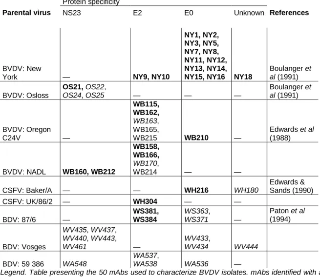
The panel comprised 31 mAbs kindly provided by Dr D.J. Paton (Central Veterinary Laboratory, Weybridge, UK) and 19 mAbs produced in our laboratory. All these mAbs have been previously characterized [13, 14]. Out of these mAbs, 31 were raised against BVDV, 3 against CSFV, 4 against BDV and 12 against atypical pestivirus strains. Table 1 summarizes their specificities.
PEROXIDASE-LINKED ASSAY OF VIRUS/MAB BINDING
An optimal dilution was determined for each mAb, as described by Edwards and colleagues [15]. Antibody binding was detected by peroxidase-linked assay (PLA), as described by Holm Jensen [16], using microplate culture cell monolayers infected with 1000 TCID50 per well of virus. For each isolate, two microplates were inoculated. After 72 h one plate was fixed with 95 % acetone for testing with our mAbs while the other was fixed in 20 % acetone for PLA with mAbs provided by Dr D. J. Paton. Staining was scored as negative, doubtful, positive or strong.
GROUPING OF ISOLATES
Isolates were grouped according to overall epitope similarities. Two approaches were used: one was a parsimony method while the other was a clustering method. For both techniques, virus/mAb interactions, measured by PLA, were scored: negative, 0; positive or strong, 1 ; doubtful, P. Doubtful results were considered as indistinguishable from 0 or from 1 (P = 0 or P = 1).
Table 1. Monoclonal antibodies used in this study and their specificities
Protein specificity
Parental virus NS23 E2 E0 Unknown References
BVDV: New
York — NY9, NY10
NY1, NY2, NY3, NY5, NY7, NY8, NY11, NY12, NY13, NY14,
NY15, NY16 NY18
Boulanger et al (1991) BVDV: Osloss OS21, OS22, OS24, OS25 — — — Boulanger et al (1991) BVDV: Oregon C24V — WB115, WB162, WB163, WB165, WB215 WB210 — Edwards et al (1988) BVDV: NADL WB160, WB212 WB158, WB166, WB170, WB214 — — CSFV: Baker/A — — WH216 WH180 Edwards & Sands (1990) CSFV: UK/86/2 — WH304 — — BDV: 87/6 — WS381, WS384 WS363, WS371 — Paton et al (1994) BDV: Vosges WV435, WV437, WV440, WV443, WV461 — WV433, WV434 WV444 BDV: 59 386 WA548 WA537, WA538 WA536 —
Legend. Table presenting the 50 mAbs used to characterize BVDV isolates. mAbs identified with a W as first letter were kindly provided by Dr D. J. Paton (CVL, Weybridge, UK). Only mAbs in bold could discriminate our isolates.)
CLUSTERING METHOD
An implementation of Algorithm AS 136 [17], developed in StatMost® (DataMost Corp., 1995), was used to segregate isolates into 6 clusters (Table 3). In this programme, grouping is based on K means, with the goal of minimizing the variability within clusters while maximizing the variability between clusters.
PARSIMONY METHOD
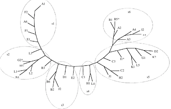
The Penny programme of the Phylip, phylogeny inference package [18] was used for analysis and drawing of a tree diagram. Sub-branches of the tree, as shown in Figure 1, were used for subgrouping isolates.
Figure 1. Epitope tree based on reactivity to Mabs assessed by parsimony method.
Legend. Parsimony tree diagram with subtrees (s1 to s6) drawn in by hand. * Indicates isolates for which subtree and cluster segregations (see Table 3) are not identical. Branch lengths are not proportional.
STATISTICAL ANALYSIS
In order to verify the hypothesis of herd specificity, groups based of antigenic similarities were compared to the groups based on the herd of origin. This was realized by cross-tabulated analysis, using for each isolate the farm of origin as first variable and the calculated cluster or tree subgroup as second variable. T-tests were performed to assess the validity of results. A1 F2 s6 B5* A6 B1 s1 A2 F1 A4 I2 A5 E1 A3 s2 L3 D2 K2 G2* H3 I1* G1 C3 B3 K1 L5 L1 B4 C2 L2 D1 E2 C1 J1 H2 s5 B6 H1 L4 B2 J2 s4 s3
Table 2. Recognition of BVDV strains by Mabs in PLA
Legend. (Strains with the same letter were isolated from IPI belonging to the same herd. Only discriminent mAbs are represented. Solid boxes indicate positive or strong reactions, hatched boxes indicate doubtful results, clear boxes are for negative reactions.)
Table 3. Segregation of isolates into clusters.
Herd Cluster 1 Cluster 2 Cluster 3 Cluster 4 Cluster 5 Cluster 6 A A1, A6 — — — — A2, A3, A4, A5 B — B3, B4 B2, B6 B5 — B1 C — — — C1 C2, C3 — D — — D1 — D2 — E E1 — E2 — — — F F1, F2 — — — — — G — — G2 — G1 — H — H3 — H1 H2 — I — — — — — I1, I2 J — — J2 — J1 — K — — — — K1, K2 — L — L1, L2, L3 — L4 L5 —
Legend. Computerized 6 cluster grouping according to overall epitope similarities (K-means clustering program, StatMost©DataMost Corp., 1995).
RESULTS
VIRUS STRAIN BINDING SPECIFICITY OF MABS
To examine the hypothesis of BVDV herd-specific strains, we compared intra-herd antigenic variation of BVDV isolates to inter-herd variation. Isolates were tested by PLA against a panel of mAbs. Resulting recognition patterns were used to assess antigenic variation. According to the PLA, we found that not all mAbs could discriminate isolates: NY16, OS22, OS24, OS25 recognized all isolates while WB163, WB170, WH180, WS363, WS371, WV433, WV434, WV435, WV437, WV440, WV443, WV444, WV461, WA536, WA537,
WA538, WA548 reacted with none of them. Non-BVDV mAbs recognized only very few isolates of particular herds. Table 2 shows the recognition pattern of isolates by the discriminant mAbs. For simplicity, different intensities of positive have been ignored.
Some isolates had similar patterns; notably L1, L2 and L3 were nearly identical. However, no recognition pattern was farm-specific nor age specific within farms and important variations were found between some strains isolated from the same herd. We had therefore to compare intra-herd to inter-herd variations.
ANTIGENIC GROUPING BY CLUSTERING AND PARSIMONY METHOD
Grouping of isolates, based on recognition patterns, was realized with two different methods. Segregation into clusters was performed with the cluster analysis program of the StatMost® package (DataMost corp., 1995). The 6 clusters obtained are shown in Table 3. The other grouping, realized with the Penny program of the Phylip package (18), produced parsimony trees. A consensus tree diagram is shown in Figure 1. Boot-strap resampling performed to assess validity of trees showed only minor variations, specially within the s5 area (see Fig. 1). Table 3 and Figure 1 showed an obvious relation between the two grouping methods, the 6 clusters corresponding to adjacent regions of the parsimony tree (s1–s6), as shown in Figure 1. On the contrary, relation between farm groups and clusters or between farm groups and subtrees were not so evident and needed therefore to be assessed by statistical analysis.
STATISTICAL COMPARISON OF FARM GROUPS, CLUSTERS AND
SUBTREES
Cross-tabulated analysis with t-test showed a significant (P = 0·03) linkage between herds of origin and subtrees as well as between herds and clusters (P= 0·02).
Within farm A, isolates A1 and A6 had a closely related pattern while A2, A3, A4 and A5 presented an other type of pattern. This suggested the coexistence of two subpopulations (sub-farm A I and sub-farm A II ) of isolates in the same herd. Figure 1 suggests the existence of subpopulations also in farms B and L: subfarm B I : B2, B6, B3 and B4 ; subfarm B II : B1, and B5 ; subfarm L I : L1, L2, L3 ; subfarm L II : L4, L5.
Cross-tabulated analysis using subfarms for A, B and L isolates gave highly significative relation between (sub) farm and subtree (P = 0·0002) and between (sub) farm and cluster (P = 0·001).
DISCUSSION
The hypothesis of the limitation of strain diversification by immune elimination of newly emerged antigenic variants [1, 9] leads to important epidemiological predictions, namely strain stability in individual IPI [8, 9] and herd specific strain [11] in herds harbouring IPI. Our purpose was to examine the existence of BVDV herd specific strains in herds where the presence of immunotolerant animals is thought to limit antigenic variation. Thirty-seven BVDV stains isolated from IPI cattle in 12 herds were tested against a panel of mAbs. Analysis of the resulting recognition patterns showed that no mAb or recognition pattern was farm specific. Antigenic diversity within herds was however, smaller than across herds as assessed by cross-tabulation analysis.
Some isolates reacted with non-BDVD mAbs. One explanation could be that these isolates belong to BVDV type II rather than type I but this was not examined.
Some of the minor variations observed could have occurred after virus isolation as all isolates underwent 5 passages in cell culture including limiting dilutions, prior to being tested against mAbs. However, this would probably not generate the important differences found among the isolates originating from the same farm.
There is no contradiction between our results and the hypothesis of strain stability in individual IPI.
Diversity of strains within the same herd can have several origins. The acute infection of the cow could favour antigenic variation before the fetus is contaminated. Different strains occuring within one herd would then have the same origin. In BVDV infections involving cattle and sheep, this seems to be the case. Limited sequence of 188 nucleotides of the E2 gene showed all isolates to be closely related although epitope mapping showed marked differences [11]. In our case, a partial sequencing of E2 performed on isolates from farms A, B and L [20] also showed a strong farm specific relationship. Barring complete sequence analysis, epitope analysis could be a more subtile tool to pick up variation.
In countries where BVDV is a very common disease, herds have been contaminated over the years and probably by several strains, including the introduction by purchased cattle. These strains might coexist and evolve for years in the same farm. In the context of high endemicity, the concept of herd specific strain could have its limitation.
ACKNOWLEDGEMENTS
We would like to thank Dr D. J. Paton, M. Loncar, Professor A. Ducamp and Professor R. Hamers, respectively for providing monoclonal antibodies, technical assistance, statistical
help and useful comments. This research was supported by grants from the Institut pour l’encouragement de la Recherche Scientifique dans l’Industrie et l’Agriculture (IRSIA).
References
1. Paton DJ. Pestivirus diversity. J Comp Path 1995 ; 112 : 215–36.
2. Liess B, Frey H-R, Kittsteiner H, et al. Observations and investigations on mucosal disease of cattle, a late stage of BVD-MD virus infection with immuno-biological explanation and criteria of a slow virus infection? Deutsche Tierarztliche wochenschrift 1974 ; 81 : 481–7.
3. McClurkin AW, Littledike ET, Cutlip RC, et al. Production of cattle immunotolerant to BVD virus. Can Comp Med 1984 ; 48 : 156–61.
4. Roeder PL, Drew TW. Mucosal disease of cattle: a late sequel to fetal infection. Vet Rec 1984 ; 114 : 309–13.
5. Brownlie J, Clarke MC, Howard CJ. Experimental production of fatal mucosal disease in cattle. Vet. Rec 1984 ; 114 ; 535–6.
6. Roeder PL, Harkness W. BVD infection: Prospects for control. Vet Rec 1986 ; 118 : 143–7. 7. Corapi WV, Donis RO, Dubovi EJ. Monoclonal antibody analyses of cytopathic and
noncytopathic viruses from fatal bovine viral diarrhea virus infections. J Virol 1988 ; 62 : 2823– 7.
8. Edwards S, Wood L, Brockman S, Ibata G. Clinical and virological observations of a mucosal disease outbreak with persistently-infected seropositive survivors. Arch Virol 1991 ; Suppl. 3 : 125–32.
9. Mignon B, Schwers A, Waxweiler S, et al. Etude de la stabilite antigenique d’une souche non cytopathogene du virus BVD chez des animaux infecte s experimentalement de maniere persistante. Ann Med Vet 1990 ; 134 : 325–9.
10. Bolin SR, Ridpath JF. Differences in virulence between two non-cytopathic bovine viral diarrhea viruses in calves. Am Vet Res 1992 ; 53 : 2157–63.
11. Paton DJ, Carlsson U, Lowings JP, et al. Identification of herd-specific bovine viral diarrhoea virus isolates from infected cattle and sheep. Vet Microb 1995 ; 43 : 283–94.
12. Onclin M, Lambot M, Limbourg B, et al. Prevalence des bovins immunotolerants infectes permanents par le pestivirus responsable de la diarrhee virale bovine, dans l’est de la Belgique, au sein de troupeaux infectes. Ann Med Vet 1995 ; 139 : 429–31.
13. Paton DJ, Sands JJ, Lowings JP, et al. A proposed division of the pestivirus genus using monoclonal antibodies, supported by cross-neutralization assays and genetic sequencing. Vet Res 1995 ; 26 : 92–109.
14. Boulanger D, Waxweiler S, Karelle L, et al. Characterization of monoclonal antibodies to bovine viral diarrhea virus: evidence of a neutralizing activity against gp48 in the presence of goat anti-mouse immunoglobulin serum. J Gen Virol 1991 ; 72 : 1195–8.
15. Edwards S, Sands JJ, Harkness JW. The application of monoclonal antibody panels to characterize pestivirus isolates from ruminants in Great Britain. Arch Virol 1988 ; 102 : 197– 206.
16. Holm Jensen M. Detection of antibodies against hog cholera virus and bovine viral diarrhea virus in porcine serum. Acta Vet Scand 1981 ; 22 : 85–98.
17. Collected Algorithms for ACM, Volume IV. A publication of the Association for Computing Machinery, Inc. New York, 1989.
18. Felsenstein J. Phylip: phylogeny inference package (version 3.2). Cladistics 1989 ; 5 : 164–6. 19. Pellerin C, Van den hurk J, Lecomte J, Tussen P. Identification of a new group of bovine viral
diarrhea virus strains associated with severe outbreaks and high mortalities. Virol 1994 ; 203 : 260–8.
20. Hamers C, Lecomte C, Kulcsar G, et al. Persistently infected cattle stabilise bovine viral diarrhea virus leading to herd specific strain. Vet Microbiol 1998 ; 61 : 177–82.