Contents lists available atScienceDirect
Biomedicine & Pharmacotherapy
journal homepage:www.elsevier.com/locate/biophaAntiviral e
ffects of selected IMPDH and DHODH inhibitors against foot and
mouth disease virus
Mei-jiao Gong
1, Shi-fang Li
1, Yan-yan Chang, Jun-jun Shao, Yue-feng Sun, Ting-ting Ren,
Yong-guang Zhang, Hui-yun Chang
⁎State Key Laboratory of Veterinary Etiological Biology, OIE/National Foot-and-Mouth Disease Reference Laboratory, Lanzhou Veterinary Research Institute, Chinese Academy of Agricultural Sciences, Lanzhou, 730046, Gansu, China
A R T I C L E I N F O Keywords: FMDV Antiviral activity IMPDH DHODH In vivo A B S T R A C T
Foot-and-mouth disease virus (FMDV) is an important pathogen that affects livestock breeding and causes huge economic losses worldwide. Currently, the development of antiviral agents to combat FMDV infection at the early stages is being explored. As viral replication critically depends on the host for nucleoside supply, host enzymes involved in nucleotides biosynthesis may represent potential targets for the development of antiviral agents. In the present study, the effects of IMP dehydrogenase (AVN-944 and mycophenolate mofetil) and di-hydroorotate dehydrogenase (teriflunomide) inhibitors were evaluated both in vitro and in vivo. The results revealed that these compounds were effective in suppressing FMDV (O/MY98/BY/2010 and A/GD/MM/2013) infection. With regard to the antiviral mechanism, time-of-addition experiments revealed that these compounds were effective when added at the early stages of viral lifecycle (0–8 h post infection). However, exogenous guanosine/uridine eliminated the antiviral activity of these compounds. Importantly, treatment AVN-944 and teriflunomide significantly improved the survival of mice that were subcutaneously treated with FMDV. Together, the results of the present study indicate the broad-spectrum activities of anti-FMDV agents targeting IMP dehydrogenase or dihydroorotate dehydrogenase, which could be useful in developing strategies to prevent FMD.
1. Introduction
Foot-and-mouth disease (FMD) is one of the highly contagious dis-eases of domestic and wild cloven-hoofed animals, such as cattle, swine, sheep, goats, and deer. The causative agent, FMD virus (FMDV), rapidly replicates in the host and spreads to susceptible animals in contact [1]. The common symptoms of FMD include fever, lethargy, and appearance of vesicular lesions in tongue, feet, snout, and teats, resulting in high morbidity and low mortality in adult animals, and high mortality in young animals because the disease can affect the heart [2]. The FMDV is the type species of the genus Aphthovirus belonging to the Picorna-viridae family, and contains a single-stranded, positive-sense RNA genome of approximately 8500 bases surrounded by an icosahedral capsid with 60 copies each of four structural proteins, VP1–4. The virus is antigenically highly variable and consists of seven serotypes, in-cluding O, A, C, Asia 1, South African Territories (SAT) 1, SAT 2, and SAT 3, with multiple subtypes in each serotype [3]. Serotype O is the most common worldwide and has been responsible for much of the
reported economically devastating disease occurrence [4,5]. The cur-rently available measures for controlling and preventing FMD mainly comprise vaccination. However, vaccines are ineffective in controlling complicated outbreaks or genetic variants of the virus. Therefore, there is an urgent need to develop specific antiviral strategies against FMDV for providing early protection from the disease, as well as determine alternative methods of applying antiviral agents to reduce the spread of FMDV in outbreak situations [6].
Cellular nucleotides, composed of purines and pyrimidines, are important constituents of RNA and DNA, and viruses heavily rely on specific host factors, such as nucleosides, to complete their lifecycles. Therefore, disruption of enzymes involved in nucleosides biosynthesis in hosts may represent a potential antiviral strategy. Inosine mono-phosphate dehydrogenase (IMPDH), a key enzyme in the de novo synthesis of purines, is widely conserved in living organisms [7]. This enzyme catalyzes a rate-limiting step essential for the de novo synthesis of guanosine monophosphate (GMP) and ultimately the nucleotide guanosine triphosphate (GTP) [8]. Two isoforms of IMPDH, type I and
https://doi.org/10.1016/j.biopha.2019.109305
Received 13 June 2019; Received in revised form 26 July 2019; Accepted 31 July 2019
⁎Corresponding author at: No.1 Xujiaping, Yanchangbao, Chengguan District, Lanzhou, 730046, Gansu, China.
E-mail address:changhuiyun@caas.cn(C. Hui-yun).
1These authors contributed equally to this paper.
0753-3322/ © 2019 The Authors. Published by Elsevier Masson SAS. This is an open access article under the CC BY-NC-ND license (http://creativecommons.org/licenses/BY-NC-ND/4.0/).
II with 84% amino acids identity, have been identified in humans. As inhibition of IMPDH can deplete guanine nucleotide pools, followed by a decrease in DNA and RNA synthesis, this enzyme could be a good candidate for use as an antiviral agent. IMPDH has been employed as a target in immunosuppressive therapy, as an antiangiogenic agent, and as an antiviral drug [9,10]. Dihydroorotate dehydrogenase (DHODH) is sequentially the fourth and rate-limiting enzyme in the de novo bio-synthesis pathway of pyrimidines. Located in the inner membrane of mitochondria, DHODH can convert dihydroorotate to orotate, which is subsequently utilized by the multifunctional uridine monophosphate (UMP) synthase to produce UMP. The inhibition of pyrimidines bio-synthesis has also been explored as a strategy in rheumatology and oncology for treating various infectious diseases, cancers, and im-munological disorders [11,12].
In the present study, we evaluated the effects of two IMPDH hibitors (AVN-944 and mycophenolate mofetil) and one DHODH in-hibitor (teriflunomide) on FMDV replication in vitro, and then identi-fied their preliminary modes of action. Besides, the antiviral activities of these compounds against FMDV infection in vivo were also de-scribed.
2. Methods
2.1. Experimental materials
IBRS-2 cells were cultured in Dulbecco’s modified Eagle’s medium (DMEM) supplemented with 10% fetal bovine serum (FBS) and 1% penicillin-streptomycin at a temperature of 37 °C with 5% CO2. The
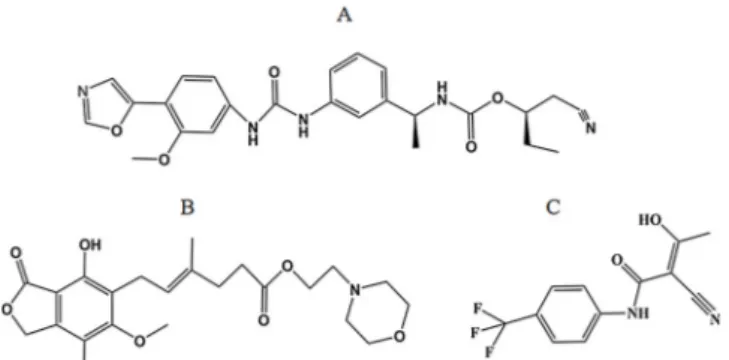
virus strains of serotype O (O/MY98/BY/2010) and A (A/GD/MM/ 2013) FMDV used in the present study were derived from Lanzhou Veterinary Research Institute, Chinese Academy of Agriculture Sciences. AVN-944, mycophenolate mofetil (MF), and teriflunomide (Fig. 1), guanosine and uridine were all products of MCE. Rabbit hyper-immune serum raised against O/MYA98/BY/2010 were prepared in our laboratory. PrimeScript™ RT reagent kit containing gDNA Eraser and SYBR Premix Ex Taq™II (Tli RNaseH Plus) used for Q-PCR experiment was purchased from TaKaRa (Dalian, China). All the other agents used in this report were commercially available.
2.2. Ethics statements
All animal experiments were performed in a Biosafety level-3 la-boratory and approved according to the requirements of the Animal Ethics Committee of Lanzhou Veterinary Research Institute, Chinese Academy of Agricultural Science (No. LVRIAEC2018-007).
2.3. Cell viability assay
The potential cytotoxicity of compounds against IBRS-2 cells was
determined by MTS assay (Abcam, USA) according to the manu-facturer’s instructions. Briefly, the IBRS-2 cells were cultured in 96-well plate at a density of 3 × 10⁴cells/well, and various concentrations of
AVN-944, mycophenolate mofetil, and teriflunomide (100–500 μM) were respectively added. After incubation for 72 h at 37℃ in 5% CO2
atmosphere, 20μL of MTS were added to each well for another 4 h at 37℃. Then, the absorbance of each well at 490 nm was determined by using ELISA reader.
2.4. Antiviral activity assay
The antiviral activity of the compounds was determined based on cytopathic effect (CPE) reduction by MTS assay. One day prior to in-fection, 3 × 10⁴IBRS-2 cells/well were seeded onto a 96-well culture plate. The next day, the medium was removed and cells were washed with PBS. Subsequently, 100 TCID50of FMDV (O/MY98/BY/2010 and
A/GD/MM/2013) were added to the cells for 1 h, respectively. Afterwards, 0.1 mL of medium supplemented with FBS containing ap-propriate concentration of the test compounds were added to the cells to generate appropriate CPE for 48 h after infection. Four wells were used as viral controls (virus-infected cells that were not treated with the compounds), while another four wells were employed as cell controls (non-infected cells that were not treated with the compounds). The absorbance of each well was determined by a plate reader at 490 nm. The results were quantified as a percentage of the controls, and the 50% effective concentration (EC50) values were graphically obtained using
GraphPad software. The percent protection (expressed in %) achieved by the test compounds in FMDV-infected cells was calculated as follows: {(ODt)FMDV − (ODc)FMDV}/{(ODc)mock − (ODc)FMDV}×100, where (ODt)FMDV is the optical density of FMDV-infected cells treated with a given test compound, (ODc)FMDV is the optical density of the control untreated FMDV-infected cells, and (ODc)mock is the optical density of the control untreated mock-infected cells. The antiviral ac-tivity was presented as % of control. DMSO was used as a negative control. The cell culture supernatants were collected at 48 h post-in-fection for the detection of FMDV 2B mRNA by Q-PCR. For nucleosides supplementation, IBRS-2 cells (3 × 10⁵cells/well) were plated in
12-well plates and treated as described earlier [13]. The samples were collected at 48 h post-infection.
2.5. Time-of-Addition assay
The time-of-addition effect was examined as previously described [14]. In brief, 2 × 10⁵ IBRS-2 cells/well were seeded onto 12-well
culture plates and incubated for 24 h. Then, the confluent monolayer was infected with FMDV for 1 h and treated with the test compounds (added into the wells) either concurrently during FMDV infection (0 h) or at intervals of 2, 4, 8, and 16 post-infection. After incubation at 37 °C for 48 h, samples were collected and the reduction in viral 2B mRNA levels and VP1 protein was determined by Q-PCR and Western blot analysis, respectively.
2.6. Indirect immunofluorescence assay
The IBRS-2 cells infected with FMDV in a 12-well plate werefixed with 4% paraformaldehyde, permeabilized with 0.5% Triton X-100 (in PBS), blocked with 1% bovine serum albumin (BSA) for 0.5 h, and then incubated with primary antibody (rabbit hyperimmune serum) for 1 h, followed by appropriate peroxidase-conjugated goat anti-rabbit IgG (H + L) for 1 h. The cell nuclei were stained with 4′,6-diamidino-2-phenylindole, washed repeatedly with PBS, andfluorescence was ob-served under afluorescence microscope (Nikon ECLIPSE TS100 fluor-escence microscope, Nikon, Japan).
Fig. 1. The chemical structure of compounds. A. AVN-944. Molecular weight = 477.51 g/mol. B. mycophenolate mofetil. Molecular weight = 433.49 g/mol. C. Teriflunomide. Molecular weight =270.51 g/mol.
2.7. Q-PCR assay
For relative quantification of viral mRNA synthesis, the total RNA from the harvested cells was isolated using TRIzol reagent (Invitrogen). The RNA was eluted in 50μL of RNase-free water and treated with PrimeScript™ RT reagent kit containing gDNA Eraser (Takara, Dalian, China) to eliminate DNase I, according to the manufacturer’s instruc-tions. Subsequently, the total RNA was reverse transcribed with random hexamer primers using Agilent Technologies Stratagene Mx3005 P in-strument (Agilent, USA) and SYBR Premix Ex TaqTMII (Tli RNaseH
Plus) (TaKaRa, Dalian, China) with a reaction volume of 25μL. The relative expression of the FMDV 2B (forward, 5′-CAACAAAACACGGA CCCGAC-3′; reverse, 5′-TTGTACCAGGGTTTGGCCTC-3′) was de-termined by normalizing against the porcine β-actin gene (forward, 5′-GACCACCTTCAACTCGATCA-3′; reverse, 5′-GTGTTGGCGTAGAGGT CCTT-3′).
2.8. Western blot analysis
The cell lysates were separated by 12% SDS-PAGE and transferred to a polyvinylidene difluoride membrane (Millipore, USA). The mem-brane was blocked with 5% non-fat milk powder in TBST buffer (20 mM Tris−HCl, pH 7.4, 150 mM NaCl, 0.1% Tween-20) and then probed with anti-FMDV VP1 antibody as described previously [13], or with anti-β-actin antibody (Sigma-Aldrich, USA). The bound antibody was detected using HRP-conjugated secondary antibodies and visualized by enhanced chemiluminescence (GE Healthcare).
2.9. Invivo Experiments
In vivo experiments were performed with specific pathogen-free 3–4 day-old BALB/c suckling mice (Lanzhou Veterinary Research Institute, China). The 50% lethal dose (LD50) of FMDV was estimated by
Reed-Muench method. The BALB/c mice were infected with 100μL of 100 LD50of FMDV O/MY98/BY/2010. Subsequently, 60μg of AVN944,
mycophenolate mofetil, and teriflunomide were respectively dissolved in PBS containing 100μM DMSO and 5% Tween-80, and sub-cutaneously administered to the mice for 2 h at the time of virus ex-posure. The placebo controls received 100μL of solvent, while the virus controls and normal controls received no viral infection and test com-pounds treatment. The mice were observed for 96 h after infection. 2.10. Histopathology
Heart tissues from solvent-treated (n = 4), solvent-treated FMDV-infected (n = 4), and AVN944-treated FMDV-FMDV-infected (n = 4) mice werefixed in 10% formalin for 48 h at room temperature, sectioned, conventionally stained with hematoxylin and eosin, and examined. 2.11. Statistical analyses
The data were processed using GraphPad Prism 5 and expressed as means ± S.D. Statistical differences between the two groups were de-termined using Student's t-test. For multiple groups, one-way ANOVA was employed to compare the means, and p < 0.05 indicated sig-nificant difference between the groups.
3. Results
3.1. Cytotoxic effects of the compounds on IBRS-2 cells
The test compounds were examined for cytotoxicity before assessing their antiviral activities. As shown inFig. 2A, D and G, the compounds were differentially cytotoxic. While mycophenolate mofetil, at the ex-amined concentrations, did not produce a cytotoxic effect on IBRS-2 cells, AVN944 (< 100μM), and teriflunomide (< 400 μM) failed to
distinctly affect cell viability following treatment for 72 h. However, a higher concentration of AVN944 and teriflunomide exhibited sig-nificant dose-dependent cytotoxic effects on IBRS-2 cells, when com-pared with the DMSO-treated control. The 50% cytotoxic concentration (CC50) of AVN944 and teriflunomide on IBRS-2 cells were 88.18 μM and
542.7μM, respectively.
3.2. Antiviral activity of the compounds against FMDV invitro
Based on the results of cytotoxicity of the test compounds, a non-toxic concentration was employed to explore the antiviral activity of these compounds against FMDV. The cells were infected with 100 TCID50of FMDV and incubated with different concentrations of the test
compounds for 48 h. The results revealed that viral replication was inhibited by the test compounds in a dose-dependent manner. The EC50
of AVN944, mycophenolate mofetil, and teriflunomide were 12.02 μM, 8.142μM, and 294.1 μM against FMDV O/MY98/BY/2010, respec-tively, and the selectivity indices (SI) were 7.33, 12.28, and 1.84, re-spectively. Besides, the test compounds also showed significant in vitro antiviral activities against another strain of FMDV (A/GD/MM/2013). The EC50values of AVN944, mycophenolate mofetil, and teriflunomide
against FMDV A/GD/MM/2013 in IBRS-2 cells were 3.953μM, 6.500μM, and 104.9 μM, yielding SI values of 22.30, 15.38, and 5.17, respectively (Fig. 2B, E, and H). Interestingly, the SI values of the test compounds against FMDV A/GD/MM/2013 were higher than those against FMDV O/MY98/BY/2010. The antiviral activities of these compounds were also analyzed by qPCR and immunofluorescence assay (IFA). As shown in Fig. 2C, F, and I, treatment with various con-centrations of the test compounds for 48 h inhibited viral replication in a dose-dependent manner, as quantified by the relative levels of the FMDV 2B gene expression. Consistently, the number of FMDV-positive cell foci presented a corresponding decrease in a dose-dependent manner, as indicated by indirect IFA (Fig. 3).
3.3. Effects of Guanosine/Uridine on the antiviral activity of the compounds To further address whether guanosine/uridine could block the ac-tivity of the test compounds, the infected cells treated with or without the test compounds were supplemented with serial dilutions of gua-nosine/uridine. Notably, FMDV VP1 protein levels recovered following guanosine supplementation in cells treated with mycophenolate mofetil and AVN944 (Fig. 4B and C), indicating that the antiviral mechanism of these compounds involved depletion of the purine nucleotide pool. Likewise, uridine also presented similar effect on teriflunomide-medi-ated suppression of FMDV replication (Fig. 4D), suggesting that in-hibition of DHODH is the major antiviral mechanism of teriflunomide against FMDV infection.
3.4. Time course assay
Time course assay was used to examine the stages of FMDV re-plication that were affected by the test compounds (Fig. 5). The test compounds were added during viral infection (0 h post-infection) or after viral infection (2–16 h post-infection). The mRNA and VP1 protein changes in the IBRS-2 cells were analyzed at 48 h post-infection. As shown inFig. 5, the compounds could considerably reduce 2B mRNA and VP1 protein levels until 8 h post-infection; however, no significant inhibitory effect on FMDV replication was observed after 16 h of viral adsorption. Taken together, these results suggested that the tested in-hibitors constrained FMDV replication in vitro at the early stage of infection.
3.5. Invivo Experiments
Based on the anti-FMDV potency of the inhibitors in vitro, their protective efficacy against lethal FMDV infection in suckling mice was
further explored. Three-day-old suckling mice were injected with FMDV O/MY98/BY/2010 strain at a dose of 100 LD50,followed by treatment
with the test compounds. As shown inFig. 6, the suckling mice in the control group began to die at 36–60 h. Among the treatment groups, the suckling mice in the AVN-944 and teriflunomide treatment groups began to die at 72 h and 60 h, respectively. The infected mice in the mycophenolate mofetil treatment group began to die at 36 h, and the death time decreased subsequently (P= 0.02). At 96 h post-infection, the survival rates of mice in the AVN-944, mycophenolate mofetil, and teriflunomide treatment groups were 25%, 8.3%, and 25%, respec-tively. Although mycophenolate mofetil injection showed a higher survival rate, no statistical significance were found between the control and the drug-treated groups (P=0.02). Larger studies are needed for a significant difference in the survival rate between the two groups. While, subcutaneous administration of AVN-944 and teriflunomide significantly improved the survival rates of infected mice, when com-pared with the virus control group. In particular, AVN944 showed su-perior activity using the same dose, when compared with the treatment of teriflunomide. Therefore, the protective effects of AVN944 against FMDV-induced tissue damage were further examined through histo-pathology analysis. As shown inFig. 7, myocardialfiber dissolution and inflammatory cell infiltration were observed in the hearts of virus
control mice without inhibitors treatment (Fig. 7C and F), whereas no obvious inflammatory symptoms were noted in AVN944-treated in-fected mice (Fig. 7B and E).
4. Discussion
In general, vaccination is considered as the gold standard for trolling viral diseases; however, the use of antiviral drugs for the con-trol of highly mutagenic viruses such as FMDV is still limited. As commercially available vaccines do not provide early protection for virus-infected animals, the World Organization for Animal Health (OIE) recommends the use of a combination of vaccines and antiviral drugs to effectively control viruses [15]. It is therefore very important to un-cover antiviral agents for FMDV treatment. In recent years, increasing evidences have indicated that targeting DHODH and IMPDH may be a promising strategy to develop broad-spectrum antiviral drugs.
AVN944 is a highly selective, non-nucleoside, specific, un-competitive IMPDH inhibitor, which was developed by Vertex (VX-944) and licensed by Avalon Pharmaceuticals (AVN944) [16]. AVN944 has been demonstrated to be well-tolerated by humans, and is currently under clinical trials on patients with hematological malignances and, in combination with gemcitabine, on patients with pancreatic cancer [17].
Fig. 2. Compounds potently inhibits FMDV infection in IBRS-2 cells. (A, D, G) Cytotoxicity of compounds on IBRS-2 cells. MTS assays were performed to assess IBRS-2 cells viability after 72 h of treatment with AVN-944 (A), mycophenolate mofetil (D), teriflunomide (G), respectively. Data are expressed as mean ± SD of the percentage values obtained from three independent experiments carried out in triplicate. (B, E, H) IBRS-2 cells were infected with 100 TCID50FMDVs (O/MY98/BY/
2010 or A/GD/MM/2013) for 1 h and then treated with increasing concentrations of compounds. 48 h after treatment, antiviral activity was determined by the reduction of the cytopathic effect in an MTS assay. Cell viability of DMSO-treated cells was set to 0% and that of uninfected cells was set 100%. (C, F, I) Total RNAs were prepared from cells in (B, E, H) and then subjected to Q-PCR for 2B region of FMDV viral RNA.β-actin mRNAs were also analyzed as a negative control. Data are expressed as mean ± SD of the percentage values obtained from three independent experiments carried out in triplicate. ***P < 0.001.
Mycophenolic mofetil, a prodrug of mycophenolic acid, is a non-com-petitive inhibitor of IMPDH, and has been approved by the FDA for immune suppression. In addition, it has been reported to inhibit H5N1 virus replication in MDCK cells and mice [18]. Teriflunomide, a prin-cipal active metabolite of leflunomide, is a novel oral im-munomodulator with anti-inflammatory properties, and is used for the treatment of patients with relapsing forms of multiple sclerosis and rheumatoid arthritis [19,20]. Besides its anti-inflammatory effects, teriflunomide has also been found to exert antiviral activities against numerous viruses, including Theiler’s murine encephalomyocarditis virus [21], Epstein-Barr virus [22], Newcastle disease virus, and Ebola virus [23]. To the best of the authors’ knowledge, the present study is thefirst to demonstrate the antiviral activity of AVN944, mycophenolic
mofetil, and teriflunomide against FMDV.
With regard to FMDV epidemic, besides type O FMDV, type A FMDV is also widely disseminated in China [24]. To explore whether the an-tiviral activity of the tested compounds is strain-dependent, the effect of these inhibitors on type A FMDV (A/GD/MM/2013) was evaluated. The results demonstrated that the compounds could also inhibit type A FMDV infection in IBRS-2 cells, supporting that the tested IMPDH and DHODH inhibitors exerted broad-spectrum antiviral activities. In future studies, the effect of these compounds on other FMDV genotypes or strains must be investigated. Considering their characteristics, both IMPDH and DHODH could be promising candidates for the treatment of other viral diseases, such as enterovirus-A71 (which belongs to the same family (Picornaviridae) as FMDV) and African swine fever (ASF;
Fig. 3. The viral proteins were determined by IFA. IBRS-2 cells were infected by 100 TCID50 FMDV O/MY98/BY/2010, with or
without treatment by various concentrations of compounds for 12 h. The green fluorescence represents the intracelluar distribution of FMDV visualized by DAPI staining. Scale bars indicate 100μm (For interpretation of the re-ferences to colour in this figure legend, the reader is referred to the web version of this article).
Fig. 4. Guanosine or uridine attenuated the antiviral activity of compounds. A. Schematic overview of de novo biosynthesis of purine and pyrimidine nucleotide. (B, C) FMDV-infected IBRS-2 cells were treated with AVN-944 (50μM) or mycophenolate mofetil (50μM) in the presence of exogenous guano-sine. (D) FMDV-infected IBRS-2 cells were treated with teriflunomide (300 μM) in the presence of exogenous uridine, as indicated. Cells were incubated for 48 h, total cell extracts were prepared from cells and subjected to Western Blot analysis with anti-VP1 antibody. β-actin was also analyzed as a loading control.
another disease listed by OIE similar to FMD). Since thefirst reported ASF case in China in 2018, over 100 ASF cases had been recorded in 23 provinces or regions of China [25]. ASF has caused a severe threat to the domestic pig population in China, resulting in huge economic loss. With the current lack of vaccines to control ASF, there is a need to consider alternative control strategies, including IMPDH and DHODH as potential targets for antiviral drug development. Interestingly, it has been indicated that targeting other catalytic steps leading to primary purine nucleotide synthesis may stimulate viral replication (e.g. hepa-titis E virus (HEV)) [26]. For example, Wang et al. reported that in-hibition of amido phosphoribosyltransferase, glycinamide ribonucleo-tide transformylase, 5-aminoimidazole-4-carboxamide ribonucleoribonucleo-tide formyltransferase, and IMP cyclohydrolase promoted HEV replication [26]. In our future study, we will test the effects of inhibition of these enzymes on FMDV infection. In fact, besides the inhibition of nucleotide synthesis, disruption of protein translation may also be a potential antiviral therapeutic strategy to control infection because viral
Fig. 5. Antiviral activity of compounds de-pending on the time of addition. IBRS-2 cells were infected with 100 TCID50 FMDV O/
MY98/BY/2010 and treated with 50μM of AVN-944 (A, B), mycophenolate mofetil (C, D) or 300μM teriflunomide (E, F) at the indicated times after virus infection, respectively. 48 h post-infection, total cell extracts were prepared from cells and subjected to Q-PCR and Western Blot analysis with anti-VP1 antibody, respec-tively.β-actin was also analyzed as a loading control. Data are expressed as mean ± SD of the percentage values obtained from three in-dependent experiments carried out in tripli-cate. *P < 0.05, ***P < 0.001.
Fig. 6. in vivoactivity of compounds. Suckling mice were challenged with 100 LD50of FMDV O/MY98/BY/2010 after 2 h pre-treatment with 60μg of
AVN-944, mycophenolate mofetil and teriflunomide, respectively. The survival rate of suckling mice was monitored for 96 h post-challenge.
Fig. 7. Histological analysis of heart tissue stained with hematoxylin and eosin (H&E) in suckling mouse. The suckling mouse was treated with the placebo or 60μg AVN-944. The pathological changes of heart tissues at 30 hpi were observed after H & E staining. Representative H&E stained samples of heart tissue from (A, D) health, (B, E) AVN-944-treated, and (C, F) placebo-treated are shown (N = 4). A, B, C (magnification, 100×); D, E, F (magnification, 400×).
replication also relies on cellular translational machinery. For instance, halofuginone, a potent prolyl tRNA synthetase inhibitor, has been de-monstrated to suppress Chikungunya and Dengue viral infections [27] as well as FMDV replication (unpublished).
In the present study, to further investigate the stage of FMDV re-plication that is sensitive to the tested inhibitors, the IBRS-2 cells were exposed to these compounds at various time points after virus entry. While the addition of these compounds to the infected cells significantly suppressed FMDV replication before 8 h post-infection, no significant inhibitory effects were noted after 16 h post-infection. The results of time-course study suggested that all the three tested compounds ex-hibited inhibitory effects on the early stages of FMDV replication cycle. One of the well-recognized antiviral mechanisms of inhibitor com-pounds is disruption of IMPDH/DHODH to deplete the intracellular GTP or pyrimidine pools [26]. To determine whether this mechanism is involved in FMDV inhibition, guanosine/uridine was added to the in-fected cells treated with inhibitors. While guanosine/uridine alone had no effect, they significantly attenuated the anti-FMDV effects of these compounds, suggesting that nucleotide depletion by inhibitors is an important mechanism in controlling FMDV infection. In recent years, other novel potential mechanisms have been reported; for example, some IMPDH and DHODH inhibitors have also been noted to enhance the innate immune responses triggered by short 5′-triphosphate RNA molecules as measured by IFN-stimulated response element reporter and IFN-stimulated expression of downstream genes, such as IFN-in-duced protein with tetratricopeptide repeats 1 and IFN regulatory factor 1, thus further contributing to their antiviral activities [28]. However, in general, treatment with individual antiviral agents cannot achieve complete inhibition of viruses owing to limitations related to viral resistance, such as amino acid substitutions against mutagens [6]. It has been proposed that treatment with a combination of antiviral agents with different mechanisms might be more advantageous in overcoming their individual limitations. Similarly, combinations of IMPDH inhibitor and nucleoside analog have been reported to present synergistic benefits in treating several viruses [29].
Although IMPDH and DHODH inhibitors have been noted to show antiviral activity against some viruses, many of them have failed to exhibit promising antiviral effects in animal models. However, in the present study, the effects of the test compounds were also observed in the animal experiment, and the suckling mice treated with these com-pounds exhibited higher survival rates, when compared with the con-trol group. Interestingly, treatment with AVN944 was more effective than treatments with mycophenolate mofetil and teriflunomide. Therefore, in future studies, the antiviral efficacy of these compounds should be further investigated in relevant species such as pigs and cattle. Choi et al. reported that the combination of ribavirin and vaccine presented a synergistic effect on the early stage of FMDV replication [30]. Hence, the effects of combinations of antiviral agents and novel FMD vaccines on possible early protection against viral infection prior to the establishment of host immunity through vaccination must be explored.
In conclusion, the results of the present study demonstrated the effective antiviral activities of AVN944, mycophenolate mofetil, and teriflunomide against FMDV, and their underlying initial antiviral me-chanisms. Future research should focus on assessing the activities of these compounds in natural hosts as well as their efficacies in combi-nation with vaccines.
Declaration of Competing Interest None.
Acknowledgments
This research was funded by the National Key Research and
Development Program of China (2017YFD0500902 and 2016YFE0204100) and the Key R&D Program of Gansu Province of China (Grant No. 17YF1NA070).
References
[1] M.J. Grubman, B. Baxt, Foot-and-mouth disease, Clin. Microbiol. Rev. 17 (2004) 465–493.
[2] E. Ryan, J. Horsington, S. Durand, et al., Foot-and-mouth disease virus infection in young lambs: pathogenesis and tissue tropism, Vet. Microbiol. 127 (2008) 258–274. [3] Y. Cao, Adjuvants for foot-and-mouth disease virus vaccines: recent progress, Expert
Rev. Vaccines 13 (2014) 1377–1385.
[4] D.J. Paton, K.J. Sumption, B. Charleston, Options for control of foot-and-mouth disease: knowledge, capability and policy, Philos. Trans. R. Soc. Lond., B, Biol. Sci. 364 (2009) 2657–2667.
[5] T.J. KnightJones, J. Rushton, The economic impacts of foot and mouth disease -what are they, how big are they and where do they occur? Prev. Vet. Med. 112 (2013) 161–173.
[6] S.M. Kim, J.H. Park, K.N. Lee, et al., Enhanced inhibition of foot-and-mouth disease virus by combinations of porcine interferon-alpha and antiviral agents, Antiviral Res. 96 (2012) 213–220.
[7] J.M. Hamilton, M.W. Harding, T. Genna, et al., A phase I dose-ranging study of the pharmacokinetics, pharmacodynamics, safety, and tolerability of AVN944, an IMPDH inhibitor, in healthy male volunteers, J. Clin. Pharmacol. 49 (2009) 30–38. [8] A. Zimmermann, J.J. Gu, J. Spychala, et al., Inosine monophosphate dehydrogenase expression: transcriptional regulation of the type I and type II genes, Adv. Enzyme Regul. 36 (1996) 75–84.
[9] A.C. Allison, T. Hovi, R.W. Watts, et al., The role of de novo purine synthesis in lymphocyte transformation, Ciba Found. Symp. (1977) 207–224.
[10] C.R. Chong, D.Z. Qian, F. Pan, et al., Identification of type 1 inosine monophosphate dehydrogenase as an antiangiogenic drug target, J. Med. Chem. 49 (2006) 2677–2680.
[11] V.K. Vyas, M. Ghate, Recent developments in the medicinal chemistry and ther-apeutic potential of dihydroorotate dehydrogenase (DHODH) inhibitors, Mini Rev. Med. Chem. 11 (2011) 1039–1055.
[12] H. Munier-Lehmann, P.O. Vidalain, F. Tangy, et al., On dihydroorotate dehy-drogenases and their inhibitors and uses, J. Med. Chem. 56 (2013) 3148–3167. [13] S.F. Li, M.J. Gong, Y.F. Sun, et al., Antiviral activity of brequinar against
foot-and-mouth disease virus infection in vitro and in vivo, Biomed. Pharmacother. 116 (2019) 108982.
[14] S.F. Li, M.J. Gong, Y.F. Sun, et al., In vitro and in vivo antiviral activity of mizor-ibine against foot-and-Mouth disease virus, Molecules. 24 (2019).
[15] J.M. Scudamore, G.M. Trevelyan, M.V. Tas, et al., Carcass disposal: lessons from Great Britain following the foot and mouth disease outbreaks of 2001, Rev. Sci. Tech. 21 (2002) 775–787.
[16] K. Ishitsuka, T. Hideshima, M. Hamasaki, et al., Novel inosine monophosphate dehydrogenase inhibitor VX-944 induces apoptosis in multiple myeloma cells pri-marily via caspase-independent AIF/Endo G pathway, Oncogene. 24 (2005) 5888–5896.
[17] D. Floryk, T.C. Thompson, Antiproliferative effects of AVN944, a novel inosine
5-monophosphate dehydrogenase inhibitor, in prostate cancer cells, Int. J. Cancer 123 (2008) 2294–2302.
[18] J. Cho, H. Yi, E.Y. Jang, et al., Mycophenolic mofetil, an alternative antiviral and immunomodulator for the highly pathogenic avian influenza H5N1 virus infection, Biochem. Biophys. Res. Commun. 494 (2017) 298–304.
[19] C. Warnke, O. Stuve, B.C. Kieseier, Teriflunomide for the treatment of multiple sclerosis, Clin. Neurol. Neurosurg. 115 (Suppl 1) (2013) S90–94.
[20] J. Bae, J.W. Park, Topical delivery of leflunomide for rheumatoid arthritis treat-ment: evaluation of local tissue deposition of teriflunomide and its anti-in-flammatory effects in an arthritis rat model, Drug Dev. Ind. Pharm. 42 (2016) 254–262.
[21] F. Gilli, L. Li, D.B. Royce, et al., Treatment of Theiler’s virus-induced demyelinating disease with teriflunomide, J. Neurovirol. 23 (2017) 825–838.
[22] A. Bilger, J. Plowshay, S. Ma, et al., Leflunomide/teriflunomide inhibit Epstein-Barr
virus (EBV)-induced lymphoproliferative disease and lytic viral replication, Oncotarget 8 (2017) 44266–44280.
[23] S. Martin, A.I. Chiramel, M.L. Schmidt, et al., A genome-wide siRNA screen iden-tifies a druggable host pathway essential for the Ebola virus life cycle, Genome Med. 10 (2018) 58.
[24] Z. Zhu, F. Yang, J. He, et al., First detection of foot-and-mouth disease virus O/ME-SA/Ind2001 in China, Transbound. Emerg. Dis. 65 (2018) 2027–2031. [25] D. Zhao, R. Liu, X. Zhang, et al., Replication and virulence in pigs of thefirst African
swine fever virus isolated in China, Emerg. Microbes Infect. 8 (2019) 438–447. [26] Y. Wang, W. Wang, L. Xu, et al., Cross talk between nucleotide synthesis pathways
with cellular immunity in constraining hepatitis e virus replication, Antimicrob. Agents Chemother. 60 (2016) 2834–2848.
[27] J. Hwang, A. Jiang, E. Fikrig, A potent prolyl tRNA synthetase inhibitor antagonizes Chikungunya and Dengue viruses, Antiviral Res. 161 (2019) 163–168.
[28] C.F. Yang, B. Gopula, J.J. Liang, et al., Novel AR-12 derivatives, P12-23 and P12-34, inhibitflavivirus replication by blocking host de novo pyrimidine biosynthesis, Emerg. Microbes Infect. 7 (2018) 187.
[29] K.L. Yeo, Y.L. Chen, H.Y. Xu, et al., Synergistic suppression of dengue virus re-plication using a combination of nucleoside analogs and nucleoside synthesis in-hibitors, Antimicrob. Agents Chemother. 59 (2015) 2086–2093.
[30] J.H. Choi, K. Jeong, S.M. Kim, et al., Synergistic effect of ribavirin and vaccine for protection during early infection stage of foot-and-mouth disease, J. Vet. Sci. 19 (2018) 788–797.