Université de Montréal
Investigation of pH-sensitive mechanism and anticancer application of switchable lipid nanoparticles
Par
Victor Passos Gibson
Faculté de Pharmacie
Mémoire présenté en vue de l’obtention du grade de M. Sc. en sciences pharmaceutiques, option technologie pharmaceutique
Décembre 2019
Université de Montréal Faculté de Pharmacie
Ce mémoire intitulé
Investigation of pH-sensitive mechanism and anticancer application of switchable lipid nanoparticles
Présenté par Victor Passos Gibson
A été évalué par un jury composé des personnes suivantes Grégoire Leclair
Président-rapporteur Jeanne Leblond Chain Directeur de recherche
Pierre Hardy Codirecteur Gaétan Mayer Membre du jury
Résumé
Les lipides « switch » - bascules - appartiennent à la famille des matériaux sensibles à un stimulus. Quand ces lipides bascules sont incorporés aux nanoparticules lipidiques (LNP), ils permettent la délivrance contrôlée grâce à un changement de conformation activé par une baisse de pH. Des expériences précédentes avaient démontré que les LNP bascules ont transfecté le petits ARN interférents (siRNA) in vitro et in vivo, silençant la protéine fluorescente verte (GFP) et la protéine hépatique Facteur VII, respectivement. La double administration de micro ARN (miRNA) et d'agent anticancéreux melphalan a également été réalisée par les LNP bascule sur un modèle de rétinoblastome murin. Ces résultats prometteurs nous ont encouragé à élargir les applications de LNP bascules en tant que vecteur de siRNA. De plus, le mécanisme par lequel les LNP bascules induisent la déstabilisation de la membrane et la libération de matériaux encapsulé au milleu acide reste obscur. La compréhension de ce mécanisme est cruciale pour cerner les avantages et les limites des LNP bascules, pour proposer des futures applications et pour prévenir leur toxicité. Dans ce mémoire, nous avons comme objectif d’évaluer le potentiel des LNP bascules pour le traitement du cancer. Nous avons évalué les LNP bascules comme vecteur de livraison du siRNA ciblant l'une des protéines cancéreuses les plus spécifiques découvertes à ce jour, la survivine. En parallèle, nous avons étudié le comportement biophysique des membranes contenant des lipides bascules dans des vésicules de taille micromètrique.
Dans la première étude, nous avons démontré que les LNP bascules ont permis le silençage de la survivine dans une gamme de lignées cellulaires cancéreuses (poumon, cervical, ovaire, sein, côlon, rétinoblastome). Dans les cellules du rétinoblastome humain (Y79), nous avons examiné plusieurs agents cytotoxiques utilisés en clinique quant à leur synergie avec le silençage de la survivine: melphalan, topotécan, téniposide et carboplatine. Le prétraitement avec les LNP chargées de siRNA-survivine a amélioré de manière synergique la cytotoxicité du carboplatine et du melphalan mais dans une moindre mesure celle du topotécan et du téniposide. Cet effet était spécifique aux cellules cancéreuses car les cellules saines (ARPE.19) n'exprimaient pas de survivine. L'inhibition de la survivine par silençage de siRNA s'est révélée plus spécifique et moins
dommageable pour les cellules saines (ARPE.19) que le YM155, un inhibiteur moléculaire de la survivine.
Dans la deuxième étude, nous avons observé par microscopie confocale que les lipides bascules induisaient rapidement le stress, la fission et une courbure positive dans les membranes des vésicules unilamellaires géantes lorsqu'elles étaient exposées à des conditions acides. La dynamique de la membrane a été confirmée par des expériences de diffusion dynamique de la lumière (DLS) et de fuite de calcéine. Ces phénomènes ont également été observés lorsque des lipides bascules ont été incorporés dans une membrane hybride polymère/lipide, fournissant des propriétés sensibles au pH aux vésicules hybrides. À notre connaissance, c'est la première fois qu'une vésicule hybride sensible au pH est reportée.
Nos résultats corroborent l'applicabilité des LNP bascules en tant qu'agents de vectorisation des siRNA pour le traitement du cancer grâce au silençage de la survivine, en particulier comme adjuvant à la chimiothérapie. L'investigation biophysique a révélé que les lipides bascules agissent sur la fluidité de la membrane, en particulier à pH acide. Cette sélectivité en pH garantit leur biocompatibilité à pH neutre ainsi que la libération efficace et rapide de leur cargo à pH acide. La compatibilité avec les vésicules hybrides polymère/lipide ouvre de nouvelles applications au niveau de vésicules biomimétiques et l'administration de médicaments.
Mots-clés : lipides bascules cationiques, nanoparticules lipidiques bascules, siRNA ciblant la survivine, rétinoblastome, vésicules géantes unilamellaires, vésicules géantes hybrides polymère/lipide unilamellaires, vesicules pH-sensibles
Abstract
Cationic switchable lipids belong to the class of stimuli-responsive materials. When incorporated in lipid nanoparticles (LNP), switchable LNP promote pH-triggered delivery of payload based on a molecular switch mechanism. Previous studies have demonstrated that switchable LNP successfully delivered small interferring RNA (siRNA) in vitro and in vivo, promoting the silencing of a reporter Green Fluorescencen Protein (GFP) protein and liver-produced factor VII, respectively. Dual delivery of micro RNA (miRNA) and anticancer agent melphalan was also achieved through switchable LNP in a retinoblastoma rat model. These promising results encouraged us to enlarge the applications of switchable LNP as siRNA carrier. Moreover, the mechanism whereby switchable LNP mediate acid-triggered membrane destabilization and, thus, payload release remains elusive. Understanding this mechanism is crucial to draw the advantages and limitations of switchable LNP, and to tailor their future applications and prevent their potential toxicity.
In this dissertation, we aimed to further understand the potential of switchable LNP for cancer treatment. We assessed switchable LNP as a siRNA delivery carrier by targeting one of the most specific cancer protein discovered to date, survivin. Meanwhile, we investigated the biophysical behavior of switchable-lipid containing membranes in micron-sized vesicles.
In the first study, we demonstrated that switchable LNP efficiently silenced survivin in a range of cancer cell line models (lung, cervical, ovary, breast, colon, retinoblastoma). In retinoblastoma (RB) cells (Y79), several clinically used cytotoxic agents were screened for their synergy with survivin silencing: melphalan, topotecan, Teniposide, and carboplatin. Pretreatment with LNP loaded with siRNA targeted against survivin synergistically enhanced the cytotoxicity of carboplatin and melphalan but in lesser extent topotecan and teniposide. This effect was specific to cancer cells since healthy cells (ARPE.19) did not express survivin. Survivin inhibition through siRNA silencing revealed more specific and less damageable for healthy cells (ARPE.19) than a molecular approach, such as YM155.
In the second study, we observed by confocal microscopy that switchable lipids rapidly induced stress, fission, and positive curvature in giant unilamellar vesicles’ membranes when submitted to acidic conditions. The membrane dynamics was confirmed by dynamic light scattering and calcein leakage experiments. Remarkably, these phenomena were also observed when switchable lipids were embedded into a hybrid polymer/lipid membrane, providing pH-sensitive properties to hybrid vesicles. To the best of our knowledge, this is the first time a pH-sensitive hybrid vesicle is reported.
Our findings corroborate with the applicability of switchable LNP as siRNA delivery agents for cancer treatment through survivin silencing, especially as an adjuvant to chemotherapy. The biophysical investigation revealed that the switchable lipids act on the membrane fluidity, specifically at acidic pH. This pH selectivity guarantees their biocompatibility at neutral pH as well as its efficient and quick release of their cargo at acidic pH. Their compatibility with hybrid polymer/lipid vesicles opens new applications in biomimetic vesicles and drug delivery.
Keywords : cationic switchable lipid, switchable lipid nanoparticle, survivin-targeted siRNA, retinoblastoma, giant unilamellar vesicles, giant hybrid polymer/lipid unilamellar vesicles, pH-sensitive vesicles.
Table of Contents
Résumé ... 5 Abstract ... 7 Table of Contents ... 9 List of Tables ... 15 List of Figures ... 17List of symbols and abbreviations ... 21
Remerciements ... 24
Chapter 1 – Introduction ... 27
1.1. Cancer ... 27
1.1.1 History and definition ... 27
1.1.2. Chemotherapeutic strategies ... 28
1.1.3. Retinoblastoma ... 29
1.2. Survivin ... 34
1.2.1. Definition ... 34
1.2.2. Survivin and cancer ... 36
1.2.2.1. Immunotherapies ... 37
1.2.2.2. Small molecules inhibitors ... 37
1.2.2.3. mRNA targeting strategies ... 38
1.3. siRNA delivery ... 40
1.3.1. Challenges and vector rational design ... 40
1.3.1.1. siRNA Complexation and serum stability ... 42
1.3.1.3. Endosomal escape ... 44
1.3.2. Methods of preparation for lipid nanoparticles (LNP) ... 47
1.3.2.1. Lipid nanoparticles (LNP) ... 47
1.3.2.2. pH-sensitive liposomes ... 50
1.4. Hybrid polymer/lipid vesicles ... 54
1.4.1. Polymersomes: definitions and structure ... 54
1.4.2. Hybrid polymer/lipid vesicles: definition, preparation and recent developments56 1.4.3. Methods of preparation for hybrid polymer/lipid vesicles ... 60
1.4.4. pH-sensitive hybrid vesicles: an opportunity for pH-sensitive lipids ... 62
1.5. Project presentation ... 64
1.5.1. Research hypothesis ... 64
1.5.2. Specific objectives ... 65
1.5.2.1. 1st publication: ... 66
1.5.2.2. 2nd publication ... 66
Chapter 2 – Paper submitted to the Journal Molecular Therapy – Nucleic Acids ... 68
2.1. Title page ... 68
2.2. Abstract ... 68
2.3. Introduction ... 69
2.4. Materials and method ... 71
2.4.1. Preparation of cationic switchable lipid nanoparticles ... 71
2.4.2. Physiochemical characterization of switchable LNP ... 72
2.4.3. siRNA complexation, characterization and encapsulation efficiency ... 73
2.4.5. Cell culture ... 73
2.4.7. Relative quantification of target genes by RT-qPCR ... 75
2.4.8. Western blot assay ... 76
2.4.9. Viability assay ... 77
2.4.10. Synergistic effect ... 78
2.4.11. Statistical analysis ... 78
2.5. Results ... 78
2.5.1. Preparation of switchable lipid nanoparticles ... 78
2.5.2. In vitro survivin silencing by switchable LNP ... 79
2.5.3. Drugs used in the RB protocol induce survivin expression differently ... 82
2.5.4. Survivin downregulation followed by drug treatment on Y79 cells ... 84
2.5.5. Specificity of survivin downregulation ... 87
2.6. Discussion ... 91
2.7. Conclusion ... 93
2.8. Supporting information ... 94
Chapter 3 – Paper published in the journal Polymers ... 99
3.1. Title page ... 99
3.2. Abstract ... 99
3.3. Introduction ... 100
3.4. Materials and methods ... 102
3.4.1. GUV and GHUV preparation ... 102
3.4.2. LUV and LHUV preparation ... 103
3.4.3. 1H NMR measurement ... 103
3.4.4. Dynamic and static light scattering ... 103
3.5. Results ... 104
3.5.1. CSL was successfully inserted into LUV or LHUV ... 104
3.5.2. Acid-related morphological modifications ... 106
3.5.2.1. DLS and ζ-measurements ... 106
3.5.2.2. Confocal observations ... 107
3.5.3. Study of the pH-Triggered Membrane Permeability to GUV and GHUV ... 110
3.5.3.1. pH-triggered calcein release from GUV ... 110
3.5.3.2. pH-triggered calcein release from GHUV ... 112
3.6. Discussion ... 114
3.7. Conclusion ... 115
3.8. Supporting information ... 116
Chapter 4 – Discussion and perspectives ... 129
4.1. Switchable lipids : a robust delivery platform ... 129
4.1.1. Targeting survivin : advantages and limitations ... 132
Advantages of survivin targeting ... 132
Limitations of survivin targeting ... 133
Other considerations into survivin targeting strategies ... 135
4.3. pH-triggered switch : a selective fluidity modulation ... 137
4.4. Perspectives ... 142
4.4.1. Optimizing survivin targeting strategy in RB ... 142
4.4.2. Optimizing pH-responisve hybrid polymer/lipid vesicles ... 143
4.4.3. Further investigation on the mechanism whereby switchable lipid mediates membrane destabilization ... 144
List of Tables
OnPattro (Patisiran) composition and role of each lipid component. ... 46 siRNA sequences used in the study ... 71 The sequence of oligos used in PCR analysis. ... 76 Physicochemical characteristics of switchable LNPs, through two preparation methods……….. ... 79 Relative (%) survivin mRNA expression in different cancer cell lines after switchable LNP transfection.. ... 95 Physico-chemical properties of LUVs and LHUVs. ... 105 Morphological changes in GUV and GHUV upon HCl or NaCl treatment. ... 109 Physico-chemical properties of LUVs and LHUVs before and after treatment with HCl or NaCl………. ... 120 Applications of switchable LNP as a drug/gene carrier. ... 130 Morphological changes in GUV-Di12diMe 50% treated with HCl. ... 141
List of Figures
Figure 1. – The Hallmarks of Cancer and tailored startegies proposed. ... 28
Figure 2. – Genetic causes of RB1-associated retinoblastoma. ... 31
Figure 3. – Retinoblastoma classification according to the IIRC and recommended therapeutic interventions…. ... 33
Figure 4. – Connectivity links between the survivin cell division and cell death networks ... 35
Figure 5. – Survivin and cancer research ... 36
Figure 6. – Mechanism of siRNA-mediated mRNA silencing ... 39
Figure 7. – General structure of unmodified siRNA. ... 41
Figure 8. – Physiological barriers faced by siRNA strategies.. ... 42
Figure 9. – Possible pathways of nanoparticles internalization. ... 44
Figure 10. – Schematic diagram of the ion-pair membrane disruption mechanism mediated by cationic lipids... ... 45
Figure 11. – Stepwise representation of LNP preparation by combining preformed liposomes with siRNA…….. ... 48
Figure 12. – Schematic representation of siRNA encapsulated LNP formulated by microfluidics technique……… ... 49
Figure 13. – Cryogenic transmission electron microscopy (Cryo-TEM) micrographs of extrusion- and microfluidics-formulated liposomes.. ... 50
Figure 14. – pH-sensitive lipid undergoes conformation change upon acidification leading to switchable LNP destabilization and siRNA delivery. ... 51
Figure 15. – Cationic switchable LNP efficiently delivered siRNA in vitro and in vivo.. ... 52
Figure 16. – Switchable LNP as a transfection vector for retinoblastoma cells (Y79).. ... 53
Figure 17. – Graphic representation of a liposome and a polymersome.. ... 55
Figure 18. – Fluorescence microscopy observation of pure giant liposomes (POPC, red), polymersomes (PB-PEO, green) and hybrid polymer/lipid vesicles (Hybrids, merged color, composed of 70:30 % mol PB-PEO:POPC) (142). ... 56
Figure 20. – Different membrane arrangements of a hybrid vesicle with regard to molar proportion between the polymer and lipid content and the thermodynamic phase of the phospholipid…………. ... 58 Figure 21. – In vitro toxicity (A) and in vivo targeting (B) of HER2/neu-targeted hybrid vesicles. Arrow indicates tumor site. ... 59 Figure 22. – Illustrative representation of the preparation of asymmetric giant hybrid vesicles containing an outer lipid layer and an inner amphiphilic diblock copolymer shell surrounding an aqueous nucleus core.. ... 60 Figure 23. – Liposomes and polymersomes can be formulated at giant scale (higher than 1 μm). SUV: small unilamellar vesicles (< 100nm); LUV: large unilamellar vesicles (100 nm < LUV < 1000 nm); GUV: giant unilamellar vesicles (GUV > 1000 nm). ... 61 Figure 24. – Schematic representation of Giant Unilamellar Vesicles (GUV) formed by the electroformation method. ... 62 Figure 25. – Confocal observation of extruded hybrid polymer/lipid vesicles composed of cholesteryl Methacrylate (pCMA)-block-poly(2-(dimethylamino)-ethyl Methacrylate) blended with POPC and the cationic lipid POEPC (7:2:1 % wt) ... 63 Figure 26. – Heat map of survivin downregulation on a range of cancer cells using siSurvivin switchable LNP. ... 80 Figure 27. – In vitro survivin downregulation in Y79 cells with switchable LNP. ... 81 Figure 28. – RT-qPCR and western blot of Y79 cells after 48 h incubation with different chemotherapeutics.. ... 83 Figure 29. – Viability of Y79 cells after survivin downregulation by siRNA/LF for 48 h followed by treatment with either (A) carboplatin, (B) topotecan, (D) melphalan or (E) teniposide for 48 h. (C) Y79 survivin protein expression of cells treated with siRNA/LF (48 h) followed by 50 µM CBDA (48 h)……….. ... 84 Figure 30. – Viability of (A) Y79 and (B) Y79-luc cells 96 hours after siSurvivin delivery by switchable LNP (48 h, siLNP) followed by carboplatin incubation (48 h). (C) Western blot of Y79-Luc cells treated or not with siSurvivin (LF, 20 nM siRNA) and/or Carboplatin (25 µM) ... 86
Figure 31. – Carboplatin cytotoxicity in (A) Y79 cells transfected with scramble (scrLNP, 20nM siRNA) or survivin-targeted siRNA switchable LNP (siLNP, 20 nM siRNA) and in (B) ARPE.19 cells transfected with siSurvivin (LF, 20 nM siRNA). (C) Survivin immunoblotting of non-treated Y79 and
ARPE.19 prior and after survivin silencing (LF, 20 nM siRNA). ... 87
Figure 32. – Viability of Y79 (A and B) and primary RB cells (C) after siLNP or scrLNP transfection followed by chemotherapeutics. ... 90
Figure 33. – Lipofectamine RNAiMAX carrying siRNA survivin (20nM) downregulates survivin protein in both Y79 and Y79-luc cells. ... 95
Figure 34. – Y79 cells viability 48 hours upon (A) carboplatin, (B) topotecan, (C) melphalan or (D) teniposide treatment. (E) IC50 before and after survivin silencing treatment with siRNA/LF (20nM)………….. ... 96
Figure 35. – Effect of survivin silencing on carboplatin and topotecan combined therapy in Y79 cells………. ... 97
Figure 36. – Viability of Y79 (A) and ARPE.19 (B) cells 48 hours after treatment with LF carrying siSurvivin (20 nM) or chemical inhibitor YM155 (2 nM). . ... 98
Figure 37. – DLS and zeta potential measurements of LUV and LHUV at pH 6.8 and 2.8.. ... 106
Figure 38. – Overall calcein fluorescence intensity in GUVs before and after acidification (pH 6.8 and 4.8)………… ... 111
Figure 39. – Overall calcein fluorescence intensity distribution in GHUVs before and after acidification………. ... 113
Figure 40. – 1H NMR spectrum of GUV-CSL 20% in CDCl3 ... 117
Figure 41. – 1H NMR spectrum of GUV-CSL 50% in CDCl3 ... 117
Figure 42. – 1H NMR spectrum of GHUV-CSL 20% in CDCl3 ... 118
Figure 43. – 1H NMR spectrum of GHUV-CSL 50% in CDCl3 ... 118
Figure 44. – Size distribution and correlogram fit of LUV and LHUV at pH 6.8 and 2.8. (a) LUV-POPC, (b) LHUV-POPC 20%, (c) LHUV-POPC 50%, (d) LHUV-CSL 20%. ... 119
Figure 45. – Morphological changes of GUVs upon HCl or NaCl treatment. ... 121
Figure 46. – Snapshots of morphological changes in GUV-CSL 50% treated with HCl. White boxes in the upper left corner indicate frame order. Scale bar: 10 µm. ... 123
Figure 47. – Morphological changes of GHUVs upon HCl treatment. Open arrows: vesicles with
outward projections. ... 123
Figure 48. – Snapshots of morphological changes in GHUV-CSL 20% treated with HCl ... 124
Figure 49. – Permeability of calcein-loaded GUV-POPC 50% treated with HCl. ... 125
Figure 50. – Snapshots of morphological changes in GUV-CSL 50% treated with HCl. ... 126
Figure 51. – Snapshots of calcein-loaded GHUV-CSL 50% treated with HCl. ... 127
Figure 52. – Comparison of in vivo dose-dependent silencing of FVII by switchable LNP (CSL3, filled circles, left) or ionizable lipid-containing LNP (1st and 2nd generations, right). ... 132
Figure 53. – Effects of transfection of siRNA survivin (20 nM) on cell cycle and proliferation in Y79 cells………. ... 135
Figure 54. – Endosomal escape pathways mediated by pH-sensitive liposomes ... 138
Figure 55. – GUV-C12diMe 50% and calcein-loaded GUV-C12diMe 50% submitted to HCl treatment………….. ... 142
Figure 56. – Lymphocyte-derived microparticles (LMPs) mediate SYK silencing (A) and induce p53 and p21 expression (B). ... 143
Figure 57. – Polymers are labelled in green (filter I), lipids are labelled in red (filter II). Filter III, merged filters.. ... 144
List of symbols and abbreviations
1H RMN Proton nuclear magnetic resonance
31P RMN Phosphorus-31 nuclear magnetic resonance
AC Electrical field
ACTB Beta actin
AFM Atomic force microscopy
AOS Antisense oligonucleotides
CBDA Carboplatin
CDCl3 Deuterated chloroform
Cryo-TEM Cryogenic transmission electron microscopy
CSL Cationic switchable lipid
DLin-MC3-DMA dilinoleylmethyl-4-dimethylaminobutyrate
DLS Dynamic Light Scattering
DMPG-PEG 1,2-dimyristoyl-rac-glycero-3-methoxypolyethylene glycol-2000
DNA Deoxyribonucleic acid
DOPE 1 ,2-dioleyl-sn-glycerol-3-phosphoethanolamine
DOTAP 1,2-dioleoyl-3-trimethylammonium-propane
DSPC 1,2-distearoyl-sn-glycero-3-phosphocholine
FDA Food and drug administration
GUV Giant unilamellar vesicles
HCl Hydrochloride acid
HSPC Hydrogenated soy phosphatidylcholine
IAC Intra-arterial chemotherapy
IAP Inhibitors of apoptosis protein
IIRC International Classification of Intraocular Retinoblastoma
ITO Indium tin oxide
IVC Intravenous chemotherapy
LF Lipofectamine
LHUV Large hybrid polymer/lipid unilamellar vesicles
LNP Lipid nanoparticle
LUV Large unilamellar vesicles
MELPH Melphalan
miRNA micro RNA
mRNA Messenger RNA
OS Overall survival
PB-PEO poly(butadiene)-b-poly(ethylene oxide)
PCC Pearson correlation coefficient
PCL Polycaprolactone
PDMS-g-PEO Poly(dimethylsiloxane)-graft-poly(ethylene oxide)
PEI Polyethylenimine
PEO-PBD Poly(ethylene oxide)-block-polybutadiene
POEPC 1-palmitoyl-2-oleoyl-sn-glycero-3-ethylphosphocholine qPCR quantitative real-time Polymerase Chain Reaction
RB Retinoblastoma
RISC RNA-induced silencing complex
RNA Ribonucleic acid
scrLNP Scrambled siRNA complexed switchable LNP
siLNP Survivin-targeted siRNA complexed switchable LNP
siRNA Small interfering RNA
SUV Small unilamellar vesicles
TENI Teniposide
TOPO Topotecan
WB Western Blot
WHO World Health Organization
Y79-Luc Luciferase-expressing Y79 cells
Remerciements
The first acknowledgment I’d like to express will never fit in this line. It would never fit in more than the 100 pages of this dissertation. It would never fit in any thesis I may write. Mom, I will be always grateful for all the support, motivation and faith you put on me. Pursuing an education abroad is hard. Being apart from the family, culture, friends is a challenge, especially if a language barrier is involved. But being apart from a son must be even harder (even though it is the third time I’m away, I guess you will never be used to it). Thank you for always being there for me. By extent, I express my gratitude to my family for allowing me to fully dedicate to my education in a fostering environment. Like you all have always said to me: you wanna see the world? “Vá estudar”! Guess what? you were right.
Secondly, I must thank you, Jeanne. You have welcomed me three times in your lab: first as an intern in 2016, then as a Master student and again in Bordeaux (there not necessarily in your lab, but thanks for the networking). Merci for these opportunities. It was of an immeasurable growth, both scientifically and personally. Thank you for your mentoring, formation, scientific discussion, and biking around Bordeaux! Proud of being part of the Laboratoire de Vectorisation Génetique. I hope I’ve met your expectations. I’m confident you will achieve great things scientifically and I wish you luck in your future endeavors. Looking forward to future collaborations.
My co-supervisor also played an important role during this M. Sc degree. Thank you, Dr. Hardy, for trusting me with this project. I extend my gratitude to all the lab for the support and formation. Part of this study would not have been done without the support and opportunity given by Prof. Jean-François Le Meins at the Laboratoire de Chimie des Polymères Organiques (LCPO) in Bordeaux. Thank you, Jeff, for the valuable contribution to this research. It was a unique experience. Thank you, Martin and Emmanuel, for the valuable training.
In the same perspective, I must express my gratitude to Mouna and Warren, two excellent researchers who trained me at Université de Montréal. Mouna, you are more than a colleague now, so I’m also thankful for your friendship. I express my acknowledgment to all the team from
the 4th floor and the personnel the Faculté de Pharmacie. Outside the university, some people
helped me through this journey; thank you, Marcela, Hudson, and Lucas.
I thank the jury for your valuable time to read this dissertation and precious consideration. Finally, thank you Mitacs for the Globalink and Fellowship studentships.
Chapter 1 – Introduction
1.1. Cancer
1.1.1 History and definition
A swelling mass accompanied by a grieving prognosis, for which no treatment was available. That might have been the first-ever recorded description of the group of diseases currently named cancer (1). It belongs to the Edwin Smith Papyrus dating back to 3000 BC, and the Egyptians might have encountered cancer many times throughout their history, making their medical records a valuable source of understanding that cancer is a long-lived unwanted brother in human history (2). It was not before Hippocrates (460-370 BC), however, that the word cancer started being fashioned, but it belongs to the Roman physician Galen (130-200 AD) the first register of the term onkos, that later coined the scientific field dedicated to the study of cancer: oncology.
The World Health Organization (WHO) classifies cancer as the second leading cause of death worldwide (3). In 2018, 18.1 million people were estimated to be diagnosed with cancer, while 9.6 million were expected to have died from the same disease (4). Interestingly, for the same year, the global incidence is higher than mortality in all WHO world regions’, but in Africa and Asia, where incidence shares were estimated to be 5.8% and 48.4%, while mortality reached 7.3% and 57.3%, respectively (5). Multiple factors can contribute to high mortality statistics, but the economy plays an important role in cancers’ fate. Late diagnosis, difficult access to health care, non-compliance with drug therapy and high rates of relapse are a few examples shared among low- and middle-income countries that deeply impact cancer’s prognosis and cure (6, 7). Such problematic becomes more evident when analyzing the statistics for pediatric cancers, which reaches 80% of cure in high-income countries, but drops to 20% in low- and middle- income ones (7). One specific type of pediatric cancer has its prognosis deeply affected by the country it is diagnosed: retinoblastoma. The implication of a late diagnosis on Retinoblastoma’s progression and treatment will be further discussed in this chapter (in section 1.1.1).
A normal cell evolves progressively towards a malignant state through the acquisition of mutations on specific pathways that provides adaptative traits to the cancer cell. In 2011, Hanahan and Weinberg (8) proposed the Hallmarks of Cancer, categorizing the acquired mutations by tumors during the neoplastic development. Dividing the complex tumorigenesis process into key altered pathways enabled researchers to exploit tumor’s characteristics by looking for vulnerabilities and specificities within the hallmarks that could differentiate malignant cells from healthy ones and, thus, allowing us to propose more efficient and tailored therapies (Figure 1).
Figure 1. – The Hallmarks of Cancer and tailored startegies proposed (8).
1.1.2. Chemotherapeutic strategies
The debut of chemotherapy is attributed to the weaponry development during wartime when researchers linked the cytotoxicity of analogs of sulfur mustards to the proliferative state of the
targeted tissue (9). The leading study fostered the introduction of a series of alkylating agents into medical practice, some of which are still in use today, as melphalan (10, 11). Around the same time, Farber & Diamond (12) published the clinical success of aminopterin in treating acute leukemia in children, giving birth to another class of chemotherapeutics: the antimetabolites (13). By then, neither Farber nor Gillman were aware they have taken advantage of one of the hallmarks of cancer (sustained proliferative signaling, Figure 1), but the wave of compounds followed by their discovery have significantly killed cancer halting the vital activated tumor intracellular machinery that enables replicative immortality. Researchers have long envisaged a universal therapy that specifically targeted cancer cells and cancer cells only. The utopic idea of the magic bullet proposed by Paul Ehrlich (14) nearly thrived with the advent of first targeted therapy in cancer research. Herceptin was the first FDA-approved monoclonal antibody against solid cancer that selectively targeted the HER2/neu surface receptor enriched in cancer cells, but not in healthy ones, a biomarker-driven drug discovery that halted the sustained proliferative signaling (Figure 1).
In 2003, the genomic era flourished with the terminus of the Human Genome Project and alongside it came the improvement of sequencing and genomic techniques, which enabled researchers to understand molecular events that define cancer at a personalized level, as well finding targets that are specifically activated and expressed by malignant cells. A valuable resource that emerged in the genomic era was the small interfering RNA (siRNA) technology. The Nobel prize-winning discovery (15) allowed scientists to interrogate virtually any target within the cell to understand or block key components of the hallmarks that might not be spatially available for antibody targeting or harbor a catalytic site for small molecule inhibition.
1.1.3. Retinoblastoma
In 1872, Hilário de Gouvêia, a Brazilian ophthalmologist practicing in Rio de Janeiro, identified retinoblastoma (RB) in a young boy and removed the eye surgically. Years later, the now-grown-man married a healthy wonow-grown-man and two of their offspring were diagnosed by Dr. Gouvêia with bilateral RB (16). Although largely obliviated by the scientific community, Dr. Gouvêia pioneer identification of a familiar case of RB laid ground to the “Two-hit-hypothesis” proposed by
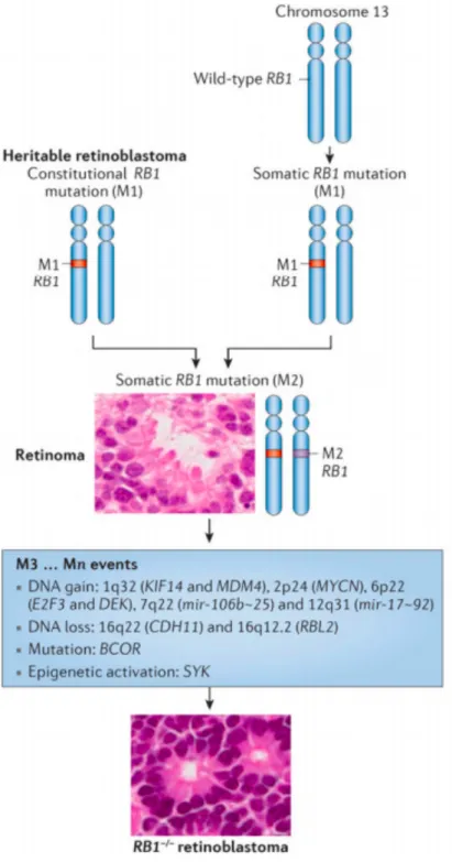
Knudson in 1971, who explained that two consecutive mutations, either one inherited or both acquired somatically, are necessary to trigger retinal tumorigenesis (17). Knudson theory was validated after the identification of the tumor suppressor Rb1 gene in the chromosome 13, a sequence that was inactivated in all patient-derived RB samples (18). Now, a multi-hit hypothesis is commonly accepted for the tumorigenesis process (19). In RB, the Rb1 gene inactivation is a tumor-driven trigger during the maturation of cone-precursor cells that culminates in a malignant state after further genetic and epigenetic alterations (20) (Figure 2).
Figure 2. – Genetic causes of RB1-associated retinoblastoma. Adapted from (20)
Heritable cases of RB are characterized by a constitutive RB1 mutation (Mutation 1, M1). A second hit (M2) is somatically acquired in a susceptible retinal cell. In non-heritable cases, the two mutations occur somatically in the same retinal cell. A benign retinoma evolves towards a malign state (retinoblastoma, RB) through a third (M3) or more (Mn) genetic or epigenetic alterations.
Although not presented in Figure 2, a third rare genetic subtype of RB is known. It is characterized by the presence of wild-type RB1 and amplification of the MYCN oncogene (RB1+/+ MYCNA). RB remains a rare type of cancer with an overall 8000 new cases per year worldwide (20). It is the most common intraocular cancer in children, being responsible for 3% of all cases of childhood malignancies. The overall survival (OS) of RB varies considerably according to the economic development of the region it is diagnosed in. Whereas the OS is higher than 95% in high-income countries (21, 22), the OS is lower than 40% in low-income ones (23, 24). Difficult access to healthcare, unavailability of clinical resources and, thus, late diagnosis and treatment, enucleation denial by the family and incompliance to chemotherapy are a few examples underlying such discrepancy (20). To tackle the difference, international engagement to promote awareness and collaboration between countries are playing an important role to fight RB in economically undeveloped regions of the globe (25, 26).
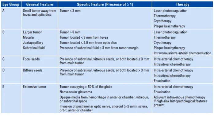
The International Classification of Intraocular Retinoblastoma (IIRC) categorizes RB in 6 stages, from a 0 phase, where no disease is detected, to stage A through E, corresponding to a minor tumor with less than 3 mm to a more severe case with diffuse vitreous seeds and potential extraocular invasion, respectively. As later the diagnosis, the worse the prognosis is, once the tumor tends to migrate back through the optic nerve to colonize the brain, when it becomes incurable. Therefore, RB staging is crucial to determine the clinical protocol. Clinical management of RB takes into consideration the probability of survival of the patient, salvage of the eye, vision preservation and reducing secondary tumors (Figure 3).
Figure 3. – Retinoblastoma classification according to the IIRC and recommended therapeutic interventions (27).
RB treatment could be classified in chemotherapy (either systemic or locally delivered), focal therapy and enucleation (27-29).
• Systemic, or intravenous, chemotherapy (IVC) is usually a combination of 2 to 3 drugs with different mechanisms of action. Standard drugs include alkylating agents (carboplatin, cisplatin), topoisomerase II inhibitors (etoposide, topotecan, and teniposide) and vinka alkaloids (vincristine). A major drawback in IVC is the reduced drug bioavailability in the vitreous due to the blood-retina barrier. As a result, IVC by itself rarely eradicates the tumor, being commonly combined with focal therapies.
• Local administration includes intra-arterial (IAC), intravitreal or periocular chemotherapy. IAC has been successfully applied for the initial stages of RB. For stages D and E, IAC can be combined with IVC. IAC requires highly skilled physicians at dedicated cancer centers, which are not usually available in developing countries. Intervention is made through a micro-catheter through the femoral artery up to the ophthalmic artery of the affected eye where melphalan, a mustard alkylating agent, is delivered. A drug combination is also preferred if extensive vitreous seeds are present. Intravitreal chemotherapy is the chosen
intervention for advanced stages with extensive vitreous seeds unresponsive to IVC or IAC, as it bypasses the drug delivery challenge of nonvascularized vitreous. Melphalan can be solely injected or in combination with topotecan. Special concern is given during needle withdrawal. It’s important to seal the site of injection with cryotherapy as the tumor could migrate through needle track after the intervention. Finally, the periocular injection of either carboplatin or topotecan allows rapid and high vitreous concentration. It can be used to control retinoblastoma or as an adjuvant of IVC for advanced stages of RB with the presence of vitreous seeds.
• Focal therapy is the primary treatment for initial stages of retinoblastoma and englobes laser therapy, cryotherapy, and plaque radiotherapy. Laser therapy is designed to either cytotoxically heat the tumor directly using an 810-nm diode laser (thermotherapy) or a 510-nm argon laser to coagulate blood vessels that supply the tumor (laser photocoagulation). Cryotherapy uses a metal probe cooled to very low temperatures to freeze and kill small tumors. Plaque radiotherapy consists of implementing a radiative robe, usually Iodine-125 and ruthenium-106, to deliver ionizing radiation to small tumors over 4-7 days.
• Enucleation remains the ultimate and most effective intervention for advanced stages of RB (group E). It is the first-line treatment in non-familiar cases of RB, as the disease is usually identified at later stages.
1.2. Survivin
1.2.1.
Definition
Baculoviral IAP repeat-containing protein 5, commonly known as survivin, is the smallest member of the inhibitors of apoptosis protein family (IAP). Survivin is a multitask protein implicated in proliferation and cell cycle progression (30), angiogenesis (31), DNA repair (32), cancer invasiveness and stemness properties (33) and mediates resistance to chemotherapeutics by inhibiting both extrinsic and intrinsic apoptosis signaling (34) through a complex mechanism yet to be fully elucidated. Possessing such a vast network of interaction, survivin’s nodal function can be viewed as two-fold: (i) a key role in cell division (mediating microtubule dynamics and their
attachment to the centrosomes – kinetochore survivin) and (ii) cell death and genomic fidelity regulator (Figure 4).
Figure 4. – Connectivity links between the survivin cell division and cell death networks. The functions of survivin intersect with mechanisms of cell division control, genomic fidelity, mitotic spindle assembly, subcellular trafficking, checkpoint regulation and apoptosis (35). CHK2 ; XIAP, X-linked inhibitor of apoptosis protein; PKA, protein kinase A; CDK1, cyclindependent kinase 1; HSP90, heat shock protein 90; MCAK, mitotic centromere-associated kinesin; TD60, telophase disk protein of 60 kD; MEN, mitotic exit network; SGO2, shugoshin 2; CRM1, chromosome region maintenance protein 1; INCENP, inner centromere protein antigens.
There is a large room for debate about survivin’s importance in cellular homeostasis. Although one may categorize its function at two levels as described above (Figure 4), we should not rule out that many of the survivin’s pathways could be connected, demanding a holistic or systems
biology interpretation of its cellular role. Such a nodal protein implicated in key cellular pathways became an obvious target for scientific investigation, especially in cancer, where it is abnormally overexpressed (36).
1.2.2.
Survivin and cancer
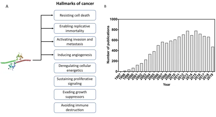
Since its discovery (37), survivin has puzzled cancer biologists: it is extensively expressed in embryonic development, silenced in most healthy adult differentiated cells, but it rises again at the malignancy stage. Survivin is deeply implicated in cancer resistance to chemotherapeutics, radiation, and usually associated with poor patient prognosis in colorectal (38), breast (39) and bladder cancer (40), as human gliomas (41), melanoma (42) and retinoblastoma (43). Besides, survivin has been linked to other hallmarks of cancer beyond its firstly reported ability to confer resistance to cell death (Figure 5A). This is why reports involving survivin considerably increased in the last 15 years (Figure 5B).
Figure 5. – Survivin and cancer research. (A) Arrows indicate scientific evidence of survivin and the hallmarks of cancer. (B) Number of publications from 1997 to 2019 retrieved from Web of Science (Thomson Scientific) using the entry “Survivin” on Title, Abstract or Keyword. Total
Resisting cell death Enabling replicative
immortality Activating invasion and
metastasis Inducing angiogenesis Deregulating cellular energetics Sustaining proliferative signaling Evading growth suppressors Avoiding immune destruc=on A Hallmarks of cancer B 19971998199920002001200220032004200520062007200820092010201120122013201420152016201720182019 0 200 400 600 800 1000 Year Number of publications
number of publications: 10.304, as of October 22 of 2019. Left side, a 3D crystallography representation of survivin protein (PDB entry: 1E31).
There is scientific evidence of survivin’s implication on the following hallmarks of cancer: resistance to cell death (44); Enabling replicative immortality (here, survivin as key player in cell division (45), which is highly exploited by cancer cells, as well as its possible contribution to stemness properties in cancer (46, 47)), were considered as a link to this hallmark); activation of metastasis and invasion (48, 49); and inducer of angiogenesis (50).
Furthermore, survivin was unveiled to be the 4th most expressed transcript in colon, brain, breast
and lung cancer, and melanoma in comparison to their corresponding normal tissues (36). Not surprisingly, many anti-survivin strategies have been pursued in vitro and in vivo with a few fruitful approaches reaching clinical trials. It must be noted, however, that targeting survivin isn’t an easy strategy. As an intracellular protein, localized in different subcompartments (nucleus, mitochondria, and cytosol), one of each possibly contributing to circumvent cell death, and lacking a catalytic site for small molecules inhibitors, survivin can easily be considered an “undruggable” target (51, 52). Therefore, scientists used to target survivin with: (i) immunotherapies that recognize survivin as a tumor-associated antigen (TAA), (ii) agents capable of regulating survivin at DNA level repressing its promoter region or (iii) at mRNA precursors. 1.2.2.1. Immunotherapies
Survivin emerged as a good candidate for immune-based strategies (53) due to its poor expression in healthy adult cells and high expression in cancer cells (37). As a result, multiple clinical trials are being carried with Survivin-targeting cytotoxic T lymphocytes (54) and survivin-directed immunization (55, 56), the later also combined with immune checkpoint blockers (NCT03349450). However, caveats to immune-based therapies remain the high cost associated to adoptive T cell therapy (57), tumor heterogeneity with regards to TAAs (58) and absence of long-lasting efficiency in survivin-directed immunization (59).
1.2.2.2. Small molecules inhibitors
The most studied survivin-targeting small molecule is the imidazolium-based compound, YM155. The molecule’s mechanism of action was first assumed to be due to inhibition of survivin’s
promoter region at the DNA level (60). Later on, it was speculated that YM155 actually induced DNA damage and resulted in survivin inhibition in an unspecific fashion (61, 62). Regardless of its true pharmacodynamics, YM155 inhibited tumor growth in several cancer models either alone or in combination with other anti-cancer strategies both in vitro (63, 64) and in vivo (65-67). YM155 sensitized human RB cells (Y79) to carboplatin, in vitro and in an orthotopic RB model (68). Finally, the molecule gathered enough evidence to move forward towards clinical trials. Although encouraging results were reported combining YM155 with CD20-targeting monoclonal antibody to treat relapsed aggressive B-cell Non-Hodgkin lymphoma (69), only modest efficacy or failure to achieve primary endpoints were reported in other trials (70-73). No clinical trial is currently active to investigate YM155 as an interventional drug to treat cancer (clinicaltrials.gov as of October 23 of 2019).
1.2.2.3. mRNA targeting strategies
Another strategy to circumvent the undruggable survivin protein is to repress its expression by cleaving the mRNA precursor. Ribozymes are enzymatic RNAs capable of recognizing an mRNA target and cleaving it, impairing protein translation (74). Survivin-targeted ribozymes sensitized prostate cancer (75), lung adenocarcinoma (A549) (76) and breast cancer cells (MCF7) (77) to etoposide, and head and neck squamous cell carcinomas to etoposide and carboplatin (78). Although interesting results were achieved in vitro, no survivin-targeting ribozyme-based strategy was further developed in vivo.
A second survivin mRNA-targeted strategy is antisense oligonucleotides (ASO). ASO are a single RNA or DNA strand complementary to a target mRNA sequence. ASO may exert their repressive effect by recruiting enzymes to cleave the ASO-mRNA complex, modulating mRNA splicing or steric blocking of ribosome-mediated translation (79). LY2181308 is an ASO against survivin that demonstrated good human tolerability (80), tumor bioaccumulation, evident survivin silencing and restored tumor apoptosis signaling (81) in a phase I human trial, but failed to achieve its primary endpoint at a subsequent trial in combination with docetaxel (82, 83). No clinical trial is currently active using LY2181308 as an anti-survivin strategy (clinicaltrials.gov).
Finally, small interfering RNA (siRNA) has been used as anti-survivin strategies. siRNA is a double-stranded RNA that, when incorporated into the cells, activates the well-conserved naturally occurring RNAi mechanism to mediate mRNA cleavage and protein silencing (84). Mechanistically, once inside the cell, the double-stranded siRNA is incorporated into the RNA-induced silencing complex (RISC) which unwinds and cleaves the sense strand through the argonaute 2 (AGO2) enzyme-containing within the RISC. The antisense strand remains in the RISC and the now active RISC-antisense strand complex seeks the complementary RNA sequence to mediate mRNA suppression (Figure 6). Long double-stranded siRNA follows the same pathway once it is cleaved in the cytoplasm into siRNA by the enzyme DICER. The silencing complex is regenerated after each mRNA cleavage, being capable of promoting gene silencing for less than a week in rapidly dividing cells, but up to 3 weeks in slow dividing ones (85).
Survivin-targeting siRNA has demonstrated tumor growth inhibition in patient-derived colorectal cancer (87) and glioblastoma (88) xenografts. In retinoblastoma, survivin-targeted siRNA inhibited human RB cells (Y79) proliferation, growth and invasiveness, in vitro (89, 90). As an adjuvant therapy, siRNA against survivin sensitized SKOV-3 cells to cisplatin (91), synergistically improved paclitaxel tumor growth inhibition in breast-cancer bearing mice (92) and reversed Apo2L/TRAIL resistance in melanoma cells (93).
Despite the remarkable success in vitro, siRNA-based therapies face considerable challenges to be translated in vivo. Biological barriers naturally evolved to prevent eukaryotic cell infection by exogenic nucleic materials. Of many naturally occurring biological barriers that need to be surmounted before clinical translation, 3 are primarily addressed during in vitro optimization: poor serum stability, low cell penetration due to electrostatic impairment and lysosomal degradation of the fraction uptaken by the cell (94). Fortunately, it is possible to circumvent the pitfalls that restrain siRNA delivery using viral or non-viral vectors (95). In fact, the latter approach has recently been responsible for OnPattro, the first RNAi-based drug to be approved by the FDA in 2018 (96), a milestone for gene therapies that shed a hopeful light into the future of gene medicines. Lipid nanoparticles (LNP), a non-viral vector approach for gene medicines delivery, as taken the siRNA strategy from the bench to the bedside.
1.3. siRNA delivery
1.3.1.
Challenges and vector rational design
Gene silencing through siRNA technology has significantly pushed the medical frontier forward. Numerous siRNA-based therapeutics are now on clinical trials (97) following the steps of the first FDA-approved RNAi-based drug, OnPattro (Patisiran). Considering non-viral vector approaches, siRNA could be delivered as lipoplexes or polyplexes, if carried through a lipid or polymer formulation, respectively. A first essential step before proposing an optimal carrier to enable siRNA delivery is to understand the chemical nature of siRNAs to identify their weakness and biologically imposed barriers.
Structurally, siRNA is a double-stranded RNA, containing ~21-23 base pairs anchored in a ribose backbone linked by anionic phosphodiester bonds (Figure 7). The hydroxyl group in the 2’ position of ribose moiety renders chemical susceptibility to siRNA, especially to serum nucleases. The 2’ position is often used for chemical modification to improve siRNA stability (98).
Figure 7. – General structure of unmodified siRNA.
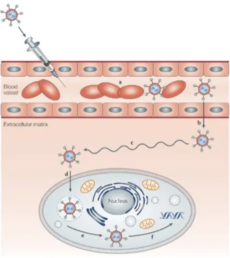
By its structure, it is possible to identify the siRNA as a highly hydrophilic and negatively charged macromolecule with susceptible instability against nucleases. The first chemical characteristic implies two additional challenges: big macromolecules as siRNAs (i) cannot cross the cell membranes due to electrostatic impairment and low lipophilicity (94), and (ii) are rapidly eliminated through renal clearance (99). Considering that an intact siRNA reaches the desired cell and is internalized, nature imposes yet another challenge; upon endocytosis, the siRNA must escape the endosomal compartment before vesicle maturation and eventual lysosomal degradation of siRNA (100). Based on these assumptions, a good vector candidate must minimally act at 3 levels: (i) complex the siRNA, protect it from nuclease degradation and avoid fast renal clearance, (ii) promote cell internalization and (iii) allow endosomal escape and cytosolic delivery of intact siRNA. The aforementioned barriers and other physiological challenges to be circumvented by siRNA strategies are summarized below (Figure 8).
U
C
G
A
3’ overhang
3’ overhang
5’
5’
Ribose backbone
O O O OH Base P O O -2’Antisense strand
sense strand
Figure 8. – Physiological barriers faced by siRNA strategies. (86). The vector carrying the siRNA should (a) avoid renal clearance, phagocytosis and protect siRNA from blood stream degradation; (b) be transported across endothelial barrier; (c) reach the target site; (d) be taken up into the cell; (e) mediate endosomal escape; (f) promote cytosolic delivery of siRNA.
Being aware of the limitations involved in siRNA delivery, we can now propose a bottom-up approach to design an optimal non-viral vector harboring the essential moieties required to overcome each challenge.
1.3.1.1. siRNA Complexation and serum stability
An ideal candidate for siRNA delivery would be able to closely pack the siRNA, protecting it from nuclease degradation. Obvious candidates would take advantage of the negatively charged phosphate groups within siRNA backbones. That’s the case of cationic lipids, as 1,2-dioleoyl-3-trimethylammonium-propane (DOTAP), 3ß - [N-(N0,N0-dimethylaminoethane) - carbamoyl]
cholesterol hydrochloride (DC-Chol) and N-[1-(2,3-dioleoyloxy)propyl] - N,N,N-trimethylammonium chloride (DOTMA), which in combination with other neutral lipids, as cholesterol and 1,2-dioleyl-sn-glycerol-3-phosphoethanolamine (DOPE), self-assemble with siRNA into lipoplexes for efficient gene delivery in vitro (101-103). The branched cationic polymer polyethylenimine (PEI), as its lipids counterpart, also self-assembles with nucleic acids into polyplexes (104). However, the resulting positively charged lipo- and polyplexes (nanocomplexes) are rapidly coated by negatively charged serum proteins in vivo, halting transfection ability and promoting its recognition and cleareance by macrophages (105). An elegant approach to hinder the nanocomplexes from reticuloendothelial system-mediated clearance and serum proteins adsorption was incorporating polyethylene glycol (PEG)-grafted lipids or polymers can provide to nanocomplexes stealth properties due to new hydrophilic surface coating (106). Caution must be considered when incorporating PEG-grafted elements into nanovectors since the steric hindrance provided by PEG might also prevent endocytosis and endosomal escape of genetic cargo, impairing siRNA transfection (107).
Another strategy to avoid opsonization and fast serum clearance is to eliminate the net positive charge of nanocomplexes. Ionizable cationic lipids are able to electrostatically complex siRNA at an acidic pH, where amino groups are protonated, but reach neutrality as the pH is graduality elevated at physiological values (108). After synthesizing a library of ionizable lipids, Jayaraman and coworkers (109) found DLin-MC3-DMA as the leading component with the highest silencing efficiency in vitro. The authors unveiled the pKa for ionizable lipids between 6.2 and 6.5 is a critical parameter effective hepatocyte gene silencing. Lipid chain length from 10 to 13 carbons was another parameter unveiled by combinatorial synthesis reported to influence siRNA efficient silencing in vivo (110, 111).
1.3.1.2. Cell internalization
The self-assembly of the cationic vector with siRNA at the right proportion (measured as the ratio between nitrogen groups per cationic lipid/polymer and phosphate moieties in the siRNA, proportion known as the N/P ratio) results in a complex able to electrostatically bind to the negatively charged cell membrane if the overall charge of the vector is positive (112, 113). After binding, different endocytic pathways could be activated to mediate nanocomplex internalization
(Figure 9) (114-116). The mechanisms whereby nanocomplexes enter the cells are still the focus of many studies, since it relies on various parameters such as nanoparticles structure, shape, size, surface charge, but also the cell type and culture conditions. Nevertheless, a deeper understanding of these mechanisms would truly contribute to the rational design of nanovectors to gene therapy.
Figure 9. – Possible pathways of nanoparticles internalization (117). 1.3.1.3. Endosomal escape
Once internalized, nanocomplexes must avoid lysosomal degradation. Intriguingly, the majority of the cargo and vector is degraded upon endosomal maturation or recycled back to the extracellular space (115, 116, 118). Therefore, the nanocomplexes require fusogenic or endosomolytic properties to allow endosomal escape and, thus, the release of siRNA within the cytosol.
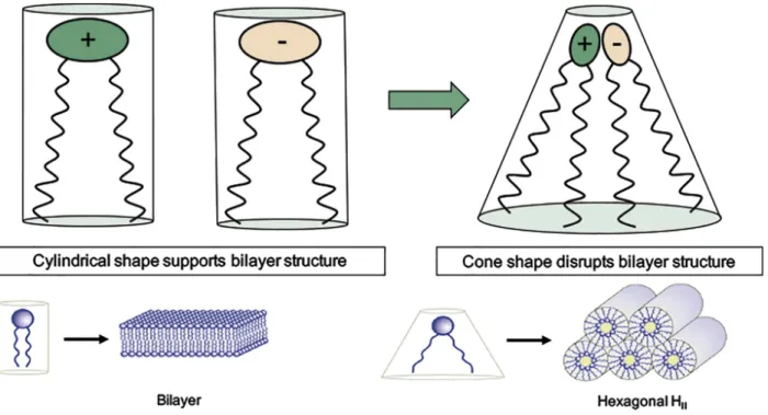
Cationic lipids are not only able to self-assemble with siRNA into lipoplexes, but also mediate endosomal escape through a mechanism known as “ion-pair” theory (119). Once inside the endosome, cationic lipids interact with the negatively charged lipids in the inner leaflet of the endosomal lumen forming an ion-pair complex, a non-lamellar (HII) structure that mediates
endosomal disruption (120). Non-lamellar lipids, like cholesterol and DOPE, known as “helper lipids”, facilitate HII transition, thus improving transfection efficiency (119) (Figure 10).
Figure 10. – Schematic diagram of the ion-pair membrane disruption mechanism mediated by cationic lipids (120). Lamellar lipids support bilayer structures whereas cone shape lipids adopt a non-bilayer conformation. The structure formed by the electrostatic interaction between cationic and anionic lipids (ion-pair) behave as a cone shape lipid, hence adopting an inverted hexagonal phase (HII) and disrupting the bilayer structure.
However, the endosomal destabilization mediated by cationic lipids is far beyond its full efficacy, as already demonstrated that only 1-2% of siRNA was released from the endosomal when encapsulated in lipid nanoparticles (LNP) containing the gold standard ionizable lipid DLin-MC3-DMA, cholesterol as helper lipid, DMG-PEG for stealth properties and disteroylphosphatidyl choline (DSPC) as membrane structural lipid (116). Therefore, endosomal entrapment is still a major barrier that prevents full translation of siRNA therapies mediated by non-viral vectors. Researchers have addressed this issue by developing responsive materials to improve endosomal escape, thus enhancing cytosolic delivery of siRNA. Adding a pH-responsive material into lipoplex composition has shown to promote cytosolic delivery of siRNA and efficient silencing in vivo (121, 122).
Based on these premises, an ideal classical non-viral vector for siRNA delivery would contain at least, but not limited to, the following components:
General:
• A cationic moiety for siRNA complexation and initial cell-binding; • A PEG-grafted component for stealth properties;
For lipoplexes:
• A “helper” lipid to ameliorate non-bilayer transition and boost endosomal escape and; • A structural lamellar-forming lipid.
Such a design was employed in the development of OnPattro (Patisiran) formulation, the first RNAi-based drug to be approved by the FDA (table 1).
Lipid component Function
DLin-MC3-DMA
• Complexes siRNA at values below pKa • Mediates endosome disruption (ion-pair
complex with negatively charged lipids inside the endosome lumen)
Cholesterol • helper lipid
DSPC1 • Structural lipid
DMPG-PEG2 • Provides stealth property
1DSPC: 1,2-distearoyl-sn-glycero-3-phosphocholine; 2DMPG-PEG:
1,2-dimyristoyl-rac-glycero-3-methoxypolyethylene glycol-2000
OnPattro (Patisiran) composition and role of each lipid component (120).
Lipid nanoparticles (LNP) are the most advanced non-viral vectors for gene medicines. Therefore, we will focus on methods of preparation for LNP, exemplifying in vitro or in vivo approaches of siRNA delivery. Next-generation of responsive LNP capable to boosting cytosolic delivery of siRNA upon endosomal acidification, pH-sensitive LNP, will also be presented.
1.3.2.
Methods of preparation for lipid nanoparticles (LNP)
1.3.2.1. Lipid nanoparticles (LNP)Liposomes are defined as spherical structures composed of an aqueous core enclosed by one or more lipid bilayers. Initially proposed by Bangham as simplified cell membrane models in the early 60s (123, 124), the potential of liposomes as drug and gene carriers has significantly advanced (94, 120, 125).
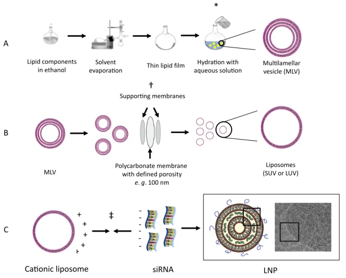
The classic method for designing liposomes to enable siRNA delivery consist of (A) formulating cationic liposomes, (B) submitting resulting lipid vesicles to size reduction and homogenization and (C) incubation with siRNA to afford LNP (Figure 11).
A. Pre-formulated liposomes are prepared by hydrating a lipid film. Briefly, the lipids are selected based on the rational design of a gene delivery vector as aforementioned discussed, and solubilized in an organic solvent, preferentially ethanol to decrease in vivo toxicity of traces of organic solvent. In a round bottom flask, the organic solvent is evaporated under reduced pressure, forming a thin lipid film, which is then hydrated with an aqueous solution of low concentrated buffer or 5 % dextrose solution to maintain human osmolarity compatibility (Figure 7A). Hydration of the lipid film will afford multilamellar vesicles (MLV).
B. MLV are subsequently submitted to size reduction by extrusion through polycarbonate membranes of defined porosity (Figure 7B). The resulting homogeneous vesicles could be classified as small unilamellar liposomes (SUV) if smaller than 100 nm or large unilamellar liposomes (LUV) if bigger than 100 nm but smaller than 1000 nm.
C. SUV or LUV are diluted in dextrose 5% to achieve the desired N/P ratio before combining with genetic cargo to afford a complex structure containing a centered aqueous core with the genetic cargo complexed between the multilayers of the lipoplex (120, 126) (Figure 7C).
The hydration of the lipid film, extrusion and self-assemble of SUV with genetic material into lipoplexes are carried at a temperature above the gel-to-liquid transition (Tm) point for the lipid
Figure 11. – Stepwise representation of LNP preparation by combining preformed liposomes with siRNA. See the text for details. Big square: left: schematic representation of PEGylated lipoplex with siRNA complexed within lipid bilayers; right: cryo-TEM micrograph of cationic lipid/siRNA complexes (bis (guanidinium)-tren-cholesterol (BGTC):siRNA) (127). Small boxes highlight the siRNA complexed within the bilayers of the lipoplex. *, † and ‡ indicate steps performed under heating.
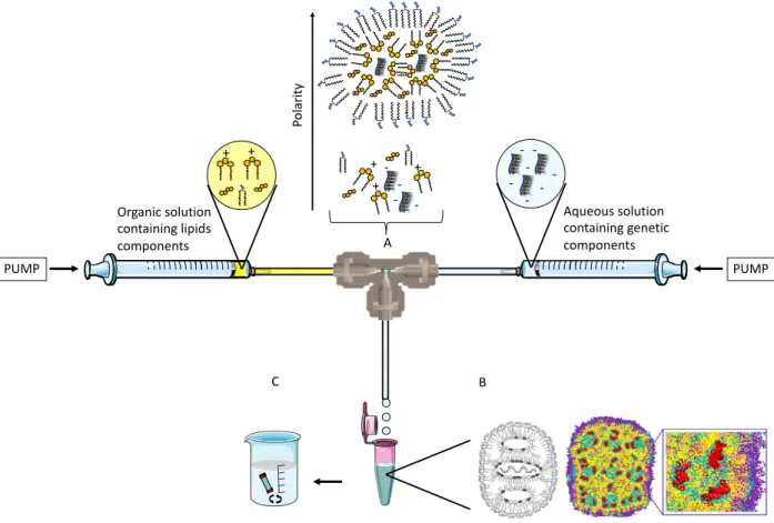
A second approach consists of an all-in-one fast mixing step of an organic (ethanol) lipid solution with an aqueous solution containing the genetic cargo to spontaneously afford lipid nanoparticles. The strategy was further optimized with the advent of microfluidic devices for the controlled and reproducible mixing process.
Lipid components
in ethanol evapora3onSolvent Thin lipid film
Hydra3on with
aqueous solu3on Mul3lamellar vesicle (MLV)
MLV Polycarbonate membrane with defined porosity
e. g. 100 nm Suppor3ng membranes
*
†
Liposomes (SUV or LUV) Ca3onic liposome + + + + + -siRNA‡
LNP A B CBriefly, two syringe pumps are oppositely placed, one loaded with an aqueous miscible organic solvent containing a mixture of desired lipids, whilst the second contains the genetic cargo solubilized in an aqueous solution. Pumps are connected into a mixing Tee, or a microfluidic chamber, with an exit tubing towards a sterile container to receive the resulting LNP (Figure 12). The lipid and nucleic acid solutions are mixed at a controlled ratio and speed and, as the polarity of the system is increased, the lipids spontaneously self-assemble onto the genetic cargo brought together by electrostatic interaction (Figure 12A). The inverted micelles containing the genetic cargo enclosed at internalized aqueous cores are finally surrounded by PEGylated lipids, which maintain the hydrophilic head group orientated towards the exterior aqueous medium (figure 12B). LNP is dialyzed against low concentration buffer or 5% dextrose to remove the ethanol (Figure 12C).
Figure 12. – Schematic representation of siRNA encapsulated LNP formulated by microfluidics technique. (A) represents the interior of the mixing tee chamber. As the polarity is increased, the siRNA precipitates at the interior of the newly formed LNP. (B) Left in black and white: schematic
PUMP PUMP -+ + + Organic solution containing lipids components Aqueous solution containing genetic components A B C + + -Po la rit y
representation of LNP. Middle, colored: Molecular representation of LNP. Yellow, cyan and pink represent lipids; red is the complexed siRNA, while blue is the PEGylated shell. Right, colored: zoom view of the LNP interior. (C) Dialysis against an aqueous solution as the final step to remove the solvent. Adapted from (120, 128).
T tube/microfluidics mixing affords smaller LNP than when prepared by pre-formulate liposomes due to all-in-one precipitation of materials at the interior of LNP rather post-complexation of siRNA within the multilayers of lipoplexes (Figure 13).
Figure 13. – Cryogenic transmission electron microscopy (Cryo-TEM) micrographs of extrusion- and microfluidics-formulated liposomes. Left: POPC:Cholesterol (1:1 mol/mol) liposomes formulated by the extrusion method. Unilamellar liposomes formulated by this method present an aqueous core surrounded by a lipid bilayer. Right: Cationic liposomes formulated by microfluidics technique (DLinKC2-DMA/DSPC/Chol/PEG-lipid (40/11.5/47.5/1 mol/mol). Adapted from (128).
1.3.2.2. pH-sensitive liposomes
Stimuli-responsive delivery systems take advantage of early endosomal acidification to promote membrane destabilization and fast cytosolic cargo delivery. It is now recognized that optimal lipid formulation requires a pH-sensitive component. To this end, numerous pH-sensitive materials,
based on polymers, peptides, or hydrolyzable chemical bonds, have been developed. Our laboratory previously demonstrated the efficiency of a pH-sensitive liposome based on a principle of a molecular switch (129). In this lipid structure, the di(methoxyphenyl)-pyridine is in a trans conformation with the pyridine nitrogen (Figure 14A). The protonation of pyridine (predicted pKa » 5.28) at a value within the early endosomal pH (∼5–6), triggers the conformation change to enable H bonding between di(methoxyphenyl) group and the protonated pyridine (Figure 14B). By consequence, the alkyl chains change from a lipid stacking bilayer-favoring position to an open bilayer-disfavoring conformation that might destabilize the endosomal membrane, promoting siRNA cytosolic delivery (Figure 14C).
Figure 14. – pH-sensitive lipid undergoes conformation change upon acidification leading to switchable LNP destabilization and siRNA delivery. (A) Left: Schematic representation of switchable LNP Right: Structural representation of cationic switchable lipid. * is the di(methoxyphenyl)-pyridine pH-switchable unit; † site of protonation under acid environment; ‡ cationic polar group responsible for complexing the genetic material. (B) protonation-induced conformational change of switchable lipid. (C) Schematic representation of the destabilized switchable LNP and siRNA delivery upon tweezer-like proton-induced conformational change. Adapted from (121).
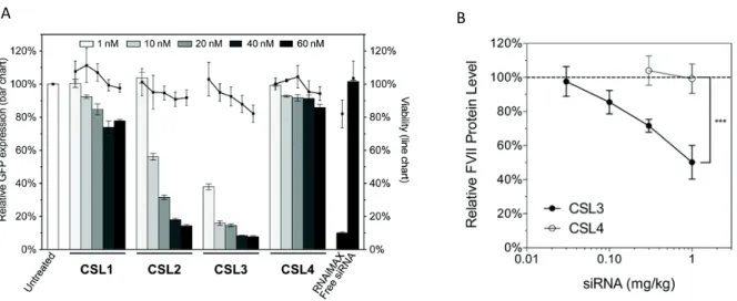
The switchable LNP successfully delivered hydrophilic cargo in vitro (130) and it was further optimized into a cationic switchable LNP, able to deliver a GFP-targeted siRNA in vitro (Figure 15A). Three different switchable lipids, CSL1, CSL3 and CSL3 were synthesized on that study. CSL3 possessed a predicted pKa within the early endosomal pH and constituted the most efficient
* †
‡
cationic switchable LNP in the in vitro assay. The transfection ability of CSL3 (from this point on in this investigation, one will refer to CSL3 only as CSL) was further evaluated in a proof-of-concept in vivo study using the cationic switchable LNP carrying the factor VII-targeted siRNA for hepatic targeting (Figure 15B) (121). The specific pH-triggered siRNA delivery through cationic switchable LNP was confirmed by the inability of the non-switchable LNP (formulated with compound CSL4) in silencing the targeted protein both in vitro and in vitro. CSL4 does not possess the two methoxy moieties necessary to initiate pH-triggered intramolecular hydrogen bonding with the protonated pyridine, thus preventing conformational switch and LNP destabilization.
Figure 15. – Cationic switchable LNP efficiently delivered siRNA in vitro and in vivo. (A) GFP In vitro knockdown and viability of GFP-HELA cells after GFP-targeted siRNA transfection by cationic switchable LNP. (B) In vivo hepatic targeting of cationic switchable LNP carrying factor VII-targeted siRNA (121).
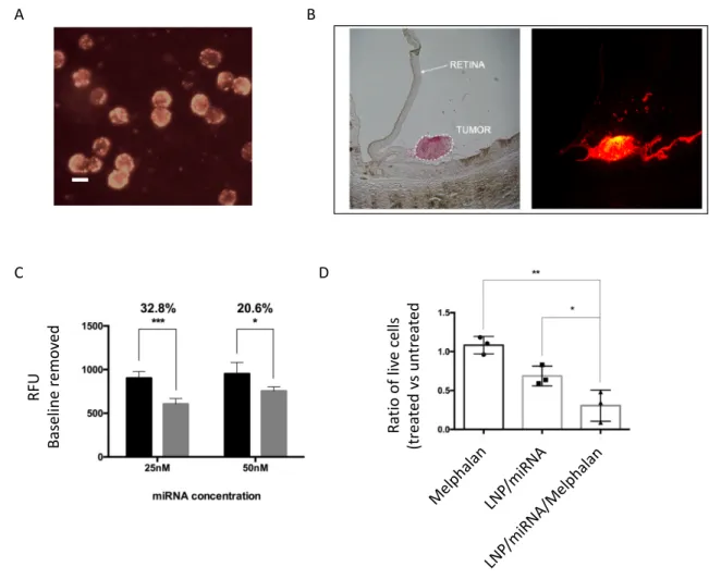
Importantly, the efficient cytosolic delivery of genetic cargo was not impaired by PEGylated liposomes formulated by either microfluidics for in vivo siRNA hepatic targeting or, as demonstrated recently, by extrusion method for intravitreal delivery of micro RNA (mir-181a) (122). In that study, Tabatabaei and coworkers demonstrated that cationic switchable lipid prepared with indocarbocyanine dye accumulated within retinoblastoma cell lines as soon as 2 hours after transfection, with strong internalization visualized 24 hours initial incubation (Figure 16A). Previously, the same group had attested that the switchable LNP preferentially accumulated within RB tumor cells, sparing retinal and adjacent tissues after intravitreal injection in an