HAL Id: pastel-00004418
https://pastel.archives-ouvertes.fr/pastel-00004418
Submitted on 4 Dec 2008
HAL is a multi-disciplinary open access
archive for the deposit and dissemination of sci-entific research documents, whether they are pub-lished or not. The documents may come from teaching and research institutions in France or
L’archive ouverte pluridisciplinaire HAL, est destinée au dépôt et à la diffusion de documents scientifiques de niveau recherche, publiés ou non, émanant des établissements d’enseignement et de recherche français ou étrangers, des laboratoires
In vivo ovulation, oocyte maturation and fertilisation in
the bitch
Alain Fontbonne
To cite this version:
Alain Fontbonne. In vivo ovulation, oocyte maturation and fertilisation in the bitch. Life Sciences [q-bio]. AgroParisTech, 2008. English. �NNT : 2008AGPT0010�. �pastel-00004418�
T H È S E
pour obtenir le grade de
Docteur
de
l’Institut des Sciences et Industries du Vivant et de
l’Environnement
(Agro Paris Tech)
Spécialité : Reproduction Animale présentée et soutenue publiquement
par
Alain, Yves, Michel Fontbonne
le 14 février 2008
OVULATION, MATURATION OVOCYTAIRE ET FECONDATION IN VIVO CHEZ LA CHIENNE
IN VIVO OVULATION, OOCYTE MATURATION AND FERTILISATION IN THE BITCH
Directeur de thèse : Sylvie Chastant-Maillard
Travail réalisé : Ecole Nationale Vétérinaire d’Alfort, UMR 1198 Biologie de la Reproduction et du Développement, F-94704 Maisons-Alfort Cedex
Devant le jury :
Pr Dr Wenche Farstad, PhD, Oslo Veterinary Faculty, Norway Rapporteur Pr Dr Cecilia Luvoni, PhD, Milan Veterinary Faculty, Italy Rapporteur Pr Dr Pierre Guérin, PhD, HDR, Ecole Nationale Vétérinaire de Lyon, France Examinateur Pr Dr Francis Fiéni, PhD, HDR, Ecole Nationale Vétérinaire de Nantes, France Examinateur Dr Marc-Antoine Driancourt, PhD, HDR, Responsable R.et D., Intervet S.A. Examinateur Pr Dr Jacques Guillot, PhD, Ecole Nationale Vétérinaire d’Alfort, France Examinateur Dr Sylvie Chastant-Maillard, PhD, HDR, Ecole Nationale Vétérinaire d’Alfort, France Directeur
A la mémoire de Noël Marseloo, qui nous a quittés vraiment trop tôt.
A Sylvie Chastant, sans qui cette thèse n’aurait probablement jamais vu le jour. En souvenir de toutes ces années passées au sein de cette sacrée Unité de Reproduction Animale. Pour sa confiance et son aide précieuse et en témoignage de l’admiration que je lui porte. A Karine Reynaud qui m’a fait partager son savoir-faire en recherche au laboratoire, et m’a notamment enseigné l’utilisation du microscope confocal. Pour son aide précieuse, sa bonne humeur et en souvenir de notre folle équipée à Rio de Janeiro. A Pierre Guérin pour les bonnes années passées ensemble au CERREC à Lyon. A Wenche Farstad et Cecilia Luvoni, mes collègues et amies de l’EVSSAR, qui m’ont fait l’honneur, l’amitié et la grande joie d’accepter de juger mon travail et de venir à Alfort pour la soutenance. A Francis Fieni en témoignage de toutes ces années où nous avons collaboré dans les congrès, les colloques, les formations ou au sein du GERES A Marc-Antoine Driancourt pour avoir gentiment accepté d’examiner ce travail A Sarah Rivière qui a coordonné avec efficacité l’étude Family et à Franck Noël Aux responsables et au personnel du service d’Imagerie de l’ENVA pour leur gentillesse et leur aide constante A Andrew Ponter pour son aide pour les dosages de LH A ceux et celles qui ont collaboré activement à cette thèse, prises de sang le soir, échographies ovariennes le week-end, ovariectomies les jours fériés..., notamment Christine Viaris de Lesegno et Giovanna Bassu. Je vous aime toutes les deux. A tous mes amis, collègues et collaborateurs du CERCA, de l’Unité de Reproduction Animale et du Laboratoire de Biologie de la Reproduction d’Alfort. Ils sont trop nombreux pour que je les cite. A mes parents et à ma sœur Annick. A Kaota, ma fidèle compagne depuis tant d’années. A Christine Guérin
A Gargamelle, Betsy, Grassouille, Mouflette, Nana, Quenotte, Salsa, Stricky, Thalia, Tithia, Windy, Youpi, Nutts, La Frousse et Nem aux crevettes; A Laistee, Sunny, Tulipe, Sanka, Sweety, Tess, Pivoine, Peggy, Taïga, Odi, Junga, Mandy, Cannelle, Mégane, Téva, Blacky, Rubby, Poppy, Réglisse, Maonia et Chelsea; A Ojissan, Savannah, Roxane, Macha, Mona, Nikita, Pistache, Simrha, Nora, Nina, Perle, Shaggie, Pantera, Tulipan, Orphée, Perak et R’huera; A la mémoire de Sumo.
« Les enfants seuls savent ce qu’ils cherchent… Ils perdent leur temps pour une poupée de chiffons, et elle devient très importante, et si on leur enlève, ils pleurent…»
Antoine de Saint Exupéry « Le Petit Prince »
« Only children know what they are searching for. They spend all their time for a rag doll, and she becomes very important, and if she is taken away from them, they cry… ».
Antoine de Saint Exupéry « Le Petit Prince »
Alain Fontbonne
1Ovulation, maturation ovocytaire et fécondation in vivo chez la chienne
2Résumé:
3La détermination précise du jour de l’ovulation est considérée par la plupart des auteurs comme l’un
4
des facteurs les plus importants pour fixer le moment optimal d’insémination artificielle chez la
5
chienne. Ceci est particulièrement important lors d’utilisation de semence congelée en raison de la
6
faible durée de survie de la semence congelée/décongelée dans l’appareil génital femelle. Notre
7
étude a tout d’abord tenté de déterminer la technique la plus précise pour identifier la survenue de
8
l’ovulation chez la chienne. Notamment, nous avons souhaité savoir si l’examen échographique des
9
ovaires pouvait être un moyen fiable et précis pour déterminer l’ovulation chez la chienne. Notre but a
10
été de détecter l’ovulation par échographie dans différentes races et d’évaluer les intérêts et la
11
précision de cette technique, en comparaison avec les taux hormonaux autour de l’ovulation. Nos
12
résultats ont confirmé que l’échographie ovarienne était une technique précise de détermination de
13
l’ovulation chez la chienne. Nous avons également démontré que cette technique n’améliorait la
14
détection de l’ovulation que chez 15,3% des chiennes en comparaison des dosages de progestérone
15
sanguine. Au bilan, les concentrations de progestérone plasmatique sont extrêmement constantes au
16
moment de l’ovulation quelle que soit la race et, de ce fait, les dosages de progestérone apparaissent
17
être une méthode suffisamment précise pour déterminer le moment de l’ovulation. Utilisant les
18
échographies ovariennes pour la détection du moment de l’ovulation chez la chienne, nous avons
19
ensuite essayé d’évaluer le déroulement exact de la maturation ovocytaire in vivo chez la chienne,
20
ainsi que le développement embryonnaire précoce. De plus, nous avons cherché à vérifier si, in vivo,
21
les spermatozoïdes canins étaient capables de pénétrer des ovocytes encore à un stade immature.
22
Nous avons démontré que le stade de la vésicule germinative (VG) était le seul présent jusqu’à 44h
23
après l’ovulation. Le premier stade métaphase II n’est observé qu’à partir de 54h. Plusieurs stades de
24
maturation ovcytaire différents sont observés au même moment pour une même chienne. In vivo, la
25
fécondation se produit dans la plupart des cas à partir de 90h post ovulation dans des ovocytes ayant
26
complété leur maturation (métaphase II). La pénétration in vivo d’ovocytes immatures par des
27
spermatozoïdes apparaît extrêmement rare.
28
Ces résultats devraient permettre une amélioration ou une simplification de la pratique vétérinaire
29
quotidienne, notamment lors d’utilisation d’insémination artificielle en semence congelée. De plus une
30
meilleure connaissance des processus impliqués dans la maturation ovocytaire in vivo, la
31
fécondation, et le développement embryonnaire précoce sont des étapes importantes pour améliorer
32
le développement des biotechnologies de la reproduction dans l’espèce canine.
34
Alain Fontbonne
35IN VIVO OVULATION, OOCYTE MATURATION AND FERTILISATION IN THE BITCH.
36
SUMMARY:
37
Timing the day of ovulation as accurately as possible is considered by most authors as one of the most important factor in order to determine when to inseminate bitches. This is especially important when using frozen semen, due to the relatively short survival of frozen/thawed semen in the female genital tract after artificial insemination.
In our study, we first tried to find the most accurate technique to determine the exact occurrence of ovulation in the bitch. We hypothesized that ovarian ultrasound examination could be a reliable and accurate method to determinate ovulation in bitches. Our aims were to try to detect ovulation by ultrasound in different breeds and to evaluate the interests and the accuracy of such a technique, compared with hormonal levels around ovulation. Our study confirmed that ovarian ultrasound was an accurate technique for timing ovulation in the bitch. We also found that, although more accurate, ovarian ultrasound examinations improved the accuracy of ovulation detection in only 15.3 % of bitches compared with progesterone. Altogether, progesterone plasma concentrations appeared fairly constant around ovulation, whatever the breed, and, as a whole, progesterone assays appeared as a precise method to time ovulation.
Using ovarian ultrasound examinations as the reference technique to time ovulation in the bitch, we then aimed to evaluate the precise kinetics of in vivo oocyte maturation in the bitch, as well as early embryonic development. Furthermore, we wanted to check if, in vivo, spermatozoa were able to penetrate oocytes still at immature stages. We found that the germinal vesicle (GV) stage was the only one present until 44 hours after ovulation. The first metaphase II stage was observed at 54 hours. Various oocyte maturation stages were observed at the same time within each bitch. Fertilisation occurred in most cases from 90 hours post-ovulation in mature oocytes (metaphase II).
In vivo penetration of immature oocytes by spermatozoa was extremely rare.
These fundamental results may lead to an improvement or a simplification in everyday veterinary practice, especially for artificial insemination with frozen-thawed semen. Furthermore, a better knowledge of the processes involved in in vivo oocyte maturation, fertilisation and early embryonic development are important steps towards the improvement of reproductive biotechnologies in the canine species.
TABLE OF CONTENTS
Introduction 11
In vivo oocyte maturation, ovulation and fertilisation in the bitch : state of the
art 13
Chapter 1. Determination of ovulation 25
Chapter 2. In vivo oocyte maturation and fertilisation 69
Chapter 3: Optimal timing for artificial insemination with frozen-thawed
semen 83
Discussion and perspectives 89
General conclusion 96
References 97
Introduction
This PhD thesis is a compilation of several presentations or publications, some of which are more ancient than others. For the past 15 years, a large part of our clinical and scientific activity has been devoted to a better understanding of the processes and the factors underlying in vivo fertilisation in the dog. At the end of the eighties, it was often stated in veterinary manuals that the optimal time of insemination in the bitch was located around the time of ovulation, which happened at the end of the pro-oestrous period or the beginning of the oestrous period. Having tried several times to inseminate bitches with frozen semen at that period with no results, it became clear for us that the timing of insemination and fertilisation in the bitch had to be further studied.
Trying to improve the fertility results obtained after artificial insemination (AI) using frozen semen, we co-published our first study related to the timing of canine in vivo fertilisation in 1993 (Badinand, Fontbonne et al. 1993). We were not able at that time to use DNA to evaluate the paternity of the puppies, and only phenotypic evaluation was used to determine the days when in vivo fertilisation took place (see Annex 1). In the meantime, we also demonstrated that, using frozen semen, intra-uterine inseminations gave better fertility results than intra-vaginal depositions of semen (Fontbonne et al. 1993). Our following data concerning AI with frozen semen tend to show that the bitch seemed to be ideally inseminated quite late in the oestrous period, when progesterone plasma level was already high (Fontbonne et al. 2000). However, the understanding of this peculiar situation remained partially unclear. In order to know the optimal time of insemination with frozen-thawed semen, and may be diminish the number of inseminations per cycle, there was a need to know precisely when oocytes are ovulated, and furthermore, when they allow fertilisation to occur. In vitro oocyte maturation had been extensively studied (Luvoni et al. 2005). But we could wonder if in vivo processes were identical with what was observed during in vitro studies.
In order to further study in vivo occurrence of events, it was essential to be able to determine ovulation as accurately as possible. Following the example of what was performed in large animals (mostly cows and mares), we aimed to precisely determine the exact time of ovulation in bitches, and to evaluate the interest of
ovarian ultrasonography (Fontbonne et al. manuscript to be submitted). We first compared ovarian ultrasound examinations with plasma LH and progesterone levels for the prediction of ovulation. Using ovarian ultrasonography as a reference in timing ovulation, and repeated intra-uterine AIs, we then studied in vivo oocytes meiosis resumption and fertilisation (Reynaud, Fontbonne et al. 2005).
The last – and the least - part of our contribution to the improvement of in vivo fertilisation concerns the sperm. We have been involved in a better understanding of dog sperm capacitation and ability to fertilise (Guerin et al. 1999). This will not be discussed in this PhD thesis.
This PhD thesis is a summary of our scientific contribution in all these aspects of canine in vivo fertilisation.
In vivo oocyte maturation, ovulation and fertilisation in the bitch: state of the
art.
1. The oestrous cycle of the bitch
The oestrous cycle of the bitch is composed of four successive periods: pro-oestrus, pro-oestrus, metoestrus (or dioestrus) and anoestrus.
Pro-oestrus is defined as the period from onset of vulvar bleeding to the first acceptance of copulation. Its average duration is 9 days. However, it displays considerable variation, as it can range from 0 to 27 days (Johnston et al. 2001). The following phase is oestrus, the period during which the bitch accepts copulation. Here also, the duration of this phase is highly variable: 3 to 21 days (Beijerink 2007).
Metoestrus (Dioestrus) begins when the bitch is no longer willing to accept the male. Its average duration is 70 days. It is associated with the presence of active corpora lutea.
Anoestrus is the phase between the end of the metoestrus and the beginning of next pro-oestrus. Its duration is variable, 2 to 10 months (Concannon 1993).
2. Pre-ovulatory events
2.1. Pre-ovulatory hormonal profiles.
Surprisingly, relatively little is known about the changes in, and temporal relationships between, reproductive hormones around the time of ovulation in the domestic bitch (De Gier et al. 2006).
2.1.1. Pituitary gonadotropins. 2.1.1.1. LH
The secretory pattern of LH at this period is characterised by frequent increases of short duration (Kooistra et al. 1999).
Unlike the other mammalian species, the duration of the LH surge in the bitch is relatively long, ranging from 1 to 5 days (Phemister et al. 1973, Wildt et al. 1978, De Gier et al. 2006). Furthermore, Hase et al. (2000) found that the period in which
LH values peaked above 10 ng/mL continued for more than 12 hours. De Gier et al. (2006) found that this LH peak had a bifurcated aspect in 4/6 Beagle bitches. LH is often stated as the ideal technique for determining with accuracy the ovulation period, as the LH peak induces ovulation and is, therefore, generally stated as being the “Day zero” of the sexual cycle of the bitch. Assaying LH is in fact not easy as it requires repeated blood samplings and a specific assay technique. Furthermore, De Gier et al. (2006) doubt that assaying LH is a good method to time ovulation, due to the individual variations among bitches.
2.1.1.2. FSH
At the beginning of pro-oestrus, the basal plasma FSH concentration is elevated, but becomes relatively low during the progression of the follicular phase (De Gier et al. 2006). Concurrent or slightly earlier than the LH peak, a pre-ovulatory surge in FSH occurs that lasts about three times longer than the pre-ovulatory LH peak (De Gier et al. 2006).
2.1.2. Oestradiol.
During pro-oestrus, as tertiary follicles develop within the ovaries, they produce oestradiol, leading to peak plasma levels in late proestrus (Beijerink 2007). These oestradiol peak levels differ considerably between oestrous cycles, both within and between individual bitches (Olson et al. 1982). De Gier et al. (2006) demonstrated that these high plasma oestradiol concentrations occur concomitantly or just before the LH peak and have a positive feedback effect on LH release, leading to the pre-ovulatory LH surge. However, as soon as the LH surge has occurred, there is a decrease in the plasma oestradiol 17β concentration (Concannon et al. 1977). Basal values occur around 80 hours after the LH peak (De Gier et al. 2006).
2.1.3. Progesterone.
During proestrus, plasma progesterone concentrations initially remain low but fluctuate. At the start of the pre-ovulatory LH surge, granulosa cells begin to luteinise and secrete progesterone (De Gier et al. 2006).
The exact temporal relationship between the initial rise in plasma progesterone concentration and the pre-ovulatory LH surge is uncertain. Wildt et al. (1978) found that there was a slight detectable rise in progesterone – 0.5 to 2.5 ng/mL – concomitantly or within 24 hours following the burst of LH. In fact, the initial rise in progesterone concentrations may occur just before, concomitantly or just after the start of the LH surge (De Gier et al. 2006). After the initial rise, De Gier et al. (2006) also found that the plasma progesterone concentration remained at about the same level for 3 to 4 days before increasing again in 4/6 bitches. At the time of the LH peak, the progesterone values are, according to different authors: 1.21 ± 0.92 ng/mL (Concannon et al. 2001), 1.6 ± 0.2 ng/mL (Concannon et al.1977), 2.2 ± 0.18 ng/mL (Kützler et al. 2003), 2.95 ± 1.2 ng/mL (Guerin et al. 1997) and 2 to 4.8 ng/mL (Wright 1990). According to England and Concannon (2002), 2.0 ng/mL is the progesterone concentration typically observed at the time of the LH surge or on the following day.
During metoestrus, plasma progesterone concentrations are high. They usually plateau at 10 to 30 days after ovulation. In non-pregnant bitches, the progesterone secretion declines slowly and reaches a basal level at about 75 days after the start of the luteal phase.
2.2. Folliculogenesis
During anoestrus, follicular growth occurs, but terminal follicular differentiation is absent and maximum follicular diameter is only 0.6-1mm (antral follicles – Andersen and Simpson 1973). At the onset of proestrus, several follicles measuring 1 to 1.5 mm are already present within the ovary, they grow and reach 1.5 to 5 mm at the end of proestrus, when progesterone concentrations are still basal : < 1 ng/mL (Hase et al. 2000).
During estrus, follicular diameters increase, reaching the pre-ovulatory stage. Diameters of pre-ovulatory follicles have been reported to range from 3 to 8 mm (Wildt et al. 1977, England and Yeager 1993, Hase et al. 2000). Prior to ovulation, the follicles undergo luteinisation. Histologically, the granulosa cells layer takes at this stage a plicated appearance (Andersen and Simpson 1973).
2.3. Pre-ovulation oocyte maturation
Pre-ovulatory oocytes are difficult to obtain because their collection requires bitches in oestrus and because it must follow a precise monitoring of the heat period to determine the exact time prior to ovulation.
In the majority of mammalian species, the pre-ovulatory LH peak represents the stimulatory signal inducing, before ovulation, both the resumption of oocyte meiosis (from prophase I to metaphase II) and the mucification of cumulus cells due to hyaluronic acid accumulation. In the bitch, this process is different: when ovulation occurs, oocytes are still at the immature germinal vesicle (GV) stage at that time. A few hours after the LH peak, mucification is clearly apparent in the granulosa cells of the cumulus (Reynaud et al. 2006). However, the two or three innermost layers of granulosa cells remain unmucified and compact around the oocyte (Phemister et al. 1973). This mucification depends on the follicular maturity and, in a pre-ovulatory ovary, all the oocytes originating from the antral follicles are not mucified after LH and a minimal follicular diameter (linked to the differentiation, i.e. the receptivity to LH) seems to be required (Reynaud et al. submitted). At the pre-ovulatory stage, oocyte diameters can range from 100 to 120 µm (Andersen and Simpson 1973).
3. Ovulation
3.1. Physiological aspects
3.1.1. Time of ovulation.
In the bitch, ovulation is assumed to occur approximately 2 to 3 days after the pre-ovulatory LH surge (Phemister et al. 1973, England and Yeager 1993). However, the period at which ovulation occurs ranges from as early as 24 hours until more than 96 hours after the pre-ovulatory LH surge (Wildt et al. 1978).
3.1.2. Ovulation mechanism
At ovulation, the rupture site of a follicle can be recognised by a red pin-point area (Andersen and Simpson 1973). This point is 0.4 to 0.8 mm in diameter. Follicular
rupture does not seem to be associated with extensive haemorrhage (Wildt et al. 1977).
3.1.3. Ovulation rate
Mean ovulation rate in the canine species can be estimated by several methods (ie: litter size or ultrasonography). However, litter size cannot take into account embryonic or early fetal losses, and it is not clear whether ovarian ultrasonography permits to correctly evaluate the number of pre-ovulatory follicles and if non-ovulated follicles remain – and, if so, in which percentage - after ovulation (Wallace et al. 1992). Some authors evaluated ovulation rate more precisely, after ovariectomy and by counting corpora lutea present on both ovaries, and reported an ovulation rate that ranged from 5.7 ± 0.3 (n=22 bitches – Tsutsui and Shimizu 1975) to 6.0 ± 0.1 (n = 192 bitches – Shimizu et al. 1990). However, the role of the size or the breed of the bitch on the size of pre-ovulatory follicles and on ovulation rate remains to be further established.
3.1.4. Duration and synchronicity of the ovulation process.
After the LH peak, ovulation occurs but its duration and synchronicity are not well known. Concannon et al. (1986) found that ovulation appeared to occur synchronously in the two ovaries about 36 to 50 hours after the LH peak. Concerning the duration of the ovulation process, Boyd et al. (1993) suggested that the ovulation process seemed to begin in the right ovary and that the whole process may take as long as 36 hours to be completed.
3.1.5. Number of oocytes released.
In the bitch, polyovular follicles are not uncommon (Telfer and Gosden 1987, McDougall et al. 1997). However, it is not known whether these follicles can reach ovulation and release one or more viable oocytes. Follicles containing more than one oocyte are frequently observed in small growing follicles, but only rarely in large pre-antral ones (Telfer and Gosden 1987). However, Bysted et al. (2001) reported the collection after flushing of more oocytes or embryos than expected
after counting the corpora lutea. We may therefore think that ovulation of more than one oocyte per follicle may occasionally occur.
3.2. Timing of ovulation
Timing the day of ovulation as accurately as possible is considered by most authors as one of the most important factor in order to determine when to inseminate bitches. This is especially important when using frozen semen, due to the relatively short survival of frozen/thawed semen in the female genital tract after artificial insemination (Concannon and Battista 1988). In this respect, many different techniques and plans for breeding have been tested by veterinarians over the past twenty years.
When inseminating a bitch with frozen semen, it is recommended to perform it at the optimal time of fertilisation which occurs between 2 to 4 days after ovulation, when the oocytes are fully mature and have not undergone degeneration (England and Concannon 2002).
None of the clinical assessments, like the vulval oedema, the quantity and aspect of the vulval discharge (more or less haemorragic), the postural signs (i.e. turning the tail aside when the veterinarian touches the perineal region) or the acceptance to be mounted by the male, are precise enough to detect the day of ovulation (Wildt et al. 1978, England and Concannon, 2002). Wright (1991) found that the precision of the time of ovulation was 12 days based on a fixed day after the beginning of heats (vulvar bleeding) and 5 days on the occurrence of positive postural reflexes.
Furthermore, it is well known that there is no reliability on a predetermined ovulation day, and consequently, a predetermined mating date. Some bitches may ovulate as early as day 5 of the heat period, and others as late as day 30 (England and Concannon 2002). In the same bitch, it has been shown that significant variations of the day of ovulation may occur among successive heat periods in around 44% of the cases (Badinand and Fontbonne 1993).
In these conditions, it is highly recommended to use complementary clinical tests for accurately timing ovulation. Vaginal cytology cannot be used to detect ovulation prospectively as it is not repeatable and precise enough. At the end of the heat
period, the “onset of vaginal metoestrus”, when there is a sudden increase in intermediate cells and parabasal cells, occurs around 5 days after ovulation (Nöthling and Volkmann 1993). But, it only helps to detect ovulation retrospectively. Wright (1991) found that the precision of the determination of the time of ovulation was 12 days based on vaginal smears (reaching an eosinophilic index of 100%) and 6 days based on the first metoestrus smear.
Vaginal endoscopy is performed by some authors to determine the “fertile period”, but once again, with this method, which requires to rely on an expensive equipment, it is impossible to be accurate in timing the exact day of ovulation. Jeffcoate and Lindsay (1989) stated that this technique may be useful to determine the fertilisation period, i.e. the period during which a mating or an artificial insemination with fresh semen may be successful, but the interpretation is likely to vary between observers.
Variations in the electrical resistance of the vaginal mucus around the time of ovulation may also be recorded using ohm-meters probes inserted at repeated intervals into the vagina during the heat period. If this technique is used in foxes (Farstad et al. 1992), data is lacking to confirm its degree of accuracy in detecting ovulation in the bitch. However, unpublished data from our laboratory may indicate that it is not highly repeatable among bitches (Fontbonne, unpublished).
Hormonal assays are therefore commonly used by veterinarians for this purpose. LH assays are ideal in theory, but timing the LH peak may require at least two blood samples per day every day, and, in most countries, no commercial assays for canine LH are available. Researchers willing to perform LH assays therefore have to rely on expensive and time consuming radio-immunoassay tests. Many authors state that ovulation occurs 48 hours after the LH peak (Wright 1991) but in fact the delay between the LH peak and ovulation may vary as much as 24 to 96 hours (Wildt et al. 1978).
Some authors estimate the day of the preovulatory LH peak using progesterone assays, and consequently deduce the occurrence of ovulation (England and Concannon 2002).
Other authors advise to continue assaying progesterone until it reaches a value considered to indicate with certainty that ovulation has occurred. According to
Arbeiter (1991), a reliable identification of mating time in bitches requires monitoring of rising progesterone concentrations up to at least 32.0 nmol/L (11 ng/mL). Wright (1991) found that ovulation, estimated as occurring 48 hours after the LH peak, happened when plasma progesterone concentrations were around 5.4 ng/ mL (range 3 to 8 ng/mL). De Gier et al. (2006) found that the rapid rise of progesterone around ovulation, secondary to the early progesterone rise which occurs at the time of the LH peak, may be a more reliable marker of ovulation than the pre-ovulatory LH surge. However, Wright (1991) found that the precision of the time of ovulation was 2 to 3 days based on an estimation of the time of the LH surge based on plasma progesterone concentrations (2 to 4 ng/mL) and 2.5 to 5 days based on plasma progesterone concentrations (4 to 10 ng/mL). According to this author, assaying progesterone to detect ovulation lacks precision.
Another technique to determine ovulation in the bitch is ovarian ultrasound scanning. Unfortunately, it is in accordance to all authors that, in the bitch, the ultrasound images of the ovaries around ovulation are more difficult to analyse than in other species. Previous studies have shown that the ovarian follicles just before and just after ovulation look very similar (England 2003), some follicles do not collapse at the time of ovulation (Hayer et al. 1993, Yeager and Concannon 1996) and, furthermore, non-ovulated follicles often remain after ovulation (Wallace et al. 1992). Considering these difficulties, some recommendations have been edicted. For example, at least two daily examinations are recommended by some authors in order to determine ovulation with accuracy (England and Yeager 1993). However, even when following a very precise protocol and frequent examinations, ovulation could only be diagnosed in 15.4 % (2/13) and 54.5 % (6/11) bitches (Hayer et al. 1993, Hase et al. 2000). This lead some people to think that, in the bitch, the accuracy of the detection of ovulation was difficult to obtain using ultrasound.
Finally, direct observation of the ovaries has been performed by some authors, using repeated laparotomies (Phemister et al. 1973, Tsutsui 1989) or laparoscopic examinations (Silva et al. 1996). These techniques are not ethically and practically usable in everyday practice.
4. Post-ovulation oocyte maturation
This aspect has been poorly studied so far in the bitch. It is now well established that the canine oocyte is ovulated at an immature stage (germinal vesicle, prophase I) and must further undergo meiosis resumption before being fertilised. This specificity has been found in foxes (Farstad et al. 1993) but it is not yet known if it is the same in all canid species.
A few hours after ovulation, oocytes are found within the uterine tubes (Phemister et al. 1973). Dense cumulus cell layers are still attached around the oocyte, and will remain until the early embryonic development. This linkage between the cumulus cells and the oocyte may participate in blocking the oocyte meiotic resumption (Luvoni et al. 2001).
Twenty-four to forty eight hours after ovulation, the oocytes are found in the proximal and medial parts of the uterine tubes. (Phemister et al. 1973, Tsutsui et al. 1975 b.). In vivo, no oocytes at the metaphase I stage were observed until 48 hours post-ovulation, and the metaphase II stage appeared even later (Tsutsui 1975 a.).
In vivo, Van der Stricht (1923) hypothesised that oocytes may be penetrated by spermatozoa at an immature stage. This author, using optical light microscopy, observed sperm heads inside the cytoplasm of oocytes at the germinal vesicle (GV) or the metaphase I (MI) stage. These findings have also been observed in vitro (Saint-Dizier et al. 2001), suggesting that sperm penetration may play a role in the resumption of oocyte meiosis. It remains to be demonstrated if this phenomenon may be confirmed in vivo using more recent techniques of microscopy.
After ovulation, oocytes may also remain fertilisable for a significant time, up to 5 days (Tsutsui and Shimizu 1975) and even 7 or 8 days (England et al. 2006), and even after the closure of the cervix (Verstegen et al. 2001). However, if the uterine tubes are flushed between 4 to 10 days following ovulation, up to 50% of non-fertilised degenerated oocytes are collected together with normal embryos (Bysted et al. 2001, Tsutsui et al. 2006). Some of these oocytes are still at an immature stage and may have been bad quality oocytes.
The end of the oocytes ability to be fertilised may widely be due to changes in the local environment (Hewitt and England 2001).
5. Sperm transport
In the dog, there are very few studies about the distribution and survival of spermatozoa in the female reproductive tract. During natural mating, canine spermatozoa are deposited in the cranial vagina. The role of the prostatic fraction of the ejaculate is unclear, but it may play a role in increasing the number of spermatozoa passing the cervix and entering the uterine body (Nöthling and Volkmann 1993). Prostasomes, small vesicles which have been found in seminal plasma in horses, men and also dogs (Fabiani et al. 1995, Polisca et al. 2002) may play an active role in the motility of spermatozoa deposited into the vagina. In the cervix of the bitch, there are very few amounts of mucus, therefore this does not seem to represent an important barrier to sperm transportation (England et al. 2006). Silva et al. (1995) demonstrated that the cervical opening and closing was related to plasma oestradiol/progesterone concentrations ratio. According to these authors, the cervix opened approximately 4 days before ovulation and closed around 5 days after ovulation. Therefore, the cervix did not seem to form a barrier to the passage of sperm during the oestrous period in the bitch.
After having passed the cervix, the spermatozoa are distributed rapidly in the genital tract mainly due to a dynamic process involving vaginal and uterine contractions. Using M-mode ultrasound, England et al. (2006) showed that spontaneous contractions of the uterine body increased between the day of ovulation and 5 days later.
The mating process at that period increased these contractions even more. Spermatozoa were found at the tip of the uterine horns within 25-50 seconds after mating (Evans 1933). Rijsselaere et al. (2004) postulated that these genital contractions may be more active in case of natural mating than in case of artificial insemination.
After 24 hours spent in the uterine lumen, spermatozoa attach themselves to the uterine epithelium, mostly within luminal crypts and glands (England et al. 2006). They especially accumulate at the utero-tubal junction, where an interaction may
take place between the sperm head and the epithelium of the uterine tube (England and Burgess 2003, Rijsselaere et al. 2004, England et al. 2006).
The utero-tubal junction may act as a barrier to spermatozoal ascent to the uterine tubes, thus reducing the risk of polyspermia (England and Pacey 1998, Rijsselaere et al. 2004). According to these authors, several factors may cause spermatozoa to be retained at the utero-tubal junction, such as a narrow lumen, the presence of a thick viscous secretion and the binding of sperm to species-specific receptors. Only motile (but not hyperactivated) sperm enter the uterine tubes (England et al. 2006).
Furthermore, the period of the cycle of the bitch may play a role in sperm distribution within the female genital tract after natural mating or artificial insemination. In vitro studies demonstrated that the addition of plasma from an oestrous bitch increased the hypermotility of spermatozoa (Iguer-Ouada 2000). Rijsselaere et al. (2004) found higher number of spermatozoa linked to the uterine epithelium in bitches inseminated around ovulation compared with bitches inseminated earlier or later during the heats.
After a certain period of time, the sperm detach from the uterine epithelium. The mechanism of this detachment has been poorly studied in canids. England and Burgess (2003) postulated that the rise of progesterone concentrations may be the signal for sperm detachment. In vitro, the calcium influx and the acrosome reaction could be induced in capacitated dog sperm exposed to progesterone (England et al. 2006).
6. Fertilisation
6.1. Timing of fertilisation
Sperm survival is very long within the female genital tract. Matings performed as early as 9 days before ovulation may still result in pregnancy and litters (England and Pacey 1998).
6.2. Optimal time of artificial insemination with frozen semen
The optimal time of insemination with frozen-thawed semen is often said to be 2 to 3 days after ovulation (Thomassen et al. 2006). Repeating daily artificial inseminations with frozen semen, England et al. (2006) found that the greatest pregnancy rates were obtained when the bitches were inseminated 2 to 5 days after ovulation detected by progesterone concentration and ovarian ultrasound. Tsumagari et al. (2003) found that better results were obtained with inseminations performed between 5 to 7 days after the LH surge. This aspect will be widely discussed later.
In the following chapters, we will investigate:
- in Chapter 1, if ovarian ultrasonography is the most accurate method to time ovulation in the bitch;
- in Chapter 2, what are the timing and kinetics of in vivo oocyte maturation and fertilisation;
- in Chapter 3, if bitches may be inseminated with frozen semen without trying to detect the time of ovulation.
Practice and accuracy of ovarian ultrasound examinations for the determination of the time of ovulation in bitches and comparison with hormonal parameters.
Fontbonne A., Viaris de Lesegno C., Rivière S., Reynaud K., Ponter A., Rault D., Marseloo N., Bassu G., Noël F., Begon D., Biourge V., Chastant-Maillard S.
This study will be divided into two sub-articles that may be submitted to two different journals.
Aims of this study:
This study is closely linked to the study presented in chapter 2. Our aim was to find the most accurate technique to determine the exact occurrence of ovulation in the bitch. We hypothesized that, using recent high-performing ultrasound machines, ovarian examination could be a reliable and accurate method to determinate ovulation in bitches. Our aims were to try to detect ovulation by ultrasound in different breeds and to evaluate the interests and the accuracy of such a technique, compared with hormonal levels around ovulation. Furthermore, we aimed to be able to give a time reference (T zero) for ovulation in order to study in vivo oocyte maturation and early embryonic steps (see Chapter 2).
Summary of the protocol.
This study was conducted in two successive steps.
In a first group of twenty-one Beagle bitches, we tried to compare the detection of ovulation using daily ovarian ultrasound examinations – performed with a standard quality machine with hormonal parameters (LH, progesterone). Furthermore, we tried to confirm that the intra-ovarian modifications visualised by ultrasound around the supposed time of ovulation were occurring at the time of ovulation. To confirm this point, the Beagle bitches in this experimental group were mated naturally. The
date of parturition was recorded and compared to the data concerning the length of pregnancy (ovulation to parturition) in beagle bitches (Tsutsui et al. 2006).
In two other groups (fifteen Beagle bitches and thirty-seven non-Beagle bitches), we tried to compare the detection of ovulation using two daily ovarian ultrasound examinations – performed with a high quality machine - with hormonal parameters (LH, progesterone). This high quality ultrasonic equipment enabled us to describe the features of ovulation observed by ovarian ultrasound.
Furthermore, all the Beagle bitches and nineteen non-Beagle bitches were ovario-hysterectomised 15 to 136 hours after ovulation. The number of ovarian structures (corpora lutea or non-ovulated follicles) counted on the surface of the ovaries after surgical removal was compared with what had been found inside the ovaries with ultrasound.
Main conclusions of this study.
This study confirmed that ovarian ultrasound was an accurate technique for timing ovulation in the bitch. In Beagle bitches, it was possible to detect the occurrence of ovulation even with only one daily examination using a standard ultrasound machine. However, even with a high quality machine, features of ovulation may be difficult to visualise in large breeds.
Ovulation was completed in both ovaries for around 50% of the bitches in less than 12 hours and appeared synchronous between the two ovaries. The features of ovulation were rarely a complete follicular collapsus but, more frequently, a persistence of hypo-echoic intra-ovarian structures. Various amounts of liquid was visualised just after ovulation around the ovaries, and non-ovulated follicles were quite frequent. Altogether, the estimation of the number of follicles using ultrasound appeared quite accurate.
We also found that, although more accurate, ovarian ultrasound examinations improved the accuracy of ovulation detection in only 15.3 % of bitches compared with progesterone. Altogether, plasma progesterone concentrations appeared fairly constant around ovulation, whatever the breed, and, as a whole, progesterone assays appeared to be a precise method to time ovulation. LH assays appeared difficult to perform, to interpret and finally less accurate to time ovulation precisely.
Introduction
Timing ovulation as accurately as possible is considered by most authors as one of the most important factors in order to determine the moment of insemination in the bitch. This is especially important when using frozen semen, due to the probably short survival of frozen/thawed semen in the female genital tract (Battista et al. 1988, Concannon and Battista 1988). In this respect, ovarian ultrasound examination in the bitch has been tested by several authors as a tool to diagnose ovulation in bitches (Inaba et al. 1984, England and Allen 1989 a. and b., Renton et al. 1992, Wallace et al. 1992, Boyd et al. 1993, England and Yeager 1993, Hayer et al. 1993, Silva et al. 1996, Hase et al. 2000, Bocci et al. 2006) as it used in women, or in cows and mares (Blanchard et al. 2003). However, some authors state that, in the bitch, the images are more difficult to analyse than in other species: the ovarian follicles just before and just after ovulation look very similar (Concannon 1986, England and Allen 1989 a., England et al. 2003), not all the follicles collapse at the time of ovulation (England and Allen 1989 a., Wallace et al. 1992, England and Yeager 1993, Hayer et al. 1993, Silva et al. 1996) and, furthermore, non-ovulated follicles often remain after ovulation (Silva et al. 1996). Considering these difficulties, England and Yeager (1993) recommend at least two daily examinations to determinate ovulation with accuracy. However, even with frequent examinations, ovulation could only be diagnosed in 15.4 % (Hayer et al. 1993), 42% (Renton et al. 1992) or 54.5 % (Hase et al. 2000) of the bitches. Nevertheless, improvement of ultrasound technology, especially resolution increase, may allow nowadays a better efficiency.
The present study was constructed in three complementary experiments designed in order to check if, using recent high-performances ultrasound machines, ovarian examination could be a reliable and accurate method to determinate ovulation in bitches. Different breeds were included in this study. We also compared the interests and the accuracy of ultrasonography, compared with hormonal blood parameters (LH, progesterone) around ovulation.
Material and methods
Animals:
- Experiment 1: Twenty-one primiparous closely related Beagle bitches (group 1),
aged 8-14 months, were followed during their first detected heats. Matings were performed by four closely related stud males, aged 2 to 4 years, and whose fertility had been confirmed (good sperm analysis and several recent litters). These dogs and bitches were housed outdoors in a large breeding kennel. They were fed ad libitum using a commercial dry food (Royal Canin, France). Water was also provided ad libitum.
- Experiment 2: Fifteen non-related Beagle bitches (group 2), aged 9 months to 8 years, were included in this study. These dogs were housed indoors at the Alfort Veterinary College by groups of 2 to 8 bitches. Most of these bitches had already produced litters, but their precise reproductive history was unknown. They were fed ad libitum using a commercial dry food (Royal Canin, France). Water was also provided ad libitum.
- Experiment 3: Thirty-seven bitches (group 3), among which 33 originated from 25 different pure breeds, and 4 were mongrels, were included in this study. These bitches could be classified into four categories according to their body weight (BW): small size (<10 kg BW; 9 bitches from 6 different breeds + 2 mongrels); medium size (10-25 kg BW; 11 bitches from 8 different breeds + 2 mongrels); large size (25 - 50 kg BWl; 13 bitches from 9 different breeds); giant size (> 50 kg BW; 4 bitches from 2 different breeds). All these bitches belonged to private owners. They were all housed indoors in individual paddocks at the Alfort Veterinary College during their heats and fed with different dry foods. Water was provided ad libitum.
Detection of heats:
The bitches were observed daily for pudendal enlargement and the presence of vulval bleeding. They entered the experimental protocol as early as heats began.
Protocols:
- Experiment 1: Two blood samples were taken daily from the brachiocephalic vein or the jugular vein on Heparin test tubes. Blood samples were centrifuged immediately and divided into 2 aliquots. One aliquot was used immediately for semi-quantitative progesterone assay (Premate®, Biovet, Canada). The other aliquot was stored at – 20°C for further LH and pro gesterone assay.
When the progesterone semi-quantitative assay indicated an early increase (around 2 ng/mL), daily ovarian ultrasound examinations were performed, at the beginning of the morning, to estimate the occurrence of ovulation. The bitches were mated naturally daily from the day of ovulation until either the female or the male refused to copulate after 30 minutes spent together in the same room, and, when pregnant, the date of parturition was recorded.
- Experiment 2: In all bitches, three blood samples were taken daily from the
brachiocephalic vein or the jugular vein for LH in Heparin test tubes. Blood samples were performed daily around 8:00 am, 2:00 pm and 8:00 pm. Blood samples were centrifuged as described above and divided into 2 aliquots. One aliquot was used immediately for quantitative progesterone assay. The other aliquot was stored at – 20°C for further LH quantit ative assay. When plasma progesterone reached 2 ng/ml, two daily ovarian ultrasound examinations were performed around 8:00 am and 8:00 pm. The ultrasound examinations were continued at least one day after the complete transformation of the ovary following ovulation. All bitches were ovario-hysterectomised using a conventional surgical procedure from 15 to 136 h after ovulation. The number of corpora lutea within each ovary was evaluated.
- Experiment 3: In 18 bitches, one blood sample was taken daily from the
brachiocephalic vein or the jugular vein. In the remaining 19 bitches, two or three blood samples were taken daily. When two blood samples were made, they were performed around 8:00 am and 8:00 pm. When three blood samples were made, they were performed around 8:00 am, 2:00 pm and 8:00 pm. All blood samples were put into heparinised vacuum test-tubes (Venoject®, Terumo Europe, Leuven, Belgium) and processed as described in Experiment 2. In all 37 bitches, when
plasma progesterone reached 2 ng/mL, ovarian ultrasound examinations were performed twice daily at 12-hour-intervals. The ultrasound examinations were continued at least one day after the complete transformation of the ovary following ovulation. Nineteen bitches were ovario-hysterectomised according to the same procedure described for Group 2.
Methods. LH assay:
LH was assayed by RIA (Canine-LH ®, Biocode S.A., Belgium). The LH peak was statistically estimated as follows: Lorentz peaks were fitted to the LH surges using Microcal Origin software (Microcal software®, Northampton, USA), and the center of the curves was used as the time reference.
Progesterone assays:
- Semi-quantitative assay (Experiment 1 only):
Progesterone was assayed by Elisa (Premate®, Biovet, Canada). - Quantitative assay:
Progesterone was assayed by chemiluminescence (Progesterone II ®, Roche diagnostics, Germany). This chemiluminescent immunoassay was validated for use in determining progesterone concentrations in canine serum and results were previously found to be comparable to those obtained by RIA.
Ovarian ultrasonography:
- Material:
Two different ultrasound machines were used: a 7.5 MHz sector transducer (Vetson Pro®, Kontron, France) in Experiment 1 and a 7.5 to 10 MHz sector transducer (ATL HDI 3500®, Philips Systèmes Médicaux, France) in Experiments 2 and 3. The two ultrasound machines differed in accuracy, which was respectively 1 mm and 0.19 mm.
- Technique:
The bitches were examined in dorso-lateral recumbency, lying in a contention cushion (Doggy-Relax®), and sometimes standing if the ovary could not be found
(Fig. 1). No medical sedation was used. Examinations always begun on the left side with the transducer placed caudally to the area of the kidney (Fig. 1). The usual procedure consisted in beginning with the left ovary, which was easier to find. The caudo-lateral area of the kidney abdominal region was carefully scanned in order to find the ovary, remembering that the ovary has a very superficial location under the skin. The same procedure was used for the right side. The right kidney being more cranial than the left kidney, the right ovary was usually located more cranially than the left kidney.
The ovarian cortex appeared a little bit less echoic - “darker” - than the renal cortex. As ultrasound examinations were performed once daily, ovulation was supposed to have begun after the last ultrasound examination showing no follicular modification (decreasing in size, shrinking or collapsing; see below) and the following examination one day later showing a clear transformation of the image of the ovaries.
Figure 1: Positions of the bitches to perform ovarian ultrasound examinations: 1 and 2: left and right dorso-lateral recumbency (respective examination of the right and the left ovary), 3: standing position ( examination of the right ovary).
1 2
Statistical analysis:
Pair-wise comparisons were used to compare the number of follicles within both ovaries and also to compare the number of follicles counted under ultrasound examination and the number of structures (corpora lutea + non ovulated follicles) at gross examination after ovariohysterectomy. Results are expressed as means ± SEM. Differences were considered as significant when p < 0.05.
Results.
Practice of ovarian ultrasound and qualitative aspects.
Groups 2 and 3:
Three successive aspects of the ovaries by ultrasound could be determined: pre-ovulation, ovulation and post-ovulation aspects.
- Pre-ovulation:
In the two to three days preceding ovulation, the pre-ovulatory follicles appeared as anechoic fluid-filled structures with a thick surrounding wall measuring around 0.1 cm. It was easy to visualise them and to measure their inner diameter. Most often, the pre-ovulatory follicles had a round shape (Fig.2). They were evenly dispatched, giving the ovary a honeycomb aspect (Fig.2). However, when numerous follicles were packed together within the same ovary, they had a rather flattened-aspect (Fig.2). The inner diameter of the pre-ovulatory follicles – measured at the last ultrasound examination before the clear transformation of the ovary - was 0.52 ± 0.07 mm (Group 2) and 0.53 ± 0.09 mm (Group 3).
Figure 2: Different aspects of pre-ovulatory follicles: a. usual round or slightly triangular aspect. Note the thick follicular wall; b. flattened aspect, c. round-shaped pre-ovulatory follicles; d. honeycomb aspect, showing pre-ovulatory follicles evenly dispatched inside the ovary.
The estimated number of pre-ovulatory follicles using ultrasound was, in the left ovary: 2.9 ± 1.2 (Group 2) and 4.6 ± 1.5 (Group 3) and, in the right ovary: 5 ± 1.9 (Group 2) and 4.6 ± 1.8 (Group 3). The number of follicles was significantly higher in the right ovary in Group 2 (p<0.05) but not in Group 3. In the 19 bitches from Group 3 that were due to be ovario-hysterectomised, the estimated number of pre-ovulatory follicles using ultrasound was, in the left ovary 4.3 ± 1.6 and in the right ovary: 4.1 ± 1.7 (p>0.05).
After ovario-hysterectomy, the number of corpora lutea counted macroscopically on the ovaries was, in the left ovary: 2.3 ± 1.1 (Group 2) and 4.3 ± 1.6 (Group 3, 19 bitches) and in the right ovary: 4.9 ± 2.8 (Group 2) and 4.2 ± 3.5 (Group 3, 19 bitches). If we considered the number of structures (corpora lutea + non-ovulated follicles) counted macroscopically on the ovaries, the results were, in the left ovary: 2.5 ± 0.3 (Group 2) and 3.3 ± 0.5 (Group 3, 19 bitches) and in the right ovary: 5.3 ± 0.8 (Group 2) and 4.8 ± 0.8 (Group 3, 19 bitches). No statistically significant
difference was found between the number of follicles estimated by ultrasound and the number of ovarian structures visualised macroscopically after ovariectomy. - Ovulation:
The process of ovulation (complete and definitive transformation of the aspect of the ovary by ultrasound) was completed in less than 12 hours – time between two successive ultrasound examinations – in 9/15 (60% - Group 2) and 16/37 bitches (43.2% - Group 3). In 2 bitches (Group 2) and 4 bitches (Group 3) there was a delay in the start of the ovulation process between the two ovaries: one ovary having begun to ovulate on one ultrasound examination and not the other. Two bitches had only one active ovary (the other ovary bearing no follicle since the beginning of the heat period until ovulation of the contralateral ovary (Figure 3). In another bitch the right ovary could not be imaged by any of the ultrasound examinations, although repeated twice daily, and despite the fact that the left ovary was easily visualised.
Figure 3: Inactive left ovary in the pro-estrous period.
By ultrasound, a clear ovarian change at the supposed time of ovulation was detected in 15/15 bitches (Group 2) and in 33/37 bitches (Group 3).
A complete or nearly complete disappearance of the follicular cavities, giving the ovary a rather homogeneous aspect under ultrasound, was observed in 18/52 bitches (34.6 %) (Fig.4).
Figure 4: Follicular collapsus at ovulation: a. and b. Ovary the day before (a.) and the day of ovulation (b.) in the same bitch. c. Complete follicular collapse at ovulation. d. Nearly complete follicular collapsus at ovulation.
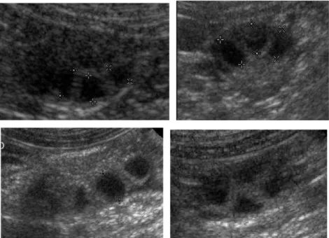
In all the 34 remaining bitches (65.4%), intra-ovarian hypoechoic structures, smaller than pre-ovulatory follicles and irregular in shape, were still observed in the ovary after collapsus of other pre-ovulatory follicles, the ovary never showing an homogeneous aspect at ovulation (Figure 5).
Figure 5: Hypoechoic structures at ovulation..
Unchanged follicles, keeping a “pre-ovulation” aspect, were observed in 9/15 bitches (Group 2) and in 17/37 (Group 3) bitches (Figure 6). In the bitches that were ovario-hysterectomised after ovulation, persistent follicles were observed on the ovaries after removal in 5/9 bitches (Group 2) and 4/17 bitches (Group 3).
a b
Figure 6: Persistence of round anechoic structures (non-ovulated follicles?) after ovulation.
Visualisation of liquid around at least one ovary (between the ovary and the ovarian bursa) was observed at ovulation in 6/16 (Group 2) and 15/37 (Group 3) bitches, in variable amounts among bitches (Figure 7.). In all bitches, this collection had disappeared within the following 12 hours.
Figure 7: presence of liquid (arrows) between the ovary and the ovarian bursa at ovulation.
A hyperechoic spot (“white spot”) was sometimes observed inside at least one follicle on ovarian ultrasound images in the 24 to 36 hours preceeding ovulation, and was less often observed 12 hours after ovulation (Figure 8).
Figure 8: Hyperechoic spot (“white spot”) visualised in some pre-ovulatory follicles and occasionally in post-ovulatory follicles.
- Post-ovulation:
In the immediate post-ovulation period – 12 to 24 hours after the complete transformation of the ovaries at ovulation – hypoechoic structures, very similar to pre-ovulatory follicles, were seen in most cases. The ultrasound aspect was very similar to the pre-ovulatory stage (Figure 9).
- Difficulties:
The right ovaries were sometimes difficult to visualise due to the proximity of intestines (Figure 10). In such a case, it was useful to examine the bitch in a standing position.
Figure 10: Bad image of the ovary due to the proximity of intestines.
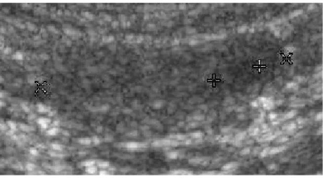
The exact details and measurement of the follicles were also more difficult to assess in 4 bitches, all belonging to large breeds (25 – 40 kg) (Dogo Argentino, Labrador Retriever, German Shepherd, Belgian Shepherd) (Figure 11).
Figure 11: Pre-ovulatory follicles in two large bitches (Dogo Argentino and German Shepherd), for which it was more difficult to assess the ultrasonographic details.
Group 1:
Using an ultrasound equipment with a lower resolution than for Groups 2 and 3, we still had a global image which allowed us to detect the changes that occurred within the ovaries around ovulation.
By ultrasound, the pre-ovulatory follicles appeared as round anechoic structures with a thick wall visualised within the ovary (Figure 12).
Figure 12: Ultrasonographic image of ovaries (Vetson-Pro ultrasound machine with a 7.5 transducer): pre-ovulatory follicles (a.), aspect of the ovary at ovulation (b.).
A clear ovarian change at the supposed time of ovulation (at least 80% of the pre-ovulatory follicles disappearing or clearly diminishing in size) was detected in 20/21 bitches, performing only one daily examination. It was considered as the day of ovulation. No significant difference in the estimated ovulation time could be seen between the left and the right ovary.
Quantitative aspects.
Group 1: Nineteen of 21 bitches conceived, with a mean litter size of 6.68 ± 2.16
pups. The interval between the estimated time of ovulation (day during which the aspect of the ovaries under ultrasound clearly changed) and parturition was 63.72 ± 1.44 days.
The LH peak could be estimated in 19/21 bitches. The average value of the LH peak was 4.85 ± 1.96 ng/mL. In those animals, the interval between this LH peak and the first ultrasound examination showing signs of ovulation was 50.47 ± 1.01 hours (extreme values: 24 - 72 hours). The delay between the day when the LH peak occurred and the start of parturition was 65.43 ± 1.60 days. Considering that in Group 1 progesterone was assayed only once daily, the mean progesterone value on the day of occurrence of the LH peak was 2.20 ± 0.53 ng/mL. When trying to correlate the LH peak with the progesterone plasma level, the delay between progesterone level reaching 2 ng/mL and ovulation was 39 ± 14.86 hours.
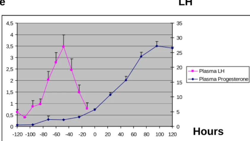
The mean progesterone value at ovulation estimated by ultrasound was 5.73 ± 1.51 ng/mL (extremes values: 3.0 - 8.09 ng/mL). In 18/20 bitches, ovulation occurred in less than 24 hours apart from the day at which progesterone reached 5 ng/mL. Progesterone LH 0 0,5 1 1,5 2 2,5 3 3,5 4 4,5 -120 -100 -80 -60 -40 -20 0 20 40 60 80 100 120 0 5 10 15 20 25 30 35 Plasma LH Plasma Progesterone
Figure 13: Plasma LH (ng/mL) and progesterone (ng/mL) before and after ovulation estimated by ultrasound (noted 0) in Group 1. (n=19 for LH ; n=21 for progesterone)
Group 2: The LH peak could be estimated in 11/15 bitches. The average value of
the LH peak was 8.85 ± 2.96 ng/mL. In those animals, the interval between this LH peak and the first ultrasound examination showing signs of ovulation was 46.16 ± 1.60 hours (extreme values: 36 to 60 hours). The delay between progesterone level reaching 2 ng/mL and ovulation was 49.38 ± 9.84 hours. If we compare the time when progesterone reached 2 ng/mL with the time of the maximum LH value, it occurred 12 hours before (3 bitches), 6 hours before (1 bitch), at the same time (5 bitches) or 12 hours after (2 bitches). In the 15 bitches of Group 2, the mean progesterone value at ovulation estimated by ultrasound was 6.36 ± 1.34 ng/mL (extreme values: 4.55 ng/mL to 7.36 ng/mL). At the time of the last ultrasound examination before the start of ovulation (which occurred 12 hours before) it was 4.64 ± 1.04 ng/mL. Taking into account that in 9/15 bitches the ovulation process was completed in less than 12 hours and that in the remaining 6/15 bitches, the ovulation process – the complete ovarian change on ultrasound - was completed after only two successive examinations at 12 hours intervals, the mean progesterone value at the end of ovulation process was 7.28 ± 1.92 ng/mL.
The day at which blood progesterone reached 5 ng/mL was determined in 12 bitches. Ovulation occurred exactly the same day in 9/12 bitches. In the other bitches, it occurred 24 hours before (1/12), 12 hours before (1/12) bitches and 12 hours later (1/12). Progesterone LH 0 1 2 3 4 5 6 7 8 9 -120 -100 -80 -60 -40 -20 0 20 40 60 0 5 10 15 20 25 30 35 Plasma LH Plasma Progesterone
Figure 14: Plasma LH (ng/mL) and progesterone (ng/mL) before and after ovulation estimated by ultrasound (noted 0) in Group 2. (n=19 for LH ; n=21 for progesterone). (n=11 for LH ; n=15 for progesterone)
Group 3: The LH peak could be estimated in 9/17 bitches. The average value of
the LH peak was 12.01± 9.08 ng/mL. In those animals, the interval between this LH peak and the first ultrasound examination showing ovulation was 51.62 ± 3.73 hours. In these bitches, the mean progesterone value on the day of occurrence of the LH peak was 2.58 ± 0.57 ng/mL.
The delay between progesterone level reaching 2 ng/mL and ovulation was 50.0 ± 14.0 hours. If we compare the time when progesterone reached 2 ng/mL with the time of the maximum LH value, it occurred 24 hours before (1 bitch), 12 hours before (2 bitches), at the same time (2 bitches), 6 hours after (1 bitch), 12 hours after (1 bitch) and 24 hours after (2 bitches).
In these 37 bitches, the mean progesterone value at ovulation estimated by ultrasound was 6.42 ± 1.53 ng/mL (extreme values: 4.43 ng/mL to 9.81 ng/mL). At the time of the last ultrasound examination before the start of ovulation (which
16/37 bitches the ovulation process was completed in less than 12 hours and that in 21/37 bitches, the ovulation process – the complete ovarian change on ultrasound - was completed after only two successive examinations at 12 hours intervals, the mean progesterone value at the end of ovulation process was 7.71 ± 1.99 ng/mL.
The day at which blood progesterone reached 5 ng/mL was determined in 27/37 bitches. It occurred on the same day of the estimated ovulation in 17/27 bitches. In the other bitches, it occurred 78 hours before (1/27), 36 hours before (1/27), 24 hours before (4/27) and 12 hours after (4/27).
0 2 4 6 8 10 12 14 -120 -100 -80 -60 -40 -20 0 20 40 60 0 5 10 15 20 25 30 35 Plasma LH Plasma Progesterone 38
Figure 15: Plasma LH (ng/mL) and progesterone (ng/mL) before and after ovulation estimated by ultrasound (noted 0) in Group 3. (n=9 for LH ; n=17 for progesterone)
Progesterone LH