OATAO is an open access repository that collects the work of Toulouse
researchers and makes it freely available over the web where possible
Any correspondence concerning this service should be sent
to the repository administrator:
tech-oatao@listes-diff.inp-toulouse.fr
This is an author’s version published in:
http://oatao.univ-toulouse.fr/20345
To cite this version:
Verheecke, Carol
and Diaz, Roxanne
and Mathieu, Florence
and Anson, Philippe
and Liboz, Thierry
Reduction of aflatoxin
production by Aspergillus flavus and Aspergillus parasiticus in
interaction with Streptomyces. (2015) Microbiology, 161 (5).
967-972. ISSN 1350-0872
Reduction of aflatoxin production by Aspergillus
flavus and Aspergillus parasiticus in interaction with
Streptomyces
C. Verheecke, T. Liboz, P. Anson, R. Diaz and F. Mathieu
CorrespondenceFlorence Mathieu mathieu@ensat.fr
Universite´ de Toulouse, Laboratoire de Ge´nie Chimique, UMR 5503 CNRS/INPT/UPS, INP-ENSAT, 1, avenue de l’Agrobiopoˆle, 31326 Castanet-Tolosan, France
The aim of this study is to investigate aflatoxin gene expression during Streptomyces–Aspergillus interaction. Aflatoxins are carcinogenic compounds produced mainly by Aspergillus flavus and Aspergillus parasiticus. A previous study has shown that Streptomyces–A. flavus interaction can reduce aflatoxin content in vitro. Here, we first validated this same effect in the interaction with A. parasiticus. Moreover, we showed that growth reduction and aflatoxin content were correlated in A. parasiticus but not in A. flavus. Secondly, we investigated the mechanisms of action by reverse-transcriptase quantitative PCR. As microbial interaction can lead to variations in expression of household genes, the most stable[act1, btub (and cox5 for A. parasiticus)] were chosen using geNorm software. To shed light on the mechanisms involved, we studied during the interaction the expression of five genes (aflD, aflM, aflP, aflR and aflS). Overall, the results of aflatoxin gene expression showed that Streptomyces repressed gene expression to a greater level in A. parasiticus than in A. flavus. Expression of aflR and aflS was generally repressed in both Aspergillus species. Expression of aflM was repressed and was correlated with aflatoxin B1 content. The results suggest that aflM expression could be a potential aflatoxin indicator in Streptomyces species interactions. Therefore, we demonstrate that Streptomyces can reduce aflatoxin production by both Aspergillus species and that this effect can be correlated with the repression of aflM expression.
INTRODUCTION
Aflatoxins (AFs) are polyketide-derived furanocoumarins. They are produced by fungi of the genus Aspergillus (including Aspergillus flavus and Aspergillus parasiticus) in agricultural foodstuffs (maize, hazelnut, peanut, etc.) (Giorni et al., 2007; Passone et al., 2010). These AFs are toxic and their main adverse effects on humans are hepatocarcinoma (Qian et al., 1994; IARC, 2014), immune system deficiency (Jiang et al., 2005), reduced child growth (Gong et al., 2004) and increased risks of stillborn or newborn jaundice (Shuaib et al., 2010). To reduce those multiple effects, many countries have implemented maximum authorized levels of AFs in food and feed (Wu & Guclu, 2012).
AF biosynthesis is coded by a 80 kb long DNA sequence. The latter is a cluster containing 30 putative genes charac-terized in both A. flavus and A. parasiticus (Yu, 2012). For structural genes, early (as aflD), medium (as aflM) and late (as aflP) genes are denominated (Fig. S1, available in the
online Supplementary Material). The gene aflD encodes a reductase enzyme involved in the conversion of norsolori-nic acid to averantin (Papa, 1982); aflM is required for the conversion of versicolorin A to demethylsterigmatocystin (Skory et al., 1992); and aflP encodes a methyltransferase converting sterigmatocystin to O-methylsterigmatocystin (Bhatnagar et al., 1988). Two cluster-specific regulators are also known: aflR encodes a transcription activator that binds a consensus sequence in the promoter regions of AF structural genes (Payne et al., 1993), and AflS is a potential co-activator of AflR (Meyers et al., 1998) (Fig. S1). Schmidt-Heydt et al. (2009) showed that the aflR/aflS ratio can also be used as an indicator of AF biosynthesis. In addition to AflR and AflS, the clustered genes are also regulated by aspecific transcriptional regulators such as LaeA or Ap-1 (Reverberi et al., 2008; Chang et al., 2012). Microbial interactions with yeast, bacteria or fungi can reduce AF production by aspergilli (Yin et al., 2008). Streptomyces are soil-borne bacteria that can develop in crops and that are known to be good biocontrol candidates (Bressan & Figueiredo, 2008). Studies have shown that Streptomyces metabolites are sources of AF repressors (Ono et al., 1997; Sakuda et al., 2000). However, until recently no studies have focused on Streptomyces–Aspergillus mutual Abbreviations: AF, aflatoxin; RT-qPCR, reverse-transcriptase
quantitat-ive PCR.
One supplementary table and two supplementary figures are available with the online Supplementary Material.
interactions and their impact on AF production and AF gene expression.
Recently, we found that Streptomyces (27 strains)–A. flavus (NRRL 62477) mutual interaction on contact can reduce the concentration of AF B1 (AFB1) and AF B2 (AFB2) in vitro by up to 4.4 % (remaining concentration) (Verheecke et al., 2014).
In this study, six of the Streptomyces strains previously used were chosen for further investigation. Our preliminary goal was to verify the interaction impact on an AF G producer, namely A. parasiticus. Our main objective was to study the impact of these interactions on AF gene expression. The methodology was applied to A. flavus and A. parasiticus on expression of five targeted genes (aflD, aflM, aflP, aflR and aflS).
METHODS
Fungal and Streptomyces strains.The fungal strains used were A. flavus NRRL 62477 and A. parasiticus Afc5. The six actinomycete strains were selected on ISP-2 medium after 10 days at 28uC, mainly based on the results from Verheecke et al. (2014): antagonism on contact with A. flavus, reduction of AFs concentration under 17 % versus control and growth on ISP-2 medium (unpublished data). Their 16S rRNA genes were sequenced according to the method described by Zitouni et al. (2005). The six strains were identified as Streptomyces roseolus S06, Streptomyces calvus S13, Streptomyces thinghirensis S17, Streptomyces sp. S27, Streptomyces griseoplanus S35 and Streptomyces caeruleatus S38. Streptomyces were kept at 220uC in cryotubes in ISP-2 medium with 20 % (v/v) glycerol.
Interaction method and AF quantification.Pre-cultures for both Aspergilli (on yeast extract peptone dextrose medium) and for Streptomyces (on ISP-2) were made for 7 days at 28uC as previously described by Verheecke et al. (2014). The culture conditions are based on Verheecke et al. (2014) with slight modifications: a sterile 8.5 cm cellophane sheet (Hutchinson) was dropped on ISP-2 (Shirling & Gottlieb, 1966) prior to inoculum and two streaks (instead of one) of Streptomyces culture were inoculated in parallel 2 cm away from Aspergillus inoculation (centre of the Petri dish). Two sets of plates (three Petri dishes each) were inoculated: one set for RNA extraction at 90 h (day 4) and one at 7 days for analysis of fungal growth and AF concentration. One day 4 (set one), the fungal biomass was separated from the bacterial biomass. Using a scalpel and with the naked eye, the mouldy cellophane was removed and used for RNA extraction (avoiding taking bacterial biomass). At day 7 (set two), the fungal biomass was removed from the cellophane sheet for measurement of dry weight (after drying: 18 h at 80uC). In the remaining media, three agar plugs (w 9 mm) were removed from the fungal growth area for AF quantification (Verheecke et al., 2014). The experiment was done twice in triplicate.
AF quantification was done as previously described (Verheecke et al., 2014). Briefly, methanol (1 ml) was added to agar plugs during a 30 min incubation period (shaken three times). This was then centrifuged for 15 min at 12470 g and the supernatant was filtered (0.45 mm, 4 mm PVDF; Whatman) into vials. AF quantification was done on an Ultimate 3000 system (Dionex- Thermo Electron) with all the RS series modules. A C18 pre-column and column were used (Phenomenex, Luna 3 mm, 20064.6 mm). Detection of AFs was done according to instructions for the Coring Cell analysis system (Coring System Diagnostix). Quantification was realized with Chromeleon software, using AFB1 and AFB2 (Sigma-Aldrich)
(detection limit: 0.5 p.p.b.) as standards. Statistical analyses were made using ‘nparcomp’ R (version 2.15.2).
RNA extraction and quantification.In total, 60 mg of mycelium was crushed in liquid nitrogen to a fine powder. The powder was then stored at 280uC until RNA isolation. Total RNA was isolated using an Aurum Total RNA kit (Bio-Rad). The manufacturer’s instructions for eukaryotic and plant cell materials were followed, except for two modifications: DNase I digestion was extended to 1 h and elution was done at 70uC for 2 min in the elution buffer. Total RNA was eluted into 80 ml and stored at 220uC. Then, 1 ml of total RNA of each sample was loaded into an RNA StSens chip (Bio-Rad) and quantified on a Nanodrop 2000 spectrophotometer (Thermo Scientific) according to the manufacturer’s instructions. Samples with RNA Quality Indicator .7, A260/280.2 and A260/230.1.3 were selected for further analysis.
Reverse-transcriptase quantitative PCR (RT-qPCR). Reverse transcription was carried out with an Advantage RT-PCR kit (Clontech) with Oligo (dT)18primer according to the manufacturer’s instructions (RNA concentration: 1 mg total RNA), with one modification: incubation at 42uC was extended to 4 h. RT-qPCR was performed in duplicate using a CFX96 Touch instrument (Bio-Rad) using SsoAdvancedTM SYBR Green Supermix (Bio-(Bio-Rad) according to the manufacturer’s instructions (annealing temperature, 59uC; concentrations: primers, 500 nM and cDNA, 100 ng). Primer pairs and associated efficiencies were validated (85–115 %) (Table S1).
Validation of reference genes. Based on the literature, six candidate genes (act1, btub, cox5, ef1, gpdA and tbp) were studied as potentially suitable reference genes (Radonic´ et al., 2004; Bohle et al., 2007). For identification of the optimal number of reference genes and stability, eight samples (randomly selected among the different conditions) were tested in triplicate. The measures of gene stability V (gene pairwise variation) and M (V of a gene with other genes) were calculated using geNorm software (Vandesompele et al., 2002). M values are represented in Fig. S2 for A. flavus and A. parasiticus, according to the geNorm software in standard configura-tion. This led to the choice of act1 and btub (for A. flavus) and act1, btub and cox5 (for A. parasiticus) as optimal reference genes.
Relative quantification. Relative quantification was determined compared with the chosen reference genes. Calculation of gene expression was via qbase+ software as well as statistical analysis (Hellemans et al., 2007).
The correlations between fungal dry weight, AF content and gene expression were determined using Pearson correlation (r, asterisks indicate statistically significant differences at P,0.05).
RESULTS
Interaction of Streptomyces with A. parasiticus and A. flavus
Interaction between Streptomyces and both Aspergillus species was monitored in Petri dishes over 7 days. On day 7, all the tested Streptomyces strains showed a mutual antagonism on contact with the aspergilli. For A. parasiticus, compared with the control dry weight (100 %), in interaction with the bacterial strains, the fungal residual dry weight (RDW) ranged from 24.7 % (S06) to 57.2 % (S17) (Table 1). For A. flavus (Table 2), RDW ranged from 60.7 % (S35) to 92.7 % (S27) of the control dry weight (100 %) when treated with the same bacterial strains.
Reduction of AF concentration
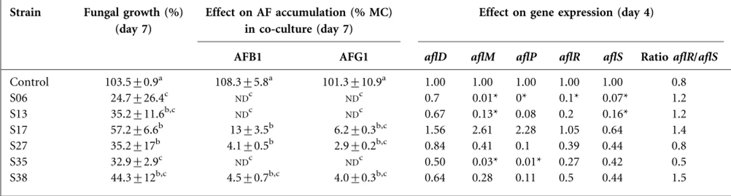
On day 7, the production of AFs by A. parasiticus and A. flavus was reduced in contact with the six Streptomyces strains tested. For A. parasiticus, AFB1 and AFG1 production was monitored (Table 1). S17 showed lower reductions of 13 and 6.2 % of the concentration in the medium as a percentage of the control) for AFB1 and AFG1, respectively. S27 and S38 showed higher reduction of 4.1 and 4.5 % for AFB1 and 2.9 % and 4.0 % for AFG1. S06, S13 and S35 reduced to the greatest extent, with no AFB1 or AFG1 detected.
For A. flavus, AFB1 and AFB2 production was monitored (Table 2). S17 showed the least reduction, with 24 and 5.3 % concentration in the medium for AFB1 and AFB2, respectively. S13 showed higher reduction of 15.6 and 9.3 % for AFB1 and AFB2, respectively. S06, S27, S35 and S38 were the greatest reducers, with no AFB1 or AFB2 detected. Pearson correlation was also applied.
AF gene expression
Gene expression was determined on day 4 with A. flavus and A. parasiticus alone (controls) and in interaction with the six Streptomyces strains. Five genes (aflD, aflM, aflP, aflR and aflS) were investigated relative to two reference genes (act1 and btub) for A. flavus and three reference genes (act1, btub and cox5) for A. parasiticus.
For A. parasiticus, aflM expression was slightly impacted by S13 (7.7-fold), moderately by S35 (33.3-fold) and very highly by S06 (100-fold) (Table 1). S35 and S06 also reduced aflP expression 83- and 250-fold, respectively. Regarding aflS and aflR, S13 significantly reduced aflS expression (6.25-fold) and S06 repressed the expression of both aflS (10-fold) and aflR (14.3-fold). The interaction did not significantly impact aflD expression.
For A. flavus, S35 repressed the expression of aflM (8.4-fold) and aflR (1.5-(8.4-fold) (Table 2). S38 repressed the
Table 1. Impact of Streptomyces strains on A. parasiticus AFs and gene expression
Data with the same letter are not significantly different (P,0.05). MC, Concentration in the media as a percentage of the control;ND, not detected. Mean values are given±SD.
Strain Fungal growth (%) (day 7)
Effect on AF accumulation (% MC) in co-culture (day 7)
Effect on gene expression (day 4)
AFB1 AFG1 aflD aflM aflP aflR aflS Ratio aflR/aflS
Control 103.5±0.9a 108.3±5.8a 101.3±10.9a 1.00 1.00 1.00 1.00 1.00 0.8 S06 24.7±26.4c NDc NDc 0.7 0.01* 0* 0.1* 0.07* 1.2 S13 35.2±11.6b,c NDc NDc 0.67 0.13* 0.08 0.2 0.16* 1.2 S17 57.2±6.6b 13±3.5b 6.2±0.3b,c 1.56 2.61 2.28 1.05 0.64 1.4 S27 35.2±17b 4.1±0.5b 2.9±0.2b,c 0.84 0.41 0.1 0.39 0.44 0.8 S35 32.9±2.9c NDc NDc 0.50 0.03* 0.01* 0.27 0.42 0.5 S38 44.3±12b,c 4.5±0.7b,c 4.0±0.3b,c 0.64 0.28 0.11 0.5 0.44 1.5 *Significant difference (P,0.05).
Table 2. Impact of Streptomyces strains on A. flavus AFs and gene expression
Data with the same letter are not significantly different (P,0.05). MC, Concentration in the media as a percentage of the control;ND, not detected. Mean values are given±SD.
Strain Fungal growth (%) (day 7)
Effect on AF accumulation (% MC) in co-culture (day 7)
Effect on gene expression (day 4)
AFB1 AFB2 aflD aflM aflP aflR aflS Ratio aflR/aflS
Control 100.0±15.4a 100.0±13.9a 100.0±17.3a 1.00 1.00 1.00 1.00 1.00 0.9 S06 64.6±8.6b 2.3±4.5c ND 0.69 0.25 1.57 2.37 0.40 2.9 S13 81.3±16.2a 15.6±9.2b 9.3±20.8b 1.60 0.45 0.41 0.82 0.70 0.5 S17 77.7±11.2a 24.0±19.8b 5.3±11.9b 0.95 0.26 3.03 1.53 0.39 1.8 S27 92.7±18.3a 8.1±5.1b ND 1.42 0.26 0.39 0.88 0.96 0.5 S35 60.7±11.4b 0.2±0.5c ND 0.50 0.12* 1.02 0.63 0.24 1.3 S38 62.4±15.2b 3.1±5.3c ND 1.44 0.14* 0.21* 0.69* 0.62 0.5 *Significant difference (P,0.05).
expression of aflP (4.8-fold) and aflR (1.45-fold). S06 enhanced the expression of aflR (2.37-fold). Expression of aflD and aflS was not significantly impacted by the six strains.
The ratio aflR/aflS was monitored in both producing strains. Both positive controls were close to 1 : 0.8 for A. parasiticus and 0.9 for A. flavus. This ratio was above 1 for A. parasiticus in interaction with S06 (1.2), S13 (1.2), S17 (1.4) and S38 (1.5) and for A. flavus in interaction with S06 (2.9), S17 (1.8) and S35 (1.3). Ratios for the other interactions were below 1.
Assessment of correlation
Independently of the Streptomyces tested, Pearson correla-tions were done between RDW and AF concentration. For A. parasiticus, the reduction of AFB1 and AFG1 concen-tration in the medium was correlated (r50.94* and 0.91*) with RDW reduction. For A. flavus, AFB1 and AFB2 concentration were not correlated with RDW reduction. Pearson correlations were also applied to gene expression versus RDW or AF concentration in the medium. For A. parasiticus, all gene expressions were correlated with RDW reduction. The strongest correlation was obtained for expression of aflP (r50.97*). Correlations were also iden-tified between the reduction of AFB1 concentration in the mediumand aflD, aflM and aflP repression (r50.91*, 0.92* and 0.86*, respectively). For A. flavus, RDW and AFB1 and AFB2 concentrations were only correlated with aflM expression (r50.86*, 0.86* and 0.83, respectively).
DISCUSSION
Six Streptomyces strains had their impact confirmed on A. flavus and tested for A. parasiticus. They all showed mutual antagonism on contact as described by Magan & Lacey (1984). This type of interaction has already been studied in Petri dishes (Sultan & Magan, 2011; Verheecke et al., 2014). The latter showed that after 10 days at 28uC on ISP-2 medium, 27 of 37 actinomycete strains showed mutual antagonism on contact with A. flavus and were able to reduce AF accumulation (residual concentration below 38 %). Here, after 7 days, the interaction with both Aspergillus species and the six chosen bacterial strains led to mutual antagonism on contact impacting fungal growth and resulting in residual AF concentration in the medium below 24 %.
In our study, for A. parasiticus, RDW reduction was correlated with AF concentration reduction. This correla-tion is generally observed in the literature (reviewed by Holmes et al., 2008; Bluma et al., 2008a, b). However, exceptions to this rule are also found. Indeed, Reverberi et al. (2008) studied the effect of Lentinula edodes CF42 filtrate (2 %, w/v) on A. parasiticus after 9 days at 30uC in potato dextrose broth. The results showed 1.90 % AF concentration while no impact on fungal growth was detected. In our study, we highlight another example in
another Aspergillus species. Indeed, for A. flavus, RDW reduction was not correlated with AF concentration reduction. In conclusion, we observed different responses to the Streptomyces interaction depending on the Aspergillus species studied. Regarding A. flavus, the results described here demonstrate that bacterial interaction did not impact AF concentration in the medium just by fungal growth reduction.
AF inhibition can occur through gene repression (Yu, 2012; Alkhayyat & Yu, 2014). Thus, we developed a methodology to monitor AF gene expression. Our preliminary work iden-tified maximum gene expression at 90 h (data not shoown). Based on those results, we monitored gene expression under the same conditions. Reference genes were then chosen based on geNorm software and the data matched the MIQE guidelines (Bustin et al., 2009). In our study, we tested six candidates genes for their stability during Aspergillus– Streptomyces interaction and the most stable genes were identified (Radonic´ et al., 2004). Nevertheless, cox5 was less stable than expected (fifth out of seven for A. flavus) and gapdh was more stable than described in the literature for other organisms (Dheda et al., 2004; Bohle et al., 2007; Radonic´ et al., 2004).
In particular, we monitored the expression of three structural genes, aflD (early), aflM (medium) and aflP (late), and two regulator-coding genes, aflR and aflS. The expression of aflM was mostly repressed (between 2.2- and 100-fold) under the conditions tested. A disruption of the aflM homologue in Aspergillus nidulans (verA) led to a reduction of sterigmatocystin production by 200- to 1000-fold (Keller et al., 1994) and versicolorin A accumulation. Here, we showed that repression of aflM expression was highly correlated with AFB1 concentration reduction in both Aspergillus species. Thus, the measure of aflM expression could be an indicator of AF concentration in our experimental conditions.
For A. parasiticus, gene expressions were correlated with growth reduction. This could be linked to a delay in fungal growth impacting gene expression. For A. flavus, RDW reduction was not correlated with gene expression. The latter were differentially modulated depending on the bacterial strain. Similar results were obtained for A. flavus with caffeic acid addition to the medium: aflD (6.6-fold), aflM (7.1-fold), aflP (9.1-fold) and aflS (1.5-fold) were repressed without affecting fungal growth (Kim et al., 2008). In our case, the same range of repression was observed in the Streptomyces–Aspergillus interaction. With regard to regulators, expression of aflR was differently impacted. It was enhanced 2.37-fold by S06 for A. flavus and repressed up to 10-fold by S06 for A. parasiticus. Variation of aflR expression was also observed in A. parasiticus after addition of Trametes versicolor filtrate in the medium. Indeed, after 3 days, aflR expression was enhanced by more than 10-fold in Czapek–Dox broth solidified with agar while AF content was reduced (Zjalic et al., 2006). In the present study, aflR expression was
enhanced in S06 interaction with A. flavus and AF pro-duction was also reduced. In the S06 interaction, aflR expression was not representative of AflR function on aflD, aflM or aflS expression.
Depending on the fungal and bacterial strains, the ratio aflR/aflS was differently impacted. It ranged for A. flavus from 2.9 by S06 to 0.5 by S35 and for A. parasiticus from 1.5 by S38 to 0.5 by S35. This ratio was first studied under various activity of water and temperatures, and a ratio above 1 would lead to an activation of AFB1 biosynthesis (Schmidt-Heydt et al., 2009). In our study, a ratio above 1 was found under most conditions but was not correlated with high AF accumulation.
Moreover, the repression of aflM expression was highly correlated with AFB1 concentration in the medium in both Aspergillus species. A further indicator besides the aflR/aflS ratio could be aflM expression in relation to AF accumu-lation in the interaction with Streptomyces.
In conclusion, we have shown that mutual antagonism on contact between Streptomyces species and species of the genus Aspergilli led to a reduction of AF accumulation by A. flavus and A. parasiticus. The AF reduction of the latter was correlated with fungal growth reduction whereas no correlation was observed for A. flavus. Here, Streptomyces species bacterial interactions mainly led to the repression of aflM and aflS but had a different impact on aflP and aflR expression. Expression of aflM was correlated with AF accumulation in both Aspergillus species and could be an indicator of AF content in the interaction with Strepto-myces. Based on this, Streptomyces griseoplanus S35 appears to be the best biocontrol candidate for further testing on maize.
ACKNOWLEDGEMENTS
We thank Dr Atika Meklat and Dr Elodie Choque for their help with the experiments. Lora Verheecke provided editorial help. We are grateful to the French National Research Agency (Aflafree no. 11 003 01) for financial support and Agri Sud-Ouest Innovation.
REFERENCES
Alkhayyat, F. & Yu, J.-H. (2014).Upstream regulation of mycotoxin biosynthesis. Adv Appl Microbiol 86, 251–278.
Bhatnagar, D., Ullah, A. H. J. & Cleveland, T. E. (1988).Purification and characterization of a methyltransferase from Aspergillus para-siticus SRRC 163 involved in aflatoxin biosynthetic pathway. Prep Biochem 18, 321–349.
Bluma, R., Amaiden, M. R., Daghero, J. & Etcheverry, M. (2008a). Control of Aspergillus section Flavi growth and aflatoxin accumula-tion by plant essential oils. J Appl Microbiol 105, 203–214.
Bluma, R., Amaiden, M. R. & Etcheverry, M. (2008b).Screening of Argentine plant extracts: impact on growth parameters and aflatoxin B1 accumulation by Aspergillus section Flavi. Int J Food Microbiol 122, 114–125.
Bohle, K., Jungebloud, A., Go¨cke, Y., Dalpiaz, A., Cordes, C., Horn, H. & Hempel, D. C. (2007). Selection of reference genes for
normal-isation of specific gene quantification data of Aspergillus niger. J Biotechnol 132, 353–358.
Bressan, W. & Figueiredo, J. E. F. (2008).Efficacy and dose–response relationship in biocontrol of Fusarium disease in maize by Streptomyces spp. Eur J Plant Pathol 120, 311–316.
Bustin, S. A., Benes, V., Garson, J. A., Hellemans, J., Huggett, J., Kubista, M., Mueller, R., Nolan, T., Pfaffl, M. W. & other authors (2009).The MIQE guidelines: minimum information for publication of quantitative real-time PCR experiments. Clin Chem 55, 611– 622.
Chang, P.-K., Scharfenstein, L. L., Ehrlich, K. C., Wei, Q., Bhatnagar, D. & Ingber, B. F. (2012).Effects of laeA deletion on Aspergillus flavus conidial development and hydrophobicity may contribute to loss of aflatoxin production. Fungal Biol 116, 298–307.
Dheda, K., Huggett, J. F., Bustin, S. A., Johnson, M. A., Rook, G. & Zumla, A. (2004).Validation of housekeeping genes for normalizing RNA expression in real-time PCR. Biotechniques 37, 112–114, 116, 118–119.
Giorni, P., Magan, N., Pietri, A., Bertuzzi, T. & Battilani, P. (2007). Studies on Aspergillus section Flavi isolated from maize in northern Italy. Int J Food Microbiol 113, 330–338.
Gong, Y., Hounsa, A., Egal, S., Turner, P. C., Sutcliffe, A. E., Hall, A. J., Cardwell, K. & Wild, C. P. (2004).Postweaning exposure to aflatoxin results in impaired child growth: a longitudinal study in Benin, West Africa. Environ Health Perspect 112, 1334–1338.
Hellemans, J., Mortier, G., De Paepe, A., Speleman, F. & Vandesompele, J. (2007). qBase relative quantification framework and software for management and automated analysis of real-time quantitative PCR data. Genome Biol 8, R19.
Holmes, R. A., Boston, R. S. & Payne, G. A. (2008).Diverse inhibitors of aflatoxin biosynthesis. Appl Microbiol Biotechnol 78, 559–572. IARC, International Agency For Research on Cancer (2014). Monograph classification. http://monographs.iarc.fr/ENG/Classification/. Jiang, Y., Jolly, P. E., Ellis, W. O., Wang, J.-S., Phillips, T. D. & Williams, J. H. (2005).Aflatoxin B1 albumin adduct levels and cellular immune status in Ghanaians. Int Immunol 17, 807–814.
Keller, N. P., Kantz, N. J. & Adams, T. H. (1994).Aspergillus nidulans verA is required for production of the mycotoxin sterigmatocystin. Appl Environ Microbiol 60, 1444–1450.
Kim, J. H., Yu, J., Mahoney, N., Chan, K. L., Molyneux, R. J., Varga, J., Bhatnagar, D., Cleveland, T. E., Nierman, W. C. & Campbell, B. C. (2008).Elucidation of the functional genomics of antioxidant-based inhibition of aflatoxin biosynthesis. Int J Food Microbiol 122, 49–60. Magan, N. & Lacey, J. (1984).Effects of gas composition and water activity on growth of field and storage fungi and their interactions. Trans Br Mycol Soc 82, 305–314.
Meyers, D. M., Obrian, G., Du, W. L., Bhatnagar, D. & Payne, G. A. (1998). Characterization of aflJ, a gene required for conversion of pathway intermediates to aflatoxin. Appl Environ Microbiol 64, 3713– 3717.
Ono, M., Sakuda, S., Suzuki, A. & Isogai, A. (1997).Aflastatin A, a novel inhibitor of aflatoxin production by aflatoxigenic fungi. J Antibiot (Tokyo) 50, 111–118.
Papa, K. E. (1982). Norsolorinic acid mutant of Aspergillus. J Gen Microbiol 128, 1345–1348.
Passone, M. A., Rosso, L. C., Ciancio, A. & Etcheverry, M. (2010). Detection and quantification of Aspergillus section Flavi spp. in stored peanuts by real-time PCR of nor-1 gene, and effects of storage conditions on aflatoxin production. Int J Food Microbiol 138, 276– 281.
Payne, G. A., Nystrom, G. J., Bhatnagar, D., Cleveland, T. E. & Woloshuk, C. P. (1993). Cloning of the afl-2 gene involved in aflatoxin biosynthesis from Aspergillus flavus. Appl Environ Microbiol 59, 156–162.
Qian, G. S., Ross, R. K., Yu, M. C., Yuan, J. M., Gao, Y. T., Henderson, B. E., Wogan, G. N. & Groopman, J. D. (1994).A follow-up study of urinary markers of aflatoxin exposure and liver cancer risk in Shanghai, People’s Republic of China. Cancer Epidemiol Biomarkers Prev 3, 3–10.
Radonic´, A., Thulke, S., Mackay, I. M., Landt, O., Siegert, W. & Nitsche, A. (2004). Guideline to reference gene selection for quantitative real-time PCR. Biochem Biophys Res Commun 313, 856–862.
Reverberi, M., Zjalic, S., Ricelli, A., Punelli, F., Camera, E., Fabbri, C., Picardo, M., Fanelli, C. & Fabbri, A. A. (2008). Modulation of antioxidant defense in Aspergillus parasiticus is involved in aflatoxin biosynthesis: a role for the ApyapA gene. Eukaryot Cell 7, 988– 1000.
Sakuda, S., Ikeda, H., Nakamura, T., Kawachi, R., Kondo, T., Ono, M., Sakurada, M., Inagaki, H., Ito, R. & Nagasawa, H. (2000).Blasticidin A derivatives with highly specific inhibitory activity toward aflatoxin production in Aspergillus parasiticus. J Antibiot (Tokyo) 53, 1378– 1384.
Schmidt-Heydt, M., Abdel-Hadi, A., Magan, N. & Geisen, R. (2009). Complex regulation of the aflatoxin biosynthesis gene cluster of Aspergillus flavus in relation to various combinations of water activity and temperature. Int J Food Microbiol 135, 231–237.
Shirling, E. B. & Gottlieb, D. (1966).Methods for characterization of Streptomyces species. Int J Syst Bacteriol 16, 313–340.
Shuaib, F. M. B., Ehiri, J., Abdullahi, A., Williams, J. H. & Jolly, P. E. (2010).Reproductive Health Effects of Aflatoxins: A Review of the Literature. Reprod Toxicol 29, 262–70.
Skory, C. D., Chang, P. K., Cary, J. & Linz, J. E. (1992).Isolation and characterization of a gene from Aspergillus parasiticus associated with
the conversion of versicolorin A to sterigmatocystin in aflatoxin biosynthesis. Appl Environ Microbiol 58, 3527–3537.
Sultan, Y. & Magan, N. (2011).Impact of a Streptomyces (AS1) strain and its metabolites on control of Aspergillus flavus and aflatoxin B1 contamination in vitro and in stored peanuts. Biocontrol Sci Technol 21, 1437–1455.
Vandesompele, J., De Preter, K., Pattyn, F., Poppe, B., Van Roy, N., De Paepe, A. & Speleman, F. (2002).Accurate normalization of real-time quantitative RT-PCR data by geometric averaging of multiple internal control genes. Genome Biol 3, H0034.
Verheecke, C., Liboz, T., Darriet, M., Sabaou, N. & Mathieu, F. (2014).In vitro interaction of actinomycetes isolates with Aspergillus flavus: impact on aflatoxins B1 and B2 production. Lett Appl Microbiol 58, 597–603.
Wu, F. & Guclu, H. (2012). Aflatoxin regulations in a network of global maize trade. PLoS ONE 7, e45151.
Yan, P. S., Song, Y., Sakuno, E., Nakajima, H., Nakagawa, H. & Yabe, K. (2004). Cyclo(L-leucyl-L-prolyl) produced by Achromobacter xylosoxidans inhibits aflatoxin production by Aspergillus parasiticus. Appl Environ Microbiol 70, 7466–7473.
Yin, Y. N., Yan, L. Y., Jiang, J. H. & Ma, Z. H. (2008).Biological control of aflatoxin contamination of crops. J Zhejiang Univ Sci B 9, 787–792. Yu, J. (2012).Current understanding on aflatoxin biosynthesis and future perspective in reducing aflatoxin contamination. Toxins (Basel) 4, 1024–1057.
Zitouni, A., Boudjella, H., Lamari, L., Badji, B., Mathieu, F., Lebrihi, A. & Sabaou, N. (2005). Nocardiopsis and Saccharothrix genera in Saharan soils in Algeria: isolation, biological activities and partial characterization of antibiotics. Res Microbiol 156, 984–993.
Zjalic, S., Reverberi, M., Ricelli, A., Mario Granito, V., Fanelli, C. & Adele Fabbri, A. (2006). Trametes versicolor: a possible tool for aflatoxin control. Int J Food Microbiol 107, 243–249.