Bacteriophages of Brevibacterium
aurantiacum:diversity,host interactions,and impact in
washed rind cheeses
Thèse
Alessandra Gonçalves de Melo
Doctorat en microbiologie
Résumé
Brevibacterium aurantiacum est l'un des principaux micro-organismes utilisés dans la
production de fromages à croûte lavée dans le monde. L’utilisation de cette bactérie est dû à sa richesse métabolique, car elle produit des composés soufrés volatils, des pigments caroténoïdes et des enzymes lipolytiques et protéolytiques, qui sont nécessaires à la maturation d’une variété de fromages. Des souches de cette espèce bactérienne sont inoculées à la surface de fromages au cours de l'affinage et sont sensibles à des infections virales. Les bactériophages (phages), virus qui infectent les bactéries, sont omniprésents dans divers écosystèmes. Dans l'industrie laitière, ils sont reconnus pour perturber les procédés de production lors de l'infection de ferments lactiques, mais leur implication sur des fromages présentant des défauts de couleur et de saveur reste à démontrer. Ces anomalies de maturation de fromages à croûte lavée ont conduit à cette thèse.
Le premier objectif de cette thèse de doctorat consistait à analyser le génome de la souche industrielle B. aurantiacum SMQ-1335 et qui est aussi sensible à des phages. Le deuxième objectif de la thèse visait à étudier les phages virulents infectant cette souche. D’ailleurs, cette étude rapporte la première description et caractérisation de phages infectant cette espèce bactérienne. Malgré la similitude entre ces phages, des répétitions en tandem d'ADN ont été identifiées dans des génomes viraux et une analyse approfondie a montré que ces segments d'ADN sont répandus parmi les phages. Le troisième objectif visait à étudier l'interaction phage-hôte via l’analyse du génome de souches mutantes insensibles aux phages. En étudiant ces souches mutantes, des gènes potentiellement nécessaires pour l'infection phagique ont été identifiés. Enfin, le quatrième et dernier objectif visait à évaluer l'impact des phages de
B. aurantiacum dans la production de fromages à croûte lavée et ce, à l'aide des caillé
modèles. À noter que le reclassement de la souche SMQ-1335, avant identifiée auparavant comme Brevibacterium linens, est décrit en annexe de cette thèse.
Malgré des décennies d'études sur les phages laitiers, les phages de B. aurantiacum étaient encore inconnus. Mes travaux auront permis le développement d’un protocole reproductible pour isoler ces phages. Ces travaux ont également apporté de nouvelles connaissances sur les interactions phage-hôte et de leur impact dans les fromages affinés en surface.
Abstract
Brevibacterium aurantiacum is one of the key players in the production of washed rind
cheeses produced worldwide. The importance of this bacterium to the dairy industry is due to its metabolic richness, as it produces volatile sulfur compounds, carotenoid pigments, and lipolytic and proteolytic enzymes, which play roles in the maturation of washed rind cheeses. As strains of this species are regularly inoculated on the cheese surface during ripening, there is a significant risk of viral attacks. Bacteriophage (phages), viruses that infect bacteria, are ubiquitous in the cheese environment. In the dairy industry, virulent phages have long been known to disrupt cheese processes by infecting lactic acid bacteria. The recent observations of color and flavor defects in washed rind cheeses suggested that phages may also infect strains of B. aurantiacum. These observations led to this thesis.
The first objective of this PhD dissertation was to study the genomics of B. aurantiacum SMQ-1335, an industrial strain used in the production of washed rind cheeses. The second objective of the thesis was to study the diversity and biology of virulent phages infecting this industrial strain. This study was the first report of phages infecting B. aurantiacum. Despite the low diversity of the isolated B. aurantiacum phages, DNA tandem repeats were found in an intragenic region of the viral genomes and extended analysis showed that these DNA segments are widespread among phages. The third objective was to investigate phage-host interactions through the genome analyses of bacteriophage insensitive mutants, which were selected by challenging SMQ-1335 with phage AGM1. Host genes likely necessary for phage infection were identified and may explain why some of these mutants are phage-resistant. Finally, the fourth objective of this thesis evaluated the impact of virulent phages on the production of washed rind cheeses using model curds. Of note, the reclassification of the strain SMQ-1335, long believed to be Brevibacterium linens, is described in the annex of the thesis.
Table of contents
Résumé ... ii
Abstract ... iii
Table of contents ... iv
List of figures ... viii
List of tables ... ix
List of abbreviations ... x
Acknowledgments ... xiii
Foreword ... xv
Introduction ... 19
Cheese and washed rind cheeses ... 19
Brevibacterium spp. ... 21
Brevibacterium aurantiacum ... 22
Bacteriophages (phages) ... 26
B. aurantiacum phages ... 31
Phage-host interactions ... 32
Phage as friends and enemies in food processing ... 35
Résumé ... 36
Abstract ... 36
Abbreviations ... 37
Introduction ... 38
Phages as a threat in food processing ... 38
Phage diversity ... 39
The arms race of phage and bacteria ... 40
Beneficial applications of bacteriophages in the food industry ... 41
Pathogenic infection of crops, fishes and farm animals ... 43
Food processing and packaging ... 43
Problems and risks associated with phage biocontrol ... 44
Conclusion ... 46
Conflicts of interest ... 46
Acknowledgements ... 46
References ... 47
Problematic, hypothesis and objectives of the study ... 50
Chapter 1 – Article 1 ... 52
Résumé ... 53
Abbreviations ... 54
Nucleotide sequence accession number ... 56
Funding Information ... 56 References ... 57 Chapter 2 – Article 2 ... 59 Résumé ... 60 Abstract ... 61 Abbreviations ... 62 Introduction ... 63 Results ... 64
B. aurantiacum phage detection ... 64
Isolation of B. aurantiacum phages ... 65
Morphological analysis ... 65
Phage lytic cycle and host range ... 66
Molecular characterization ... 66
Genome termini ... 66
Genome sequencing ... 66
Genome and comparative analysis ... 67
DNA tandem repeats and phage groups ... 72
TR analysis in other phage genomes ... 74
Comparative genome and proteomic analysis with other Brevibacterium spp. phages 75 Discussion ... 77
Experimental Procedures ... 81
Bacterial culture and growth conditions ... 81
Phage detection ... 81
Isolation, propagation and titration of B. aurantiacum phages ... 82
Phage morphology ... 82
DNA isolation and restriction ... 83
Genome sequencing and primer walking ... 84
Determination of phage genomic termini ... 84
Genome analysis and annotation ... 84
Phage structural proteins analysis ... 85
Comparative genome analysis ... 85
DNA tandem repeat analyses in B. aurantiacum phage isolates ... 86
Chapter 3 – Article 3 ... 94
Résumé ... 95
Abstract ... 96
Abbreviations ... 97
Introduction ... 98
Results and Discussion ... 99
Generation of bacteriophage-insensitive mutants (BIMs) ... 99
Characterization experiments show BIMs with three different phenotypes ... 102
BIMs are derivatives of SMQ-1335 ... 104
The new genome sequence of SMQ-1335 wild-type has divergences in comparison to the previously sequenced reference genome ... 104
Mutations in the genome of BIMs from different phenotypic groups ... 105
Adsorption-blocking BIMs do not share exclusive mutations in similar genes or operons ... 107
An ATP-Binding Cassette (ABC) transport system is likely involved in phage insensitivity ... 110
Analysis of the ABC-transport system of B. aurantiacum SMQ-1335 ... 113
Lactococcus lactis MG1363 as a model to study the role of permeases in phage resistance ... 115
Conclusion ... 116
Materials and Methods ... 117
Bacterial growth and phage assays ... 117
Generation of bacteriophage insensitive mutants (BIMs) ... 117
Confirmation of BIMs as derivatives of SMQ-1335 ... 119
Adsorption assays ... 119
DNA isolation, genome sequencing and mutations mapping ... 120
Verification of mutations using PCR and Sanger sequencing ... 120
In silico analysis of ABC transport system and other mutated regions in the BIMs .. 121
Genome editing of L. lactis MG1363 ... 121
References ... 125 Chapter 4 – Article 4 ... 128 Résumé ... 129 Abstract ... 130 Abbreviations ... 131 Introduction ... 132
Results and Discussion ... 133
Microbiological development on the cheese rind ... 133
B. aurantiacum phages on the cheese rind ... 138
Phage tolerance to thermal treatments ... 141
Culture methods and phage assays ... 143
Model curd preparation ... 143
Rind care ... 144
pH measurements of the model curds ... 144
Microbial composition of the rind ... 144
Development of the rind color ... 146
Thermal stability of Brevibacterium aurantiacum phages ... 146
Acknowledgements ... 146
References ... 147
Conclusion and perspectives ... 149
References ... 153
Annex A: Mobilome of Brevibacterium aurantiacum sheds light on its genetic diversity and its adaptation to smear-ripened cheeses ... 158
List of figures
Figure 1. Examples of washed rind cheeses from where B. aurantiacum has been isolated……..4
Figure 2. B. aurantiacum growth……….5
Figure 3. Current phage families according to ICTV latest report………10
Figure 4. Schematization of phage life cycles………...12
Figure 5. Examples of phage defense mechanisms and counter defenses………16
Figure 6. Graphical abstract for “Phages as friends and enemies in food processing” …………17
Figure 7. Development of phage cocktails for the biocontrol of pathogens in food…………...27
Figure 8. Electron micrograph of siphophage AGM1…………...………47
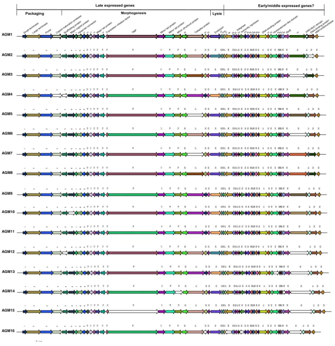
Figure 9. Schematic representation and comparisons of 16 B. aurantiacum phage genomes…...52
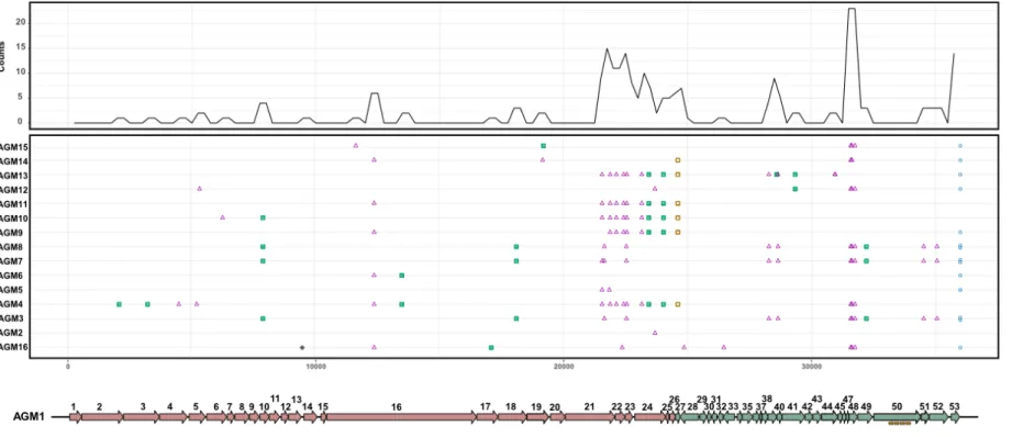
Figure 10. Mutation mapping using complete genome alignment of B. aurantiacum phages compared to phage AGM1……….53
Figure 11. Schematic representations of repeat groups in the orf50……….55
Figure 12. Schematic representation of the genome alignment and organization of phage AGM1 compared to other phages………..58
Figure 13. Schematic representation of mutants’ selection from liquid media………...82
Figure 14. Schematic representation of mutants’ generation and selection in solid media...83
Figure 15. Characterization of phage insensitive mutants……….85
Figure 16. Schematic representation of the ABC-transport system of SMQ-1335 and mutations in the BIMs………..………..94
Figure 17. Deacidification of the model curd rinds during the first 8 days of ripening………..115
Figure 18. The growth of the yeast Cryptococcus albicans and Geotrichum candidum during model cheese ripening………...116
Figure 19. Development of secondary bacteria on the model cheese surface……….117
Figure 20. Development of B. aurantiacum and its phages during cheese ripening………...…118
Figure 21. Development of the color of the rind of model curds………120
Figure 22. PCR of the tandem repeats region of isolated plaques from the model curds………121
Figure 23. Stability of B. aurantiacum phages (AGM1 – AGM16) after heat treatment at 60 to 90°C for 5 min in Elliker medium………...123
List of tables
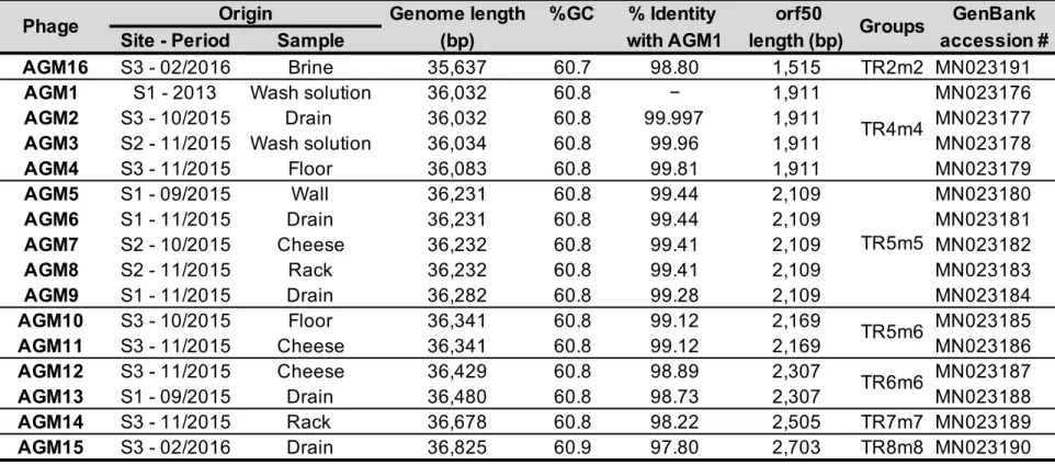
Table 1. Examples of commercially available bacteriophage products….………...24 Table 2. General genomic characterizations of B. aurantiacum phages….……….51 Table 3. Divergences between the reference genome of SMQ-1335 (GenBank accession CP017150.1) and the new sequence from Illumina….……….87 Table 4. Mutations shared among all BIMs….………..………...87 Table 5. Non-shared mutations in the genome of the BIMs….………....89 Table 6. Codon usage for WT and mutated codons of BIMs synonymous mutations…..…91 Table 7. List of B. aurantiacum strains and phages used in this study….………..100 Table 8. List of primers used for B. aurantiacum SMQ-1335 and its derivatives….…….101 Table 9. Primers designed for the genome editing of MG1363….…..………..…….104 Table 10. List of bacterial strains, phages and plasmids used in the genome editing of MG1363….………..………...105 Table 11. Analyses of the model cheeses during ripening………..126
List of abbreviations
Abi abortive infection
anti-CRISPR proteins that inhibit CRISPR-Cas systems
attB Attachment site in the bacterial genome
attP Attachment site in the phage genome
BIM Bacteriophage insensitive mutant
bp Base pair
BreLI Brevibacterium lanthipeptide island
BREX Bacteriophage exclusion system
Cas CRISPR associated
CRISPR Clustered regularly interspaced short palindromic repeats
DISARM Defense islands system associated with R-M
DNA Deoxyribonucleic acid
ds Double stranded
HGT Horizontal gene transfer
HNH A motif containing conserved histidine and arginine present in
the catalytic site of nucleases
ICTV International Committee on Taxonomy of Viruses
kb Kilo bases (1000 base pairs)
kDa Kilo Dalton
LAB Lactic acid bacteria
mRNA Messenger RNA
NaCl Sodium chloride
ORF Open reading frame
RBP Receptor binding protein
RM Restriction modification
RNA Ribonucleic acid
rRNA Ribosomal RNA
16S rRNA Small subunit of ribosomes
RUSTI iRon Uptake/Siderophore Transport Island
ss Single strand
TA toxin-antitoxin
“Education does not change the world. Education changes people. People change the world.”
– Paulo Freire
Acknowledgments
This journey into my PhD was part of dream that took a long road until it happened. As long as it took to start and finish this journey, it truly led to a process that changed me as a student, researcher and human being. I would do it again. Certainly, it would not have been the same without wonderful and supporting people around me.
First of all, I would like to thank CNPq for funding my PhD studies, granting me a fellowship to perform my PhD studies. I also thank the Canadian consortium CALDO, which had a partnership with CNPq, for helping with my PhD application at Université Laval as I felt supported during the entire process. I also thank my advisory committee, Michel Frenette and Steve Labrie, for the assistance, time and helpful suggestions during the doctorate. I have not enough words to thank my advisor Sylvain Moineau for having accepted me as a graduate student in his lab. This journey was very precious, and you helped to develop my potential with your guidance, leadership, kindness and scientific brilliance. I feel very privileged for having learned from you, as a scientist, leader and human being.
Sylvain’s lab is a very dynamic, cooperative and friendly environment. In this almost five years, I could learn with great scientists and this was priceless. Among them, I would like to give a special thank you to Denise Tremblay and Geneviève Rousseau. I wish all labs could have people like you that teach and inspire. I really appreciated all the time that you put to teach me something new, suggest troubleshooting or give me support when I needed. It was really a pleasure to work with you. I thank also Stephanie Loignon and Bruno Martel that came later to the lab but were also available to answer questions and help as needed.
During the course of five years, I had many lab mates, from whom I learned so much by listening to during lab meetings, sharing protocols, discussing results or collaborating with. Be around you guys, helped me to grow as a scientist and I appreciate the time you spent
welcomed me in a totally new environment and made me feel comfortable. Also, André Xavier, my Brazilian lab mate that became a great collaborator and close friend, with whom I enjoy spending hours talking about science. My Portuguese-speaking lab mates, Jessica Carvalhais (Brazil) and Priscila Pires (Portugal), which were there in different times of my life, but with whom I was able to speak Portuguese and feel a bit home; thank you for your support. Also, the sweet Honghui Liu, with whom I learned by example and it was always good to be around. And, of course, Cécile Philippe, who became a dear friend in and outside the lab. Many thanks to you all.
To my friends and people that were close to me at some moment outside the lab, I thank you for all the love and support. Thanks also for those that encouraged me when everything was only a plan, so this dream could become a reality. In special, I would like to thank my former house mate and friend, Elisa, who became like family for me. I cannot thank you enough for your friendship.
To my family, I thank you for always being there for me, encouraging and supporting this adventure. I thank especially my mom, Rute, that even with all the physical distance was present in every step of my journey and, since I was very young, stamped on me the value of pursuing higher education as a transforming experience. To my father, Elder; my brother, Alexandre; and my “little” sister, Victoria, thank you for being there for me.
Foreword
This PhD dissertation is organized in an introduction, which contains a review article; four chapters, each organized as a research article, and a conclusion. In addition, a collaborative article within the subject and relevant for the discussion has been included as an annex. Introduction
Cheeses and washed rind cheeses are presented in the introduction of the thesis. I also present the genus Brevibacterium and its importance in cheese production. In particular, the species
Brevibacterium aurantiacum is detailed and highlighted for its role in the ripening of washed
rind cheeses. Phages are described in detail, including an overview on B. aurantiacum phages. I also discussed the interaction between phages and their hosts as well as their two sides in the review "Phage as friends and enemies in food processing”. In this review, we discussed that these bacterial viruses may represent a risk to industrial fermentations, but they may also be valuable tools to control foodborne pathogens.
This review article was published in the journal Current Opinion in Biotechnology, in February 2018, volume 49, from page 185 to 190. I am the first author of this review. In collaboration with Sébastien Levesque and Sylvain Moineau. I performed the literature review, wrote the article and revised the manuscript. At the time of the publication, all authors were affiliated to the Département de biochimie, de microbiologie et de bio-informatique of Université Laval and to the Groupe de recherche en écologie buccale. Sylvain Moineau was also affiliated to the Félix d'Hérelle Reference Center for Bacterial Viruses. Figure, tables and references were modified to be in accordance to the dissertation format.
The problematic, hypothesis and objectives of this study were also presented at the end of the introduction section.
I am the first author of this article. I planned and performed the experiments, data analysis and also wrote the manuscript. Simon J. Labrie performed bioinformatics analysis. Jeannot Dumaresq carried out experiments to test antibiotic resistance of SMQ-1335. Richard J. Roberts performed analysis for the presence of restriction-modification systems. Denise Tremblay and Sylvain Moineau participated in the design and supervision of the study as well as manuscript revision. At the time of publication, Richard J. Roberts was affiliated to the New England Biolabs and Jeannot Dumaresq to the Département de microbiologie et d’infectiologie of the Centre hospitalier affilié universitaire Hôtel-Dieu de Lévis. All the other authors were affiliated to the Département de biochimie, de microbiologie et de bio-informatique of Université Laval, to the Groupe de recherche en écologie buccale. Denise Tremblay and Sylvain Moineau were also affiliated to the Félix d’Hérelle Reference Center for Bacterial Viruses.
Second research article (Chapter 2)
DNA tandem repeats contribute to the genetic diversity of Brevibacterium aurantiacum phages. This article was accepted for publication in the journal Environmental
Microbiology, on June 8th, 2020.
I am the first author of this article. I conceived and performed the experiments, bioinformatics and data analysis. I also wrote and revised the manuscript. Geneviève Rousseauparticipated in the supervision and elaboration of experiments as well as revision of the manuscript. Denise Tremblay sequenced the genome of the phages. Simon Labrie performed bioinformatics analysis and phage genome assembly. Sylvain Moineau participated in the design and supervision of the study, as well as in the writing and revision of the manuscript. At the time of the publication, Simon Labrie was affiliated to SyntBioLab. All the other authors were affiliated to the Département de biochimie, de microbiologie et de bio-informatique of Université Laval, to the Groupe de recherche en écologie buccale. Denise Tremblay and Sylvain Moineau were also affiliated to the Félix d’Hérelle Reference Center for Bacterial Viruses of Université Laval. Figure, tables and references were modified to be in accordance to the dissertation format. The supplementary information is available online at doi: 10.1111/1462‐2920.15113.
Third research article (Chapter 3)
Mutations in an ATP-binding cassette transporter confer phage resistance in Brevibacterium aurantiacum. This article is in preparation for submission.
I am the first author of this article. I conceived and performed the experiments, bioinformatics and data analysis. I also wrote and revised the manuscript. Geneviève Rousseauparticipated in the design of experiments as well as the revision of the manuscript. Denise Tremblay sequenced the genome of the Brevibacterium aurantiacum SMQ-1335 and its phage- insensitive derivatives. She also participated in the design of the experiments. Simon Labrie and Pier-Luc Plante performed bioinformatics analysis, BIMs genome assembly and mutations mapping. Sylvain Moineau participated in the supervision and design of the study as well as in the writing and revision of the manuscript. At the moment of the publication, Simon Labrie was affiliated to SyntBioLab. Pier-Luc Plante was affiliated to the Centre de recherche en infectiologie de l'Université Laval, Centre de recherche du CHU de Québec, Centre de recherche en données massives and also to Département de médecine moléculaire. All the other authors were affiliated to the Département de biochimie, de microbiologie et de bio-informatique of Université Laval, to the Groupe de recherche en écologie buccale. Denise Tremblay and Sylvain Moineau were also affiliated to the Félix d’Hérelle Reference Center for Bacterial Viruses.
Fourth research article (Chapter 4)
The impact of virulent phages of Brevibacterium aurantiacum in the production of smear surface-ripened cheeses. This article is in preparation for submission.
I am the first author of this article. I conceived and performed experiments and data analysis. Hany Geagea performed experiments of thermo-stability and revised the manuscript. Sylvain
Fifth research article (Annex A)
Mobilome of Brevibacterium aurantiacum sheds light on its genetic diversity and its adaptation to smear-ripened cheeses. This research article was published in the journal
Frontiers in Microbiology, in June 2019.
I am the second author of this article. Sébastien Levesque planned and performed the experiments, data analysis and also wrote the manuscript. I performed experiments, data analysis and participated in the manuscript writing and revision. Simon Labrie performed bioinformatics analysis. Sylvain Moineau designed and supervised the study and participated in the revision of the manuscript. At the time of the publication, all the other authors were affiliated to the Département de biochimie, de microbiologie et de bio-informatique of Université Laval, to the Groupe de recherche en écologie buccale. Sylvain Moineau was also affiliated to the Félix d’Hérelle Reference Center for Bacterial Viruses.
Introduction
Cheese and washed rind cheeses
Cheese is one of the oldest fermented foods created by man (Irlinger et al., 2015). It is believed that cheese evolved in a region known as the “Fertile Crescent,” around 8000 years ago, where the so-called “Agricultural Revolution” occurred with the domestication of plants and animals (Fox and McSweeney, 2017). Bacterial growth and acid production would have likely occurred during storage or from attempts to dry milk produced by domesticated animals in a warm and dry climate, with the goal of preserving milk into a stable product (Fox and McSweeney, 2017). When sufficient acid is produced, the major milk proteins (i.e. caseins) coagulate to form a gel in which the milk fat is entrapped (Fox and McSweeney, 2017). Consequently, cheese microbial communities have been “in culture” for thousands of years, with the knowledge of how to grow these organisms passed down by generations of cheesemakers (Dutton and Wolfe, 2013).
Although cheesemaking is an ancient art, modern cheese production now relies on state-of-the-art science and technology, including the use of industrial enzymes, complex microbial fermentations, and a dynamic biochemistry during cheese ripening (McSweeney et al., 2017b). Even if derived from the same raw material, a variety of cheese types can be made around the world as the result of milk fermentation and transformation. The composition of the cheese ecosystem is defined by three elements that are the ripening agents (microorganisms and enzymes), the composition of the fresh milk, and the environmental conditions during aging (Almena-Aliste and Mietton, 2014). These factors will define the composition of the cheese ecosystem that affects not only the sensory quality but also the diversity of cheeses worldwide (Almena-Aliste and Mietton, 2014).
Cheese is often the habitat of at least two different microbial communities. The first is found in the cheese core, containing starter cultures, primarily lactic acid bacteria, while the second is found
and Leuconostoc); propionic acid bacteria; molds; bacteria and yeast, which grow on the surface of smear-ripened cheeses (Cotter and Beresford, 2017). In addition to the microbial ecosystem, the variability among processing and ripening influence both the chemical composition of the fresh cheese and its enzymatic potential during ripening, which altogether contribute to the diversity and differentiation of cheese types (Almena-Aliste and Mietton, 2014).
The ripening cheese process is complex and involves microbiological and biochemical changes to the curd resulting in the flavor and texture characteristics of a particular variety (McSweeney, 2004). Microbiological changes during cheese ripening include the death and lysis of starter cells, the growth of nonstarter lactic acid bacteria and, in many varieties, the development of molds in mold-ripened varieties and a complex Gram-positive bacterial flora in smear cheeses (McSweeney, 2004). Moreover, the characteristics of the ripening conditions (temperature, relative humidity, and rates of O2, CO2, and NH3) ultimately influence the character and diversity
of the cheese microbiota (Almena-Aliste and Mietton, 2014).
Bacterial surface-ripened (“smear-ripened,” “washed-rind”) cheeses are a diverse group of varieties characterized by the growth of a complex and adapted Gram-positive bacterial flora on the cheese surface during ripening (McSweeney et al., 2017b). During manufacture, the surface of the cheeses is washed periodically with a brine solution, a process referred to as “smearing” (McSweeney et al., 2017b). The heterogeneous physicochemical composition of the surface of washed rind cheeses offers the possibility of simultaneous occupation by multiple microbial groups through the utilization of different carbon sources (Irlinger and Mounier, 2009). During the first days of ripening, yeasts and mold, such as Geotrichum candidum and Debaryomyces hansenii, dominate the surface microflora, where they metabolize the lactate present in the curd and release growth factors (Irlinger and Mounier, 2009). They also produce alkaline metabolites, such as ammonia from deamination of amino acids, which leads to an pH increase enabling the growth of acid-sensitive bacteria such as Arthrobacter spp., Brevibacterium spp., Corynebacterium casei, micrococci and staphylococci (Arfi et al., 2006; Corsetti et al., 2001; Irlinger and Mounier, 2009; Rattray and Fox, 1999). This dynamic and complex secondary microflora (molds, yeasts and surface bacteria) contribute with proteolytic and lipolytic enzymes for cheese texture, flavor, and quality (Bonomo and Salzano, 2012; Caridi et al., 2003). As such, these cheeses are characterized by a strong aroma and high levels of proteolysis and lipolysis (McSweeney et al., 2017b).
Brevibacterium spp.
The metabolism and physiology of the microorganisms will determine their growth on smear surface-ripened cheeses (Motta and Brandelli, 2008). The ability to grow on the cheese surface depends on various properties such as salt tolerance and iron acquisition systems as well as the ability to use the energy compounds present in the cheese (Motta and Brandelli, 2008). Due to its enzymatic richness, Brevibacterium spp. strains are widely used for the manufacturing of orange-pigmented surface-ripened cheeses, contributing to the breakdown of lipids and proteins as well as producing volatile sulfur compounds (Pham et al., 2017).
Brevibacterium linens is the type species of the genus Brevibacterium that belongs to the Brevibacteriacea family, order Actinomycetales, and class Actinobacteria (Breed et al., 1957).
Species of the Brevibacterium genus present similarities with members of others coryneform genera, such as Arthrobacter, Corynebacterium and Rhodococcus, for having meso-diaminopimelic acid in the cell wall or showing a rod-coccus growth cycle (Onraedt et al., 2005). However, the presence of teichoic acids in the polysaccharide cell wall of Brevibacteria distinguishes them from all others coryneform bacteria (Motta and Brandelli, 2008).
For a long time, B. linens was considered the major component of the bacterial flora present in the smear of a variety of surface-ripened cheeses, such as Appenzeller, Brick, Gruyère Limburger, Münster, Romadour, Tilsit, and other cheeses produced worldwide (Bonnarme et al., 2000; Onraedt et al., 2005). However, as Brevibacterium taxonomic classification previously indicated that B. linens was the only member of the genus to produce orange-pigmented colonies, such brevibacterial strains were usually assigned to this species (Gavrish et al., 2004). Since the taxonomy of Brevibacterium has been updated (Gavrish et al., 2004), the role of B. linens as the major Brevibacterium species in cheeses has been reconsidered. Strains of B. linens, B.
Brevibacterium aurantiacum
The B. aurantiacum species was first described by Gavrish et al. (2004) in a taxonomic study of orange-pigmented strains that were phenotypically close to B. linens. The study also included the new species B. antiquum and B. permense (Gavrish et al., 2004). The B. linens group was divided into these new species on the basis of their physiological and biochemical characteristics, the sugar and polyol composition of their teichoic acids, their 16S rRNA sequence and the DNA-DNA hybridization levels (Forquin et al., 2009). While the strain ATCC 9172 remains B. linens, the strain ATCC 9175 isolated from cheese represents the type strain of the B. aurantiacum species (Forquin et al., 2009; Gavrish et al., 2004). The isolation of B. aurantiacum strains have been reported in cheeses such as Camembert, Beaufort, Langres, Reblochon, Romadur, Limburger, Havarti, Livarot, Danbo and Gubbeen (Mounier et al., 2017; Pham et al., 2017). Figure 1 shows some examples of cheeses in which strains of B. aurantiacum have been isolated.
Figure 1. Strains of B. aurantiacum have been isolated from several washed rind cheeses. Adapted from Cogan et al. 2014. (a) Gubbeen; (b) Limburger; (c) Livarot; (d) Reblochon; and (e) Tilsit.
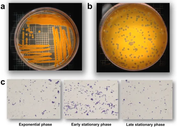
B. aurantiacum is a Gram-positive, non-motile, and non-spore-forming bacterium (Gavrish et al.,
2004). It produces orange colonies and undergoes a rod-coccus growth cycle, with older cultures dominated by coccoid cells (Figure 2). Furthermore, B. aurantiacum is aerobic and its optimal growth temperature is around 24-26°C, while being able to grow at 7°C but not at 37°C (Gavrish et al., 2004). Moreover, this species grows in 15% NaCl, hydrolyzes casein, and uses several carbon sources, such as cellobiose, fructose, galactose, glucose, glycerol, mannitol, mannose, and xylose. The peptidoglycan contains meso-diaminopimelic acid and its teichoic acids are composed of galactose, glucose and glycerol (Gavrish et al., 2004).
Figure 2. B. aurantiacum growth. (a) B. aurantiacum produces orange pigments during growth, a desirable feature in cheese production. (b) Phage plaques on a lawn of B. aurantiacum SMQ-1335. (c) B. aurantiacum cells transform from bacilli to coccoid shape during growth.
9175T). These strains were investigated for key genetic determinants known to be important for
growth in cheeses, such as the ability to use energy compounds, iron acquisition, salt tolerance, and bacteriocin production (Pham et al., 2017). Regarding the catabolism of energy compounds present in cheeses, no beta-galactosidase gene was found in the Brevibacterium genomes, which is consistent with the fact that most Brevibacterium spp., including B. aurantiacum, are not able to consume lactose (Gavrish et al., 2004; Pham et al., 2017). On the other hand, several strains of
B. aurantiacum, including the type strain (ATCC 9175), encode enzymes involved in the Leloir
pathway for galactose utilization and two of them (8(6) and CNRZ 920) have the complete pathway for D-galactonate catabolism (Pham et al., 2017).
Biochemical reactions, which occur in cheese during ripening, are conventionally grouped into three major categories: (1) glycolysis of residual lactose and catabolism of lactate and citrate; (2) lipolysis and the catabolism of free fatty acids; and (3) proteolysis and the catabolism of amino acids (McSweeney, 2017). During the manufacture of cheese curd, lactose is converted to lactic acid (or lactate) by starter bacteria and the resulting lactate is a substrate for a series of reactions during cheese ripening (McSweeney et al., 2017a). Enzymes involved in the lactate catabolism (NAD-independent lactate dehydrogenases) and transport (lactate permeases) have been identified in the genome of B. aurantiacum (Pham et al., 2017). Additionally, B. aurantiacum genomes encode enzymes involved in the catabolism of ethanol (alcohol dehydrogenase and acetaldehyde dehydrogenases), acetate (monocarboxylic acid transporter MctC and acetyl-CoA synthase), and also in the import of citrate (Pham et al., 2017).
Lipid catabolism involves the release of free fatty acids and glycerol as well as the subsequent breakdown of these compounds (Pham et al., 2017). As they are hydrophobic, lipids are excellent carriers and also precursors of numerous flavor compounds, such as esters, methyl ketones, lactones, and secondary alcohols (Thierry et al., 2017). The B. aurantiacum genomes analyzed encode several proteins with putative lipase or esterase activity in addition to enzymes involved in the catabolism of glycerol and short chain fatty acids transporters (Pham et al., 2017).
Brevibacterium lipases release volatile fatty acid components from milk triacylglycerol,
influencing the taste of ripened cheese (Motta and Brandelli, 2008; Onraedt et al., 2005).
proteolytic microorganisms (Pham et al., 2017). Brevibacterium strains synthesize highly active proteases and peptidases during its growth (Onraedt et al., 2005; Rattray and Fox, 1999). Indeed, the genome analysis of B. aurantiacum strains revealed several genes coding for proteolytic enzymes (9-12), with some strains encoding extracellular proteases, one of which was previously characterized for its activity on caseins (Pham et al., 2017). The free amino acids resulted from proteolysis of caseins (Gavrish et al., 2004) can be used as an energy source by the cheese ripening microorganisms while they also have a crucial role in cheese aroma formation as precursors of volatile flavor compounds produced by a range of catabolic reactions (Ardö et al., 2017; Pham et al., 2017). The production of volatile sulfur compounds arises mostly from the degradation of the sulfur-carbon bound of L-methionine to form methanethiol (Arfi et al., 2006; Bonnarme et al., 2000). B. aurantiacum strains encode in their genomes enzymes for methionine degradation and also de degradation pathways for several amino acids, such as proline, alanine, arginine, glutamate, histidine, phenylalanine, serine, threonine, and tyrosine (Pham et al., 2017).
In order to survive under hyperosmotic stress in this environment, Brevibacterium strains accumulate compatible solutes to maintain a positive cellular turgor (Onraedt et al., 2005). B.
aurantiacum strains have been shown to encode enzymes involved in the synthesis of the
osmoprotectants ectoine, glycine-betaine and trehalose, which may help to protect these strains of the osmotic stress resulting from high salt concentrations in cheeses (Pham et al., 2017). Bacteriocins are antibacterial peptides that are inhibitory to microorganisms that are usually, but not always, closely related to the producer strain (Motta and Brandelli, 2008). Predicted bacteriocin biosynthesis genes are present in most of the strains, and one of the corresponding gene clusters is located in a probable conjugative transposon found only in cheese-associated strains and named Brevibacterium Lanthipeptide Island (BreLI) (Pham et al., 2017). This region was later identified also in the genome of B. aurantiacum SMQ-1417 and shown to circularize, although the bacteriocin activity could not be confirmed in the strains tested (Levesque et al., 2019).
also part of the cheese rind community (Levesque et al., 2019). Growing evidence suggests that the availability of iron is a driving force in adaptation of microorganisms that grow on cheeses (Bonham et al., 2017). As iron availability is limited in cheeses (Monnet et al., 2012), iron acquisition genes obtained through horizontal gene transfer (HGT) are very abundant in B.
aurantiacum and other cheese bacteria (Bonham et al., 2017; Levesque et al., 2019; Pham et al.,
2017). For instance, a large region of ~47 kb and 34 genes, named RUSTI (i.e. iRon Uptake/Siderophore Transport Island), was found in a study investigating the extensive HGT in cheese-associated bacteria, including Brevibacterium (Bonham et al., 2017). In addition to iron acquisition genes, mobile genetic elements contributed to the expansion of B. aurantiacum pan-genome and to its genetic diversity, with significant differences found between the mobilome of
B. aurantiacum dairy strains and environmental strains of B. linens and B. epidermidis (Levesque
et al., 2019).
Regarding defense strategies against foreign nucleic acids, no CRISPR-Cas system has been identified in the genome of B. aurantiacum strains (Levesque et al., 2019; Pham et al., 2017). However, a new type I restriction-modification (R-M) system was identified in the genome of B.
aurantiacum SMQ-1335 and this strain may contain additional R-M systems (types II, III, and IV)
(de Melo et al., 2016). Of note, the phage-sensitive strain SMQ-1335 was previously identified as
Brevibacterium linens (Chapter 1), but phylogenetic analysis described in Annex A showed that
this strain belongs to the B. aurantiacum species (Levesque et al., 2019). Additionally, an anti-plasmid system Wadjet was recently described (Doron et al., 2018) and orthologs of its proteins were identified in the genome of several B. aurantiacum strains, which could be partly responsible for the absence of plasmids in the referred strains (Levesque et al., 2019).
Bacteriophages (phages)
Because high numbers of bacterial cells are cultivated each day in large vats, cheese manufacturing can be disrupted by strictly lytic phages. In fact, most bacterial fermentation industries have experienced phage contaminations at varying frequencies (Labrie et al., 2010; Marcó et al., 2012). As with all viruses, phages are made of at least two components: nucleic acids and proteins (Campbell, 2003; Mc Grath et al., 2007). They are the most abundant biological entities on our planet, often outnumbering coexisting bacterial cells in ecosystems (Brüssow and Hendrix, 2002).
In the non-sterile environment of heat-treated milk, the added cultures will come into contact with virulent phages naturally contaminating milk (Garneau and Moineau, 2011). Although the phage concentration is usually low in milk, a specific phage population can increase rapidly if phage-sensitive cells are part of the added bacterial culture used to ferment milk (Garneau and Moineau, 2011). Fluctuating phage titers can be observed if a starter strain rotation system is adopted (Chibani-Chennoufi et al., 2004). The occurrence of phage outbreaks leads to economical loss as the lysis of a larger number of sensitive bacterial cells will delay the milk fermentation process and lead to low-quality products (Garneau and Moineau, 2011; Mc Grath et al., 2007). Over the past decades, the dairy industry has adopted different strategies to control phage propagation in the industrial settings, including the adaptation of factory design and processes, improved sanitation, strain rotation, and the use of phage-resistant strains (Garneau and Moineau, 2011). Despite these efforts, new viral variants keep emerging as a result of phage evolution (Garneau and Moineau, 2011).
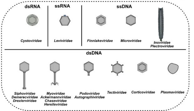
Historically, phages were classified into ten families based on characteristics such as genome type (ssDNA, ssRNA, dsDNA or dsRNA), morphological properties of the virion and host range (Dion et al., 2020). However, in the past few years, phage taxonomy has been revisited by the Bacterial and Archaeal Viruses Subcommittee of the International Committee on Taxonomy of Viruses (ICTV) (Adriaenssens et al., 2018, 2020) and it is currently undergoing a major overhaul, moving from a mostly morphology-based approach to a genome-based taxonomy (Dion et al., 2020). Since then, nine new families of bacterial viruses were added and many more are expected to be described in the coming years.
The most predominant bacterial viruses belong to the Caudovirales order, which are tailed phages containing double strand DNA (dsDNA) (Brüssow and Hendrix, 2002; Dion et al., 2020; Hatfull, 2008). Previously, the Caudovirales order comprised three families based on the tail morphology, which were Myo-, Sipho- and Podoviridae. Phages from the Myoviridae family have a long,
morphology, whereas phages from the Autographiviridae family have a podoviral morphology (Adriaenssens et al., 2018, 2020). The minimal genome of tailed phages encodes DNA packaging, capsid, tail, DNA replication, transcription regulation, and lysis genes (Brüssow and Hendrix, 2002). The virion structure and assembly genes typically encompass at least 15 kb of genome space. Therefore, the majority of siphophages have genomes larger than 20 kb (Hatfull, 2008). Other apparently less common phages can pack their single-stranded DNA (ssDNA), dsDNA, single-stranded RNA (ssRNA) or double-stranded RNA (dsRNA) genome into non-tailed virions (Dion et al., 2020) that can be polyhedral (Microviridae, ssDNA; Corticoviridae, dsDNA;
Tectiviridae, dsDNA; Leviviridae, ssRNA; Cystoviridae, dsRNA; and Finnlakeviridae, ssDNA),
filamentous (Tubuvirales order, ssDNA; Inoviridae, Plectroviridae), or pleomorphic (Plasmaviridae, dsDNA) (Adriaenssens et al., 2020; Dion et al., 2020).
Phages have evolved diverse strategies to exploit prokaryotic host cells for their own reproduction (Koskella and Brockhurst, 2014). Based on their genetics and interactions with the host bacteria, phages can undergo distinct cycles, such as lysogenic, lytic (Feiner et al., 2015; Mc Grath et al., 2007) or chronic infection (Koskella and Brockhurst, 2014) (Figure 4). In general, lytic phages immediately enter in a productive cycle upon infection, in which the phage genome is replicated and packaged into new phage particles that are then released through bacterial lysis (Bondy-Denomy and Davidson, 2014; Feiner et al., 2015). On the other hand, phages can enter in a symbiotic cycle with the host bacteria, during which the phage genome is integrated into the bacterial chromosome in a lysogenic state, or forms an extrachromosomal plasmid in pseudolysogeny (Bondy-Denomy and Davidson, 2014; Feiner et al., 2015). Additionally, filamentous phages have the ability to establish a chronic infection in which the viral genome resides within the cell in either an exclusively episomal state or integrated into the host chromosome, while virions are continuously released from the productive cycle without killing the host (Roux et al., 2019).
The adsorption, the first step of phage infection (Duplessis and Moineau, 2001), involves recognition of receptors located on the bacterial cell surface by the phage receptor-binding proteins (RBPs) (Duplessis et al., 2006; Mahony and van Sinderen, 2012). Secondly, the phage genome is then injected through the cell wall into the bacterial cytoplasm, in a process facilitated by peptidoglycan hydrolases, enzymes that are able to locally degrade the peptidoglycan without lysing the cell (Kenny et al., 2009; Oliveira et al., 2013). Phage genome entry generates a cycle of phage production initiated by phage-specified proteins, which are translated from phage mRNA after infection (Campbell, 2003). Subsequently, the components of the biosynthetic apparatus (such as ribosomes and ATP generators) are diverted from their normal tasks in bacterial growth to the production of phages, in which the replication of nucleic acids occurs first, followed by the synthesis of phage structural proteins (Campbell, 2003). Afterwards, the new phage capsids are then assembled, and the genetic material is packaged into them. In most phages, a tail will be
described as a prophage and the bacterial cell harboring a prophage is known as a lysogen (Campbell, 2003). The excision and integration of a phage genome are mediated by phage-encoded DNA recombinases, such as integrases and excisionases, and take place at specific attachment sites, which are identical in both bacterial (attB) and phage genomes (attP) (Feiner et al., 2015). Prophages are replicated together with the bacterial chromosome, and this lysogenic state is maintained by the repression of phage lytic genes (Feiner et al., 2015). Moreover, the action of the repressor proteins expressed by prophages leads to resistance to superinfection by the same phage (Bondy-Denomy and Davidson, 2014; Marcó et al., 2012). Hence, the lysogenic cell can survive and replicate without the production of phage particles or deleterious phage proteins, and even be “immune” to another infection (Bondy-Denomy and Davidson, 2014; Feiner et al., 2015; Marcó et al., 2012). Ultimately, certain conditions can induce the phage genome excision, and lead lysogenic cells to re-enter the lytic cycle (Campbell, 2003).
Figure 4. Schematization of phage cycles.
Most phages will perform a lytic (a) or lysogenic (b) life cycles. Filamentous phages can perform chronic infections (c) and some phages can have episomal genomes in a pseudolysogenic state (d).
B. aurantiacum phages
It was recently pointed out that the phage sensitivity of ripening cultures containing
Brevibacterium strains was unknown, and the impact of phages on these cultures was probably
overlooked (Pham et al., 2017). In fact, studies on Brevibacterium phages are rare. To date, two different virulent phages of Brevibacterium iodinum and Brevibacterium fuscum has been described, which are phage LuckyBarnes (Siphoviridae) (Underwood et al., 2019) and phage Cantare (Myoviridae). Besides these two phages, two seemingly complete prophages have been identified in the genome of the strains B. linens VCM10 (Melo et al., 2020) and B. aurantiacum SMQ-1419 (Levesque et al., 2019). Except for B. aurantiacum SMQ-1419, none of the prophages or phages have been associated to the dairy environment, being instead isolated from environmental samples, such as soil.
Recently, virulent phages infecting a sensitive dairy strain of B. aurantiacum have been isolated in cheese manufacturing facilities after defective color and flavor development in smear surface-ripened cheeses (Melo et al., 2020). These observations suggested that these cheese defects might be related to the presence of virulent phages in this environment. Our group led the investigation of this phenomenon and the discovery of the first B. aurantiacum virulent phages (Chapter 2). We isolated 16 highly related virulent Siphoviridae phages, named AGM1 to AGM16, which all seem to have evolved from a prophage that lost the ability to integrate in the bacterial genome. B.
aurantiacum virulent phages are unrelated to the prophage found in the cheese-associated strain B. aurantiacum SMQ-1419, which is closer to LuckyBarnes, but their genes coding for the phage
structural proteins are related to the structural genes found in the prophage VCM10 (Melo et al., 2020). Although there is a narrow diversity among the sixteen phages, the major contributor to their genetic diversity is the presence of tandem repeats in their genomes.
Phage-host interactions
The reciprocal evolution of bacteria and phages is an important driver of ecological processes in microbial communities (Koskella and Brockhurst, 2014). This continuous cycles of co-evolution leads to the emergence of phage-insensitive hosts that help to preserve bacterial lineages, whereas counter-resistant phages threaten these new bacterial strains (Labrie et al., 2010). Bacteria have evolved numerous immune mechanisms, both innate and adaptive, to cope with this evolutionary pressure (Hampton et al., 2020). Different bacterial defense mechanisms can interfere with each step of phage infection while phages have evolved ways to impede the interference and proceed with the infection (Figure 5). An understanding of phage and host genetics and biology is essential to define phage requirements for infection and to determine how they co-evolve to counteract and adapt to the threat posed by the other (Mahony et al., 2012).
The nature and location of the phage receptors at the cell surface vary greatly depending on the phage and host. The receptors may be polysaccharides, surface proteins and other structures such as flagella, pili and capsules (Bertozzi Silva et al., 2016). The adsorption of phage particles to a bacterium leads to the activation of molecular mechanisms of infection in the virion (tail contraction, DNA ejection, etc.). The interaction between viral proteins and receptor structures on the bacterial cell surface is necessary for the conformational triggering of phage infection (Letarov and Kulikov, 2017). To prevent adsorption, bacteria can alter or disguise receptors through surface modification (Hampton et al., 2020). As such, phages lose the ability to effectively infect its host as the receptors become inaccessible or non-complementary to the phage receptor-binding protein (Bertozzi Silva et al., 2016).
Phage DNA entry can also be prevented by either mutating the bacterial components used during genome injection or through superinfection exclusion (Sie) systems. The latter are membrane proteins that block the entry of phage DNA into host cells, thereby conferring immunity against specific phages (Labrie et al., 2010). The genes encoding these membrane anchored proteins are often found in prophages (Labrie et al., 2010). They also inhibit subsequent infection of related phages with mechanisms that include interacting with the cytoplasmic membrane and blocking phage genome injection as well as interacting with the phage receptor on the bacterial outer membrane and blocking phage adsorption (Bondy-Denomy et al., 2016).
The ability to cleave phage DNA in a sequence-specific manner is shared by both restriction-modification (R-M) and clustered regularly interspaced short palindromic repeats (CRISPR)– CRISPR-associated protein (Cas) systems (Hampton et al., 2020). However, other defense systems also act during the DNA replication, including systems recently discovered, such as BacteRiophage Exclusion (BREX) (Goldfarb et al., 2015), and the Defense Islands System Associated with R-M (DISARM) (Ofir et al., 2018). Although the modes of action have yet to be elucidated, nine families of new anti-phage defense systems (i.e. Thoeris, Hachiman, Shedu, Gabija, Septu, Lamassu, Zorya, Kiwa and Druantia) were recently discovered by searching for enriched gene families within the so-called defense islands (Doron et al., 2018), which are clusters of defense genes associated to transposable mobile elements (Makarova et al., 2011). Additionally, bacteria carry “altruistic” defense systems, such as abortive infection (Abi) and toxin-antitoxin (TA) systems, that provide population resistance by sacrificing infected cells. Abi systems provide resistance through the abortion of phage infection targeting a crucial step of phage multiplication such as replication, transcription or translation, leading to the death of the infected cell (Labrie et al., 2010). In the TA systems, a toxic molecule is produced by the cell and neutralized by the antitoxin product, but when the balance between the two regulatory halves is altered, an available toxin can kill the bacterium (Labrie et al., 2010). Taken altogether, there is a clear diversity of phage resistance mechanisms (Hampton et al., 2020).
As phages face a wide range of antiviral mechanisms when infecting bacterial cells, they have evolved multiple tactics to avoid, circumvent or subvert these mechanisms (Labrie et al., 2010). For example, phage can produce their own antitoxin that acts by inactivating the host toxin (Samson et al., 2013). Additionally, phages have the ability to overcome CRISPR–Cas defenses through point mutations in the protospacer-adjacent motif (PAM) or protospacer, deletions or modifications of the DNA to prevent cleavage by Cas complexes and also by encoding anti-CRISPR proteins that can interfere with anti-CRISPR immunity (Hampton et al., 2020). More recently, it was shown that some (jumbo) phages form a protein shell that protects viral DNA from DNA
(Samson et al., 2013).
Figure 5. Examples of phage defense mechanisms and counter defenses.
Several other strategies used by phages to circumvent the anti-phage defense systems have been described (Hampton et al., 2020; Samson et al., 2013) and surely new ones will be discovered in the years to come as phage research gains more interest. A better understanding of phage–host interactions has already benefited applications that rely on phage-resistant bacteria to produce foods and biotechnological products (Samson et al., 2013). Additionally, as phage-resistant strains are required in different industries, the fundamental research into phage defense systems has supported the development of applications, such as gene editing and diagnostics (Hampton et al., 2020).
Phage as friends and enemies in food processing
Alessandra Gonçalves de Melo 1, Sébastien Levesque 1, and Sylvain Moineau 1,2*
1 Département de biochimie, de microbiologie, et de bio-informatique, Faculté des sciences et de
génie, Groupe de recherche en écologie buccale, Faculté de médecine dentaire, Université Laval, Québec City, QC, G1V 0A6, Canada
2 Félix d'Hérelle Reference Center for Bacterial Viruses, Université Laval, Québec City, QC, G1V
0A6, Canada
Résumé
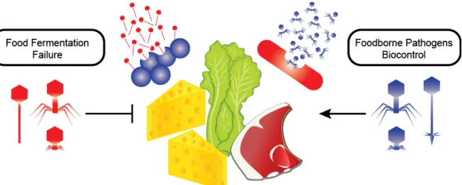
Les phages peuvent infecter tous les genres bactériens expliquant ainsi leur grande abondance dans les écosystèmes. Bien que ces virus bactériens puissent représenter un risque pour les fermentations industrielles, ils peuvent également être des outils précieux pour contrôler les bactéries pathogènes d'origine alimentaire. Dans cette opinion, nous avons révisé ces deux aspects des phages. Comme les fermentations bactériennes sont constamment menacées par des attaques phagiques, nous discutons de la diversité des phages et des stratégies employées par l'industrie pour réduire leur impact négatif sur la transformation des aliments. De plus, nous explorons l'utilisation de phages afin de réduire les risques associés aux maladies bactériennes d'origine alimentaire.
Abstract
Phages can infect all bacterial genera on the planet leading to their abundance in ecosystems. While these bacterial viruses may represent a risk to industrial fermentations, they may also be valuable tools to control foodborne pathogens. Here we review these two sides of phage replication. As bacterial fermentations are constantly under threat of phage attacks, we will discuss phage diversity and the strategies employed by the industry to reduce their impact on food processing. Furthermore, we will explore the use of pathogen-infecting phages to reduce the risks associated with foodborne diseases over the steps of the food chain.
Abbreviations
Anti-CRISPR Proteins that acts to inhibit CRISPR-Cas systems
ARGs Antibiotic resistance genes
BIMs Bacteriophage insensitive mutants
CaCO3 Calcium carbonate
CRISPR-Cas Clustered regularly interspaced short palindromic repeats and CRISPR-associated proteins
CRISPR-Cas9 Gene-editing tool using the nuclease Cas9
LAB Lactic acid bacteria
MTP Major capsid protein
mtp Gene encoding for the major capsid protein
NPS Neck passage structure
tpeX Gene encoding a tail extension protein
TpeX Tail protein extension
Introduction
Bacteriophages or phages are recognized as the most abundant biological entities on our planet, outnumbering bacteria by an estimated ten-fold (Brüssow and Hendrix, 2002). They are ubiquitous and, as such, can be found in natural and man-made environments, especially those in which their bacterial host thrives (Chibani-chennoufi et al., 2004). Because many food fermentation processes rely on the metabolic richness of bacterial cells to transform products, they are constantly threatened by the prospect of phage contamination (Mc Grath et al., 2007). Sensitive host encounters with lytic phages can result in fermentation arrest or delay, leading to low-quality products and at worst, production loss (Emond and Moineau, 2007). Although phages are usually acknowledged as a menace, they can also be considered allies against pathogenic bacteria that represent a persistent food safety hazard. This review will highlight the character of phages as the enemies of food manufacturing, using dairy phages as examples. Furthermore, we will also focus on the role of phages as biocontrol agents in several steps of the food chain and the advances made for the application of phage cocktails to control bacterial pathogens.
Phages as a threat in food processing
Due to the nature of food fermentation processes, performed at large scale, under non-sterile conditions (Samson and Moineau, 2013) and often relying on the same bacterial cultures in successive batches (Emond and Moineau, 2007), it is not surprising that virulent phages can cause fermentation failures (Samson and Moineau, 2013). The food industry has adopted several antiphage strategies as countermeasures to prevent and diminish contamination. Example strategies include developing bacteriophage insensitive mutants (BIMs), establishing bacterial rotations as well as using chemical and physical treatments to decontaminate the factory and the substrates (Moineau and Lévesque, 2004; Samson and Moineau, 2013). The state of knowledge on phage-host interaction to prevent viral contamination in food processing (Mahony and van Sinderen, 2015) as well as standard phage control strategies have been extensively reviewed lately (Emond and Moineau, 2007; Moineau and Lévesque, 2004). Only very recent developments will be highlighted here.
(Campagna et al., 2014). However, several dairy phages are thermal-stable, surviving for example pasteurization (Murphy et al., 2014). Moreover, whey proteins can provide an additional protective effect to phages (Geagea et al., 2017). Because thermal inactivation of phages would require higher temperatures and lead to protein denaturation, other alternatives, such as membrane filtration, are been investigated as a mean to reduce phage levels without changing protein structure (Samtlebe et al., 2017).
Several biocides have proven to be effective antivirals to clean industrial environs (Campagna et al., 2014; Hayes et al., 2017; Murphy et al., 2014). However, increasing resistance to chemical compounds has been observed, leading to phage persistence in manufacturing sites (Hayes et al., 2017). For example, some members of the highly prevalent Lactococcus lactis phage genus Sk1virus (formerly 936) are broadly tolerant to multiple classes of compounds found in commonly used sanitizers (Hayes et al., 2017). A similar conclusion was made in another study, in which the virulent lactococcal phages CB13 (isolated in Canada) and P1532 (isolated in Germany) were less sensitive to biocides than other phages (Campagna et al., 2014). These observations, and the phylogenetic separation between resistant and non-resistant phages, suggest that biocide resistance is due to the intrinsic robustness of the phage, rather than to the biocide’s mechanism of action (Hayes et al., 2017). Therefore, rotation of sanitizers should be a recommended precautionary measure to avoid selection of increasingly resistant viruses (Campagna et al., 2014; Murphy et al., 2014).
Phage diversity
Dairy phages are by far the best-characterized food-related phages due to their global impact on milk fermentation processes. Carefully selected strains of lactic acid bacteria (LAB), e.g., L. lactis,
Lactobacillus sp., Leuconostoc sp., and Streptococcus thermophilus, are repetitively added to milk
The understanding of phage evolution and diversity at food processing sites can shed light on phage population dynamics to ensure more efficient strategies to control them (McDonnell et al., 2016). For instance, a novel emerging group of S. thermophilus phages, the 987 phages, was recently discovered (McDonnell et al., 2016) and compared to the three other known S.
thermophilus phage groups (Mahony and van Sinderen, 2014). Interestingly, genomic relatedness
of 987-type phages to the morphogenesis and replication modules from some lactococcal phages (P335 group) and S. thermophilus phages, respectively, suggests genetic exchanges that resulted in a new mosaic architecture and emerging phage group. Additionally, adsorption of the 987 phages to both S. thermophilus and L. lactis indicated that cell surface molecules might be shared between both bacterial species (McDonnell et al., 2016). The recent industrial practice of mixing
L. lactis and S. thermophilus strains in a single starter culture may have led to the rise of this new
phage group. Another example involves recent lactococcal phage isolates of the Sk1virus genus that are able to cross-infect various groups of L. lactis strains, thereby displaying a more dynamic host range than previously described for this group of phages (Murphy et al., 2013). Clearly, phage populations are dynamic in industrial environments and they need to be constantly monitored in order to adapt the antiviral strategies, particularly for bacterial strain rotation.
A comparative genomic analysis of ninety siphophages has established the core genome and variable elements of the Sk1virus genus (Murphy et al., 2016). Interestingly, a probable hotspot for gene rearrangements was identified surrounding the major tail protein-encoding gene (mtp), including the presence or absence of a gene encoding the neck passage structure (NPS) and/or a gene encoding a tail protein extension (tpeX). For the latter phenotype, MTP-TpeX was arranged as a spiral structure on the phage tail (Murphy et al., 2016). Both structures were also observed on phage persisting over several years in whey powders, suggesting that they might be beneficial for phage structural stability in harsh conditions (Wagner et al., 2017).
The arms race of phage and bacteria
Whenever they encounter each other, bacteria and phages are continuously outcompeting in an antagonistic co-evolution process. In this arms race, bacterial derivatives acquire resistance to phages using several strategies (frequently through receptor mutations), whereas phages counter-attack to overcome antiviral barriers in order to infect and propagate (Labrie et al., 2010). Bacteria
can also harbor phage-resistance mechanisms, such as restriction modification systems, abortive infections, CRISPR-Cas systems and others. These mechanisms have been extensively reviewed elsewhere as well as the strategies used by phages to respond to these challenges (see (Labrie et al., 2010; Samson et al., 2013)). Of particular interest in the past decade, the CRISPR-Cas system allows the “immunization” of bacterial strains against several phages. While this can be observed by challenging bacteria with virulent phages, the system can also be “programmed” via a plasmid to protect bacteria against multiple phages in a single assay (Hynes et al., 2016). This programming was described for S. thermophilus, using a high copy plasmid with highly conserved protospacers and specific protospacer adjacent motifs targeting the greatest number of virulent phages (Hynes et al., 2016). This strategy was very efficient at conferring resistance to multiple phages at once and offers the possibility of rapidly developing specific BIMs in strains carrying active CRISPR-Cas systems.
This natural coevolution can sometimes be directly observed when analyzing mixed starter cultures (Spus et al., 2015). Food fermentations can be performed using a limited number of defined bacterial strains as indicated above but also with mixed starter cultures. The latter are composed of undefined numbers and ratios of bacterial strains that will drive the fermentation process. These mixed cultures will also coexist with diverse phages leading to a complex dynamic of phage-host interactions. It is usually not recommended to use both defined and mixed cultures in the same industrial environment.
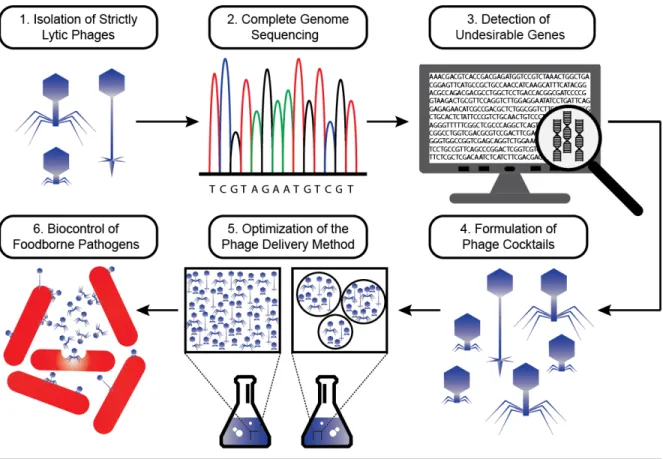
Beneficial applications of bacteriophages in the food industry
Phages are also prospective antimicrobial alternatives to prevent contamination by bacterial pathogens at different stages of the commercial food chain. Diseases caused by foodborne bacteria affect millions of people annually on a global scale (Amarillas et al., 2016; Schmelcher and Loessner, 2016). While the intensive use of antibiotics led to the emergence of resistant bacteria