En vue de l'obtention du
DOCTORAT DE L'UNIVERSITÉ DE TOULOUSE
Délivré par :Institut National Polytechnique de Toulouse (INP Toulouse)
Discipline ou spécialité :
Pathologie, Toxicologie, Génétique et Nutrition
Présentée et soutenue par :
M. SAJID UMAR le jeudi 12 janvier 2017
Titre :
Unité de recherche : Ecole doctorale :
Avian influenza and co-infections: investigation of the interactions in the
poultry models
Sciences Ecologiques, Vétérinaires, Agronomiques et Bioingénieries (SEVAB)
Interactions Hôtes - Agents Pathogènes (IHAP)
Directeur(s) de Thèse : M. MAXENCE DELVERDIER
MME MARIETTE DUCATEZ Rapporteurs :
Mme CATHERINE BELLOC, ONIRIS NANTES Mme GINETTE DAMBRINE, UNIVERSITE DE TOURS
Membre(s) du jury :
1 Mme ELENA CATELLI, UNIVERSITA DEGLI STUDI DE BOLOGNE, Président 2 M. JEAN-LUC GUERIN, ECOLE NATIONALE VETERINAIRE DE TOULOUSE, Membre 2 M. MAXENCE DELVERDIER, ECOLE NATIONALE VETERINAIRE DE TOULOUSE, Membre 2 Mme MARIETTE DUCATEZ, ECOLE NATIONALE VETERINAIRE DE TOULOUSE, Membre 2 M. PASCAL ARNE, ENV MAISON ALFORT, Membre
i
Acknowledgements
I am very grateful to my supervisors Maxence Delverdier and Mariette Ducatez for accepting me as their student and for their scientific supervision and support. I thank their guidance throughout my PhD, their invaluable advice and comments for this thesis. I would also like to express my sincere thanks to Jean Luc Guerin for his excellent care and support throughout this time. I consider working with them, one of the greatest privileges of my professional life. I also would like to express my genuine gratefulness to Stéphane Bertagnoli, Christelle Camus, Romain Volmer, Guillaume LeLoch. I also wish to thank Isabelle Pardo and Céline Bleuart, the technician in Department of Pathology, National Veterinary School of Toulouse France for their technical support with the histopathology work. I am extremely grateful to Guillaume Croville, Angélique Teillaud, Charlotte Foret, Josyane Loupias and Brigitte Peralta for introducing me basic lab techniques. We were a perfect team in planning the experiments, preparation and execution of lab-work, journeys to conferences and Ph.D courses. I would like to thank all members of the virology group for the pleasant working atmosphere and all the fun we had inside and outside the lab. In particular, I thank Guillaume Croville, Sokhuntea Top, Elias Salem, Florian Grard, Matias Delpont, Sakhia Belkasmi as well as my former colleagues Etienne Liais and Clement Fage. I wish to thank the Plateau de Génomique GeT-Purpan, UDEAR UMR 5165 CNRS/UPS, CHU Purpan, Toulouse, France, for performing the sequencing of my PCR samples. I would like to thank the GFA de Pierpont, Castelnau de Montmirail, France, for providing commercial turkeys for experimental studies. I also thank Eric Oswald (Purpan University Hospital, Toulouse, France) for providing E. coli seed, used in this study.
I thank the Almighty for enabling me to complete my PhD studies. My vocabulary is not sufficient to express accolades to my revered parents, brothers and sister who have brought me to this stage, and nor are words alone enough to express special feelings to my wife for her love, care, and constant moral support. Her inspiration, encouragement and faith in me at the times of pain, hurdles, and happiness have always energized me to perform my best.
Finally, I am deeply grateful to the Higher Education Pakistan and Institute Carnot Santé Animale (ICSA), project RESPICARE for providing me with an Overseas Scholarship and also for generous funding.
ii
Scientific publications and communications
Articles in peer reviewed scientific journals
Article 1: S.Umar, J.L.Guerin, M. Ducatez (2017) Low pathogenic avian influenza and
co-infecting pathogens: a review of experimental infections in avian models. Avian Diseases, 61:000–000, 2016.
Article 2: S.Umar, M.Delverdier, M.Delpont, S. Belkasmi, J.L.Guerin, M. Ducatez (2017)
Co-infection of turkeys with E. coli (O78) and H6N1 avian influenza virus (in preparation for Avian Pathology)
Article 3: S.Umar, A. Teillaud, H. Aslam, J.L. Guerin, M. Ducatez (2017) Molecular
epidemiology of avian respiratory viruses in Pakistan (2014-2015) (Under review in BMC
veterinary Research)
Article 4: S.Belkasmi, S. Fellahi, S. Umar, M.Delpont, M. Delverdier, M.N.Lucas, C.
Bleuart, F. Kichou, S. Nassik, J.L. Guerin, M. Ducatez, O.F.Fihri, M. El Houadfi (2017) Protection conferred by H120 vaccine against IBV Moroccan Italy 02 in commercial broilers and SPF chickens (in preparation for Avian Pathology)
Poster presentations
• S.Umar, M.Delverdier, J.L.Guerin, M. Ducatez (2015) Low pathogenic avian influenza virus experimental infection in the turkey model: effect of the route of inoculation on the course of disease.7th Orthomyxoviraidae conference in Toulouse, France.
• S. Belkasmi, S. Fellahi, S. Umar, M. Delpont, M. Delverdier, M.N. Lucas, C. Bleuart, F.Kichou, S.Nassik, J.L. Guerin, M. Ducatez, M. El Houadfi (2016) Protection conferred by H120 vaccine against IBV Moroccan Italy 02 in commercial broilers and SPF chickens 9th International symposium on Avian corona- and pneumoviruses and complicating pathogens, Utrecht (Leusden, 21-24 June), The Netherlands.
iii
• S.Umar, A. Teillaud, H. Aslam, J.L. Guerin, M. Ducatez (2016) Molecular epidemiology of avian infectious bronchitis and metapneumoviruses in Pakistan (2014-2015) 9th International symposium on Avian corona- and pneumoviruses and complicating pathogens, Utrecht (Leusden, 21-24 June), The Netherlands
Conference proceedings
• S.Umar, A. Teillaud, H. Aslam, J.L. Guerin, M. Ducatez (2016) Molecular epidemiology of avian infectious bronchitis and metapneumoviruses in Pakistan (2014-2015) 9th International symposium on Avian corona- and pneumoviruses and complicating pathogens, Utrecht (Leusden, 21-24 June), The Netherlands
• S.Belkasmi, S. Fellahi, S. Umar, M.Delpont, M. Delverdier, M.N.Lucas, C.Bleuart, F.Kichou, S.Nassik, J.L. Guerin, M. Ducatez, M. El Houadfi (2016) Protection conferred by H120 vaccine against IBV Moroccan Italy 02 in commercial broilers and SPF chickens 9th International symposium on Avian corona- and pneumoviruses and complicating pathogens, Utrecht (Leusden, 21-24 June), The Netherlands
iv
Table of contents
Title Page
Acknowledgements I
Scientific publications and communication II
Table of contents IV
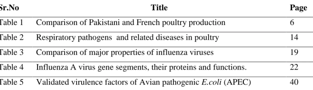
List of tables V
List of figures VI
List of abbreviations VII
Introduction 1
General introduction 2
Avian respiratory system 6
Defense mechanism and immune responses in birds 9
Respiratory diseases and associated pathogens in turkeys and chicken 13
Avian Influenza 16
E. coli infection in poultry 39
Multi-causal respiratory diseases 57
Objectives of the study 62
Article 1: S.Umar, J.L.Guerin, M. Ducatez (2017) Low pathogenic avian
influenza and co-infecting pathogens: a review of experimental infections in avian models ( accepted Avian Diseases)
66
Presentation of research work 106
Article 2: S.Umar, M.Delverdier, M.Delpont, S. Belkasmi, J.L.Guerin, M.
Ducatez (2017) Co-infection of turkeys with E. coli (O78) and H6N1 avian influenza virus (under review in Avian Pathology)
107
Article 3: S.Umar, A. Teillaud, H. Aslam, J.L. Guerin, M. Ducatez (2017)
Molecular epidemiology of avian respiratory viruses in Pakistan (2014-2015) (Under review in BMC Veterinary Research)
138
General discussion & Conclusions 167
References 173
Annexure 211
v
List of Tables
Sr.No Title Page
Table 1 Comparison of Pakistani and French poultry production 6
Table 2 Respiratory pathogens and related diseases in poultry 14
Table 3 Comparison of major properties of influenza viruses 19
Table 4 Influenza A virus gene segments, their proteins and functions. 22 Table 5 Validated virulence factors of Avian pathogenic E.coli (APEC) 40
vi
List of Figures
Sr.No Title Page
Figure 1 Respiratory system of chickens 8
Figure 2 Mechanism of respiration in birds 9
Figure 3 Genomic structure of influenza A virus 20
Figure 4 Structure of influenza virus ribonucleoprotein (vRNP). 21
Figure 5 Schematic diagram of influenza virus A particle. 23
Figure 6 Overview of receptor predilections of avian and mammalian influenza viruses 24
Figure 7 Life cycle of influenza viruses. 26
Figure 8 Schematic diagram of the antigenic shift process. 29
Figure 9 Schematic diagram of antigenic drift process 30
Figure10 Schematic showing chicken’s inflammatory response to APEC in the respiratory tract.
vii
List of abbreviations
AI avian influenza
AIV(s) avian influenza virus(es)
ABSL‐2 animal biosafety level 2
aMPV Avian metapneumovirus
Ct cycle threshold
DMEM Dulbecco’s Modified Eagle’s Medium
Dpi days post‐inoculation
EID50 mean egg infective dose
ELISA enzyme‐linked immunosorbent assay
HA Hemagglutinin
HE hematoxylin/eosin
HI hemagglutination inhibition
HPAI highly pathogenic avian influenza
HPAIV(s) highly pathogenic avian influenza virus(es)
IHC Immunohistochemistry
IBV Infectious bronchitis virus
ILTV Infectious laryngotracheitis virus
LPAI low pathogenic avian influenza
LPAIV(s) low pathogenic avian influenza virus(es)
MDCK Madin‐Darby canine kidney
M1 matrix protein
M2 membrane ion channel protein
NEPs nuclear export proteins
NA Neuraminidase
NP Nucleoprotein
NS1 nonstructural protein 1
NS2 nonstructural protein 2
NDV Newcastle disease virus
OIE World Organization for Animal Health
PA polymerase acidic protein
PBS phosphate buffer saline
PB1 polymerase basic protein 1
PB2 polymerase basic protein 2
qRRT‐PCR quantitative real time RT‐PCR
SHS Swollen head syndrome
µg Microgram µM Micromolar µl Microlitre µm Micrometre mL Milliliter Min Minute(s)
1
2
1. General introduction
Respiratory disease is a multifactorial problem in poultry, with viral and bacterial respiratory pathogens often concurrently present and most probably influencing one another (Marien et al., 2007). A community of mucosal dwelling microorganism colonize healthy upper respiratory tract including both commensals and potential pathogens kept under control by the host immune system. There are some evidence demonstrating that bacterial colonization can be enhanced by viral priming (Brealey el al., 2015).There are two hypotheses which can explain the underlying mechanism. One hypothesis is that viral infections may lead to bacterial superinfection by damaging the respiratory tissue, characterized by loss of cilia and ciliated cells (Bakaletz, 1995; Matthijs et al., 2009), decreased ciliary activity and mucociliary clearance (Wilson et al., 1996), and leading to efficient bacterial attachment with damaged respiratory tissue (El Ahmer et al., 1999). A second hypothesis is that the dysfunction of immune system may increase the colonization of bacterial infections after viral infection. Previous virus infection led to decreased phagocytic activity of macrophages and heterophils (Engelich et al., 2002; Navarini et al., 2006). Moreover, bacterial colonization may also be enhanced by severe granulotoxic effects of the innate anti-viral responses (type I interferons (IFN)) (Navarini et al., 2006; Matthijs et al., 2009). A significant higher number of pathogens are seen in tissues of superinfected animals than in tissues of animals infected with only one pathogen. Furthermore, inflammatory cytokines can be over produced during superinfection leading to exacerbated immune responses and damage to host tissue (Beadling & Slifka, 2004; van der Sluijs et al., 2006; Speshock et al., 2007).
Usually opportunistic bacterial pathogens are detected during respiratory virus infections such as Escherichia coli (E. coli). Uptil now, unidirectional view of avian influenza virus (AIV) /bacterial interactions has been studied, where viral infection proved beneficial to bacterial infection and led to bacterial superinfection at the respiratory tract. Respiratory viruses and bacteria may interact in a bidirectional way, where bacteria may also influence host
3
susceptibility to viral infection. Moreover, cotransmission of virus and bacteria may be possible as infectious agents of respiratory tract are transmitted through aerosol or direct contact with respiratory excretions (Brealey et al., 2015). Low pathogenic Avian influenza virus (LPAIV) infection is an emerging respiratory problem, isolated from different birds from a number of countries and has been reported to have zoonotic potential (Swayne, 2008; Liu et al., 2014; Umar et al., 2016a). LPAIV may be transmitted from aquatic birds to domestic poultry leading to economic losses (Swayne, 2008). Turkeys are an important host in influenza virus ecology because they are susceptible to infection with these viruses and are often involved in inter species transmission. Several previous studies reveal that waterfowl-origin influenza viruses can be more easily transmitted to domestic turkeys than to chickens (Abid et al., 2016).
An important natural route of avian influenza (AI) infection in farms is inhalation of contaminated dust. Aerosols may contribute to the transmission of AI between birds in addition to the faecal-oral route. For example, some high pathogenic avian influenza virus (HPAIV) (H5N1) can replicate in feather follicles of waterfowl, which may serve as a potential source for aerosol transmission (Yamamoto et al., 2007). Spread of viruses in the air has been
suspected when outbreaks of AI have occurred downwind from infected flocks or when contaminated manure has been spread on land in the proximity of poultry buildings (Lv et al., 2012). Experimental studies have shown that some subtypes of AIV could be transmitted between flocks of chickens via the air (Tsukamoto et al., 2007; Shi et al., 2010; Yao et al., 2011; Guan et al., 2013), and more information on virus infection by aerosols and the corresponding host immune response is needed to improve the understanding of aerosol transmission of AIV. Viruses that travel in the air can be carried as aerosols; aerosols contain particles < 5 μm in size. Aerosols can stay in the air longer and travel farther than large droplets and hence are more likely to be responsible for airborne transmission of viruses (Nicas et al., 2005). Commonly used methods of experimental infection, such as intranasal and intratracheal inoculations, bypass the deep air sac access of virus particles. In addition, the minute size of virus aerosols facilitates their reach into the lower respiratory tract and causes more severe
4
disease (Tellier, 2006; Guan et al., 2015). A human clinical study of influenza infection reported that the 50% infectious dose (ID50) by aerosol inhalation was approximately 100-fold less than that by inoculation with intranasal drops (Tellier, 2006). In accord with that report, studies in chickens have shown that the ID50 values of both HPAIV (H5N1) and LPAIV (H9N2) were substantially lower by aerosol inoculation than by intranasal drops (Guan et al., 2013; Sergeev
et al., 2013). Sergeev et al. (2013) found that aerosolised HPAIV was rapidly spread to various
organs via respiratory infection in chickens. LPAIV H9N2 is currently widespread in domestic poultry and occasionally transmitted to mammalian species, including humans (Alexander, 2007; Kwon et al., 2008; Liu et al., 2014). Respiratory tract infections in turkeys due to viruses and bacteria frequently result in considerable financial losses due to increased production losses, mortality rates and medication costs. Respiratory diseases in turkeys are triggered by several pathogens, alone or in combination with the support of other non-infectious factors. The respiratory viruses influenza virus type A, paramyxovirus types 1, 2 and 3 and avian metapneumovirus (avian pneumovirus, turkey rhinotracheitis virus) (aMPV) have been shown to induce respiratory problems. However, clinical signs following experimental inoculation with these viruses are less severe than those observed in the field (Marien et al., 2007). It is generally accepted that secondary bacterial pathogens are often involved, with amongst others
E. coli. These bacterial agents differ from the viral pathogens in that it is not always
straightforward to reproduce clinical signs following experimental infection. E. coli, a notorious infectious pathogen in poultry, illustrates this apparent paradox.
LPAIV H6N1 has been circulating in French Poultry industry and may cause severe economic losses especially due to secondary bacterial infection by E. coli (Prof.Jean Luc Guerin personal communication) and E. coli infections often occur between production onset and slaughter. Thus, in the current field situation bacterial infections appeared to be the predisposing agents rather than AIV. Whether this virus, which belongs to the genus Orthomyxovirus and this Gram-negative bacterium merely act separately or in a synergistic or additive way remains to be elucidated. Only a few experimental studies have been undertaken in chickens to study
5
possible mutual interactions between AIV and E. coli (Barbour et al., 2009). The lack of documentation in scientific journals is surprising since AI and E. coli co-infections are serious emerging issues in the poultry industry. Previous studies have shown that pathogenicity of the LPAIV was typically enhanced by secondary bacterial infections, resulting in chicken morbidity (Kishida et al., 2004). Recently, one H3N8 virus, Dk/BJ/40/04, caused a fatal disease when coinoculated with E. coli indicating H3N8 subtype viruses can be pathogenic to chickens under field conditions (Pu et al., 2012). Moreover, Barbour et al. (2006) and Stipkovits et al. (2012ab) reported that clinical signs were aggravated during mixed infections of AIV and
Mycoplasma in poultry (Barbour et al., 2006; Stipkovits et al., 2012ab).
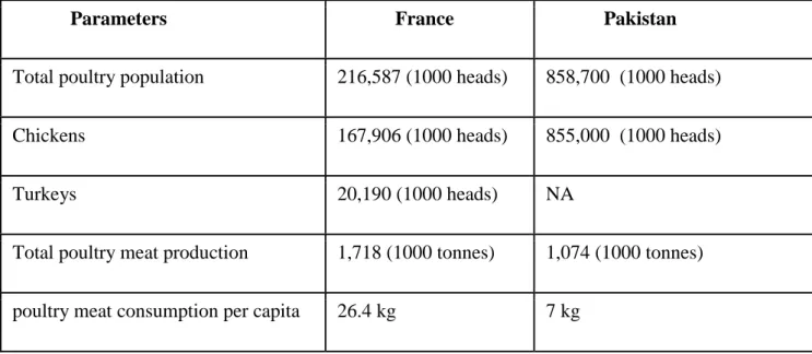
Poultry industry is one of the biggest industry in Pakistan and France. However, turkey industry is much bigger and developed in France than in Pakistan. Percapita meat consumption is lower in Pakistan than in France. Therefore, poultry industry in Pakistan needs big improvement to fulfill the demands of increasing human population. Comparison of poultry production for both countries is presented in Table 1 (FAOSTAT, 2014). The present study was undertaken to study the molecular epidemiology of respiratory viruses in Pakistani poultry and to develop a dual infection model for AIV and E. coli in turkeys using the intratracheal and aerosol inoculation routes. During our experimental work on turkeys, we performed different pilot experiments to compare aerosol and intratracheal route of inoculation. We found that aerosol route of inoculation is better than intratracheal route of inoculation in that it better mimics field conditions. Aerosol inoculation may help to shorten the gap between field and laboratory conditions. However, it is still difficult to generate 100% field conditions in laboratory settings because there are some other factors which contribute in the production of diseases e.g. dust, pollution, humidity, temperature, ammonia production, housing stress etc. Different settings were tested between infections, with clinical signs, gross lesions, histology and bacterial/viral titration as parameters for evaluating possible synergistic/additive potential between both agents.
6
Parameters France Pakistan
Total poultry population 216,587 (1000 heads) 858,700 (1000 heads)
Chickens 167,906 (1000 heads) 855,000 (1000 heads)
Turkeys 20,190 (1000 heads) NA
Total poultry meat production 1,718 (1000 tonnes) 1,074 (1000 tonnes)
poultry meat consumption per capita 26.4 kg 7 kg
Table 1: Comparison of Pakistani and French poultry production (FAO STAT, 2014)
2
.
The avian respiratory system
The principal function of the respiratory system in birds is exchanging oxygen (O2) and carbon dioxide (CO2) between atmosphere and blood, but also temperature regulation and phonation. Because of its role in gas exchange, the respiratory system is regularly in direct interaction with the outside environment. Every day many liters of air go through the lungs, providing a constant challenge of airborne particles and microbes. In complete contrast to the tidally ventilated mammalian respiratory system, where fresh inhaled air is mixed with residual stale air in the respiratory airways, the avian lung is a flow-through system (Reese et al., 2006). The respiratory tract begins at the nares, consists of passages between conchae in the head and subsequently leads inhaled gas to the larynx. The trachea extends from the larynx, and branches into two extrapulmonary primary bronchi. From each primary bronchus, four groups of secondary bronchi (medioventral, mediodorsal, lateroventral and laterodorsal) arise and from the secondary bronchi multiple parabronchi develop (Fedde, 1998) (Figure 1). Ventilation is achieved through a unique action of the air sacs as there is no diaphragm in birds. Therefore, avian lungs have a highly complex structure and are fixed in thoracic walls. Air sacs occupy every available space in the body cavity which is not occupied by other viscera. There are nine
7
air sacs in most of the birds: paired cervical air sacs, an unpaired clavicular air sac that is connected to each lung, paired cranial thoracic air sacs, paired caudal thoracic air sacs, and paired abdominal air sacs. The cervical, clavicular, and cranial thoracic air sacs arise from the medioventral secondary bronchi, and they are often called the cranial air sacs. The caudal thoracic and abdominal air sacs (the caudal air sacs) arise from the lateroventral and mediodorsal secondary bronchi and from the continuation of the intrapulmonary primary bronchus. The air sacs are auxiliary structures that pump air through the respiratory tract, but do not contribute to the gas exchange with the blood (Fedde, 1998; Reese et al., 2006). The O2 and CO2exchange only occurs in the lungs. During inspiration, active contraction of some muscles of the body wall causes an increase in the volume of the air sacs which results in pressure in the air sacs less than that in the atmosphere and gas moves through the lungs into the air sacs. The inspired air completely bypasses the cranially lying openings of the medioventral secondary bronchi, a process which is called inspiratory aerodynamic valving (Reese et al., 2006). In contrast, during the inspiratory phase as well as the expiratory phase, air flows in the mediodorsal and lateroventral secondary bronchi. About one half of the inspired volume passes through the paleopulmonic parabronchi and in this way in the cranial air sacs, and the remainder passes through the much smaller neopulmonic parabronchial network to the caudal air sacs, and through the direct connection from the intrapulmonary primary bronchus to the abdominal air sacs. During expiration, reduction in coelomic volume (decrease in the volume of the air sacs) increases the pressure in the air sacs and air moves out of the air sacs. Some of the air from the caudal air sacs again traverses the neopulmonic parabronchi and most of the air enters the paleopulmonic parabronchi, travelling in the same direction as during inspiration. Air from the cranial air sacs flows through the medioventral secondary bronchi to exit the lung without contacting any parabronchial gas exchanging surfaces (Figure 2). Thus exchange of O2and CO2between air and blood occurs both during inspiration and expiration in birds and nearly all of the air that was inhaled, has passed over paleopulmonic parabronchial gas exchanging surfaces during some part of the respiratory cycle. The walls of the parabronchi
8
are perforated by numerous openings that lead to the respiratory atria. Funnel-shaped infundibulae arise from the atria and open into the air capillaries. The inhaled air flows through the parabronchial lumen and then into the exchange tissue through the atria, the infundibulae, and the network of air capillaries. A complex network of blood capillaries closely surrounds air capillieries forming efficient gas exchange system in birds (Reese et al., 2006). The blood-gas barrier in the avian lung is approximately 56-67% thinner than that of a mammal of the same body mass and the respiratory surface area is approximately 15% greater (Maina et al., 1989). Respiratory efficiency is enhanced by large surface area and thin tissue barrier. However, these structural features make birds more susceptible towards pulmonary injury from environmental toxicants and invasion by pathogenic organisms (Reese et al., 2006).
Figure 1: Respiratory system of chickens (Clav. AS= clavicular air sac; Cran. Th. AS=/cranial
thoracic air sac; Caud. Th. AS=caudal thoracic air sac; Abd. AS=abdominal air sac). There is expansion of airsacs during inhalation drawing air from trachea and primary bronchi towards the the caudal air sacs and paleopulmonic parabronchi (Fedde, 1998).
9
Figure 2: Mechanism of respiration in birds. Mechanism and pathway of gas flow and
exchange through the pulmonary system during inspiration and expiration in right paleopulmonic lung and air sacs of chicken. A: Inspiration. B: Expiration (Source:
http://www.people.eku.edu/ritchisong/birdrespiration.html)
2. Defense mechanisms and immune responses in birds
The divergence of mammals and birds from a common reptilian ancestor occurred 200 million years ago. Despite this evolutionary time period, the fundamental principles of both the innate and adaptive immune systems of mammals and birds are the same. The availability of the chicken genome has helped improve our understanding of the avian immune system. The
10
respiratory system harbours the most extensive and thinnest surface across which the body is exposed to the external environment. Due to this characteristic, a vast array of proteins and pathogens are challenging this system on a daily basis. To cope with these pathogens, birds have well-developed defence mechanisms.
3.1. Innate resistance
The first non-specific arm of the avian immune system is known as the innate System. As in mammals, the most well characterised family of non-specific pattern recognition receptors (PRRs) are membrane-bound Toll-like receptors (TLRs) expressed by various cell types including epithelial cells and sentinel cells such as antigen presenting cells (APCs), dendritic cells and macrophages. In the chicken, TLRs are also expressed by heterophils, a polymorphonuclear leukocyte and homologue of mammalian neutrophils. TLRs detect structurally conserved microbial specific motifs. Thirteen TLRs have been described in the chicken; 11 are also present in mammals while two are chicken-specific (TLR-15 and TLR-21) (Temperley et al., 2008). Despite differences in TLR families, similar microbial motifs are recognised by both TLR repertoires. The initial line of defense for the airway is the nasal and tracheal epithelium, which prevents pathogens from entering the body. Multiple mucous glands within the pseudostratified ciliated columnar epithelium produce mucus which forms a layer on top of the cilia of the epithelial surface. Particulate material that is caught in the mucus gets transported by the movement of the cilia in an oral direction, where it is swallowed and digested or excreted by coughing and sneezing (Sharma, 2003; Koch et al., 2009). Furthermore, mucus contains antibacterial enzymes which impede the attempts of pathogens to colonize.
Phagocytic cells that include heterophils and macrophages, and natural killer (NK) cells are important components of innate immunity. Monocytes-macrophages, cells belonging to the mononuclear phagocytic system, are considered to be the first line of immunological defense. These cells originate from the bone marrow and subsequently enter the blood circulation. Upon migration to various tissues, monocytes mature and differentiate into tissue macrophages (Dietert et al., 1991; Qureshi et al., 2000). Macrophages then get involved in innate and
11
acquired immunity (Qureshi et al., 2000). Since the respiratory surface is in proportion much larger than that of mammals and the tissue barrier is much thinner (Maina, 1989; Maina et al., 1989), one can expect that, as stated above, the avian respiratory tract is relatively more easily attacked by pathogens than the mammalian one (Nganpiep & Maina, 2002). One would hence assume that for a similar defense competence, more residing avian respiratory phagocytes (ARP = macrophages and polymorphonuclear leukocytes such as heterophils) would arise on the surface of avian lungs. Paradoxically, the normal, steady-state avian respiratory system has very low numbers of residing ARP in comparison to the mammalian system, and as a consequence birds must rely heavily on the influx of ARP into the site of infection for non-specific defense against bacteria and other pathogens (Ficken et al., 1986; Qureshi et al., 1994, 2000; Klika et
al., 1996; Toth, 2000). Interestingly, ARPs the were present atria and the infundibulae and were
never found on the surface of the air capillaries which represent the functional equivalent to the mammalian alveoli (Nganpiep & Maina, 2002; Reese et al., 2006). Thus, macrophages seem to be located at strategic check points where fresh air is distributed into the gas exchange areas and where particles can be trapped and removed (Marien et al., 2007). The paucity and even lack of ARPs in birds has been used to explain a purported high susceptibility of poultry to respiratory diseases. Nganpiep & Maina (2002), however, showed that a composite defense armament has additionally developed in the avian respiratory system. A highly lytic upper airway epithelium endowed with lysosomes (apparently lacking in mammals), generally robust ARPs, and efficient translocation of subepithelial macrophages onto the respiratory surface, play a role in the protection of the respiratory system (Nganpiep & Maina, 2002). In the air sacs, being thin walled and lacking an elaborate ciliated epithelium, particle clearance is largely accomplished by phagocytic cells albeit significantly lower than in the lungs (Nganpiep & Maina, 2002; Reese et al., 2006).
3.2. Adaptive immunity
The second arm of the chicken immune system is the adaptive one involved in both cellular and humoral (antibody) responses, as well as the production of memory cells. The avian
12
antigenic repertoire is more compact than that of the mammalian system; one reason for this is that birds only possess 2 of the Major Histocompatability complex class (MHC), compared to the 6 of mammals (Kaufman, 2000). When pathogens cannot be withheld by physical barriers nor controlled by innate immune defense mechanisms, adaptive immunity (specific immune response) is required to specifically focus defense mechanisms on that particular antigen resulting not only in the elimination of the pathogen but also in protecting in case of a repeat encounter with the same pathogen (memory). Adaptive immunity is mediated by a variety of cells, of which T lymphocytes, B lymphocytes, and macrophages are the most important.
In poultry as in mammals, adaptive immunity is critically dependent on regulation by T lymphocytes (T cells), the coordinators of the immune response. Maturation of the T cells takes place in the thymus, a feature shared with mammalian species (Arstila et al., 1994). Before T cells can initiate and participate in an adaptive immune response to a pathogen, the antigen has to be presented by host cells in the context of their MHC molecules, i.e., as an antigenic peptide bound to the MHC molecule. The MHC molecules come in two forms: the MHC class I is expressed by essentially all nucleated cells, whereas the MHC class II is expressed mainly by cells of the immune system, the so-called antigen presenting cells (APC) such as macrophages, dendritic cells and B lymphocytes (B cells). These APC also deliver other signals equally important to the T cell activation, the so-called second or costimulatory signals (Arstila et al., 1994). Activation of T cells results in proliferation of the activated T cells and their differentiation into subpopulations of diverse effector cells, helper T cells (CD4+), suppressor T cells, and cytotoxic T cells (CD8+), or memory cells. Effector functions of T helper cells primarily involve production of cytokines (soluble molecules secreted to the extracellular space), and expression of membrane-bound cell-surface molecules, all affecting other cells of the immune system. The cytotoxic T cells, in contrast, are mostly killers that are specialized in the elimination of intracellular antigens. The latter include those that have entered cells via the endocytic pathway (exogenous antigens; e.g., phagocytosed bacteria) or were produced within the cell such as viral proteins and proteins resulting from neoplastic transformation of the cell
13
(endogenous antigen) (Erf, 2004). Another lineage of T cells exists (γδ T cells), but their physiological significance remains largely a matter of speculation (Marien et al., 2007).
Besides the T lymphocytes, other cells important to the cellular immune response include macrophages, dendritic cells, NK cells, and effector cells of antibody dependent cellular toxicity (Sharma, 2003). NK cells can also be regarded effector cells of specific cell-mediated immunity as they greatly benefit from T helper mediated activity (Erf, 2004). Unlike mammals, birds have a special organ, the bursa of Fabricius, where the development of B lymphocytes (B cells) from their immature precursors takes place. For humoral immunity, B cells differentiate into plasma cells that secrete antigen-specific antibodies. Antibodies can prevent disease caused by pathogens and provide protection, but they are primarily effective in preventing entry of pathogens through mucosal surfaces (e.g., secretory IgA) and in eliminating extracellular antigens (Koch et al., 2009). Most organisms stimulate both cell-mediated immunity and humoral immunity, although the type of immunity most critical for defense may vary with the organism (Vandaveer et al., 2001; Sharma, 2003; Erf, 2004).
3. Respiratory diseases and associated pathogens in turkeys and
chickens
Respiratory diseases are continuing to cause heavy economic losses in the poultry industry due to high production losses, mortality and medication costs (Van Empel & Hafez, 1999). Respiratory disease in poultry is a multifactorial problem, with viral and bacterial respiratory pathogens often concurrently present and most probably influencing one another (Marien et al., 2007). In addition to these infectious organisms, non-infectious factors, such as climatic conditions (e.g. inadequate ventilation, high ammonia levels, too high or too low temperature) can also help in diseases progress. The severity of clinical signs, duration of the disease and mortality are extremely variable and are influenced by many factors such as a virulence and
14
pathogenicity of the infectious agent as well as by many environmental factors. In many cases, respiratory disease observed in a flock may be a component of a multisystemic disease or it may be the predominant disease with lesser involvement of other organ systems. Respiratory tract infections increase the overall cost of production in terms of the provision of services of qualified veterinary personnel and the cost of medication for possible treatment. It is therefore important to reduce if not eliminate, respiratory infections among poultry flocks to the barest minimum to have good production and maximize profit of the producer. Various pathogens including a variety of viruses, bacteria, and fungi may initiate respiratory diseases in poultry. Environmental factors may augment these pathogens to produce the clinically observed signs and lesions. Poultry respiratory diseases are known to be caused by many pathogens (Table 2) including Newcastle disease virus (NDV), AIV, Infectious Bronchitis Virus (IBV), aMPV,
Mycoplasma gallisepticum (M. gallisepticum), Mycoplasma synoviae (M. synoviae) Mycoplasma meleagridis (M. meleagridis), Mycoplasma iowae (M. iowae), Ornithobacterium rhinotracheale (O. rhinotracheale), Pasteurella multocida and Avibacterium paragallinarum, Bordetella avium, Chlamydophila psittaci and E. coli with associated significant economic
losses to the industry (Van de Zande et al., 2001).
Disease Aetiology Main Clinical signs /lesions Prevention /control Avain influenza (AI) Avian influenza virus (AIV) Mild to severe respiratory signs depend on virus subtype Vaccine available Good biosecurity measures NewCastle disease (ND) Newcastle disease virus (NDV) Variable: mild to severe respiratory clinical signs and lesions Vaccine available Good biosecurity measures Infection bronchitis (IB) Infection bronchitis virus (IBV) Tracheitis, airsacculitis, pneumonia, nephritis Vaccine available Good biosecurity measures Infectious laryngotracheitis (ILT) Infectious laryngotracheitis (ILTV)
Teacheitis Vaccine available
Good biosecurity measures Swollen head syndrome/ turkey rhinotracheitis Avian metapneumovirus (aMPV) Swollen head, tracheitis, airsaculitis,pneumonia Vaccine available Good biosecurity measures Mycoplasmosis Mycoplasma gallisepticum Chronic tracheitis; chronic Mycoplasma free progeny.
15 airsacculitis Vaccination Possible Mycoplasmosis Mycoplasma synoviae Moderate tracheitis and airsacculitis. Arthritis Mycoplasma free progeny
Infectious Coryza Avibacterium
paragallinarum
Conjunctivitis, sinusitis, airsacculitis
Vaccination possible
Colibacillosis E. coli, often asso-
ciated with other respir
atory pathogens, e.g. IBV, NDV, mycoplasma Fibrinous pericarditis, airsacculitis, tracheitis Vaccine available against some E. coli serotypes.
Reduce dust in shed
Pasteurellosis (Fowl cholera)
Pasteurella multocida
in chronic form e.g. conjunctivitis, tracheitis; in acute form septicaemia Vaccination possible Good biosecurity measures Ornithobacteriosis Ornithobacterium rhinotracheal Tracheitis, airsacculitis Vaccination possible Good biosecurity measures
Table 2: Respiratory pathogens and related diseases in poultry
These respiratory pathogens are of major importance because they can cause disease independently, in alliance with each other or in association with other bacterial and viral agents (Ali & Reynolds, 2000; Yashpal et al., 2004). Viral agents are mostly being attributed a triggering role, since the clinical signs following experimental inoculation with these viruses are less severe than those observed in the field. Viral infections generally cause rather acute respiratory problems from which birds usually can recover fairly easily. The problems, however, become more critical when bacterial pathogens are involved. With these bacterial agents, it is not always straightforward to reproduce clinical signs following experimental infection. This has led to a still contemporary discussion point whether the different bacterial agents are primary or rather secondary pathogens (Marien et al., 2007). In the present thesis, the experimental research focuses on the co-infection in turkeys with AIV, and E. coli. Hence, the most important literature data on these agents will be discussed below.
16
4.
Avian influenza
Avian influenza (AI) was first identified as a distinct disease entity of poultry in 1878, in Italy. It was called “fowl plague” and was defined as a highly lethal, systemic disease of chickens. From the 1870s into the early 1900s, fowl plague spread from Northern Italy into the rest of Europe, and by the 1930s it was endemic in parts of Europe and Africa (Schafer, 1955). Likewise, in the United States the disease was reported in 1924‐1925 and 1929 (Stubbs, 1948). Into the mid‐twentieth century, fowl plague had been diagnosed in North Africa, South America, North America, and most of Europe (Swayne & Halvorson, 2008). The agent responsible for the human influenza initially isolated from pigs in 1931 and later from humans in 1933 (W. Smith et al., 1933; Shimizu, 1997). Prior to that, one of the most devastating influenza pandemics in human history, the “Spanish Flu” (H1N1 subtype), hit the population in 1918, causing thousands of deaths (Taubenberger et al., 2000). Even if Centanni & Savonuzzi (1901) had already demonstrated the existence of a filter‐passing agent, the viral etiology of fowl plague was unknown until 1955, when the disease was determined to be caused by influenza A virus (Schafer, 1955).
In gallinaceous birds (i.e., chickens and turkeys), AI viruses are classified as being highly pathogenic AI (HPAI) or low pathogenic AI (LPAI) viruses. Although molecular criteria have been established by the World Organization for Animal Health (OIE) for the identification of the HPAI virus based on the protein sequence of the HA proteolytic cleavage site, in vivo testing used to be the gold standard. An AI virus isolate is classified as being HPAI if it kills at least 75% of susceptible 4- to 6-week-old chickens within 10 days postinoculation by the intravenous route. Some isolates will cause 100% mortality by 36–48 hours postinoculation. All other isolates are considered to be LPAI viruses. Biologically, the difference between HPAI and LPAI is that HPAI is a systemic infection and LPAI remains localized to the respiratory and intestinal tracts. For unknown reasons, all HPAI viruses have been either H5 or H7 HA subtypes (David L. Suarez & Swayne, 2008). Nevertheless, mild clinical forms of AI, characterized by respiratory disease and drops in egg production, were first recognized in 1949 in chickens and,
17
subsequently, in several domestic poultry species (Easterday & Tumova, 1972). Therefore, since 1971, H5 and H7 viruses have been isolated and characterized not only as HPAIVs but also as LPAIVs (Smithies et al., 1969). Although wild birds were already suspected to participate in fowl plague transmission, it was not until 1961 that the first proof of AIV infection in wild birds arose, in an outbreak in South Africa affecting common terns (Sterna hirundo) with high mortality (Becker, 1966). Since then, and particularly during the recent past decades, numerous surveys have been conducted in migratory waterfowl, confirming asymptomatic infection by AIV of healthy wild aquatic birds, especially in the orders Anseriformes and
Charadriiformes (Hinshaw & Webster, 1982).
Type A influenza strains are classified by the serological subtypes of the primary viral surface proteins, the hemagglutinin (HA) and neuraminidase (NA). The HA has 16 subtypes (H1–H16) and contains neutralizing epitopes. Antibodies against the NA are not neutralizing, and there are nine neuraminidase or “N” subtypes. The “H” and N subtypes seem to be able to assort into any combination, and many of the 144 possible combinations have been found in natural reservoir species, although some combinations are more common than others. All 16 subtypes have been found in ducks, gulls, or shorebirds, the natural reservoir host species of the virus and two of HA and NA (HA17–HA18 and NA10–NA11) have been isolated from bats (Tong
et al., 2012, 2013). However, in these species certain subtypes are more common than others;
for example, H3, H4, and H6 are most common in ducks in North America and although there is no clear association between host range or host restriction based on HA subtype, some subtypes are more common in some species than others, i.e., H1 and H3 in swine, H3 in horses, and H5 and H7 in chickens (David L. Suarez & Swayne, 2008).
It was not until 1997 that AI became considered a disease not only of birds, when the occurrence of fatal disease in poultry and humans in Hong Kong was associated with the HPAIV H5N1 strain (Claas et al., 1998). This episode increased the international interest in HPAIV among the veterinary and public health community, because it was the first indication that AIV (H5N1) could potentially be the precursor to a human pandemic viruses (Sims &
18
Brown, 2008). Indeed, over the next decade, HPAIV (H5N1) in poultry spread across three different continents with unprecedented socioeconomic consequences. These concerns were amplified because of the reassortment possibility with a human influenza A virus, which could create a new virus capable to produce the next human influenza pandemic (David L. Suarez & Swayne, 2008).
4.1. Etiology and Classification
Influenza viruses belong to the Orthomyxoviridae family (orthos, Greek for "straight"; myxa, Greek for "mucus") and are classified into five different genera: influenza A, influenza B, influenza C, Thogotovirus, and Isavirus (Cheung & Poon, 2007). The most serious types that cause dangerous outbreaks with high morbidity and mortality are influenza A viruses because they mutate more rapidly and have a wider range of hosts (Khanna et al., 2008). Influenza A viruses infect animals, including birds, pigs, horses, whales, seals, and also humans (Ito & Kawaoka, 2000; Reperant et al., 2009). Type B and C are generally found in humans, in addition to some mammals like seals, with less severity than influenza A. The main differences between the three main types of influenza viruses (A, B and C) are outlined in table 3. Wild aquatic birds of the order of Anseriformes (ducks, geese and swans) and Charadriformes (gulls, terns, surfbird and sandpiper) are considered to be the natural reservoir of all types of influenza A viruses. In these hosts, viral replication occurs mainly in the gastrointestinal tract, and to a lesser extent in the respiratory tract. The infected birds generally have no apparent signs of illness, with some exceptions after infection with highly pathogenic avian influenza viruses (Munster et al., 2007).
19
Features Influenza A virus Influenza B virus Influenza C virus Number of gene segments 8 8 7 Surface glycoproteins Haemaglutinin and neuraminidase (HA and NA)
HA and NA HEF ( Haemagglutination
esterase Fusion)
Host range Wide range of hosts (humans, pigs, horses, whales, seals and birds)
Humans and seals Mainly humans (also found in swine)
Table 3: Comparison of major properties of influenza viruses (Cheung & Poon, 2007).
4.2. Morphology and molecular organization
Influenza viruses are roughly spherical with a size of around 100 nm or filamentous in shape, often in excess 300 nm in length (Bouvier & Palese, 2008). Morphological structure is known to be affected by several viral proteins (HA, NA) and matrix proteins (M1 and M2), in addition to the nature of the host cells (Cheung & Poon, 2007). Influenza viruses are enveloped with surface glycoprotein spikes and a segmented RNA genome of negative sense (complementary to mRNA). RNA of influenza A virus is organized into 8 segments, in total around 13600 nucleotides long (Webster et al., 1992). These are the polymerase basic (PB1 and PB2), the polymerase acidic (PA), haemagglutinin (HA), nucleoprotein (NP), neuraminidase (NA), matrix (M), and non-structural (NS) genes (Samji, 2009). Influenza A viral gene segments are known to encode at least ten proteins which are the RNA polymerase complex proteins (PA, PB1, and PB2), surface glycoproteins (HA, and NA), nucleoprotein (NP), matrix proteins (M1 and M2), and nonstructural proteins (NS1, NS2) (Samji, 2009; Wang & Taubenberger, 2010). In addition, PB1–F2 and a new viral protein (N40) which is translated from segment 2 have been recently identified in some influenza A virus isolates (Wise et al., 2012). Moreover, two more proteins, PA-X and M42 which are translated from segment 3 and 7, respectively, have been recently found (Jagger et al., 2012; Wise et al., 2012) (Figure 3).
20
Figure 3: Genomic structure of influenza A virus
RNA segments (in nucleotides) shown in positive sense and their encoded proteins (in amino acids). The lines at the 5´ and 3´ termini represent the coding regions. The PB1 segment encodes three proteins, two of them (PB1 and N40) translated from ORF 0, and PB1–F2 protein translated from ORF 1. The M2, M42 and NEP/NS2 proteins are encoded by spliced mRNAs (the introns are indicated by the V–shaped lines) (Jagger et al., 2012; Wise et al., 2012).
Each viral RNA segment is surrounded by nucleoprotein (NP) forming ribonucleoprotein (RNP) and encapsidated by one copy of trimeric polymerase (PB1–PB2–PA complex) which is essential for viral replication (Digard et al., 1999). The structural organization of viral ribonucleoprotein can be seen in figure 4.
21
Figure 4: Structure of influenza virus ribonucleoprotein (vRNP).
Green spheres represent NP monomers, and the black line shows the associated single–stranded vRNA molecule. Influenza RNP folds into a double–helical hairpin structure. A short duplex formed between the 5´ and the 3´ ends provides the binding site for the heterotrimeric RNA– dependent RNA polymerase (Portela & Digard, 2002).
Four virus proteins (PB2, PB1, PA, and NP) are responsible for virus transcription and replication of the viral genome in the nuclei of infected cells. PB1–F2 protein plays a role in pro–apoptotic activity, while N40 protein, which is encoded by the same gene (PB1), interacts with the polymerase complex in the cellular environment but does not contribute to transcription function (Wise et al., 2012). PA-X protein has been shown to modulate host response and viral virulence (Jagger et al., 2012). Haemagglutinin (HA or H) plays a role in virus attachment to the host cell and subsequent fusion with cell membranes, while neuraminidase (NA or N) supports the release of viruses from the host cell surface by hydrolyzing sialic acid from glycoproteins which helps to release the progeny virus particles from host cells (McCauley & Mahy, 1983). Non–structural protein 1 (NS1) has a major role in inhibition of host immune response via limitation of interferon (IFN) production (Hale et al., 2008). NS2 (also called nuclear export protein or NEP) plays a role in the export of RNPs from the nucleus to the cytoplasm during viral replication, in addition, it also regulates virus transcription and replication processes (Robb et al., 2009). Matrix protein 1 (M1), the major structural protein, is the dominant protein in determining virus morphology and also plays an
22
important role in virus assembly and budding (Rossman & Lamb, 2011). Matrix protein 2 (M2) is the ion channel that regulates the pH, and is responsible for virus uncoating, the step that follows virus entry into the host cell (Holsinger et al., 1994). In addition, this protein also plays an important role in membrane scission in the last stage of virus life cycle (Roberts et al., 2013). Matrix protein 42 (M42) can functionally replace M2 and support efficient replication in null M2 influenza viruses (Wise et al., 2012). Table 4 summarizes the length of each viral segment and the function of protein(s) encoded by each segment.
Segment Segment size (nt) Encoded proteins Protein length (a.a) Protein roles
1 2341 PB2 759 Polymerase subunit; plays a role in
RNA
replication by mRNA cap recognition
2 2341 PB1 757 Polymerase subunit; RNA elongation
during replication
PB1–F2 87 Pro–apoptotic activity
N40 718 Polymerase complex interaction
3 2233 PA 716 Polymerase subunit, endonuclease
activity
PA-X 252 modulates the host response and viral
virulence
4 1778 HA 550 Major surface antigen, receptor binding
and fusion activities, main target for neutralizing antibodies
5 1565 NP 498 RNA binding protein, nuclear import
regulation
6 1413 NA 454 Minor surface glycoprotein for
neutralizing
antibodies; sialic acid activity, cleavage of progeny virions from host cell receptors and virus release
7 1027 M1 252 Major component of virion, RNA
nuclear export regulation, viral assembly and budding
M2 97 Ion channel for controlling pH during
virus
uncoating and HA synthesis (viral release)
M42 99 functionally replace M2 in M2-null
viruses
8 890 NS1 230 Interferon antagonist protein,
regulation of hostgene expression
23
Table 4: Influenza A virus gene segments, their proteins and functions. Typical gene and
protein sizes are shown, though strain variation occurs (Bouvier & Palese, 2008).
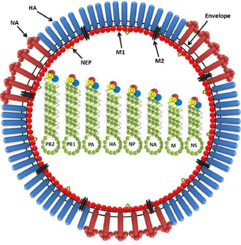
The viral envelope is made of a lipid bilayer which is derived from the host cell’s plasma membrane. Three surface viral antigens are embedded in the lipid bilayer: the HA spike, which has a rod like–shape, represents approximately 80% of the total surface proteins; the NA spike, which is almost mushroom–shaped, represents 17%; with minor components of M2 represented by few molecules (only 16 to 20 molecule per virion) (Schroeder et al., 2005; Nayak et al., 2009). Underneath the lipid bilayer, the M1 protein forms a layer that separates the viral segments from the virus membrane. Inside the virion, 8 segments of different length are associated with the nucleocapsid protein (NP) and three large proteins (PB1, PB2, and PA). NEP is also associated with the virus but in low amounts (Cheung & Poon, 2007). Figure 5 illustrates the typical structure of influenza A virus.
24
The RNA is segmented and each segment encodes one or more proteins. The segments are not identical in length (ranging from 2341 to 890 nucleotides). The longest segment encodes PB2 protein and the shortest encodes NS protein. The RNA segments are coated with nucleoprotein forming ribonucleoprotein (RNP), and a small amount of transcriptase (polymerase complex) represented by PB1, PB2, and PA is also associated with it. The haemagglutinin (HA), neuraminidase (NA), and M2 proteins are inserted into the host–derived lipid envelope. The matrix (M1) protein underlies the lipid envelope. A nuclear export protein (NEP) is also associated with the virus (Al-Mubarak, 2014).
4.3. Replication of influenza A viruses
The first step of viral replication is virus attachment to its host cell. The host specificity of each type of influenza virus is mainly determined by differences in the host cell receptors (Naeve et
al., 1984). There are two main types of host cell receptors with which influenza viruses have
the affinity to bind. The linkage between neuraminic acid and the sugar (galactose) determines whether influenza virus binds to avian or mammalian cells (Figure 6). Avian influenza viruses preferentially bind to the neuraminic acid α 2,3 galactose receptors while mammalian influenza viruses bind to neuraminic acid α 2,6 galactose (Auewarakul et al., 2007; Pillai & Lee, 2010)
Figure 6: Overview of receptor predilections of avian and mammalian influenza viruses (Nelli et al., 2010).
25
Once a host cell is infected with influenza virus, the HA glycoprotein is produced as a precursor, HA0, which is cleaved into two subunits (HA1 and HA2) by host serine proteases before virus particles become infectious (Klenk et al., 1975). The HA1 portion contains the antigenic sites (the receptor binding domain), while the HA2 portion mediates fusion of the virus envelope and cell membranes (Steinhauer, 1999). Virulent and avirulent avian influenza A viruses can be differentiated by the sequence of a few basic amino acids at the point where the HA0 is cleaved (cleavage site); the so–called cleavage sequence (Zambon, 1999). The virus enters the host cell via receptor (clathrin) mediated endocytosis at the inside face of the plasma membrane forming an endosome (Rust et al., 2004). The endosome has a low pH of around 5 to 6, which induces a conformational change in HA0, displaying the HA2 fusion peptide. This fusion peptide inserts itself into the endosomal membrane and mediates the fusion of the viral envelope with the endosomal membrane. This mechanism is not only important for inducing the conformation change in HA0, but also opens up the M2 ion channel during fusion of viral and endosomal membranes, allowing the virion interior to become acidic which releases the vRNP from M1. This permits the vRNP to enter the host cell’s cytoplasm (Pinto & Lamb, 2006). Transcription and replication occur inside the nucleus. Because of the negative sense of the viral genome, the viral RNA is copied into positive sense mRNA by the polymerase complex to act as a template for the production of the viral RNAs. The polymerase complex responsible for viral transcription and replication is formed by PB1, PB2, and PA. The viral RNA transcription is catalyzed by the RNA dependent RNA polymerase. The resultant positive sense viral mRNA is exported to the cytoplasm through nuclear pores to start viral translation by ribosomes. Positive sense viral mRNA also serves as a template to produce the negative sense RNA that is packaged into new virions (Swayne, 2008). Polymerase basic (PB1 & PB2), nonstructural (NS1 & NS2), NP, PA, and M1 proteins are synthesized in the host cell cytoplasm then transported to the nucleus to participate in matrix and nonstructural splicing, transcription and replication. Surface glycoproteins (HA and NA) are synthesized by ribosomes and then enter the endoplasmic reticulum (ER), where they are glycosylated, and then folded in the Golgi
26
apparatus. These proteins are incorporated in the cell membrane and assembled with vRNP complex (Sidorenko & Reichl, 2004). Progeny RNPs are released to the cytoplasm and packaged into new virus particles. New virions get enveloped with the plasma membrane with integrated virus proteins through budding (Palese & Shaw, 2007). Progeny virions are released from the cell surface using NA, which cleaves the sialic acid residues from the cell surface (Roberts et al., 2013). The stages of influenza virus replication start from attachment of the virus onto host cells and end with the release of the progeny particles (Figure 7).
Figure 7: Life cycle of influenza viruses.
Stages involved in the replication process are:1. Attachment to host receptor and entry to host cell via endocytosis.2. Virus uncoating and releasing RNPs to the cytoplasm.3. Transcription and translation of viral RNA. 4. Replication of viral RNA.5. Production of nucleoprotein, non-structural, matrix, polymerase acidic, and polymerase basic proteins.6. Production of envelope proteins (surface glycoproteins HA and NA, and M2) and their transportation to cell
27
membrane.7. Viral RNPs packaging and assembly.8. Virion budding and release from the cell membrane (Al-Mubarak, 2014).
4.4. Pathogenicity of influenza A viruses
According to the pathogenicity and severity of the disease in chickens, avian influenza A viruses can be classified into two pathotype groups: HPAIV and LPAIV. The mortality rates of the poultry flocks infected with HPAIV may reach 100%, while infection with LPAIV cause only milder and primarily respiratory disease (Capua & Marangon, 2000). In HPAIV, the region that encodes the cleavage site of the surface glycoprotein (HA) molecule contains multiple basic amino acids (arginine and lysine) which allows cleavage of the HA molecule by cellular endogenous proteases widely distributed throughout the cells of the body (Wood et al., 1993). This molecular structure is important in determining the virulence of these strains because it allows the virus to replicate in a considerably broader tissue range, causing widespread damage in tissues and death of the bird, with a mortality rate approaching 100% (Kim et al., 2009; Adams & Sandrock, 2010). The most pathogenic subtypes of avian influenza are restricted to subtypes H5 and H7. On the other hand, LPAIV have only one basic amino acid (arginine) in the cleavage site of the HA molecule. This limits the site for the viral cleavage by trypsin–like host proteases, and as a consequence, the replication process occurs in limited tissues and organs, particularly in respiratory and digestive tracts, causing only mild disease (Alexander, 2000). LPAIV which cause asymptomatic or low pathogenic infection may mutate and convert to HPAIV through an adaptation process after infection of poultry (Mundt et al., 2009).
4.5. Influenza A viruses evolution
During influenza viral replication, genetic variations occur frequently. This is due to the structure of the viral RNA (segmented) and the low fidelity of the RNA dependent RNA polymerase which generates replication errors during virus life cycle (Zambon, 1999; Zambon, 2001). Consequently, influenza A viruses can undergo recurrent antigenic changes. The resultant change in structure allows the virus to evade neutralizing antibody, the main
28
mechanism of protective immunity against influenza infection. Such changes may lead to the creation of a new virus strain distinctive from those previously circulating viruses (Zambon, 1999).
Antigenic shift is a result of reassortment and it occurs when two or more different influenza A viruses subtypes infect a single cell simultaneously. Because influenza A viruses are segmented, it is possible to produce new viruses with a variety of segment combinations by the acquisition of entirely new gene segments. The newly assembled progeny virions may have mixed genes from the two parent viruses (Holmes et al., 2005; Nelson et al., 2008). This may result in the emergence of new subtypes which may be more pathogenic than the original parent viruses and may be associated with pandemics (Neumann et al., 2009; Van-Tam & Sellwood, 2010). Pigs are thought to play an important role in influenza virus ecology because of their ability to become infected with different types of influenza A viruses (avian and human viruses), and thus they act as an intermediate host, or mixing vessel (Figure 8). The new reassortant strain may cause a pandemic or panzootic because the hosts (humans or birds) have little or no immunity against it (Van-Tam & Sellwood, 2010). Such a scenario happened recently in April 2009 with the H1N1 pandemics caused by swine origin quadruple reassortant virus with of avian, swine and human origins (Michaelis et al., 2009).
29
Figure 8: Schematic diagram of the antigenic shift process.
Genetic change in influenza A virus also occurs by ‘antigenic drift’. This is due to the accumulation of point mutations over time, which results from a lack of proofreading mechanism in the RNA polymerase, leading to incorrect ribonucleotide insertions during replication (Zambon, 1999; Adams & Sandrock, 2010). Such changes occur progressively over a period of time accompanied by a gradual change in surface glycoproteins (HA and/ or NA). The accumulation of basic amino acids in the HA gene product may result in the transition of low pathogenic viruses to high pathogenic forms (Adams & Sandrock, 2010). Antigenic drift gives rise to immune-escape variants and can decrease a vaccine’s efficacy (Figure 9). There are circumstantial evidences indicating that viruses may have drifted to escape vaccine induced
30
immunity in poultry (Connie Leung et al., 2013). As a result of this, influenza vaccines must be updated each year with changes in the circulating influenza viruses to achieve the best match with the circulating strain possible
Figure 9: Schematic diagram of antigenic drift process (Al-Mubarak, 2014)
This occurs when the genes encoding viral surface antigens undergo progressive mutation which leads to antigenic changes in the protein. Such changes allow the newly formed viruses to infect the host because of the absence of the specific antibodies against the altered surface antigen.
4.6. Mode of transmission of influenza A viruses
All influenza A subtypes can be transmitted in two main ways: inhalation of contaminated aerosols and by direct contact. Many studies have shown that inhalation of aerosol
31
and infectious respiratory droplets play an essential role in the spread of the disease (Tellier, 2006, 2009). Transmission by contact may occur directly from the infected persons or animals or indirectly by touching contaminated tissues and surfaces (Collier & Oxford, 1993; Tellier, 2006; Van-Tam & Sellwood, 2010). Persons who are in contact with infected birds may be infected with the highly pathogenic strains (Khanna et al., 2008). Such transmission could happen in wet markets where live birds are sold, leading to direct close contact with infected poultry, via feather plucking and preparation of poultry for consumption, as well as poultry slaughtering facilities, commercial poultry farms, and eating of raw or poorly cooked animal parts (Tambyah & Leung, 2006). Transmission between birds usually occurs by the faeco–oral route which is the predominant mean of spread in wild bird reservoirs. The stability of avian influenza viruses in water may enable transmission of the virus to other birds such as shore birds and also to aquatic mammals such as seals and whales (Stallknecht et al., 1990). Mallard ducks are of great interest because they are widely distributed and can travel large distances carrying the viruses from one region to another (Achenbach & Bowen, 2011). Transmission also occurs through inhalation of respiratory secretions contaminated with influenza virus particles (Zambon, 1999).
4.7. Clinical signs and pathology of influenza A viruses in birds
The incubation period of influenza A in birds extends from one to seven days and is followed by the appearance of clinical signs (Swayne, 2008). Clinical signs displayed by birds that are infected with avian influenza viruses can differ considerably. Factors influencing the course of the disease include: 1.) Strain (Low/High Pathogenicity AI but also subtype) 2.) Host family and subfamily (for example turkey vs. chicken) 3.) Gender 4.) Age 5.) Presence of secondary pathogens (respiratory or digestive tract pathogens) 6.) Management conditions 7.) Route of inoculation. The main clinical signs which appear in poultry infected with HPAIV include decreased food and water consumption, sudden drop in egg production, rales, sinusitis, ruffled feathers, excessive lacrimation, respiratory signs, cyanosis of the head and skin (purplish–blue coloring), edema of the face and head, diarrhea and nervous system disorders,